Article

Purification Process of Mangiferin from Mangifera indica L. Leaves and Evaluation of Its Bioactivities

by
Nguyen Thi Truc Loan
1,
Dang Thanh Long
2,
Pham Nguyen Dong Yen
3,
Truong Thi Minh Hanh
1,
Tri Nhut Pham
4,5 and
Dung Thuy Nguyen Pham
4,5,*
1
Department of Food Technology, The University of Danang, University of Science and Technology, Danang 550000, Vietnam
2
Institute of Biotechnology, Hue University, Hue 530000, Vietnam
3
Institute of Applied Materials Science, Vietnam Academy of Science and Technology, Ha Noi 100803, Vietnam
4
Center of Excellence for Biochemistry and Natural Products, Nguyen Tat Thanh University, Ho Chi Minh City 70000, Vietnam
5
NTT Hi-Tech Institute, Nguyen Tat Thanh University, Ho Chi Minh City 70000, Vietnam
*
Author to whom correspondence should be addressed.
Processes 2021, 9(5), 852; https://doi.org/10.3390/pr9050852
Submission received: 29 April 2021 / Revised: 8 May 2021 / Accepted: 10 May 2021 / Published: 12 May 2021

Abstract

:
Mangiferin (C19H18O11) is a C-glucoside xanthone that is mainly found in the leaves of mango (Mangifera indica L). The compound has been recognized for several pharmacological values, including antioxidant and antibacterial activities. Therefore, the present study aims to utilize the old leaves of mango as a potential source for mangiferin extraction and purification, and evaluate its antimicrobial and antioxidant properties. In the present study, mangiferin was extracted from a local variety of mango leaves using ultrasonic-assisted extraction methods and ethanol as the extraction solvent. The crude ethanolic extract of mangiferin was then purified by the liquid–liquid fractionation method with dichloromethane and ethyl acetate, then further separated by HPLC. The crude extract, ethyl acetate fractions and purified mangiferin were investigated for anti-microorganism activity against Escherichia coli, Salmonella spp. and Aspergillus flavus by using the paper disc diffusion assay. The results have shown that the extraction efficiency was 14.17%. The obtained mangiferin was 1.97 g of content and 94.2% of purity. The oxidation resistance of purified mangiferin was 1.77 times higher than the crude extract and 1.05 times higher than the standard mangiferin (IC50 = 13.841 μg/mL). However, the purified mangiferin has shown no inhibitory action against the experimental strains of microorganisms. The findings from the present study suggest an effective scheme of extraction and purification to obtain mangiferin from the local variety of mango with high purity and antioxidant potential.

1. Introduction

Mangifera indica L. or mango is one of the common species of the genus Mangifera (family Anacardiaceae) [1]. It is a medium-to-large evergreen tree with various parts (roots, barks, leaves, ripe and unripe pulps, seeds, and flowers) that have long been used in ethnomedicines [2]. More than 1000 varieties of mango have been identified and distributed mainly in the tropical areas of South and Southeast Asia [3,4]. During the last decade, the global production and export volume of mango took the leading position in the tropical fruit section, reaching approximately 47.1 and 1.69 million tons, respectively [5,6]. Nowadays, in the situation where the consumers are actively searching for health-promoting alternatives for traditional crops, mango is one of the tropical fruits that has been in high demand [7,8]. The pharmaceutical activities of mango are tremendously diverse, which is considered to derive from the bioactive compounds present in different mango plant parts, including protein, vitamin A, vitamin C, carotenoids, phenolic compounds (e.g., mangiferin, catechin, quercetin, gallotannins, and iriflophenones), gallic acid, benzoic acid, fiber, carbohydrates, and minerals [9,10,11]. The phytochemical profile of mango, and consequently its biological effects, can be greatly varied according to the variety, maturity stage and cultivation areas [12].
One of the major constituents of mango leaves, barks, peel and flesh is C-2-β-d-glucopyranosyl-1,3,6,7-tetrahydroxyxanthone or mangiferin (C19H18O11) [13]. Numerous studies have reported a broad range of biological activities displayed by mangiferin, leading to its role as a potential therapeutic agent for pharmaceutical and cosmetic fields [14]. The abundant presence of mangiferin in mango urges for an effective and sustainable extraction process that can utilize all plant parts, especially old leaves, in order to carry the economic implications in minimizing agricultural wastes [15,16,17]. Previously, mangiferin was extracted by conventional methods such as Sohlex, heat reflux and maceration extraction [18]. However, these methods consumed a significant amount of time and solvents, and the obtained compound was highly sensitive to the operation conditions [19]. With the recent development of novel extraction methods, such as microwave-assisted extraction, ultrasonic extraction, and subcritical fluid, several studies have reported the effectiveness of mangiferin extraction from various sources [20,21,22,23]. For instance, ultrasonic extraction alone and coupled with three-phase partitioning have produced a significantly high extraction efficiency of mangiferin, which were 58.46 ± 1.27 mg/g and 41 mg/g, respectively, under optimal conditions [24,25]. These studies confirmed that the use of ultrasonic extraction improved the mangiferin extraction and shortened the extraction time period, thus this method is economically feasible and highly applicable on an industrial scale. However, the extraction efficiency can be significantly interfered with by the presence of impurities in the crude extract. Furthermore, the biological activities of the desired compound, as well as the safety and production cost, would also be affected, especially in large-scale production [26]. For these reasons, it is highly demanding to develop an effective purification scheme to associate with the extraction process. In this regard, column chromatography is favorably used due to cost effectiveness and applicability of a wide range of stationary phases [27,28]. To our updated knowledge, ultrasonic extraction using the leaves of mango trees grown in Vietnam has not been performed, and a compatible process for mangiferin purification from this source has not been established.
In addition, mangiferin has been well-recognized for its antioxidant and anti-microorganism potentials [29,30]. The antioxidant activity of the compound lies on the formation of mangiferin–iron chelating complexes and the generation of reactive oxygen species, whereas the mechanism of action of its anti-microorganism activity has remained unexplored [31]. Mangiferin exhibited antibacterial activity against a wide range of Gram-positive and Gram-negative bacteria and has also been employed in combinatory therapy to potentiate the anti-staphylococcal activity of several antibiotics [32,33]. Besides, mangiferin and its derivatives at a high concentration (30%) effectively prevented Thermoascus aurantiacus and Aspergillus flavus [34]. However, the effect of mangiferin purity on its anti-microorganism and antioxidant activities was not highlighted. Although it is known that both the antioxidant and anti-microorganism activities of a compound are closely linked to its purity, to our best knowledge, there are little to no studies on evaluating these activities of mangiferin extracted from a local variety of mango leaves.
Considering all the aforementioned gaps, the present study aims to purify mangiferin from extracts obtained from the ultrasonic extraction of local old mango leaves using liquid–liquid fractionation and the high-performance liquid chromatography (HPLC) technique, then evaluate its antimicrobial and antioxidant capabilities.

2. Materials and Methods

2.1. Plant Materials

Approximately 300 g of fresh and healthy dark-green colored old leaves were harvested from a local orchard located in Da Nang (Vietnam). The harvested leaves were washed thoroughly with tap water for three times, allowed to dry at 45–50 °C for 24 h, ground into fine powder of 1–2 mm in size and stored in zip bags at room temperature to avoid material degradation.

2.2. Chemical and Reagents

The reference mangiferin was purchased from Central Institute of Drug Quality Control of Vietnam (Hanoi, Vietnam) in the form of light yellow powder with 98.6% of purity. Ethanol (EtOH) (≥96%), dichloromethane, ethyl acetate, chloroform, NaOH, FeCl3, HCl, 2.2-diphenyl-1-picrylhydrazyl (DPPH), and ascorbic acid (vitamin C) were obtained from Sigma-Aldrich (St. Louis, MO, USA). Deionized water used in all experiments was prepared by using a Milli-Q water purification system (EMD Millipore, Billerica, MA, USA). Luria–Bertani (LB) medium, Hansen liquid medium, potato dextrose agar (PDA) and ampicillin (50 μg/mL) were purchased from Thermo Fisher Scientific (Waltham, MA, USA). All reagents and chemicals used in the experiments were of analytical grade.

2.3. Ultrasonic Extraction

Ultrasonic extraction was performed in an ultrasonic bath (Elmasonic S100H model, 37 kHz, 600 W) as previously described by Zou et al. [24]. The mango leaves powder (3 g) was dissolved in ethanol 60% in a glass vessel, which was then immersed into water and sonication was proceeded under the following conditions: the liquid-to-solid ratios of 5/1, 10/1, 20/1, 30/1, 40/1 and 50/1 v/w, ethanol concentration of 20°, 40°, 60°, 80° and 96°, extraction temperatures of 15 °C, 30 °C, 45 °C, 60 °C and 75 °C and extraction time of 1, 2, 3, 4, 5, 7, 10 and 15 min. After the extraction process had completed, the extract was centrifuged at 13,000 rpm for 15 min and then subjected to an absorbance measurement at the wavelength of 318 nm. The experiment was repeated three times and results were the average values presented as mean ± standard deviation (S.D.). The yield of crude extracts was calculated using the following formula:
E x t r a c t i o n   y i e l d = t o t a l   m a s s   o f   c r u d e   e x t r a c t   g t o t a l   m a s s   o f   m a n g o   l e a v e s   p o w d e r   g × 100 .

2.4. Fractionation and Purification of Crude Extract

The crude mango leaves extract was subjected to fractionation and purification by using liquid–liquid fractionation combined with column chromatography as previously described by Singh et al. (2012) [31]. Briefly, the crude extract was dissolved in 60% ethanol and extracted with dichloromethane (1:1 v/v) three times for 24 h, forming upper and bottom layers. The upper layer was then continuously extracted with ethyl acetate (1:1 v/v) three times for 24 h, forming upper and bottom layers. Both layers were collected for HPLC analysis and the layer that had a high mangiferin content was selected for normal-phase column chromatography, using a glass column filled with silica gel 60 (0.04–0.063 mm) as stationary phase and chloroform:ethanol with increasing polarity (90:10 to 50:50 v/v) as mobile phase. Then, mangiferin crystals were collected from vacuum evaporation and impurities were washed with methanol and acetone [35]. The purified mangiferin was analyzed by HPLC.

2.5. High-Performance Liquid Chromatography (HPLC) Analysis

Identification of mangiferin content in the crude extracts, as well as its dicholoromethane and ethyl acetate fractions was performed by using HPLC, following the previous protocol with some modifications [33]. Briefly, 0.01 g of crude mango leaves extract, 0.03 g of upper and 0.095 g of bottom layers of ethyl acetate fraction, as well as 1 mg of standard mangiferin were accurately weighed. Each sample was then dissolved in 60% ethanol and filtered through a 0.22-μm nylon filter (Sigma-Aldrich, St. Louis, MO, USA).
The presence of mangiferin was detected by using Agilent 1260 Infinity II HPLC coupled with a UV/Vis detector and C-18 column (4.6 × 50 mm, 5 µm). Acetonitrile and acetic acid 0.5% (1:1 v/v) were used as the mobile phase. The flow rate and temperature were 0.6 mL/min and 25 °C, respectively. A total of 5 µL of each prepared sample was injected onto the column. The detection wavelength was 318 nm. The chromatogram processing involved the use of Agilent ChemStation software (Agilent Technologies, Santa Clara, CA, USA). The mangiferin content and retention time (R.T.) were calculated using a standard curve obtained from reference mangiferin and results were represented as milligrams mangiferin per milliliter of extracts (mg·mL−1).

2.6. DPPH Scavenging Activity Assay

The antioxidant activity of mango leaves crude extract, ethyl acetate fraction, purified mangiferin and standard mangiferin was tested by DPPH scavenging assay following the procedure by Alam et al. (2019) [36]. In brief, the sample was firstly diluted with ethanol 60 to 1/1700, 1/1500, 1/1300, 1/1100, 1/900, 1/700, 1/300 and 1/100 of the initial concentrations. Then, 2 mg of DPPH was dissolved in 20 mL of 99.7% ethanol. Afterwards, 1 mL of sample and 1 mL of DPPH solution were mixed thoroughly with 99.7% ethanol, vortexed, incubated in the dark at room temperature for 30 min and measured for absorbance (Abs) at 517 nm by using UV–Vis spectrometer (Hitachi, Tokyo, Japan). The negative and positive controls were pure 60% ethanol and vitamin C, respectively. The DPPH scavenging activity (in %) was calculated using Formula (2) as follows. The results were presented as half maximal inhibitory concentration (IC50 value) [37].
%   i n h i b i t i o n   o f   t h e   D P P H = A b s   c o n t r o l     A b s   s a m p l e A b s   c o n t r o l   × 100
where Abs control was the Abs of the blank and DPPH mixture and Abs sample was the Abs of the fraction and DPPH mixture.

2.7. Antimicrobial Assay

The antimicrobial activities of purified mangiferin, reference mangiferin, crude mango leaves extract, and its ethyl acetate fraction were investigated by using disc diffusion method as previously described by Mostafa et al. (2018) [38]. E. coli (ATCC 25922), Salmonella spp. (ATCC 700623), and A. flavus (NBRC 33021) were kindly provided by the Institute of Biotechnology, Hue University, Vietnam. The bacterial strains and the fungal strain were maintained in LB broth and liquid Hansen medium (pH = 5.6) at −80 °C, respectively. Before the experiment, a seed culture of each strain was prepared by inoculating a single colony in fresh media and incubating for 12 h at 37 °C under continuous agitation (100 rpm). The suspensions were then diluted with the same types of media to yield a final cell density of approximately 106 colony-forming units (CFU) per mL at 600 nm wavelength.
The prepared cultures of E. coli, Salmonella spp. and A. flavus (10 µL) were uniformly spread onto the surface of LB agar plates (for E. coli and Salmonella spp.) and PDA plates (for A. flavus) using a spreader and allowed to dry for 5 min. Meanwhile, the purified mangiferin, reference mangiferin (which had been dissolved in distilled water), crude extract, and ethyl acetate fraction at concentrations of 12.5, 25, 50, 100 and 200 mg/mL were pipetted onto sterilized Whatman paper discs (6 mm in diameter) which were placed on an empty sterile Petri dish and allowed to dry for about 20 min. Ampicillin antibiotic (50 μg/mL) which acted as positive control for the assay was also applied following the same procedure. Subsequently, these sample- and control-loaded paper discs were carefully and aseptically transferred onto the inoculated agar surface. The plates were incubated at 37 °C for 24 h. After 24 h of incubation, the diameter of the inhibition zone (mm) surrounding the paper discs was measured. The experiment was performed in triplicates and the results of inhibition zone measurements were presented as mean ± S.D.

2.8. Statistical Analysis

Statistical analysis in the present study was performed using Excel 2010 and Minitab software version 16 (Minitab, Pennsylvania State University, PA, USA). All results from the present study were subjected to one-way ANOVA and were expressed as mean ± S.D. The difference between means was calculated by using Tukey’s multiple comparison test and was considered to be significant at p-value < 0.05.

3. Results and Discussion

3.1. Effects of Extraction Methods on the Mangiferin Content

3.1.1. Effect of Liquid-to-Solid Ratio

The effect of different liquid-to-solid ratios on the mangiferin content was examined while the other conditions were set as follows: extraction temperature of 60 °C and extraction time 10 min. The results demonstrated in Figure 1A have shown that the content of mangiferin decreased significantly from 0.814 to 0.372 Abs as the liquid-to-solid ratio decreased from 5/1 to 50/1. The mangiferin content at 10/1 of liquid-to-solid ratio was not significantly different to that of 5/1 (p < 0.05). Furthermore, this ratio allowed the compound to dissolve in a sufficient quantity of solvents, thus facilitating the subsequent collection of the extract. Therefore, the liquid-to-solid ratio of 10/1 was selected for the consecutive experiment.

3.1.2. Effect of Extraction Temperature

The effect of different extraction temperatures on the mangiferin content was examined while the other conditions were set as follows: liquid-to-solid ratio of 10/1 and extraction time 10 min. Overall, as shown in Figure 1B, the mangiferin content increased significantly from 0.778 to 0.842 Abs when the extraction temperature increased from 15 °C to 60 °C. However, as the temperature continued to rise to 75 °C, the content started to decline to 0.79533 Abs. Previous studies have also reported that the temperature of 60 °C was suitable to promote the solubility and desorption of mangiferin [24]. In the present study, increasing the extraction temperature level to higher than 60 °C has resulted in solvent evaporation and mangiferin decomposition. For this reason, the extraction temperature of 60 °C was selected for consecutive experiments.

3.1.3. Effect of Extraction Time

The effect of different extraction times on the mangiferin content was examined while the other conditions were set as follows: liquid-to-solid ratio of 10/1, ethanol concentration 60° and extraction temperature of 60 °C. The results are demonstrated in Figure 1C. It can be observed that the mangiferin content reached the maximum level (0.87 Abs) after 4 min of extraction. However, as the extraction time prolonged from 5 to 15 min, the mangiferin content slowly declined. This observation could be explained by the fact that as mangiferin has achieved the equilibrium state of dissolution at 4 min, extending the extraction time to more than 4 min would cause evaporation of the solvent as well as exposure to the environmental degradation of the compound [39]. Therefore, 4 min was selected as a suitable extraction time for consecutive experiments.

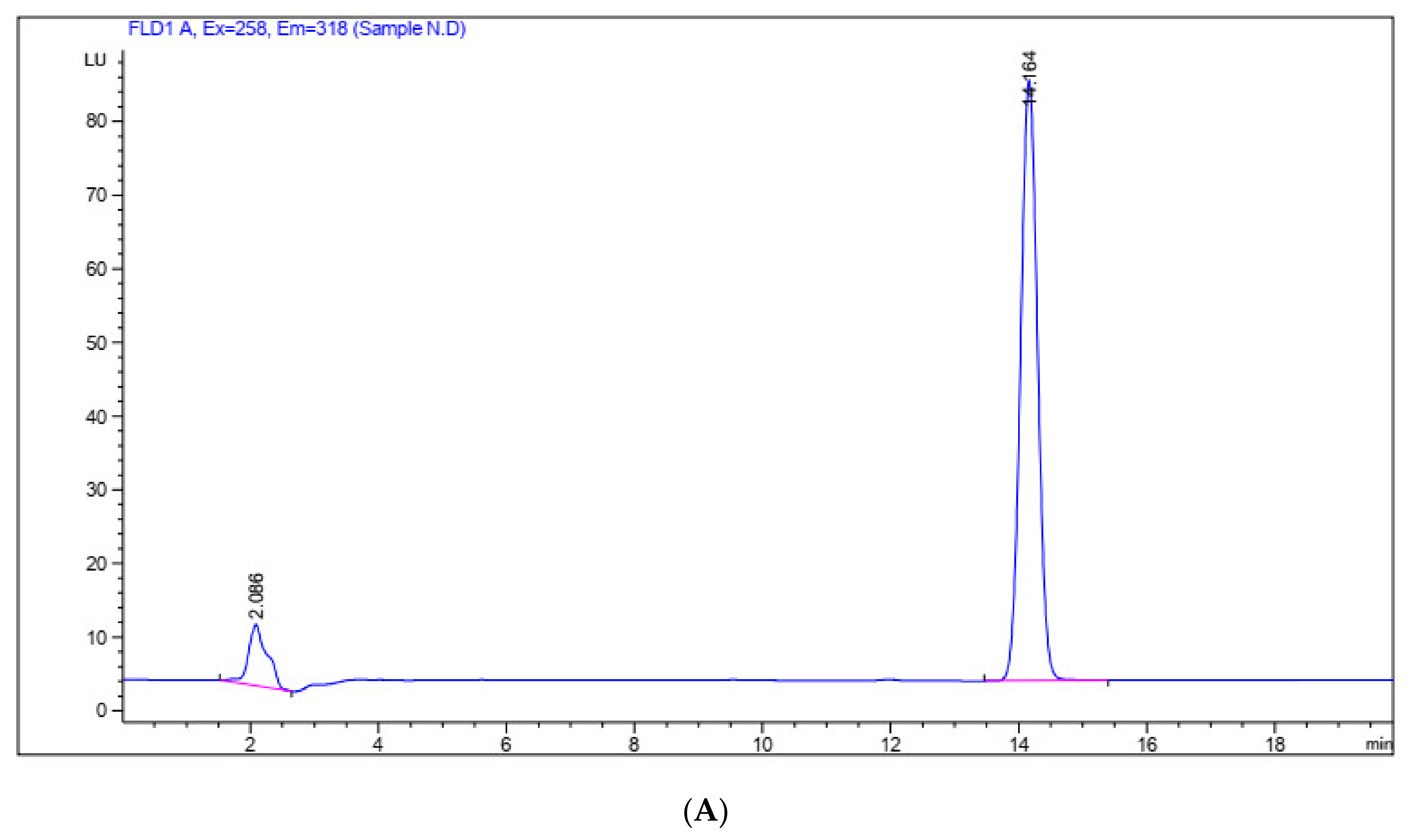
3.2. Mangiferin Quantification by HPLC Analysis

In the present study, the crude extract of local mango leaves has been produced by using the ultrasonic extraction method and ethanol as the extraction solvent. The extraction efficiency as calculated from Equation (1) was 14.17% (w/w), which was marginally higher than the results of a previous work by Akinpelu and Onakoya (2006) (13.93% w/w) [40]. However, our procedure showed superiorities in terms of the extraction time and economic efficiency. To be specific, Akinpelu and Onakoya (2006) selected methanol as the solvent and theextraction process was prolonged for 4 days, while the current process with the combination of ethanol solvent and ultrasonic extraction yielded a similar efficiency (14.17%) and only required 4 min of extraction time [40]. For this reason, the current process can be considered as an improvement in terms of the extraction efficiency and time, thereby providing an essential insight for future studies on optimization and industrial-scaled experiments.
The results of the HPLC analysis of mangiferin in the crude extract of mango leaves obtained from UE are presented in Table 1 and Figure 2. Briefly, when analyzing the crude extract, there are seven peaks shown with different R.T. and peak areas (Figure 2A). Two out of these seven peaks were shown at 2.132 and 14.296 min, which were relatively close to the peaks of the standard mangiferin, shown at 2.086 and 14.164 min (Figure 2B). Similar results have been obtained by Fernández et al. (2015) [41], confirming the presence of mangiferin the crude mango leaves extract. However, the peaks shown at 2.132 min and at 2.086 min of crude extract and standard mangiferin, respectively, were most likely corresponding to mangiferin isomers such as isomangiferin and homomangiferin, which are present in mango leaves extract in a minor quantity [2,42]. Therefore, based on the concentration and the peak area of the crude mango leaves extract shown at 14.296 min, the presence of mangiferin was identified at a content of 9.510 mg/g.
In the present study, purification was performed to purify mangiferin from the crude extract of old mango leaves. This step aims to remove the impurities (e.g., colorants and compounds with weak polarity), which interfere with the efficiency of the extraction process as well as the biological activities of mangiferin [27,43]. Previously, mangiferin has been successfully purified from Chinese mango cultivars by using macroporous HPD100 resin chromatography combined with high-speed counter-current chromatography (HSCCC) [43]. However, studies on mangiferin purification from old leaves of mango trees grown in Vietnam have not been established. Therefore, in this study, mangiferin was purified from Vietnamese mango leaves extract by liquid–liquid fractionation (e.g., dichloromethane and ethyl acetate) and column chromatography.
The results from the HPLC analysis revealed the presence of mangiferin in the upper layer of the dichloromethane fraction, as well as both layers of the ethyl acetate fraction (Table 1). Firstly, for the dichloromethane fraction, the bottom layer mostly contained impurities while the upper layer showed eight peaks, and the peak shown at 14.286 min of R.T. corresponded to mangiferin presence (Figure 2C). The mangiferin content of the upper layer of the dichloromethane fraction was calculated as 42.325 mg/g and was taken for subsequent fractionation with ethyl acetate. Secondly, for the ethyl acetate fraction, mangiferin presence was detected in both the upper and bottom layers, as shown by the peaks at 14.271 and 14.286 min, respectively (Figure 2D,E). However, as the mangiferin content was more concentrated in the upper layer than the bottom layer (Table 1), the upper layer of the ethyl acetate fraction was selected for column chromatography. After the process had finished, washing with acetone and methanol helped remove the remaining solvent and impurities without dissolving the mangiferin crystals. As evidenced by the HPLC results, the mangiferin obtained after the two-step purification process showed two peaks at 2.088 and 14.103 min, which approached closely to the peaks of standard mangiferin at 2.086 and 14.164 min (Figure 2F). Therefore, similar to the standard mangiferin, the purified mangiferin may also contain a small quantity of mangiferin isomers, which corresponded to the peak shown at 2.088 min. Therefore, based on the peak shown at 14.103 min, the content of the purified mangiferin was identified at a content of 1.97 g (Table 1).
In the present study, a total amount of 1.97 g of mangiferin was obtained from the extraction and purification of a local variety of mango leaves. The compound recovery was 0.82% and the purity was 94.2%. This result was compared to previous studies, which were also performed on the leaves of mango trees grown in Vietnam yet employed macroporous D101 and solvents of a different polarity [44,45]. In these studies, it can be seen that the recovery and purity of the obtained mangiferin were ranged from 0.37–0.49% and 68–72%, respectively, which were lower than the present study. The purity of the mangiferin in the present study can be comparable to Nian et al. (2016), in which mangiferin of 92.15% purity was extracted from Anemarrhenae Rhizome by using polyamide and macroporous HPD400 adsorption resins chromatography [46], as well as Luo et al. (2012), in which mangiferin (99.13% purity) was purified from Chinese mango cultivars using macroporous HPD100 resin chromatography associated with HSCCC [43]. Based on the obtained results, the present study proposed that liquid–liquid fractionation can be combined with column chromatography to form a cost-effective purification scheme for mangiferin from mango leaves extraction. Furthermore, HPLC analysis has confirmed that with well-developed extraction and purification techniques, the local mango leaves appeared to be a highly available source of mangiferin, thus adding to the value of this by-product and reducing agricultural waste.

3.3. Antioxidant Activity of Purified Mangiferin

The antioxidant activity of crude ethanol extract, ethyl acetate fractions, standard mangiferin, and purified mangiferin obtained from UE assisted with the purification, as shown in Table 2.
As shown in Table 2, the purified mangiferin displayed a significantly low IC50 value (IC50 = 15.548 μg/mL), as compared to the standard mangiferin (IC50 = 16.383 μg/mL), upper and bottom layers of the ethyl acetate fraction (IC50 = 15.548 and 68.769 μg/mL) and the crude extract (IC50 = 27.522 μg/mL) (p < 0.05). In comparison with previous studies, the free radical scavenging activity exhibited by mangiferin obtained from the present study was 2.48 and 1.43 times higher than the mangiferin purified by macroporous D101 resin (IC50 = 38.5 μg/mL) and by solvents with varied polarization (IC50 = 22.2 μg/mL) [45,46]. It is widely accepted that the evaluation of the biological activities of plant-based extracts such as antioxidant and anti-microorganism could provide essential insights into the efficiency of the extraction and purification methods. Therefore, from the results of the present study, it can be concluded that the combination of ultrasonic extraction, liquid–liquid fractionation and column chromatography have effectively produced mangiferin with a high antioxidant potential. Along with the benefits of lowered energy and extraction time consumption, this combinatory method is therefore highly recommended for mangiferin extraction.

3.4. Antibacterial Activity of Purified Mangiferin

The crude extracts, ethyl acetate fraction (upper layer), purified and standard mangiferin at various concentrations (12.5–200 mg/mL) were evaluated for anti-microorganism activity against E. coli, Samonella spp. and A. flavus by measuring the diameter of the inhibition zone which appeared after 24-h incubation at 37 °C.
As shown in Table 3, the crude extract inhibited the growth of E. coli in a concentration-dependent manner, with the inhibition zones ranging from 7.7 ± 0.3 to 15.6 ± 0.5 (mm). In particular, at the concentration of 25 mg/mL, this activity of mango leaves extract was lower than the methanol extract from mango stem bark, whose inhibition zone was measured as 12 mm at the concentration of 20 mg/mL [40]. In contrast, the upper layer of the ethyl acetate fraction of the mango leaves extract showed a concentration-dependent inhibition against both E. coli and Samonella spp., indicated by the range of inhibition zones from 11.3 ± 0.4 to 21.6 ± 0.5 (mm) and from 11.5 ± 0.5 to 23.5 ± 0.5 (mm), respectively. As compared to Sija (2009), the antibacterial activity of the ethyl acetate fraction of mango leaves extract in the present study was improved against Samonella spp., while being comparably effective against E. coli [47]. The inhibitory activity by the ethyl acetate fraction of the mango leaves extract has been proposed to derive from the presence of mangiferin as well as several flavonoids and alkaloids that were previously shown to exhibit a high antibacterial effect [2]. No inhibition against E. coli and Samonella spp. was observed in the presence of both purified and standard mangiferin. All of the tested samples were unable to inhibit the growth of A. flavus. The inability of mangiferin to inhibit the fungal species in this study was contradictory to previous reports by Singh et al. (2012) [31], Stoilova et al. (2008) [34] and Raju et al. [48], where a high concentration of mangiferin was able to exert a killing effect on A. flavus and Thermoascus aurantiacus. Overall, these results have indicated that (1) the ethyl acetate fraction of mango leaves extract exhibited strong antibacterial activity against E. coli and Samonella spp. at 12.5–200 mg/mL, and (2) this action was not attributed to the presence of a mangiferin component.

4. Conclusions

Mangiferin is one of the major constituents of mango that contributes to several valuable biological activities of the plant. The old leaves from a local variety which are often considered as wastes were utilized as the raw materials for the present study. By carrying out UE and developing an effective purification method that consisted of liquid–liquid fractionation and column chromatography, a total amount of 1.97 g of pure mangiferin with 0.82% of recovery and 94.2% of purity was obtained from the crude extract of the old mango leaves. The purified mangiferin showed improved scavenging activity against the DPPH free radicals, with IC50 = 13.841 ± 0.81 μg/mL, which was higher than the crude extracts and its ethyl acetate fractions. However, the purified compound alone exhibited no inhibition against E. coli, Salmonella spp. and A. flavus. The findings from the present study provide a helpful insight into the utilization of the old leaves of mango as a promising source of mangiferin extraction to reduce agricultural wastes, and they show that ultrasound combined with fractionation and column chromatography are effective extraction and purification methods to obtain mangiferin with a high purity and antioxidant potential.

Author Contributions

Investigation, N.T.T.L., D.T.L., P.N.D.Y., T.T.M.H. and D.T.N.P.; supervision, N.T.T.L., D.T.L., P.N.D.Y. and T.N.P.; writing—original draft, N.T.T.L., D.T.L. and D.T.N.P.; writing—review & editing, D.T.N.P. All authors have read and agreed to the published version of the manuscript.

Funding

This research is funded by the Funds for Science and Technology Development of the University of Danang under grant number B2018-ĐN02-45.

Institutional Review Board Statement

Not applicable.

Informed Consent Statement

Not applicable.

Data Availability Statement

The data presented in this study are available on request from the corresponding author.

Conflicts of Interest

The authors declare no conflict of interest.

References

  1. Ediriweera, M.K.; Tennekoon, K.H.; Samarakoon, S.R. A Review on Ethnopharmacological Applications, Pharmacological Activities, and Bioactive Compounds of Mangifera indica (Mango). Evid. Based Complementary Altern. Med. 2017, 2017, 1–24. [Google Scholar] [CrossRef] [PubMed] [Green Version]
  2. Shah, K.; Patel, M.; Patel, R.; Parmar, P. Mangifera indica (Mango). Phcog Rev. 2010, 4, 42. [Google Scholar] [CrossRef] [PubMed]
  3. Solís-Fuentes, J.A.; del Carmen Durán-de-Bazúa, M. Mango (Mangifera indica L.) Seed and Its Fats. In Nuts and Seeds in Health and Disease Prevention; Elsevier: Amsterdam, The Netherlands, 2011; pp. 741–748. ISBN 9780123756886. [Google Scholar]
  4. Tharanathan, R.N.; Yashoda, H.M.; Prabha, T.N. Mango (Mangifera indica L.), “The King of Fruits”—An Overview. Food Rev. Int. 2006, 22, 95–123. [Google Scholar] [CrossRef]
  5. Food and Agriculture Organization (FAO). Global Prospects for Major Tropical Fruits-Short-Term Outlook, Challenges and Opportunities in a Vibrant Global Marketplace. 2017. Available online: http://www.fao.org/fileadmin/templates/est/COMM_MARKETS_Monitoring/Tropical_Fruits/Documents/Tropical_Fruits_Special_Feature.pdf (accessed on 10 January 2021).
  6. Evans, E.A.; Ballen, F.H.; Siddiq, M. Mango Production, Global Trade, Consumption Trends, and Postharvest Processing and Nutrition. In Handbook of Mango Fruit; Siddiq, M., Brecht, J.K., Sidhu, J.S., Eds.; John Wiley & Sons, Ltd.: Chichester, UK, 2017; pp. 1–16. ISBN 9781119014362. [Google Scholar]
  7. Testa, R.; Tudisca, S.; Schifani, G.; Di Trapani, A.; Migliore, G. Tropical Fruits as an Opportunity for Sustainable Development in Rural Areas: The Case of Mango in Small-Sized Sicilian Farms. Sustainability 2018, 10, 1436. [Google Scholar] [CrossRef] [Green Version]
  8. Lauricella, M.; Emanuele, S.; Calvaruso, G.; Giuliano, M.; D’Anneo, A. Multifaceted Health Benefits of Mangifera indica L. (Mango): The Inestimable Value of Orchards Recently Planted in Sicilian Rural Areas. Nutrients 2017, 9, 525. [Google Scholar] [CrossRef] [PubMed]
  9. Coelho, E.M.; de Souza, M.E.A.O.; Corrêa, L.C.; Viana, A.C.; de Azevêdo, L.C.; dos Santos Lima, M. Bioactive Compounds and Antioxidant Activity of Mango Peel Liqueurs (Mangifera indica L.) Produced by Different Methods of Maceration. Antioxidants 2019, 8, 102. [Google Scholar] [CrossRef] [PubMed] [Green Version]
  10. Núñez Sellés, A.J.; Vélez Castro, H.T.; Agüero-Agüero, J.; González-González, J.; Naddeo, F.; De Simone, F.; Rastrelli, L. Isolation and Quantitative Analysis of Phenolic Antioxidants, Free Sugars, and Polyols from Mango (Mangifera indica L.) Stem Bark Aqueous Decoction Used in Cuba as a Nutritional Supplement. J. Agric. Food Chem. 2002, 50, 762–766. [Google Scholar] [CrossRef]
  11. Fitmawati, F.; Resida, E.; Kholifah, S.N.; Roza, R.M.; Almurdani, M.; Emrizal, E. Phytochemical Screening and Antioxidant Profiling of Sumatran Wild Mangoes (Mangifera Spp.): A Potential Source for Medicine Antidegenerative Effects. F1000Res 2020, 9, 220. [Google Scholar] [CrossRef]
  12. Maldonado-Celis, M.E.; Yahia, E.M.; Bedoya, R.; Landázuri, P.; Loango, N.; Aguillón, J.; Restrepo, B.; Guerrero Ospina, J.C. Chemical Composition of Mango (Mangifera indica L.) Fruit: Nutritional and Phytochemical Compounds. Front. Plant. Sci. 2019, 10, 1073. [Google Scholar] [CrossRef] [PubMed]
  13. Iseda, S. On Mangiferin, the Coloring Matter of Mango (Mangifera indica Linn.). V. Identification of Sugar Component and the Structure of Mangiferin. BCSJ 1957, 30, 629–633. [Google Scholar] [CrossRef] [Green Version]
  14. Telang, M.; Dhulap, S.; Mandhare, A.; Hirwani, R. Therapeutic and Cosmetic Applications of Mangiferin: A Patent Review. Expert Opin. Ther. Pat. 2013, 23, 1561–1580. [Google Scholar] [CrossRef]
  15. Sagar, N.A.; Pareek, S.; Sharma, S.; Yahia, E.M.; Lobo, M.G. Fruit and Vegetable Waste: Bioactive Compounds, Their Extraction, and Possible Utilization. Compr. Rev. Food Sci. Food Saf. 2018, 17, 512–531. [Google Scholar] [CrossRef] [PubMed] [Green Version]
  16. Jahurul, M.H.A.; Zaidul, I.S.M.; Ghafoor, K.; Al-Juhaimi, F.Y.; Nyam, K.-L.; Norulaini, N.A.N.; Sahena, F.; Mohd Omar, A.K. Mango (Mangifera indica L.) by-Products and Their Valuable Components: A Review. Food Chem. 2015, 183, 173–180. [Google Scholar] [CrossRef] [PubMed]
  17. Alañón, M.E.; Palomo, I.; Rodríguez, L.; Fuentes, E.; Arráez-Román, D.; Segura-Carretero, A. Antiplatelet Activity of Natural Bioactive Extracts from Mango (Mangifera indica L.) and Its By-Products. Antioxidants 2019, 8, 517. [Google Scholar] [CrossRef] [PubMed] [Green Version]
  18. Lerma-Torres, J.M.; Navarro-Ocaña, A.; Calderón-Santoyo, M.; Hernández-Vázquez, L.; Ruiz-Montañez, G.; Ragazzo-Sánchez, J.A. Preparative Scale Extraction of Mangiferin and Lupeol from Mango (Mangifera indica L.) Leaves and Bark by Different Extraction Methods. J. Food Sci. Technol. 2019, 56, 4625–4631. [Google Scholar] [CrossRef]
  19. Zhang, Q.-W.; Lin, L.-G.; Ye, W.-C. Techniques for Extraction and Isolation of Natural Products: A Comprehensive Review. Chin. Med. 2018, 13, 20. [Google Scholar] [CrossRef] [Green Version]
  20. Salomon, S.; Sevilla, I.; Betancourt, R.; Romero, A.; NuevasPaz, L.; AcostaEsquijarosa, J. Extraction of Mangiferin from Mangifera indica L. Leaves Using Microwaveassistedtechnique. Emir. J. Food Agric. 2014, 26, 616. [Google Scholar] [CrossRef] [Green Version]
  21. Kulkarni, V.M.; Rathod, V.K. Mapping of an Ultrasonic Bath for Ultrasound Assisted Extraction of Mangiferin from Mangifera indica Leaves. Ultrason. Sonochemistry 2014, 21, 606–611. [Google Scholar] [CrossRef]
  22. Fernández-Ponce, M.T.; Casas, L.; Mantell, C.; Rodríguez, M.; Martínez de la Ossa, E. Extraction of Antioxidant Compounds from Different Varieties of Mangifera indica Leaves Using Green Technologies. J. Supercrit. Fluids 2012, 72, 168–175. [Google Scholar] [CrossRef]
  23. Kulkarni, V.M.; Rathod, V.K. A Novel Method to Augment Extraction of Mangiferin by Application of Microwave on Three Phase Partitioning. Biotechnol. Rep. 2015, 6, 8–12. [Google Scholar] [CrossRef] [Green Version]
  24. Zou, T.-B.; Xia, E.-Q.; He, T.-P.; Huang, M.-Y.; Jia, Q.; Li, H.-W. Ultrasound-Assisted Extraction of Mangiferin from Mango (Mangifera indica L.) Leaves Using Response Surface Methodology. Molecules 2014, 19, 1411–1421. [Google Scholar] [CrossRef]
  25. Kulkarni, V.M.; Rathod, V.K. Extraction of Mangiferin from Mangifera indica Leaves Using Three Phase Partitioning Coupled with Ultrasound. Ind. Crop. Prod. 2014, 52, 292–297. [Google Scholar] [CrossRef]
  26. Tandon, S. Distillation technology for essential oils. In Extraction Technologies for Medicinal and Aromatic Plant; Handa, S.S., Khanuja, S.P.S., Longo, G., Rakesh, D.D., Eds.; International Centre for Science and High Technology: Trieste, Italy, 2008; pp. 115–127. [Google Scholar]
  27. Zhang, Z.; Pang, X.; Xuewu, D.; Ji, Z.; Jiang, Y. Role of Peroxidase in Anthocyanin Degradation in Litchi Fruit Pericarp. Food Chem. 2005, 90, 47–52. [Google Scholar] [CrossRef]
  28. Altemimi, A.; Lakhssassi, N.; Baharlouei, A.; Watson, D.; Lightfoot, D. Phytochemicals: Extraction, Isolation, and Identification of Bioactive Compounds from Plant Extracts. Plants 2017, 6, 42. [Google Scholar] [CrossRef] [PubMed]
  29. Matkowski, A.; Kus, P.; Goralska, E.; Wozniak, D. Mangiferin—a Bioactive Xanthonoid, Not Only from Mango and Not Just Antioxidant. Mini Rev. Med. Chem. 2013, 13, 439–455. [Google Scholar] [CrossRef] [PubMed]
  30. Pardo-Andreu, G.L.; Sánchez-Baldoquín, C.; Avila-González, R.; Delgado, R.; Naal, Z.; Curti, C. Fe(III) Improves Antioxidant and Cytoprotecting Activities of Mangiferin. Eur. J. Pharm. 2006, 547, 31–36. [Google Scholar] [CrossRef]
  31. Singh, S.K.; Tiwari, R.M.; Sinha, S.K.; Danta, C.C.; Prasad, S.K. Antimicrobial Evaluation of Mangiferin and Its Synthesized Analogues. Asian Pac. J. Trop. Biomed. 2012, 2, S884–S887. [Google Scholar] [CrossRef]
  32. Mazlan, N.A.; Azman, S.; Ghazali, N.F.; Yusri, P.Z.S.; Idi, H.M.; Ismail, M.; Sekar, M. Synergistic antibacterial activity of mangiferin with antibiotics against Staphylococcus aureus. Drug Invent Today 2019, 12, 14–17. [Google Scholar]
  33. Schieber, A.; Berardini, N.; Carle, R. Identification of Flavonol and Xanthone Glycosides from Mango (Mangifera indica L. Cv. “Tommy Atkins”) Peels by High-Performance Liquid Chromatography-Electrospray Ionization Mass Spectrometry. J. Agric. Food Chem. 2003, 51, 5006–5011. [Google Scholar] [CrossRef]
  34. Stoilova, I.; Gargova, S.; Stoyanova, A.; Krastanov, A.I.; Gargova, S.; Ho, L. Antimicrobial and antioxidant activity of the polyphenol mangiferin. Electron. J. Environ. Agric. Food Chem. 2008, 7, 2706–2716. [Google Scholar]
  35. Jutiviboonsuk, A.; Sardsaengjun, C. Mangiferin in leaves of three thai mango (Mangifera indica L.) varieties. Indian J. Pharm. Sci. 2010, 6, 122–129. [Google Scholar]
  36. Alam, M.K.; Rana, Z.H.; Islam, S.N.; Akhtaruzzaman, M. Total Phenolic Content and Antioxidant Activity of Methanolic Extract of Selected Wild Leafy Vegetables Grown in Bangladesh: A Cheapest Source of Antioxidants. Potravin. Slovak J. Food Sci. 2019, 13, 287–293. [Google Scholar] [CrossRef] [Green Version]
  37. González, A.F.; Balmaseda, I.H.; Gaitén, Y.I.G.; Lizama, R.S.; Carmenate García, L.M.; Pieters, L.; Rodeiro, I.; Hernandez, R.D.; Garrido, G. Phytochemical study and antioxidant capacity of three fractions from the stem of Caesalpinia bahamensis Lam. J Pharm. Pharmacogn. Res. 2018, 7, 12–20. [Google Scholar]
  38. Mostafa, A.A.; Al-Askar, A.A.; Almaary, K.S.; Dawoud, T.M.; Sholkamy, E.N.; Bakri, M.M. Antimicrobial Activity of Some Plant Extracts against Bacterial Strains Causing Food Poisoning Diseases. Saudi J. Biol. Sci. 2018, 25, 361–366. [Google Scholar] [CrossRef] [PubMed]
  39. Falleh, H.; Ksouri, R.; Lucchessi, M.-E.; Abdelly, C.; Magné, C. Ultrasound-Assisted Extraction: Effect of Extraction Time and Solvent Power on the Levels of Polyphenols and Antioxidant Activity of Mesembryanthemum Edule L. Aizoaceae Shoots. Trop. J. Pharm Res. 2012, 11, 243–249. [Google Scholar] [CrossRef] [Green Version]
  40. Akinpelu, D.; Onakoya, T. Antimicrobial Activities of Medicinal Plants Used in Folklore Remedies in South-Western. Afr. J. Biotechnol. 2006. [Google Scholar] [CrossRef]
  41. Fernández-Ponce, M.T.; Casas, L.; Mantell, C.; Martínez de la Ossa, E. Use of High Pressure Techniques to Produce Mangifera indica L. Leaf Extracts Enriched in Potent Antioxidant Phenolic Compounds. Innov. Food Sci. Emerg. Technol. 2015, 29, 94–106. [Google Scholar] [CrossRef]
  42. Wei, X.; Liang, D.; Wang, Q.; Meng, X.; Li, Z. Total Synthesis of Mangiferin, Homomangiferin, and Neomangiferin. Org. Biomol. Chem. 2016, 14, 8821–8831. [Google Scholar] [CrossRef]
  43. Luo, F.; Lv, Q.; Zhao, Y.; Hu, G.; Huang, G.; Zhang, J.; Sun, C.; Li, X.; Chen, K. Quantification and Purification of Mangiferin from Chinese Mango (Mangifera indica L.) Cultivars and Its Protective Effect on Human Umbilical Vein Endothelial Cells under H2O2-Induced Stress. IJMS 2012, 13, 11260–11274. [Google Scholar] [CrossRef]
  44. Nguyen, T.M.P. Purification of Mangiferin from Mango (Mangifera indica L.) Leaves by Using D101 Absorbent Chromatography and Evaluation of Antioxidant and Antibacterial Activitites. Master’s Thesis, The University of Danang (University of Science and Technology), Da Nang, Vietnam, 2019. [Google Scholar]
  45. Tran, T.D. Purification of Mangiferin from Mango Leaves by Traditional Method and Investigation of Biological Activitites. Master’s Thesis, The University of Danang (University of Science and Technology), Da Nang, Vietnam, 2019. [Google Scholar]
  46. Nian, S.; Liu, E.; Fan, Y.; Alolga, R.N.; Li, H.; Li, P. Orthogonal Separation Protocol for the Simultaneous Preparation of Four Medically Active Compounds from Anemarrhenae Rhizoma by Sequential Polyamide and Macroporous Resin Adsorbent Chromatography: Sample Preparation. J. Sep. Sci. 2016, 39, 3195–3204. [Google Scholar] [CrossRef]
  47. Sija, S.L. In Vitro Antioxidant Activity and Antibacterial Activity of Ethyl Acetate Extract of Medicinal Tree Species, Anacardium occidentale L. and Mangifera indica L. IJAEB 2019, 12. [Google Scholar] [CrossRef]
  48. Raju, N.V.; Sukumar, K.; Reddy, G.B.; Pankaj, P.K.; Muralitharan, G.; Annapareddy, S. In-Vitro Studies on Antitumour and Antimicrobial Activities of Methanolic Kernel Extract of Mangifera indica L. Cultivar Banganapalli. Biomed. Pharm. J. 2019, 12, 357–362. [Google Scholar] [CrossRef]
Figure 1. Effects of different parameters of extraction method on the mangiferin content, as follows: (A) effect of liquid-to-solid ratio (v/w) on the mangiferin content; (B) effect of extraction temperature (°C) on the mangiferin content; and (C) effect of extraction time (min) on the mangiferin content. Values showing different letters in the same column indicated significant difference (p < 0.05).
Processes 09 00852 g001
Figure 2. Results of HPLC analysis of (A) standard mangiferin, (B) crude mango leaves extract, (C) upper layer of dichloromethane fraction, (D) bottom layer of ethyl acetate fraction, (E) upper layer of ethyl acetate fraction, and (F) purified mangiferin.
Processes 09 00852 g002aProcesses 09 00852 g002bProcesses 09 00852 g002c
Table 1. HPLC analysis of mangiferin content (mg/g) in standard mangiferin, crude mango leaves extract, upper layer of dichloromethane fraction, bottom and upper layers of ethyl acetate fraction and purified mangiferin.
SampleConc. (g/mL)Retention Time (min)Peak Area (LU*s)Mangiferin Content (mg/g)
Standard mangiferin0.00114.1641506.5421000
Crude leaves extract0.0114.296143.2699.51
Dichloromethane fraction (upper layer)0.0114.286637.63742.325
Ethyl acetate fraction (upper layer)0.0314.2864846.101107.223
Ethyl acetate fraction (bottom layer)0.09514.271125.460.877
Purified mangiferin0.814.1031135.188941.882
Table 2. Antioxidant activity of crude extract, ethyl acetate fractions, standard mangiferin and purified mangiferin from mango leaves.
SampleIC50 (μg/mL)Regression Equationr2
Crude extract27.522y = 2.3945 x0.91670.9918
Ethyl acetate fraction (bottom layer)68.769y = 8.0215 x0.69640.9915
Ethyl acetate fraction (upper layer)15.548y = 4.7598 x0.85710.997
Standard mangiferin16.383y = 4.1919 x0.88650.9925
Purified mangiferin13.841y = 1.7396 x0.79380.9978
Vitamin C2.551y = 33.427 x0.430.9915
Table 3. Anti-microorganism activity of purified mangiferin against E. coli, Samonella spp. and A. flavus by using disc diffusion assay.
ExtractConc. (mg/mL)Inhibition Zone (mm)
E. coliSamonella spp.A. flavus
Crude extract20015.6a ± 0.5--
10011.4b ± 0.4--
5010.4c ± 0.2--
259.5d ± 0.3--
12.57.7e ± 0.3--
Ethyl acetate fraction (upper layer)20021.6a ± 0.523.5a ± 0.5-
10017.5b ± 0.517.9b ± 0.3-
5014.6c ± 0.515c ± 0.4-
2513.2d ± 0.313.9d ± 0.4-
12.511.3e ± 0.411.5e ± 0.5-
Purified mangiferin200---
100---
50---
25---
12.5---
Standard mangiferin200---
100---
50---
25---
12.5---
Ampicilin (positive control)2001219-
1001219-
501219-
251219-
12.51219-
The experiment was performed in triplicates. Values showing different letters in the same column indicated significant different (p < 0.05). “-“ indicated no inhibition zone in case of bacteria or sparse distribution in case of fungal spores.
Publisher’s Note: MDPI stays neutral with regard to jurisdictional claims in published maps and institutional affiliations.

