Article

Therapeutic Effect of Cinnamomum osmophloeum Leaf Extract on Oral Mucositis Model Rats Induced by 5-Fluororacil via Influencing IL-1β and IL-6 Levels

1
School of Dentistry, Baiturrahmah University, Kuranji, Padang 25155, Indonesia
2
Department of Food Science and Biotechnology, Da-Yeh University, Dacun, Changhua 51591, Taiwan
3
Department of Biomedical Sciences, Chung Shan Medical University, Taichung 40201, Taiwan
4
Department of Food Science and Biotechnology, National Chung Hsing University, Taichung 40227, Taiwan
5
Department of Medical Research, Chung Shan Medical University Hospital, Taichung 40201, Taiwan
*
Authors to whom correspondence should be addressed.
Processes 2021, 9(4), 615; https://doi.org/10.3390/pr9040615
Submission received: 14 March 2021 / Revised: 26 March 2021 / Accepted: 26 March 2021 / Published: 31 March 2021
(This article belongs to the Section Pharmaceutical Processes)

Abstract

:
Oral mucositis (OM) is the oral inflammation as manifestation of chemotherapy and/or radiotherapy. Cinnamomum osmophloeum (CO), of which the constituents possess anti-inflammatory activities, may have potential to alleviate OM. In this study, laboratory rats were injected with 5-Fluororacil and their oral mucosa were irritated by 18-gauge needle pouching to induce OM. Rats were randomly divided into six experimental groups: without treatment (WT), only 100 mg/mL CO leaf extract (COLE) treatment (100-only), only 5-Fluororacil treatment (5-FU), 5-FU then treated with Triamcinolone acetonide orobase (5-FU+G), 5-FU then treated with 50 mg/mL COLE, and 5-FU then treated with 100 mg/mL COLE (5-FU+100). Body weights and food and water intakes during the experimental period were recorded. Macroscopic examination, histopathological analyses, and serum tumor necrosis factor-α (TNF-α), interleukin-1β (IL-1β), and interleukin-6 (IL-6) levels of these rats were evaluated or determined. No significant difference was found between the WT and 100-only groups. Results of macroscopic examinations, histopathological analyses, body weight changes, food and water intakes, and serum IL-1β and IL-6 levels showed significant therapeutic effects of the 5-FU+100 group compared to the 5-FU group. These finding suggest that COLE can be one of potential remedies for OM therapy through influencing proinflammatory cytokine levels.

1. Introduction

Oral mucositis (OM) is the inflammation of oral mucosa as a result of chemotherapy and radiotherapy. OM clinically manifested as atrophy, swelling, erythema and ulceration [1]. OM occurrence might increase the budget of treatment and reduce the oral health quality of life [2,3,4]. Five stages of the development of OM can be described as initiation, primary injury response, signal strengthening, ulceration and healing. The first stage (initiation) occurs after radiotherapy and/or chemotherapy exposure. It involves two actions: DNA collapse and the generation of reactive oxygen species (ROS). In the second stage of the development (primary injury response), productions of proinflammatory cytokines including tumor necrosis factor-α (TNF-α), interleukin-6 (IL-6), and interleukin-1 beta (IL-1β) play important roles in mediating damage and signaling pathways [5]. The tissue and peripheral blood were affected by these cytokines and the severity of OM connects to the increases of proinflammatory cytokines. During third stage (signal strengthening), proinflammatory cytokines bring a positive feedback to develop and accelerate remedial process. In the fourth stage (ulceration) bacterial re-infection provokes macrophage accretion to cover additional numbers of proinflammatory cytokines. During the fifth stage (healing), signs from the connective tissue to the bordering epithelium can stimulate the cells movement, proliferation, and differentiation, resultant in healed mucosa. [6].
Cinnamomum osmophloeum (CO), the indigenous Cinnamomum from Taiwan, had been reported comprised anti-inflammation, wound repair, antibacterial activities, antioxidation, antihyperglycemic activities, antidyslipidemia activities, anticancer and antitumor, renal disease therapy, and antihyperuricemia activities [7]. CO plays a significant role in anti-inflammatory activities by reducing proinflammatory cytokines including IL-1β [8], IL-6 and tumor necrosis factor receptor (TNF-R) [9]. Previous review explained that CO could be potential in OM alleviation because of its anti-inflammatory effect, as the activation of several transcription factors might release the inflammatory reaction in the second phase of OM [7].
The recent investigations of Cinnamomum on OM potential therapy have been performed in vitro and in vivo [10,11]. The cinnamon bark fractions were biocompatible to oral epithelial cells and possessed the effect on inflammatory cells and can prevent the adherence of Candida albicans [11]. However, a previous study found adverse oral reactions due to cinnamon-flavoured chewing gum consumption [12] Cinnamaldehyde, one of cinnamon’s major chemical constituents, can alleviate OM rats induced by radiotherapy and has an effect on salivary antioxidant capacity [10,13]. Constructed from the background and the above previous studies, the purpose of this study is to analyze the protective effect of CO leaf extract on OM model rats induced by 5-FU both clinically and histopathologically.

2. Materials and Methods

2.1. Sample Preparation

The CO leaves were extracted using lyophilization. The leaves were collected and stored at –20 °C. The chemical contents of the leaves were extracted using 95% ethanol. The ratio of frozen leaves and the volume of ethanol (w/v) is 1:10 and were taken out with a Waring blender. The mixture was incubated at room temperature for three days to extract the active ingredients. The extractions were performed three times and combined. The supernatant of the extraction was collected with centrifugation at 6000 rpm for 20 min. A vacuum evaporator was used to remove the ethanol, and the ethanol-free extracts were freeze-dried. The dried powder was weighed and dissolved in ethanol to obtain the certain experimental concentration and sterilized by passing through a 0.2-μm filter.

2.2. Experimental Methods

2.2.1. Animal Preparation

Thirty-six male Wistar rats (seven weeks old; 200–225 g) were purchased from BioLASCO Taiwan Co., Ltd. (Taipei, Taiwan) These animals were fed with a standard rodent diet (R5001) and sterilized water ad libitum and housed in a temperature-controlled (20–24 °C), humidity-controlled (60 ± 5%), and photoperiod-controlled (12 h light/12 h dark cycle) animal room. All animal experimental protocols were followed in accordance with the guidelines of the Institutional Animal Care and Use Committee of Da-Yeh University (protocol number: 109004). After adaption for one week, rats were housed in small groups of two in standard show box cages. The animals were randomly divided into six groups (n = 6): treated with the same volume of PBS as the control animals (WT), treated by 100 mg/mL CO extracts only (100-only), injected with 5-FU only (5-FU only), injected with 5-FU then treated with 1 mg/kg Triamcinolone acetonide orobase as a positive control (5-FU+G), injected with 5-FU then treated with 50 mg/mL CO extracts (5-FU+50), and injected with 5-FU then treated with 100 mg/mL CO extracts (5-FU+100). Excluding rats with the highest and lowest values in each group, a sample size of four rats per group was calculated power (1) of 80% with alpha-error threshold of 0.05 based on results of wound healing in the study of Preethi and Kuttan.

2.2.2. 5-FU and Sodium Pentobarbital Injections

A 5-FU dose of 150 mg/kg for rats can cause OM (Codacci-Pisanelli et al., 1997). 5-FU at 50 mg/mL was purchased from Nang Kuang Pharmaceutical Co., Ltd., Tainan, Taiwan. The dose calculation of each rat was:
Dose of 5-FU for rat:
Rat   weight   ( kg )   ×   150   mg / kg   rat   weight 50   mg / mL
A dose of 50 mg/kg rat weight of sodium pentobarbital (Sigma-Aldrich Co., Ltd., St. Louis, MO, USA) was used to cause anesthetize the rats. About 1.0 g sodium pentobarbital was dissolved in 20 mL phosphate-buffered saline (PBS) (50 mg/mL preparation). The dose calculation of each rat was:
Dose of sodium pentobarbital for rat:
Rat   weight   ( kg )   ×   50   mg / kg   rat   weight 50   mg / mL

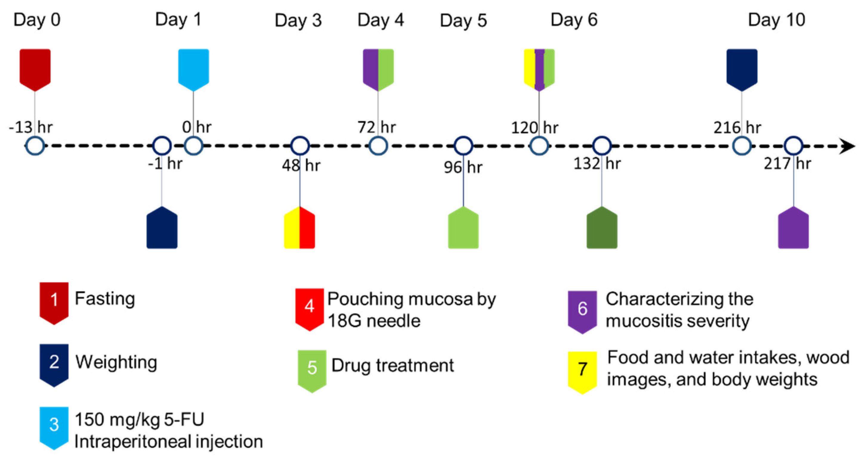
2.2.3. Animal Protocol

The animal study was done in two batches. Each batch was 10 days of experiment. After the rats arrived, we gave the food and water ad libitum for one week. The treatments were started after the animal adaptation for a week. To mimic the clinical effect of chronic irritation, the cheek pouch mucosa was irritated by superficial scratching with the tip of an 18-gauge needle. All rats were sacrificed in the last day of the experiment. The animal protocol is provided in Figure 1.

2.2.4. Rat Body Weight Changes and Water and Food Intakes

Body weights of all rats were recorded at before and after treatments. The body weight changes were calculated. On day 3 of the study, each rat was provided about 200 g food and 300 mL sterilized water. At the end of experiment, the residual foods and water were measured. The consumptions of food and water during treatments were calculated and defined as water and food intakes per rat. Five sterilized wood pieces were put in the cage on the day 3 of study and the pictures of original woods were taken. In the end of the experiment, the pictures of wood pieces bitten by the rats were taken.

2.2.5. Clinical Evaluation

Rats were observed for clinical evaluation on day 4, 6, and 10 by an investigator (doctor of dental surgery) who was blinded to the study groups. Cheek pouch mucosa was photographed for the characterization of the mucositis severity.

2.2.6. Histopathologic Examination

We cut off the oral mucosal tissues of rats in different experimental groups, and then fixed these tissues in 10% buffered natural formalin (Tonyar Biotech. Inc., Taoyuan, Taiwan) for more than 16 h, then paraffin embedded, sectioned, and hematoxylin and eosin (H&E) stained (Rapid Science Co. Ltd., Taichung, Taiwan). The stained tissue sections were scanned by TissueFAX Plus (TissueGnostics, Vienna, Austria) then observed by TissueFAXS 4.0 Viewer (TissueGnostics).

2.2.7. Serum Proinflammatory Cytokine Levels by ELISA

Plasma concentrations of TNF-α and IL-1β were quantified by rat-specific ELISA kits (DuoSet kits, P247489 and P247538, R&D Systems Inc., Minneapolis, MN, USA) as the manufactural instructions. Plasma concentration of IL-6 was quantified using rat-specific ELISA kit (900-M86) and ABTS ELISA Buffer kit (900-K00) (PeproTech Inc., Rocky Hill, NJ, USA) as the manufactural instructions.

2.3. Statistical Analysis

Results were presented as the mean ± SD (standard deviation). Statistical analyses of the data were analyzed via one-way ANOVA followed by least significant difference (LSD) to compare different treatments using IBM SPSS statistics 26. Statistical significance was expressed with p < 0.05, p < 0.01 and p < 0.001.

3. Results

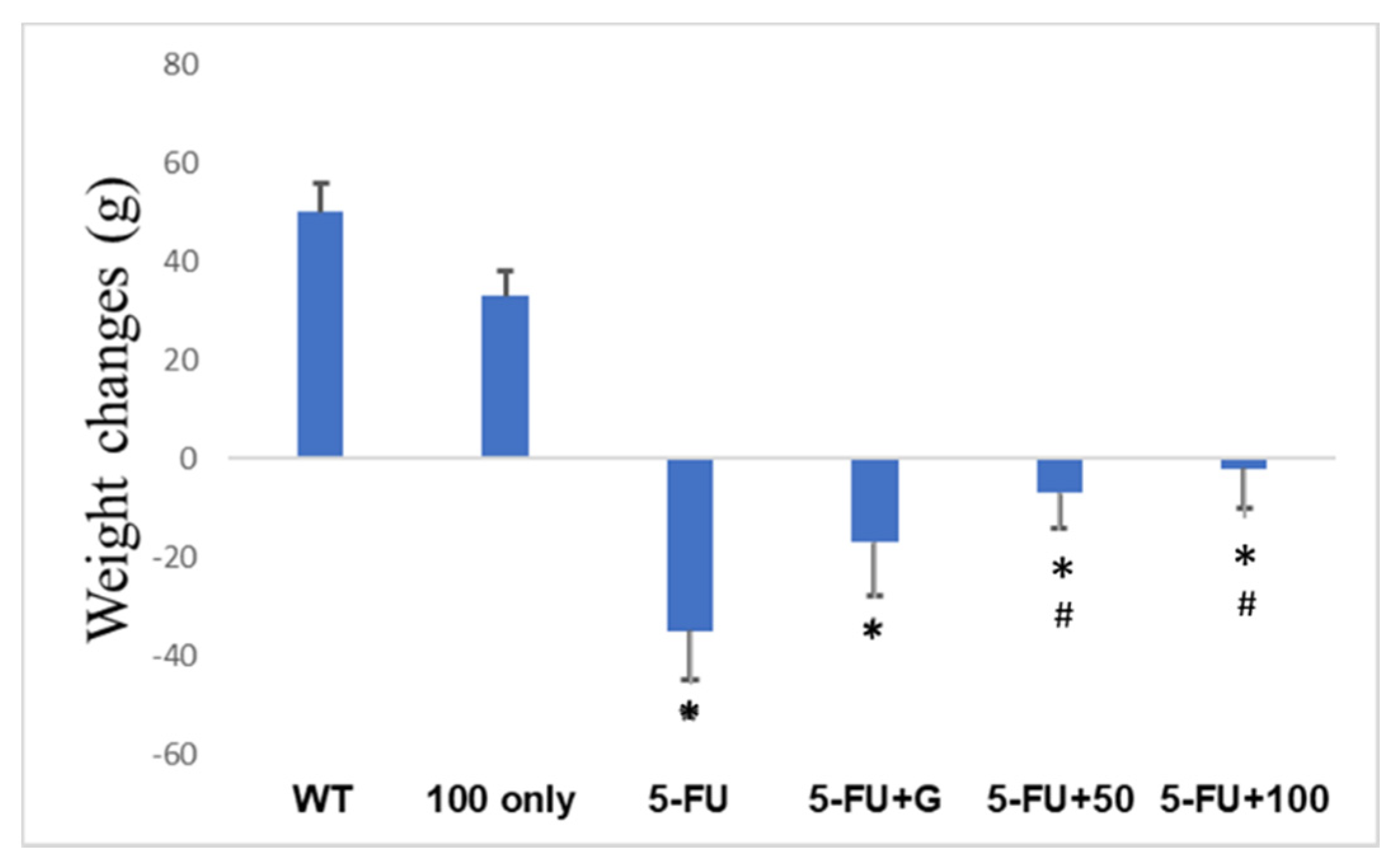
3.1. Body Weight Changes of Rats

Results of Figure 2 showed the increase in body weight of nearly 16 % over a period of 10 days was observed in the WT group (50 ± 6 g) and whereas the 5-FU-only treatment (5-FU) considerably reduced the body weight about 12 % during the study period (−34 ± 10 g). Treatment with 100 mg/mL CO only (100-only) showed increase in body weight of about 10 % (32 ± 5 g) similar to the WT group and no significant difference. Treatment with Triamcinolone acetonide 0.1 % (1 mg/kg) after 5-FU treatment (5-FU+G) showed decrease in body weight of about 5 % (−17 ± 11 g). Moreover, rats treated with 50 mg/mL CO (5-FU+50) and 100 mg/mL CO (5-FU+100) after 5-FU injection showed decreases in body weights about 2 % (−7 ± 7 g) and 1 % (−2 ± 8 g), respectively.
One-way ANOVA and post hoc testing using Tukey analysis shows statistically significant difference between the 5-FU and WT groups in body weight changes (p < 0.001). Moreover, significant difference was also found between the 5-FU+G and WT group (p < 0.001), the 5-FU+50 and WT group (p < 0.001) and the 5-FU+100 and WT group (p < 0.001). Body weight changes of rats in both of the 5-FU+50 and 5-FU+100 groups were significantly lower than the 5-FU group (p < 0.05). However, there was no significant difference between the 5-FU+G and 5-FU groups. Results of body weight changes imply that CO shows therapeutic effect against 5-FU-induced damages, and CO only shows no significant effect in the rat development.

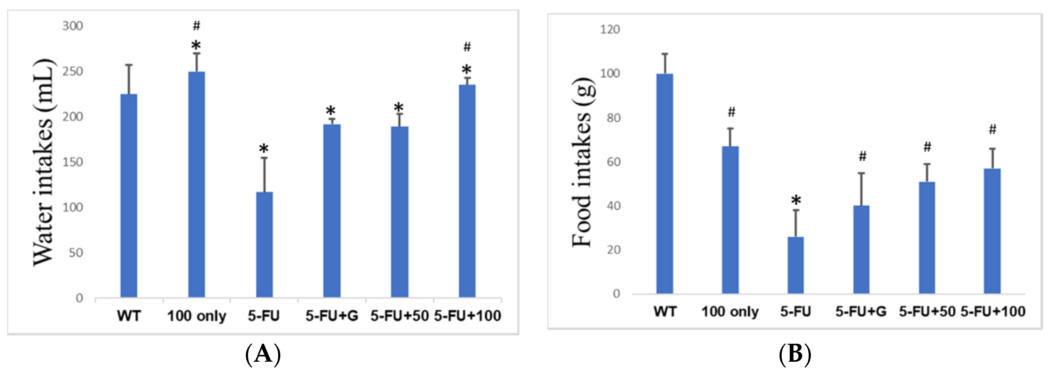
3.2. Food and Water Intakes

Results of Figure 3A showed that the average water intakes per rat during treatments in the WT, 100-only, 5-FU, 5-FU+G, 5-FU+50 and 5-FU+100 groups were 225 ± 32 mL, 250 ± 20 mL, 117 ± 37 mL, 192 ± 6 mL, 189 ± 14 mL and 236 ± 8 mL, respectively. Parametric analyses using one-way ANOVA had been conducted. Only the 5-FU group showed a significant decrease of water intakes compared to the WT group (p < 0.001). Significant differences were also found in the 5-FU group compared to the 100-only group (p < 0.001) and both the 5-FU+50 and 5-FU+G groups compared to the 100-only group (p < 0.05). Furthermore, rats in the 5-FU+50 and 5-FU+100 groups showed progressive increases in water intakes compared to the 5-FU group (p < 0.05). However, there was no significant difference between the WT and 100-only groups, and between the 5-FU+G and 5-FU+50 groups.
The average food intake per rat in the WT group showed the highest (100 ± 9 g) and in the 5-FU group showed the lowest (25 ± 12 g) among the six experimental groups during treatments (Figure 3B). Rats in the 100-only, 5-FU+G, 5-FU+50 and 5-FU+100 groups showed average food intakes of 67 ± 8 g, 40 ± 15 g, 51 ± 8 g and 57 ± 8 g, respectively. The significant decreases of food intakes were reported in both the 5-FU group and 5-FU+G groups compared to the WT group (p < 0.001). Furthermore, the significant decreases of food intakes were also found in the 100-only, 5-FU+50 and 5-FU+100 groups compared to the WT group (p < 0.05). There were no significant differences between the 5-FU, 5-FU+G and 5-FU+50 groups. However, the food intakes of rats in both the 100-only and 5-FU+100 groups were significantly higher than those in the 5-FU group (p < 0.05).
In combination of results of body weight changes (Figure 2), and food and water intakes (Figure 3), we found that 5-FU treatment significantly damaged rats and caused body weight decrease through dramatic decreases in both food and water intakes. Although high dose of CO treatment (100-only) decreased the food intakes of rats, there were no significant differences in water intakes and body weight changes between the 100-only and WT groups. Furthermore, the high-dose CO treatment (5-FU+100) showed more significant therapeutic potential than the low-dose CO (5-FU+50) and Triamcinolone acetonide 0.1% (5-FU+G) in diminishing the damage of 5-FU, because the 5-FU+100 group showed significant increases in all body weights, food and water intakes compared to the 5-FU group.

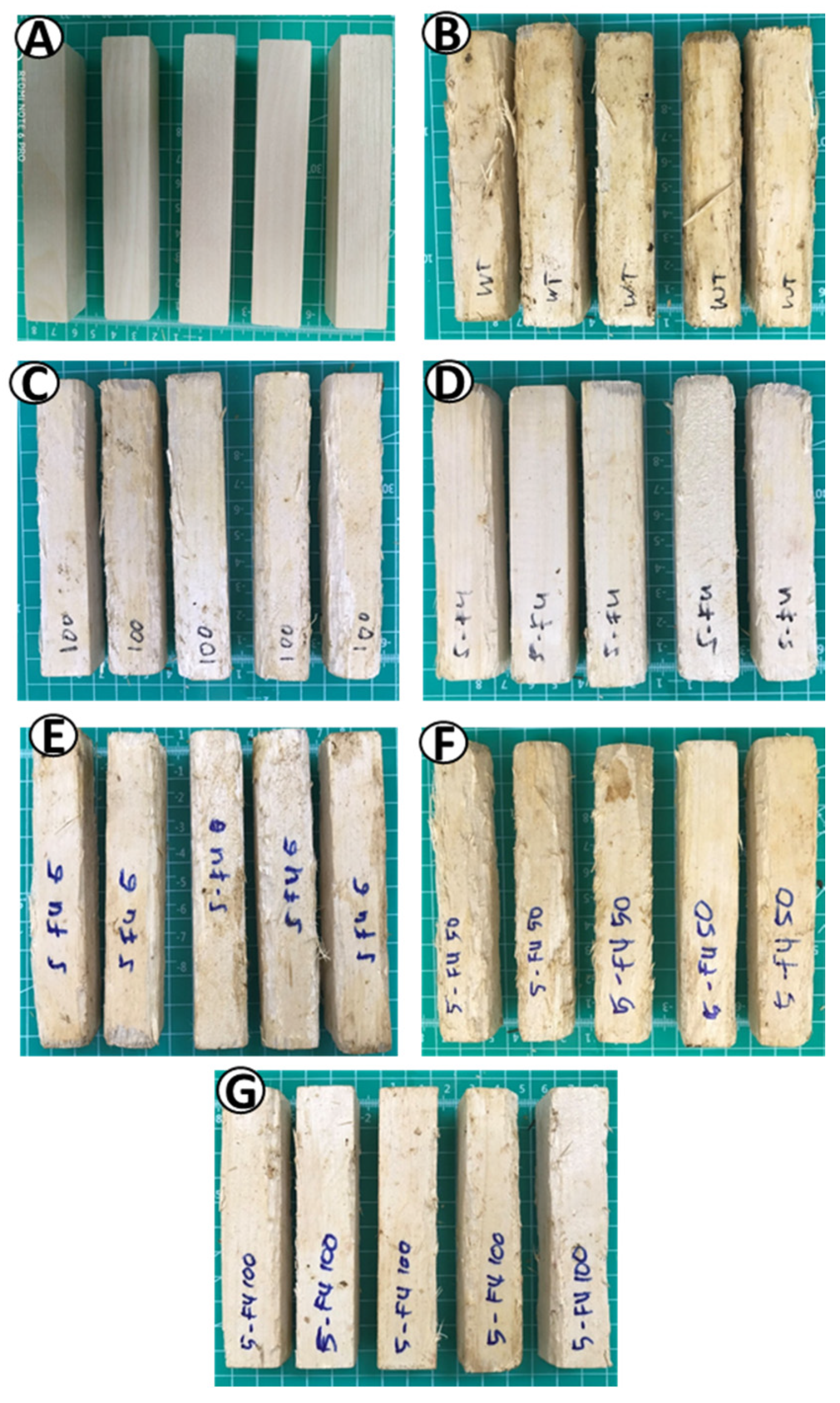
3.3. Wood Appearances

Figure 4 presented the wood appearances in six experimental groups in the end of the study. Wood blocks put in the cage of the WT group showed bite marks surrounding the block area and there were several attached pieces of woods in the cage. The woods looked yellow and contained saliva, and they were darker compared to those in other groups. In the 100-only, 5-FU+G, 5-FU+50 and 5-FU+100 groups also showed bite marks surrounding the blocks. However, the 5-FU group only showed bite marks in both side tips of the blocks. Blocks in the 5-FU group look whiter than those in other groups. From the appearances of blocks in rat cages, rats in the 5-FU group bit the blocks the least, and then the 5-FU+G, 5-FU-50, 5-FU-100, 100-only groups, and the WT group, in order.

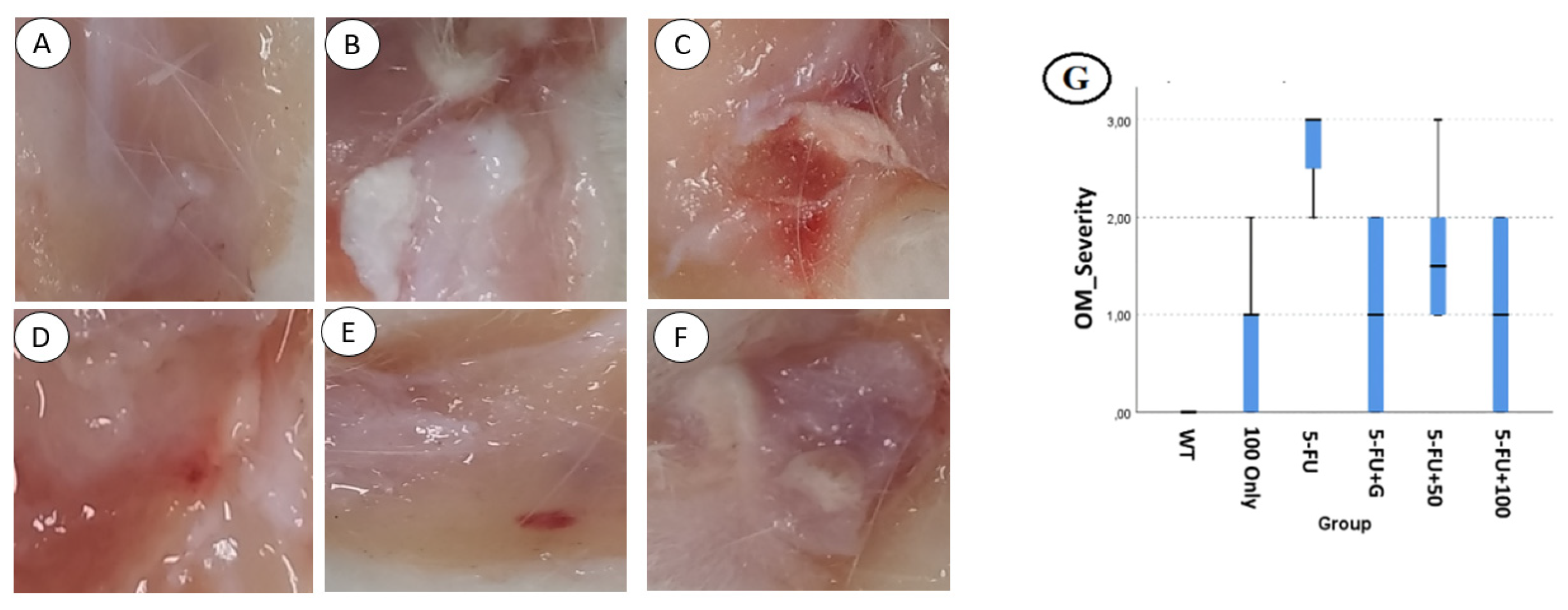
3.4. OM Clinical Appearances

Figure 5 showed the clinical features of oral skin (right buccal mucosa) of a representative rat from each group. The clinical evaluation from the WT group performed normal healthy mucosa. The features were pink and absence of erythema or ulcer (A). The features of rats in the 100-only group were pink and more likely dark compared to those in the WT group (B). The extensive erythema was clinically showed in the 5-FU group (C). Slightly erythema was found in the 5-FU+50 group (E). Moreover, both the 5-FU+G (D) and 5-FU+100 (F) groups showed no erythema or ulcer.
The bar charts of OM severities in rats of the six experimental groups in the end of the study were analyzed (Figure 5G). Rats in the 5-FU group developed OM to score 3 (presence of ulcer) or score 2 (presence of extensive erythema). Rats in the 5-FU+G group showed reduces in the severities of OM. Three rats in the 5-FU+G group had developed OM to score 1 (presence of slightly erythema) and two rats to score 2. Development of OM into score 3 in the 5-FU+G group was not found. OM severities were slightly lower in the 5-FU+50 group compared to the 5-FU group. The identical decline of OM severities was showed in rats of the 5-FU+100 group compared to both the 5-FU+G and 5-FU+50 groups. No OM to score 3 was found in the 100-only group. There was one rat with no OM, two rats with slightly erythema, and two rats with extensive erythema in this group.

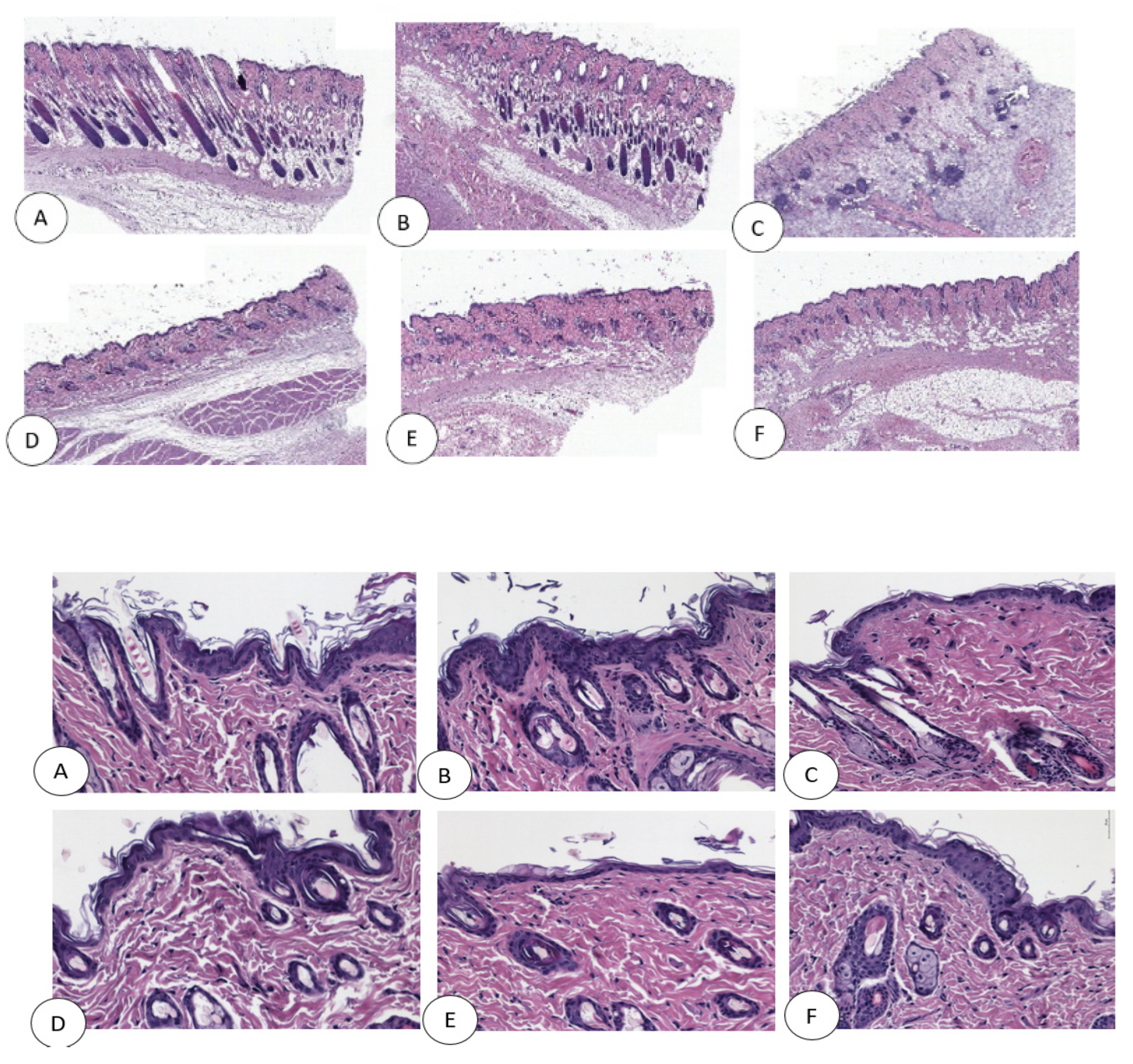
3.5. Histopathology of Oral Mucosa

Results of histopathological analyses (Figure 6) showed that oral mucosa were normal (absence of inflammation) in the WT group, and few epithelial changes found in the 100-only group showed. Severe epithelial changes with the loss of epithelium and depletion of stratum corneum (keratinized layer) were observed in the 5-FU group. The depletion of stratum corneum was also found in the 5-FU+50 group. The alteration in the basal cell layer without the disruption of the keratinized layer, fibroblast and leucocyte proliferation was observed in both the 5-FU+G and 5-FU+100 groups.

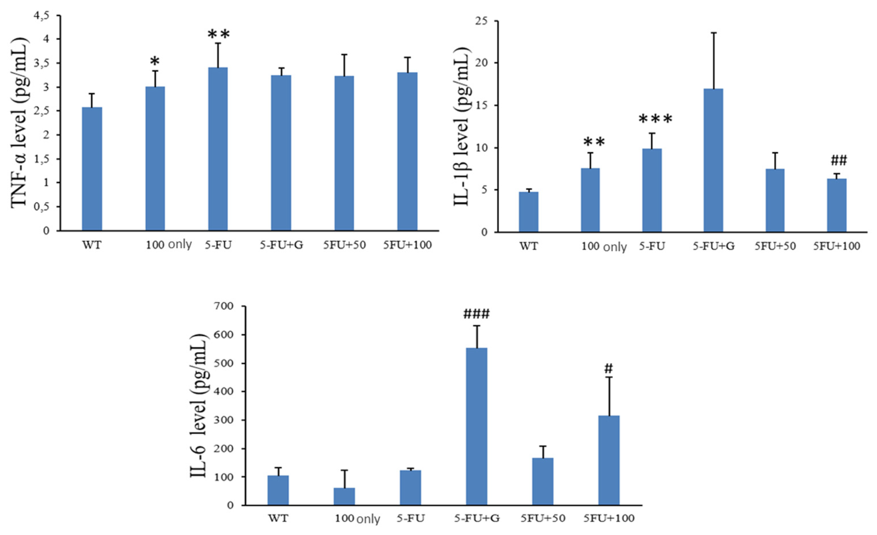
3.6. Serum Proinflammatory Cytokines

Serum concentrations of the major proinflammatory cytokines in rats have been assessed (Figure 7). TNF-α levels of the 100-only and 5-FU groups were significantly higher than the WT group (p < 0.05). IL-1β levels of the 100-only and 5-FU groups were significantly higher than the WT group (p < 0.01). Only the 5-FU+G group showed significant increases in IL-6 levels compared to the WT group. Furthermore, both IL-1β and IL-6 levels in the 5-FU+100 group showed significant differences with those of the 5-FU group. IL-1β level of the 5-FU+100 group was significantly lower compared to the 5-FU group (p < 0.01) and IL 6 level of the 5-FU+100 group was significantly higher compared to the 5-FU group (p < 0.05). The 5-FU+G group showed dramatic increases in IL-6 levels and increases in IL-1β levels compared to the 5-FU group (p < 0.001). Both IL-1β and IL-6 levels in rat serums showed dose-dependent effects in the 5-FU+50 and 5-FU+100 groups. These results indicated that 5-FU treatment significantly increased TNF-α and IL-1β levels but not IL-6 levels in rat serums. Furthermore, only treatment with 100 mg/mL CO extracts (5-FU+100) could significantly reduce the 5-FU-induced IL-1β levels. However, both treatments with 1 mg/kg Triamcinolone acetonide orobase (5-FU+G) and 100 mg/mL CO extracts (5-FU+100) significantly increased IL-6 levels compared to the 5-FU group.

3.7. Mortality Rates

In the normal control group (WT), all animals survived with body weight increases during the entire study period. In the OM group (5-FU), rats showed obvious body weight losses and 33.3% mortality was observed on the 10th day. In the Triamcinolone acetonide treated group (5-FU+G), rats still showed obvious body weight losses and one rat died on the sixth day with 16.7% mortality. Although rats still showed limited body weight losses, no rat death was observed in both the 5-FU+50 and 5-FU+100 groups during the entire study period.
Figure 8 showed the clinical finding of rats treated with 5-FU only. We observed some abnormal changes after 5-FU administration at day 6 and 10. Some rats showed “old-looking” bodies with the bent down appearance from the side view and the rat’s hair looking dull. The area around their eyes looked reddish which may have included scarring and periorbital edema. In the dead rat, we dissected the rat’s body and found colon edema, damage the in kidneys and dark lungs.

4. Discussion

In this study, we demonstrated that CO leaf extract had a therapeutic effect against 5-FU-induced OM in rats. The results of the oral clinical features of the 5-FU+100 and 5-FU+G groups showed a healing process of OM on buccal mucosa. Our finding corroborates previous study that reported the potential remedy of CO leaves as tyrosinase suppressor, which could exhibit notable effect on wound healing [13]. The wound healing effect of CO leaf extract was also observed histopathologically. Presence of hemorrhagic and the depletion of keratinized layer induced by 5-FU was significantly reduced after CO leaf extract treatment, and the absence of hemorrhagic was also showed after Triamcinolone acetonide treatment.
Previous study reported that proanthocyanidins, one of cinnamon’s chemicals, played an important role in declining two proinflammatory cytokines, IL-6 and IL-8, which assist as considerable cytokines in the OM development [11]. On the contrary, our study found the increase of serum IL-6 after not only 100 mg/mL CO leaf extract but also 0.1% Triamcinolone acetonide treatments. The decrease of serum IL-1β level after CO leaf extract treatments explained its OM alleviation effect. TNF-α and IL-1β are suggested to play key roles in OM development [6]. IL-1β inhibition by CO leaf extract treatments in this study also supports previous study that reported the anti-inflammatory effect via declining IL-1β [8]. In contrast, our study found the different results from previous study which reported cinnamaldehyde, one of CO constituents, inhibited the production of IL-6 [14]. The finding that IL-6 levels were increased after CO leaf extract and Triamcinolone acetonide treatments demonstrate the influence of therapy in serum IL-6. It suggests the future study to determine IL-6 levels in gingival crevicular fluid (GCF). IL-6 in GCF of periodontal diseases were inhibited after scaling and root canal therapy combined with laser treatment [15]. IL-6 is a main modulator of both of the inflammatory and healing processes including in the differentiation, stimulation and propagation of leukocytes, endothelial cells, keratinocytes and fibroblasts [16]. The initiation of the acute stage response by IL-6 has been observed as part of an attempt to uphold homeostasis. A previous review explained that IL-6 seems to contribute to hepatocyte propagation and initiation of protecting pathways [17]. The increase of IL-6 in the 5-FU+100 and 5-FU+G groups may mediate the healing process of OM through proliferation of leukocytes, keratinocytes and fibroblasts, which observed in histopathologic assessment.
Results of nonparametric analyses showed that 0.1% Triamcinolone acetonide and 100 mg/mL CO had therapeutic effects on 5-FU-induced OM. This study supports previous study that considered the therapy effect of Triamcinolone acetonide to reduce the mucositis score in the patients with OM [18]. Results of this study reported that 50 mg/mL CO did not show a significant therapeutic effect as 100 mg/mL CO in 5-FU-induced OM. This finding suggests a dosage-dependent manner of CO leaf extract used in OM therapy.
The presence of OM caused the difficulty in biting and chewing that reduced the body weight of rats during the experiment [19,20] and decreased the oral health quality of life [2,4]. On the first day and the end of experiments, all rats were weighed and compared their body weight changes after treatments. Our finding shows significant eliminating effects on loses of body weight, food and water intakes induced by 5-FU, a common chemotherapy agent. The effects of 5-FU in losses of food intake and body weight in this study support a previous animal study stating that methotrexate-induced OM caused similar effects [21]. The weight-loss effect can be explained by the reduces of food intake and water intake in rats of the 5-FU group. Previous panel data analysis reported weight loss as the side effects in patients who received chemotherapy for lung cancer or mesothelium [21]. Moreover, weight losses were also found in rats with 5-FU administrations and then treated topically using both Triamcinolone acetonide and CO. We also analyzed the differences in weight losses between rats with 5-FU administration only and 5-FU administration with topical interventions. Our findings demonstrate that the topical interventions using 50 mg/mL and 100 mg/mL CO may protect the OM model rats from significant weight loss.
The results showed that chemotherapy of 5-FU treatment reduced the water intake of the rats. However, the topical treatments in buccal mucosa using 0.1% Triamcinolone acetonide, 50 mg/mL CO and 100 mg/mL CO increased water intakes after 5-FU administration. We also found that rats giving only 100 mg/mL CO treatment (100-only) had less food intake and similar water intake than the WT rats. Moreover, the body weight increases were similar in rats of these two groups and no significant difference was obtained.
Our findings showed other side effects of 5-FU chemotherapy in rats. Some rats with 5-FU administrations were observed with periorbital inflammation, intestinal organs failure, and abnormal body appearance. A previous review about side effects of 5-FU treatment, which was used for such malignancies as breast and gastrointestinal cancer therapy, described that 5-FU may cause myelosuppression, nausea and stomatitis. Ocular side effects were not infrequent and the most common of which was increased lacrimation [22]. Patients receiving chemotherapy such as imatinib mesylate appeared to be caused in part by periorbital oedema [23]. Moreover, gastrointestinal side effects, particularly with high-dose administration of chemotherapy, may cause gingivitis, pharyngitis, stomatitis, anorexia, hematemesis, melena, gastrointestinal ulceration and bleeding. In rare cases, chemotherapy using methotrexate may induce colitis and toxic megacolon [24]. This study reports the mortalities of 5-FU administration on the day 6 and day 10. In line with the result, previous study reported mortality rate which was determined during the study period of 12–15 days in animal mucositis control [13]. The in vitro investigation showed that CO had antioxidant and antibacterial activities against several bacterial strains associated with OM. The results of our animal study showed the effect of CO leaf extract in alleviation of OM. The findings of an in vitro study and our in vivo animal study corroborate several previous studies related to natural agents and confirm the mechanisms for OM’s protection are antibacterial activity, antioxidant activity, modulation of inflammation and wound-healing activity [11,25,26,27,28,29,30]. To the best of our knowledge, this is the first study investigating the potential use of CO, a hardwood forest product in Taiwan, in alleviating 5-FU-induced OM.

5. Conclusions

Our investigations showed the therapeutic effect of CO leaf extract on OM model rats induced by 5-FU in vivo. The finding demonstrated the significant differences on OM development, food intakes, water intakes, body weight changes between OM control rats (5-FU) and OM rats that treated with CO leaf extract, especially with 100 mg/mL. Thickening of the stratum corneum confirmed the clinical outcome of OM healing in which extensive erythema was not found in rats of CO-treated groups. The inhibition of a major proinflammatory cytokine, IL-1β, explained the therapeutic effect of CO leaf extract and reported the potential remedy of CO intervene the second stage of OM. The increase of IL-6 may mediate the healing process of OM through proliferation of leukocytes, keratinocytes and fibroblasts. This report is a pilot study, and further studies may provide the identification of specific chemical constituent of CO leaf extract that plays a role in alleviation of OM, especially in human patients.

Author Contributions

Author Contributions: A.B., S.-C.L., and M.-S.T. conceived and designed the work; A.B., V.N., M.-S.T., and C.-T.L. collected the data (handled experimental procedures); M.-S.T., S.-C.L., and S.-H.W. verified the analytical methods; A.B., C.-W.H. and S.-H.W. analyzed the findings of this work; A.B., S.-H.W., C.-W.H. and M.-S.T. organized the paper. All authors discussed the results and contributed to the final manuscript. All authors have read and agreed to the published version of the manuscript.

Funding

This work was supported in part by the Ministry of Science and Technology of Taiwan (MOST 107-2628-B-005-002-MY3). Histopathology analysis was performed by TissueFAXS system (Tissue Gnostics, Austria) in the Instrument Center of Chung Shan Medical University, which is supported by the Ministry of Science and Technology, the Ministry of Education, and Chung Shan Medical University. The funders had no role in the study design, data collection and analysis, decision to publish, or the preparation of the manuscript.

Institutional Review Board Statement

The study was conducted according to the guidelines of the Declaration of Helsinki, and approved by the Institutional Review Board (or Ethics Committee) of Da-Yeh University (protocol number: 109004).

Informed Consent Statement

Not applicable.

Data Availability Statement

The data presented in this study are available on request from the corresponding author.

Acknowledgments

We acknowledge to Dwieyanto Aryo Pangestu, Stefanie Azaria S, Kristina Yuliani and Fachri Yafelli who helped us in animal experiment.

Conflicts of Interest

The authors declare no conflict of interest.

References

  1. Raber-Durlacher, J.E.; Elad, S.; Barasch, A. Oral mucositis. Oral Oncol. 2010, 46, 452–456. [Google Scholar] [CrossRef]
  2. Elting, L.S.; Keefe, D.M.; Sonis, S.T.; Garden, A.S.; Spijkervet, F.K.L.; Barasch, A.; Tishler, R.B.; Canty, T.P.; Kudrimoti, M.K.; Vera-Llonch, M. Patient-reported measurements of oral mucositis in head and neck cancer patients treated with radiotherapy with or without chemotherapy: Demonstration of increased frequency, severity, resistance to palliation, and impact on quality of life. Cancer 2008, 113, 2704–2713. [Google Scholar] [CrossRef]
  3. Elting, L.S.; Cooksley, C.D.; Chambers, M.S.; Garden, A.S. Risk, Outcomes, and Costs of Radiation-Induced Oral Mucositis among Patients with Head-and-Neck Malignancies. Int. J. Radiat. Oncol. Biol. Phys. 2007, 68, 1110–1120. [Google Scholar] [CrossRef]
  4. Murphy, B.A.; Beaumont, J.L.; Isitt, J.; Garden, A.S.; Gwede, C.K.; Trotti, A.M.; Meredith, R.F.; Epstein, J.B.; Le, Q.T.; Brizel, D.M.; et al. Mucositis-Related Morbidity and Resource Utilization in Head and Neck Cancer Patients Receiving Radiation Therapy With or Without Chemotherapy. J. Pain Symptom Manag. 2009, 38, 522–532. [Google Scholar] [CrossRef] [PubMed]
  5. Sonis, S.T. The pathobiology of mucositis. Nat. Rev. Cancer 2004, 4, 277–284. [Google Scholar] [CrossRef]
  6. Sonis, S.T.; Hospital, W. Pathobiology of Oral Mucositis: Novel Insights and Opportunities. J. Supportive Oncol. 2007, 5, 3–11. [Google Scholar]
  7. Bakar, A.; Yao, P.-C.; Ningrum, V.; Liu, C.-T.; Lee, S.-C. Beneficial Biological Activities of Cinnamomum osmophloeum and Its Potential Use in the Alleviation of Oral Mucositis: A Systematic Review. Biomedicines 2020, 8, 3. [Google Scholar] [CrossRef] [Green Version]
  8. Lee, S.C.; Xu, W.X.; Lin, L.Y.; Yang, J.J.; Liu, C.T. Chemical composition and hypoglycemic and pancreas-protective effect of leaf essential oil from indigenous cinnamon (Cinnamomum osmophloeum Kanehira). J. Agric. Food Chem. 2013, 61, 4905–4913. [Google Scholar] [CrossRef] [PubMed]
  9. Tung, Y.T.; Huang, C.C.; Ho, S.T.; Kuo, Y.H.; Lin, C.C.; Lin, C.T.; Wu, J.H. Bioactive phytochemicals of leaf essential oils of Cinnamomum osmophloeum prevent lipopolysaccharide/d -galactosamine (LPS/d -GalN)-induced acute hepatitis in mice. J. Agric. Food Chem. 2011, 59, 8117–8123. [Google Scholar] [CrossRef]
  10. Molania, T.; Moghadamnia, A.A.; Pouramir, M.; Aghel, S.; Moslemi, D.; Ghassemi, L.; Motallebnejad, M. The effect of Cinnamaldehyde on mucositis and salivary antioxidant capacity in gamma-irradiated rats (a preliminary study). DARU J. Pharm. Sci. 2012, 20, 1–5. [Google Scholar] [CrossRef] [Green Version]
  11. Veilleux, M.P.; Grenier, D. Determination of the effects of cinnamon bark fractions on Candida albicans and oral epithelial cells. BMC Complement. Altern. Med. 2019, 19, 303. [Google Scholar] [CrossRef] [Green Version]
  12. Calapai, G.; Miroddi, M.; Mannucci, C.; Minciullo, P.L.; Gangemi, S. Oral adverse reactions due to cinnamon-flavoured chewing gums consumption. Oral Dis. 2014, 20, 637–643. [Google Scholar] [CrossRef]
  13. Lee, M.G.; Kuo, S.Y.; Yen, S.Y.; Hsu, H.F.; Leung, C.H.; Ma, D.L.; Wen, Z.H.; Wang, H.M.D. Evaluation of Cinnamomum osmophloeum kanehira extracts on tyrosinase suppressor, wound repair promoter, and antioxidant. Sci. World J. 2015, 2015, 7. [Google Scholar] [CrossRef] [Green Version]
  14. Chao, L.K.; Hua, K.F.; Hsu, H.Y.; Cheng, S.S.; Lin, I.F.; Chen, C.J.; Chen, S.T.; Chang, S.T. Cinnamaldehyde inhibits pro-inflammatory cytokines secretion from monocytes/macrophages through suppression of intracellular signaling. Food Chem. Toxicol. 2008, 46, 220–231. [Google Scholar] [CrossRef] [PubMed]
  15. Calderín, S.; García-Núñez, J.A.; Gómez, C. Short-term clinical and osteoimmunological effects of scaling and root planing complemented by simple or repeated laser phototherapy in chronic periodontitis. Lasers Med. Sci. 2013, 28, 157–166. [Google Scholar] [CrossRef] [PubMed] [Green Version]
  16. Johnson, B.Z.; Stevenson, A.W.; Prêle, C.M.; Fear, M.W.; Wood, F.M. The role of IL-6 in skin fibrosis and cutaneous wound healing. Biomedicines 2020, 8, 101. [Google Scholar] [CrossRef]
  17. Streetz, K.L.; Luedde, T.; Manns, M.P.; Trautwein, C. Interleukin 6 and liver regeneration. Gut 2000, 47, 309–312. [Google Scholar] [CrossRef] [PubMed] [Green Version]
  18. Pakravan, F.; Ghalayani, P.; Emami, H.; Isfahani, M.; Noorshargh, P. A novel formulation for radiotherapy-induced oral mucositis: Triamcinolone acetonide mucoadhesive film. J. Res. Med. Sci. 2019, 24, 63. [Google Scholar] [PubMed]
  19. Vissink, A.; Jansma, J.; Spijkervet, F.K.L.; Burlage, F.R.; Coppes, R.P. Oral sequelae of head and neck radiotherapy. Crit. Rev. Oral Biol. Med. 2003, 14, 199–212. [Google Scholar] [CrossRef]
  20. Patel, A.; Biswas, S.; Shoja, M.H.; Ramalingayya, G.V.; Nandakumar, K. Protective effects of aqueous extract of Solanum nigrum Linn. leaves in rat models of oral mucositis. Sci. World J. 2014, 2014, 345939. [Google Scholar] [CrossRef] [PubMed]
  21. Ross, P.J.; Ashley, S.; Norton, A.; Priest, K.; Waters, J.S.; Eisen, T.; Smith, I.E.; O’Brien, M.E.R. Do patients with weight loss have a worse outcome when undergoing chemotherapy for lung cancers? Br. J. Cancer 2004, 90, 1905–1911. [Google Scholar] [CrossRef]
  22. Vizel, M.; Oster, M.W. Ocular Side Effects of Cancer Chemotherapy. Cancer 1982, 49, 1999–2002. [Google Scholar] [CrossRef] [Green Version]
  23. Esmaeli, B.; Diba, R.; Ahmadi, M.A.; Saadati, H.G.; Faustina, M.M.; Shepler, T.R.; Talpaz, M.; Fraunfelder, R.; Rios, M.B.; Kantarjian, H. Periorbital oedema and epiphora as ocular side effects of imatinib mesylate (Gleevac). Eye 2004, 18, 760–762. [Google Scholar] [CrossRef]
  24. Boussios, S.; Pentheroudakis, G.; Katsanos, K.; Pavlidis, N. Systemic treatment-induced gastrointestinal toxicity: Incidence, clinical presentation and management. Ann. Gastroenterol. 2012, 25, 106–118. [Google Scholar] [PubMed]
  25. Hawley, P.; Hovan, A.; Mcgahan, C.E. A randomized placebo-controlled trial of manuka honey for radiation-induced oral mucositis. Supportive Care Cancer 2014, 22, 751–761. [Google Scholar] [CrossRef] [PubMed]
  26. Miranzadeh, S.; Adib-hajbaghery, M.; Soleymanpoor, L.; Ehsani, M. A New mouthwash for Chemotherapy Induced Stomatitis. Nurs. Midwifery Stud. 2014, 3, e20249. [Google Scholar] [CrossRef] [Green Version]
  27. Tiemann, P.; Toelg, M.; Ramos, F.M.H. Administration of Ratanhia-based herbal oral care products for the prophylaxis of oral mucositis in cancer chemotherapy patients: A clinical trial. Evid Based Complement. Altern. Med. 2007, 4, 361–366. [Google Scholar] [CrossRef]
  28. Aoyama, T.; Nishikawa, K.; Takiguchi, N.; Tanabe, K. Double-blind, placebo-controlled, randomized phase II study of TJ-14 (hangeshashinto) for gastric cancer chemotherapy-induced oral mucositis. Cancer Chemother Pharm. 2014, 73, 1047–1054. [Google Scholar] [CrossRef] [Green Version]
  29. Rashad, U.M.; Al Gezawy, S.M.; El Gezawy, E.; Azzaz, A.N. Honey as topical prophylaxis against radiochemotherapy-induced mucositis in head and neck cancer Main Article Honey as topical prophylaxis against radiochemotherapy-induced mucositis in head and neck cancer. J. Laryngol. Otol. 2009, 123, 223–228. [Google Scholar] [CrossRef] [PubMed]
  30. Baharvand, M.; Jafari, S.; Mortazavi, H. Herbs in oral mucositis. J. Clin. Diagn. Res. 2017, 11, ZE05–ZE11. [Google Scholar] [CrossRef]
Figure 1. The animal protocol in this study.
Processes 09 00615 g001
Figure 2. The body weight changes (before and after treatment) in six experimental rat groups. WT: rats injected with the same volume of PBS as the control animals; 100-only: rats treated with 100 mg/mL CO extracts only (100-only); 5-FU: rats injected with 5-FU only; 5-FU+G: rats injected with 5-FU then treated with 1 mg/kg Triamcinolone acetonide orobase as a positive control; 5-FU+50: rats injected with 5-FU then treated with 50 mg/mL CO extract; 5-FU+100: rats injected with 5-FU then treated with 100 mg/mL CO extract. All values are expressed as mean ± SD of each group. *: p < 0.05 versus the WT group and #: p < 0.05 versus the 5-FU group.
Processes 09 00615 g002
Figure 3. The food intakes (A) and water intakes (B) per rat in six experimental groups during treatments. All values are expressed as mean ± SD in each group. *: p < 0.05 versus the WT group and #: p < 0.05 versus the 5-FU group.
Processes 09 00615 g003
Figure 4. Images of wood blocks at the beginning and end of the experiment for the six experimental groups. (A) The wood blocks at the beginning of the experiment. Images of wood pieces in WT group (B), 100-only group (C), 5-FU group (D), 5-FU+G group (E), 5-FU+50 group (F), and 5-FU+100 group (G).
Processes 09 00615 g004
Figure 5. The representative oral buccal mucosa clinical features of rats in each experimental group after sacrifice. Images from WT group (A), 100-only group (B), 5-FU group (C), 5-FU+G group (D), 5-FU+50 group (E), and 5-FU+100 group (F). (G): Box and whisker chart of independent-samples Kruskal–Wallis test (nonparametric test) of OM severities in the six experimental groups. All values are expressed as mean ± SD of each group.
Processes 09 00615 g005
Figure 6. Representative histopathological images of skin sections from oral buccal mucosa of rats in the six experimental groups. (A): WT group; (B): 100-only group; (C): 5-FU group; (D): 5-FU+G group; (E): 5-FU+50 group; (F): 5-FU+100 group. Skin sections were hematoxylin and eosin (H&E) stained. The upper region shows images with 200 times magnification; the lower region shows enlarged images of 5 μm.
Processes 09 00615 g006
Figure 7. The serum levels of TNF-α, IL-1β, and IL-6 in the six experimental groups. *, **, and *** show significant differences with the WT group with p <0.05, p <0.01, and p <0.001, respectively. #, ##, and ### show significant differences with the 5-FU group with p <0.05, p <0.01, and p <0.001, respectively.
Processes 09 00615 g007
Figure 8. The clinical findings of rat eye, mouth, body shape, and organs on the sixth day and tenth day of experiment. (A) The appearance of a rat in the5-FU+100 group like rats in the WT group; (B) the appearance (arched back) of a rat in the 5-FU group; (C) the perioral edema of a rat found in the 5-FU group; (D) the periocular tissue changes including scarring and periorbital edema in a rat of the 5-FU group; (E) the colon size and intestinal organs in a rat of the WT group; (F) the abnormal enlargement of colon (megacolon), kidney rupture, and the dark lung found in a dead rat in the 5-FU+G group.
Processes 09 00615 g008
Publisher’s Note: MDPI stays neutral with regard to jurisdictional claims in published maps and institutional affiliations.

