Article

Development of Alkaline Reduced Water Using High-Temperature-Roasted Mineral Salt and Its Antioxidative Effect in RAW 264.7 Murine Macrophage Cell Line

1
Department of Environmental Medical Biology, Wonju College of Medicine Yonsei University, Wonju 26426, Korea
2
Department of Global Medical Science, Wonju College of Medicine, Yonsei University, Wonju 26426, Korea
3
Department of Laboratory Medicine, Wonju College of Medicine, Yonsei University, Wonju 26426, Korea
4
BIOCERA Section, Biocera Co., Ltd., Seongnam-Si 13488, Korea
5
Hydrogen Fuel Cell Parts & Applied Technology Regional Innovation Center, Woosuk University, Jeonju 55315, Korea
*
Author to whom correspondence should be addressed.
These authors contributed equally to this study and share first authorship.
Processes 2021, 9(11), 1928; https://doi.org/10.3390/pr9111928
Submission received: 29 September 2021 / Revised: 19 October 2021 / Accepted: 26 October 2021 / Published: 28 October 2021
(This article belongs to the Section Biological Processes and Systems)

Abstract

:
Oxidative stress (OS) plays an important role in many diseases, and its excessive increase affects human health. Although the antioxidant effect of sea salt can be strengthened through special processing, it is scarcely studied. This study confirmed the antioxidative effect of high-temperature roasted mineral salt (HtRMS) produced through repeated roasting of sea salt at high temperature in a ceramic vessel. The dissolved HtRMS exhibited properties such as high alkalinity, rich mineral content, and a high concentration of hydrogen (H2). To detect the antioxidative effect of HtRMS, OS was induced in RAW 264.7 murine macrophage cells with hydrogen peroxide (H2O2) and lipopolysaccharide (LPS), and then treated with HtRMS solution at different concentrations (0.1, 1, and 10%). Cell viability, reactive oxygen species (ROS), nitric oxide (NO), and antioxidant enzymes such as catalase (CAT) and glutathione peroxidase (GPx), Ca2+, and mitogen-activated protein kinase (MAPK) pathway-related proteins (p-p38, p-JNK, and p-ERK) were measured. OS was significantly induced by treatment with H2O2 and LPS (p < 0.001). After treatment with HtRMS, cell viability and GPx activities significantly increased and ROS, NO, Ca2+, and CAT significantly decreased in a concentration-dependent manner compared to H2O2 and LPS-only groups, which was not observed in tap water (TW)-treated groups. Similarly, p-p38, p-JNK, and p-ERK levels significantly decreased in a concentration-dependent manner in HtRMS groups compared to both H2O2 and LPS-only groups; however, those in TW groups did not exhibit significant differences compared to H2O2 and LPS-only groups. In conclusion, our results suggest that HtRMS may have antioxidant potential by regulating the MAPK signaling pathway.

1. Introduction

Oxidative stress (OS) represents a pathological imbalance between the antioxidant system and the production of reactive oxygen species (ROS) [1]. ROS, a by-product of metabolism, can participate in some important physiological functions, such as cell signaling, immune functionality, and response to pathogens [2,3]; however, excessive ROS presence leads to cell and tissue injury, leading to OS-related pathological processes [4]. It is also known that various stimulants such as hydrogen peroxide (H2O2) and lipopolysaccharide (LPS) can activate ROS-induced signaling pathways [5,6], and that the OS process is modulated by the mitogen-activated protein kinase (MAPK) pathway, which is closely linked to cell proliferation [5].
To combat the detrimental effects of OS-related disorders, various studies and attempts are continuously being conducted, particularly in the nutritional field. This includes several in vivo and in vitro studies on the positive therapeutic effects of mineral salts, such as bamboo salt [7,8]. Cumulative evidence has shown that trace elements, such as calcium (Ca2+), potassium (K+), phosphorous (P3−), and sulfur (S2−), exert antioxidative effects on murine macrophage cell lines [7,8,9]. Todorka et al. reported that minerals can help control the OS response [10]. Consistent with this, a new concept of mineral salt was developed: the high-temperature-roasted mineral salt (HtRMS) (BIOCERA Co., Ltd., Seongnamsi, Geonggido, Korea), rich in 25 essential minerals and hydrogen (H2), with low oxidation-reduction potential (ORP) values and high alkalinity when dissolved in tap water (TW) [11], is expected to have beneficial effects.
HtRMS contains abundant trace elements such as K (7838 ppm), S (4220 ppm), Ca (1722 ppm), Mg (764 ppm), P (122 ppm), Cl (56.91%), Na (37.94%), Sr (67.8 ppm), I (67 ppm), Si (45.6 ppm), Fe (26.2 ppm), B (18.2 ppm), Li (14.2 ppm), and Be (14.2 ppm). These mineral ions have central roles in cell functions such as cell viability, DNA synthesis, ROS reduction and antioxidative effects [7,8,9]. S2− typically participates in inflammation and OS and supports the antioxidant system through the formation of glutathione peroxide (GPx) [10]. When dissolved in TW, HtRMS also releases abundant H2, which has known therapeutic potential for OS-related diseases, and has been clinically applied in countries such as Japan, China, the USA, and Korea [12,13,14,15]. H2 reportedly exhibits high antioxidant efficiency in medicine [16], likely by inducing an increase in antioxidant enzymes, such as catalase (CAT), superoxide dismutase (SOD), and heme-oxygenase-1 (HO-1) [2]. H2 was experimentally administered in many ways such as drinking, inhaling, injecting, through eye drops, skin smears, and baths [17]. H2 can be produced from the reaction between water and particular compounds such as Mg2+ and Ca2+, as well as through water electrolysis. HtRMS is completely dissolved in TW and simultaneously produces H2 through an electrochemical reaction with H2O. In addition, the dissolved HtRMS increases the alkalinity of the solution according to a principle similar to that of water electrolysis. The bio-effects of high alkalinity have been extensively studied and reportedly have relevant antioxidative effects [18,19].
Macrophages are key factors in infections and injuries. They are ubiquitous, phenotypically heterogeneous, and have complex functions, including their role in innate and acquired immunity [20], extracellular stimulation, and intracellular responses related to the MAPK signaling pathway [21], which includes phospho-p38 (p-p38), phospho-c-Jun amino-terminal kinases (p-JNK), and phospho-extracellular signal-regulated kinases (p-ERK). These enzymes have diverse biological functions, including nucleosome regulation, gene expression, mRNA stabilization and translation, and cell proliferation and survival [22].
In this study, the antioxidative effect of a fortified alkaline water was tested using HtRMS, which was produced through repeated roasting at a high temperature, in a murine macrophage cell line exposed to different stress stimulants, such as H2O2 and LPS, and compared with TW effects. We also analyzed the potential mechanism of action of HtRMS by examining its effects on the MAPK signaling pathway.

2. Materials and Methods

2.1. Experimental Materials

Dulbecco’s modified Eagle’s medium (DMEM) was purchased from Cytiva Hyclone (South Logan, OH, USA). The penicillin/streptomycin (antibiotics) solution was purchased from GibcoTM (Invitrogen Corporation, Auckland, NY, USA); fetal bovine serum (FBS) was purchased from Hyclone Laboratories, Inc. (South Logan, OH, USA). LPS (serotype O111:B4) was purchased from Sigma-Aldrich (St. Louis, MO, USA), and 30% H2O2 was purchased from Daejung (Siheung-si, Gyeonggi-do, Korea). A Cell Counting kit-8 (CCK-8) was purchased from Quanti-MaxTM. The CCK-8 assay kit was purchased from Dojindo Molecular Technologies (Rockville, MD, USA); 2′7′-dicholodihydrofluorescein diacetate (DCFH-DA) reagent was purchased from Sigma Chemical Co. (Sigma, St. Louis, MO, USA); nitric oxide (NO) reagent (Griess reagent kit) was purchased from iNtRON Biotechnology (Sungnam, South Korea); a CAT assay kit, GPx assay kit, and Ca2+ colorimetric assay kit was obtained from BioVision Inc. (Milpitas, CA, USA). The TakaRa BCA protein assay kit was obtained from Takara Bio Inc. (Shimadzu, Tokyo, Japan). Antibodies recognizing phospho-p38 (p-p38), phospho-c-Jun N-terminal kinase (p-JNK), phospho-extracellular signal-related kinase (p-ERK) 1 and 2 and β-actin (dilution 1:2000) were rabbit monoclonal IgM, whereas secondary antibodies (dilution 1:5000) were horseradish peroxidase-linked anti-rabbit IgG, all obtained from Cell Signaling Technology (Danvers, MA, USA).

Mineral Contents and Properties of HtRMS

The experimental material, HtRMS (Hydrogen A.A Mineral Salt, BIOCERA Co., Ltd., Seongnamsi, Geonggido, Korea), is water soluble and was produced through a high-temperature roasting process at 750–800 °C for 30 min in a bio-ceramic jar (diameter 15 cm, height 15 cm, thickness 1 cm), this process was repeated 10 times and then the final salt was pulverized in the size of 0.1–0.2 mm. The bio-ceramic jar used in the experiment was made through a particular manufacturing process including a high-temperature firing process at 900–1250 °C for 1 h. The mineral content (Table 1) and properties (Table 2) of HtRMS were analyzed.

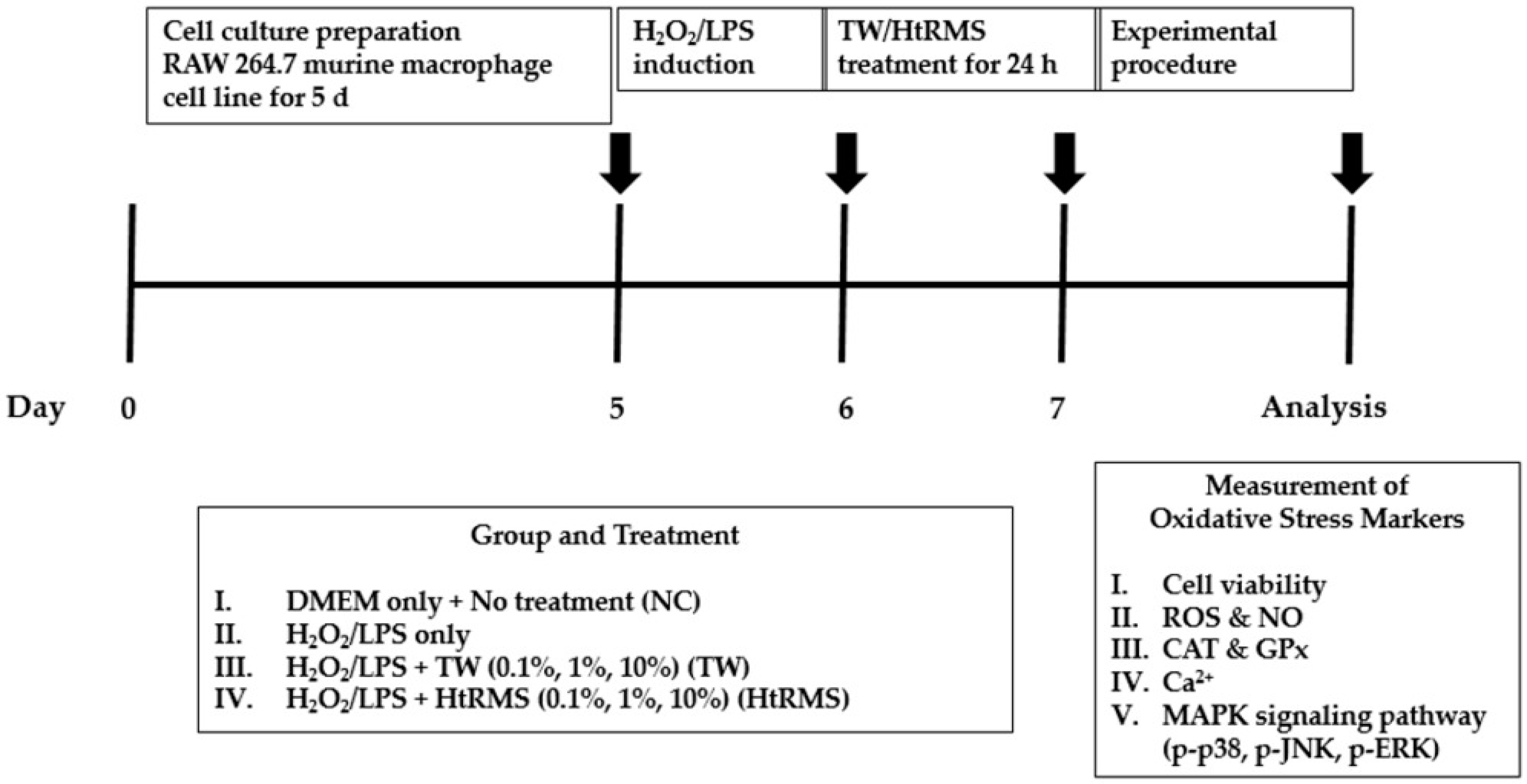
2.2. Experimental Design

RAW 264.7 murine macrophage cell line (American Type Cell Culture Collection, Manassas, VA, USA) was cultured and treated for 7 d according to the experimental protocol (Figure 1). The cells were cultured for 5 d and then treated with 200 µM/mL H2O2 for 2 h and 10 µg/mL LPS for 24 h to induce OS. The induction of OS was confirmed by detecting cell viability compared to that in untreated. OS-induced cells were treated with different concentrations (0.1, 1, and 10%) of HtRMS (HtRMS-treatment group) and TW (TW-treatment group) for 24 h. On the 7th day of the experiment, the cells were collected, and OS-related biomarkers such as ROS, NO, CAT, GPx, and Ca2+ were measured. MAPK signaling pathway proteins such as p-p38, p-JNK, and p-ERK were also evaluated via Western blotting.

2.3. Cell Culture and LPS Stimulation

To identify the ideal concentration of LPS its stimulation time, RAW 264.7, a macrophage cell line, was grown in DMEM with 10% FBS and 1% antibiotic and cultured in 75 mm flasks at 37 °C with 5% CO2 in a humidified atmosphere. RAW 264.7 cells (5 × 10³ cells/well) were used in these experiments. The cells were treated with different concentrations of LPS (0.4, 2, and 10 μg/mL) for 6, 12, 24, and 48 h after reaching 80% confluence. Cell viability was evaluated using the CCK-8 assay kit, following the manufacturer’s protocol. Finally, the results were analyzed by IC50 to select the ideal concentration and time of LPS stimulation to induce OS.

2.4. Cell Culture and H2O2 Stimulation

To identify the ideal concentration and time of H2O2 stimulation, RAW 264.7 cells (5 × 10³ cells/well) were treated with 50, 100, and 200 μM/mL of H2O2 for 30, 60, 90, and 120 min. Cell viability was measured using the CCK-8 assay kit, following the manufacturer’s protocol. Similarly, the result was analyzed by IC50 to determine the ideal concentration and time of H2O2 stimulation to induce OS.

2.5. Cell Proliferation Assay

CCK-8 was used to evaluate cell viability following the manufacturer’s protocol. Briefly, RAW 264.7 macrophage cells (1 × 10⁴ cells/well) were seeded in a 96-well plate and incubated at 37 °C in 5% CO2 for 24 h. After washing 2 times with PBS 1×, the cells were treated with LPS or H2O2 for the indicated time and concentration. Subsequently, the cells were treated with HtRMS (0.1, 1, and 10%, respectively) for 24 h. Briefly, 10 µL/well CCK-8 was transferred to each well, and the cells were incubated for 2 h at 37 °C. An absorbance microplate reader (Molecular Devices, San Jose, CA, USA) was used to measure the optical density of each well at 380 nm.

2.6. ROS Assay

DCFH-DA reagent was used to determine intracellular ROS levels following the manufacturer’s instructions. RAW 264.7 cells (1 × 10⁴ cells/well) were seeded in a 96-well blank plate before treatment with HtRMS. Afterward, the cells were washed twice with PBS and placed in a mixture of 20 µL lysis buffer and 30 µL PBS 1×. Finally, 100 µL of 10 µM DCFH-DA was added to each well and incubated for 30 min at 37 °C. A DTX multi-mode micro plate reader (Beckman Coulter Inc., Brea, CA, USA) was used to measure the fluorescence at 488 nm excitation/525 nm emission.

2.7. NO Assay

Griess reagent (iNtRON) was used to measure NO. The cells were seeded in 96-well plates (1 × 10⁴ cells/well) and treated with HtRMS. Subsequently, the cells were washed twice with PBS 1× and replaced with a mixture of 20 µL lysis buffer and 30 µL PBS 1×. Finally, following the manufacturer’s protocol, Griess reagent was added to all wells and incubated at RT for 15 min. An absorbance microplate reader (Molecular Devices, CA, USA) was used to measure the absorbance of each well at 540 nm.

2.8. Endogenous Antioxidant Enzyme Activities

The endogenous antioxidant enzymes (CAT and GPx) were evaluated using the BioVision kit (Milpitas, CA, USA). The cells were seeded into 6-well plates (0.2 × 10⁶ cells/well) and then lysed by assay buffer before centrifugation at 10,000 rpm for 15 min at 4 °C. The cell supernatant was used to measure CAT and GPx activities according to the manufacturer’s instructions. An absorbance microplate reader (Molecular Devices, CA, USA) was used to measure the optical density of CAT (570 nm) and GPx (340 nm).

2.9. Ca2+ Assay

Intracellular Ca2+ levels were measured using a colorimetric assay kit (BioVision, Milpitas, CA, USA). The cells were seeded into 100 mm culture dishes (2 × 10⁴ cells/well). After collection, the cells were lysed using the Ca2+ assay buffer and centrifuged at 10,000 rpm for 10 min at 4 °C. Ca2+ levels in the supernatants were measured according to the manufacturer’s protocol. Samples were added to the calcium standards; the reaction mix was added into a 96-well microplate, which was then incubated. An absorbance microplate reader (Molecular Devices, San Jose, CA, USA) was used to measure the optical density at 575 nm.

2.10. Western Blot Analysis

After normalizing the protein concentration, the cell supernatant was loaded and separated via SDS-polyacrylamide gel electrophoresis. The protein bands in the gel were electrophoretically transferred to polyvinylidene difluoride membranes (Sartorius, Bohemia, NY, USA). The membrane was blocked with blocking buffer (Takara Bio Inc., Shiga, Japan) at room temperature (RT) for 2 h and incubated with the following primary antibodies: p-p38, p-JNK, p-ERK, and β-actin (dilution: 1:2000; Cell Signaling Technology, Danvers, MA, USA) in Tris-buffered saline/Tween 20 (TBS-T 1×) containing 5% bovine serum albumin overnight at 4 °C. The secondary antibody used was anti-rabbit (dilution 1:2000; Cell Signaling Technology) and incubated at RT for 2 h. Antibodies were detected via chemiluminescence (ECL Pierce Biotechnology) UVP Bio spectrum 600 Imaging System (UVP, LLC, Upland, CA, USA). β-actin (dilution 1: 2000, Cell Signaling Technology) was used as a loading control for total protein content. Band intensity was analyzed using ImageJ software (Version 150-win Java, USA).

2.11. Data Management and Statistical Analysis

Data standards were taken with the mean value ± standard error of the mean (SEM). All data in each marker were normalized, and fold change was computed according to normal control and were analyzed and compared by one-way analysis of variance (ANOVA) followed by a multiple comparison test with GraphPad Prism 8.0 software package (GraphPad, La Jolla, CA, USA). Differences were considered statistically significant at p < 0.05.

3. Results

3.1. Effect of HtRMS on H2O2- and LPS-Induced Cell Viability of Murine Macrophage RAW 264.7 Cells

Cell viability was significantly reduced after treatment with H2O2 (p < 0.001) and LPS (p < 0.001) compared to the normal control (NC) (Figure 2A,B). Upon HtRMS treatment, cell viability significantly increased in the HtRMS 10% group (p < 0.001) compared to the H2O2-only group, whereas cell viability in the TW group decreased significantly at 1% (p < 0.01) and 10% (p < 0.001) as compared to the H2O2-only group (Figure 2A). The induction of LPS also exhibited a similar trend, wherein the cell viability of the HtRMS 10% group significantly increased (p < 0.01) (Figure 2B).

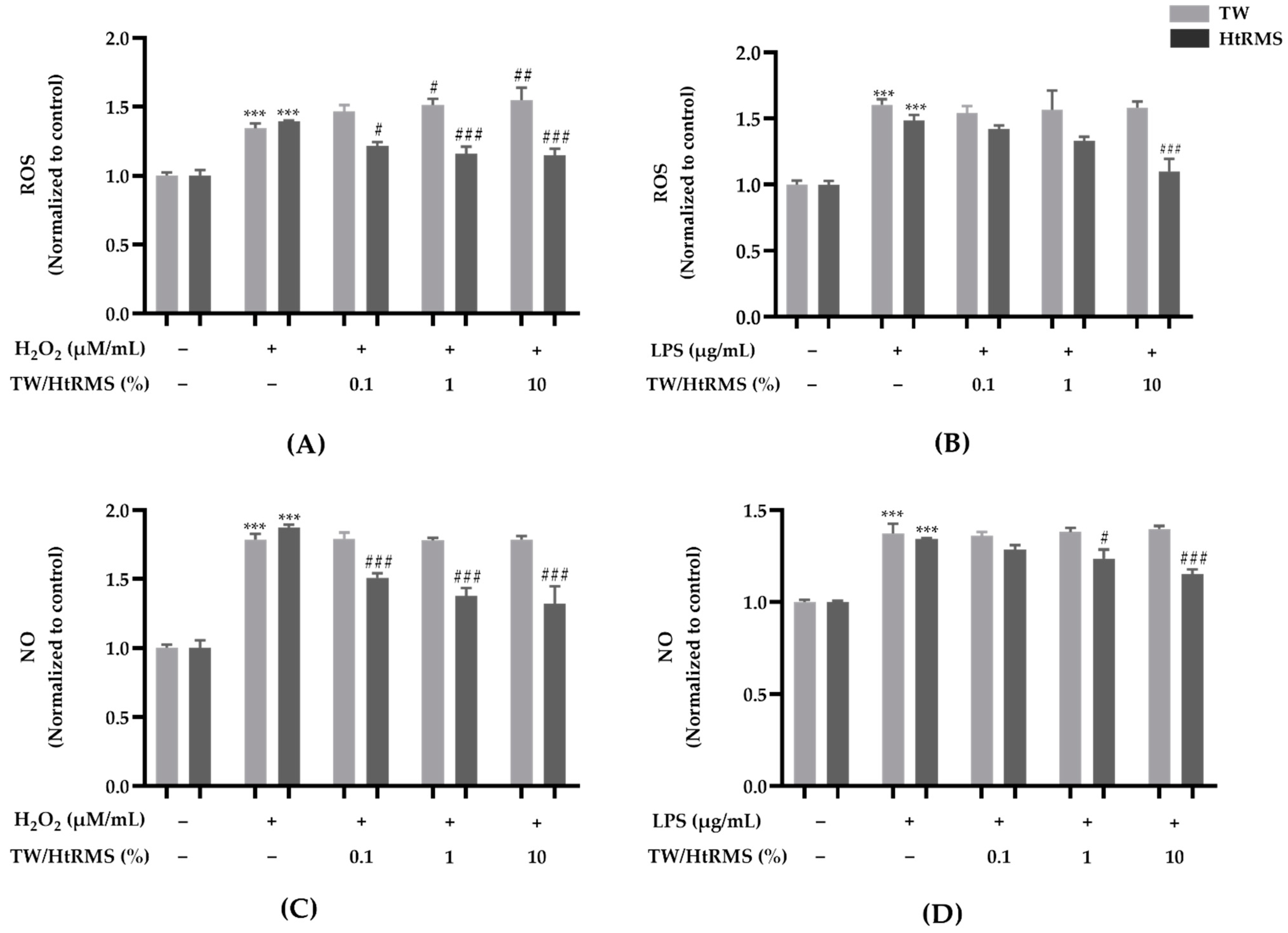
3.2. Effect of HtRMS on OS Production of H2O2- and LPS-Induced Murine Macrophage RAW 264.7 Cells

ROS and NO levels significantly increased compared to the NC group after induction by H2O2 (p < 0.001) and LPS (p < 0.001) (Figure 3). After HtRMS treatment, ROS decreased in a concentration-dependent manner in HtRMS 0.1% (p < 0.05), 1% (p < 0.001), and 10% (p < 0.001) groups compared to the H2O2-only group; however, ROS in the TW groups increased significantly at 1% (p < 0.01) and 10% (p < 0.001) concentrations (Figure 3A). The induction by LPS also exhibited a similar trend to that by H2O2, and ROS levels significantly decreased in the HtRMS 10% group (p < 0.01) compared to LPS-only group (Figure 3B). In addition, NO drastically decreased in a concentration-dependent manner at 0.1, 1, and 10% concentrations (p < 0.001) in the HtRMS groups compared to that in the H2O2-only group (Figure 3C). Similarly, after LPS induction, NO significantly decreased in the HtRMS 1% (p < 0.05) and 10% (p < 0.001) groups, and there was no significant difference in the TW groups (Figure 3D).

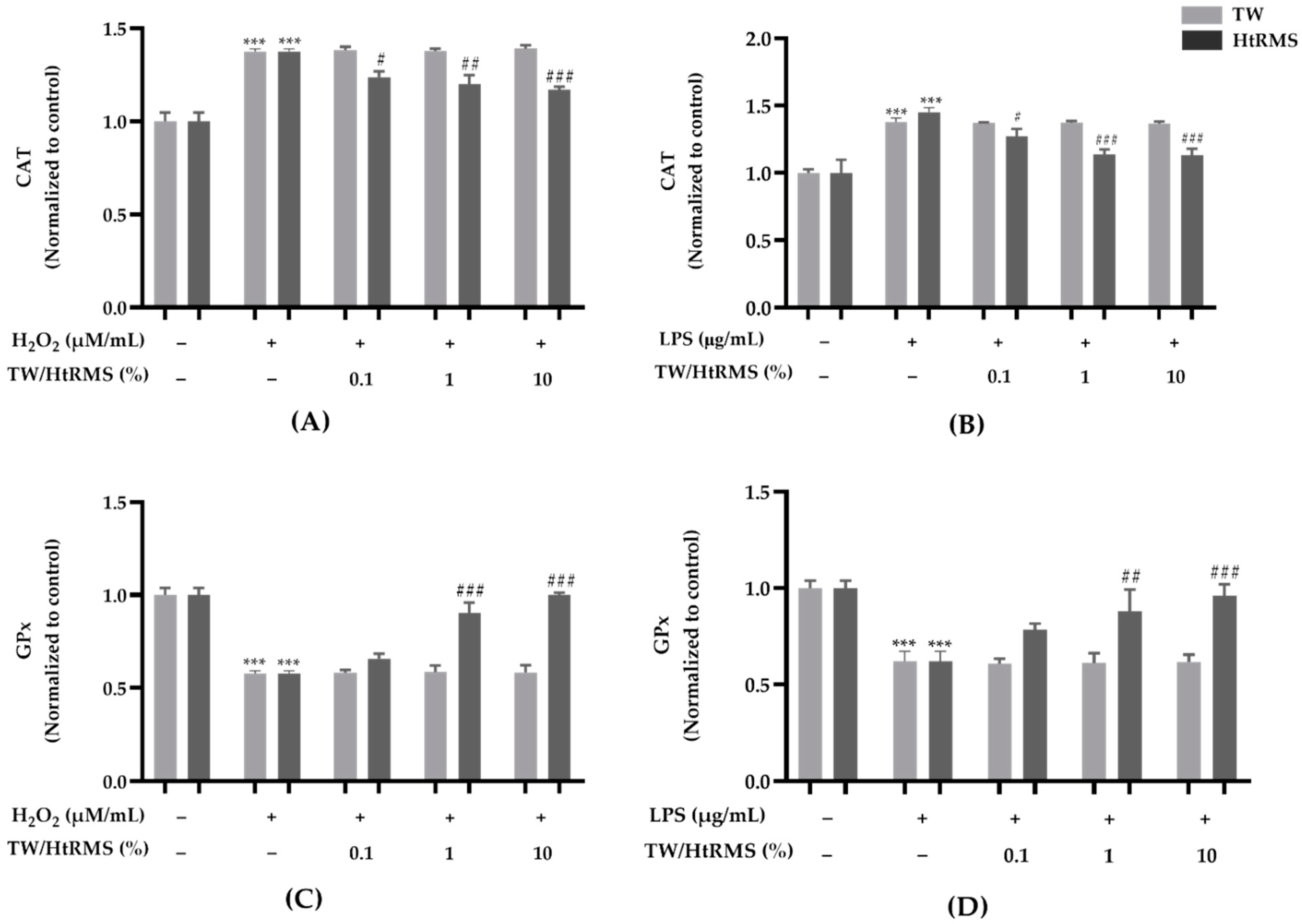
3.3. Effect of HtRMS on Intracellular Antioxidant Enzyme Levels of H2O2- and LPS-Induced Murine Macrophage RAW 264.7 Cells

CAT levels significantly increased after induction by H2O2 and LPS compared to that in the non-treated group (p < 0.001) (Figure 4A,B). After HtRMS treatment, CAT significantly decreased in a concentration-dependent manner at 0.1% (p < 0.05), 1% (p < 0.01), and 10% (p < 0.001) compared to the H2O2-only group (Figure 4A); however, the TW group exhibited no significant difference. LPS-induced cells also exhibited a similar trend to the one consequent to H2O2 induction. CAT significantly decreased in the HtRMS 0.1% (p < 0.05), 1% (p < 0.001), and 10% (p < 0.001) groups compared to the LPS-only group (Figure 4B). In contrast, GPx drastically increased in the HtRMS 1% (p < 0.001) and 10% (p < 0.001) groups compared to the H2O2-only group (Figure 4C) and to the LPS-only group (p < 0.01 and p < 0.001, respectively) (Figure 4D).

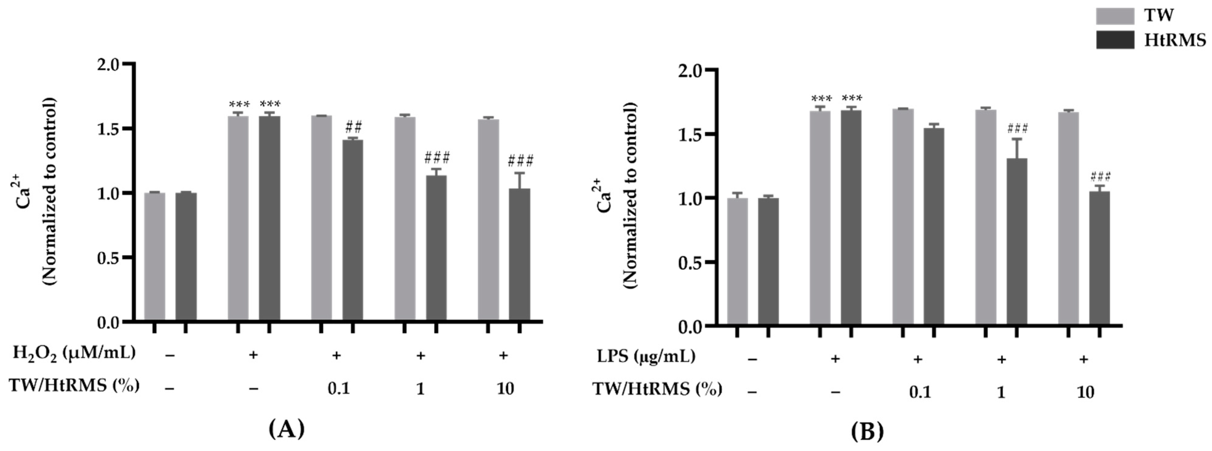
3.4. Effect of HtRMS on the Intracellular Ca2+ Level of H2O2- and LPS-Induced Murine Macrophage RAW 264.7 Cells

Ca2+ activity significantly increased after the induction of H2O2 and LPS compared to that in the non-treated group (p < 0.001) (Figure 5A,B). Upon HtRMS treatment, Ca2+ levels decreased significantly at 0.1% (p < 0.01), 1% (p < 0.001), and 10% (p < 0.001) concentrations compared to that in the H2O2-only group (Figure 5A), and similarly at 1% (p < 0.001) and 10% (p < 0.001) concentrations compared to that in LPS-only group (Figure 5B); however, TW treatment groups did not exhibit significant differences.

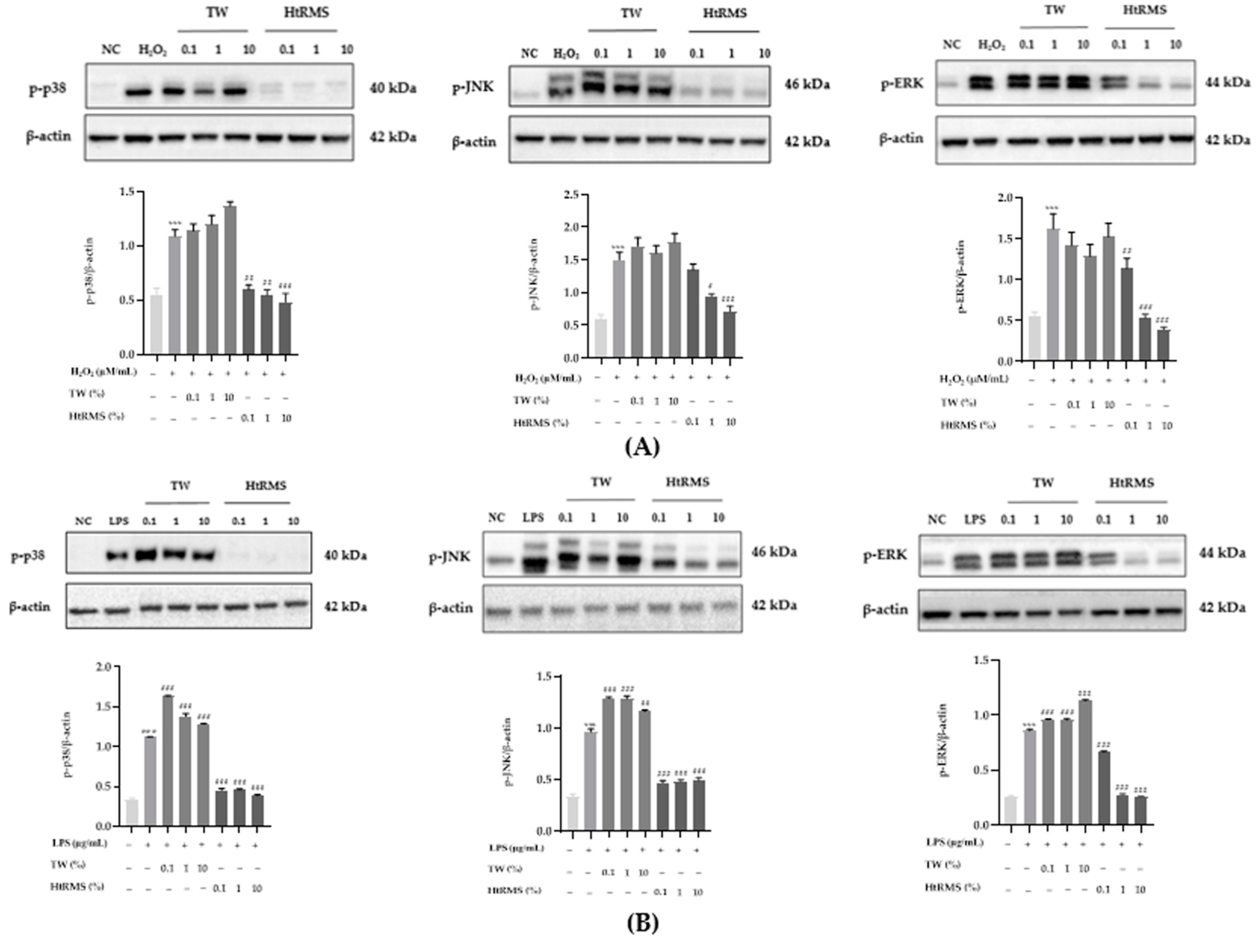
3.5. Effect of HtRMS on p-p38, p-JNK, and p-ERK of H2O2- and LPS-Induced Murine Macrophage RAW 264.7 Cells

p-p38, p-JNK, and p-ERK significantly increased after H2O2 and LPS induction compared to the NC group (p < 0.001) (Figure 6A,B). After HtRMS treatment, p-p38, p-JNK, and p-ERK exhibited a significantly decreasing trend in a concentration-dependent manner compared to the H2O2-only groups; however, the TW group did not exhibit any significant difference. p-p38 expression increased at 1% (p < 0.01) and 10% (p < 0.001) treatments, p-JNK at 1% (p < 0.05) and 10% (p < 0.001) treatments, and p-ERK at 0.1% (p < 0.01), 1% (p < 0.001), and 10% (p < 0.001) treatments compared to that in the H2O2-only groups (Figure 6A). Similarly, in LPS-induced cells, p-p38, p-JNK, and p-ERK significantly decreased at any HtRMS concentration (p < 0.001) compared to LPS-only groups; however, they significantly increased in the TW groups compared to LPS-only groups (Figure 6B).

4. Discussion

This study evaluated the antioxidative effect of HtRMS in a murine macrophage cell line, RAW 264.7. Compared to the properties of TW [11], HtRMS is enriched in mineral contents (25 essential minerals) and has higher H2, lower ORP, and higher pH. Previous studies have also revealed that the way the minerals are administered significantly impacts their antioxidative effect [8,23,24]. Specifically, minerals such as Zn2+, Cu2+, and Mg2+ are known to play an essential role in the reduction of free radicals and in pathological processes, and therefore they may possibly sustain immune defense [8,24]. Our study identified a positive active role of HtRMS in inhibiting the MAPK signaling pathway. We particularly evaluated the concentration-dependent improvement of parameters such as cell viability, antioxidant enzyme levels, and concentration of OS-related markers, such as ROS, NO, and Ca2+, and MAPK signaling pathway-related proteins, such as p-p38, p-JNK, and p-ERK after HtRMS treatment in RAW 264.7 murine macrophage cells.
First, we evaluated the effect of HtRMS on cell viability after H2O2 or LPS induction. H2O2 is commonly used to stimulate cellular OS [6], while LPS is recognized by specific host cell receptors and activates the innate immune system through an inflammatory response [25]. As expected, cell viability decreased upon induction by H2O2 and LPS; however, the application of HtRMS in murine macrophage cells significantly rescued both H2O2- and LPS-induced cell viability (Figure 1).
To assess the antioxidative effect, we measured ROS and NO production in H2O2- and LPS-induced murine macrophage cells after HtRMS treatment. ROS levels play an important role in signal transduction and cellular physiological functions in macrophage cell lines [26,27,28]: ROS and NO are produced and released in response to phagocytosis involved in bacterial killing, which is implicated in inflammation and tissue damage [29,30]. However, the overproduction of ROS influences NO production and consequently leads to stress and inflammatory responses in the immune system [31,32]. Consistent with previous studies on H2 and electrolysis-reduced water [33,34,35], our results revealed that HtRMS induced a decrease in ROS and NO levels in a concentration-dependent way (Figure 2) after H2O2 and LPS induction in murine macrophage cells. The present results suggest that HtRMS may be employed in therapeutic approaches to reduce the overproduction of harmful free radicals in murine macrophage cells. Furthermore, consistent with our ROS and NO analyses, HtRMS exhibited efficacy in enhancing antioxidant effects against H2O2-induced OS and LPS-mediated inflammation. CAT and GPx protect cells against free radicals [36]. By removing them, thereby preventing and reducing oxidation-induced cell damage [37,38]. The protective effects of antioxidants are still being studied globally [38]. Evidently, cells treated with HtRMS mediated CAT and GPx levels after H2O2 and LPS induction exhibiting its antioxidant capacity (Figure 3A). It is established that antioxidants contribute to the reduction of oxidative effectors, such as ROS and NO, which are associated with inflammation in macrophage cells [2,33]. These results were consistent with the findings of previous studies, wherein electrolyzed reduced water and H2 also increased antioxidant levels in in vivo models [39,40,41,42]. However, further studies are necessary to identify the mechanism through which HtRMS acts as an antioxidant to comprehensively understand and apply its clinical significance.
Moreover, we assessed intracellular Ca2+ levels. Ca2+ is a ubiquitous intracellular messenger that controls diverse cellular functions [43], wherein Ca2+ becomes a mediator of cell distress, but can even be toxic, if its concentration and movement inside the cell are not regulated carefully [44]. Among these, one study revealed that intracellular Ca2+ concentration activates the MAPK signaling pathway [45]. The present results showed that HtRMS salt protected the cells from high Ca2+ concentrations. Therefore, our results suggest that HtRMS treatment for 24 h can diminish ROS and NO production and reduce Ca2+ levels. Consistent with our results, several studies have proven that minerals releasing H2 at high concentrations have beneficial biological effects on Ca2+ responses [2].
Finally, to better understand the therapeutic effect of HtRMS against H2O2- and LPS-induced OS responses, the present study focused on the MAPK signaling pathway, which is one of the three major pathways involved in early OS response and cell survival, including p-p38, p-JNK, and p-ERK [46,47,48]. In addition, our Western blot results indicated that HtRMS inhibited H2O2- and LPS-induced MAPK signaling pathways, including p-p38, p-JNK, and p-ERK. This suggests that HtRMS significantly reduced the OS response via regulation of the MAPK signaling pathway.

5. Conclusions

Overall, our results suggest that HtRMS can act as an antioxidant and may further protect the cell from the OS process through the regulation of the MAPK signaling pathway. This might be the first evidence of the antioxidative effect of HtRMS in murine macrophage cells. This study suggests the potential application of HtRMS to combat stress-related disorders. Moreover, our results suggest that fortified alkaline water can be developed using HtRMS, which evidently showed its antioxidative effect. However, there are some limitations in the current study. First, the study only presented results with the antioxidative effects of HtRMS. However, it is unclear which factor of salt, either combination of minerals or alkalinity, or both has antioxidative effects. Second, this study is limited to the use of only one macrophage cell line (RAW264.7). Therefore, further research is needed with different types of organ-specific cell lines in addition to in vivo and clinical studies. Third, we only explored the MAPK signaling pathway in this study, to confirm the antioxidative effects. Hence, other signaling pathway studies are necessary to fully claim the molecular mechanism of the antioxidative effect of this salt product.

Author Contributions

Conceptualization, H.-T.J.; writing—original draft preparation, T.T.T.; writing—review and editing, K.-J.L., A.F., S.-H.Y. and J.B.; methodology T.T.T., A.F., S.S. and M.H.R.; data curation, T.T.T., A.F., C.-S.K.; supervision, H.-T.J. All authors have read and agreed to the published version of the manuscript.

Funding

The research received no external funding.

Institutional Review Board Statement

Not applicable.

Informed Consent Statement

Not applicable.

Data Availability Statement

The data presented in this study are available in the article (tables and figures).

Acknowledgments

This research was supported by Biocera Co., Ltd., Republic of Korea.

Conflicts of Interest

The authors declare no conflict of interest.

References

  1. Hussain, T.; Tan, B.; Yin, Y.; Blachier, F.; Tossou, M.C.B.; Rahu, N. Oxidative Stress and Inflammation: What Polyphenols Can Do for Us? Oxid. Med. Cell. Longev. 2016, 2016, 7432797. [Google Scholar] [CrossRef] [PubMed] [Green Version]
  2. Begum, R.; Kim, C.; Fadriquela, A.; Bajgai, J.; Jing, X.; Kim, D.-H.; Kim, S.-K.; Lee, K.-J. Molecular hydrogen protects against oxidative stress-induced RAW 264.7 macrophage cells through the activation of Nrf2 and inhibition of MAPK signaling pathway. Mol. Cell. Toxicol. 2020, 16, 103–118. [Google Scholar] [CrossRef]
  3. Rahman, H.; Bajgai, J.; Fadriquela, A.; Sharma, S.; Trinh, T.T.; Akter, R.; Jeong, Y.J.; Goh, S.H.; Kim, C.-S.; Lee, K.-J. Therapeutic Potential of Natural Products in Treating Neurodegenerative Disorders and Their Future Prospects and Challenges. Molecules 2021, 26, 5327. [Google Scholar] [CrossRef]
  4. Pizzino, G.; Irrera, N.; Cucinotta, M.; Pallio, G.; Mannino, F.; Arcoraci, V.; Squadrito, F.; Altavilla, D.; Bitto, A. Oxidative Stress: Harms and Benefits for Human Health. Oxid. Med. Cell. Longev. 2017, 2017, 8416763. [Google Scholar] [CrossRef]
  5. Park, J.; Min, J.-S.; Kim, B.; Chae, U.-B.; Yun, J.W.; Choi, M.-S.; Kong, I.-K.; Chang, K.-T.; Lee, D.-S. Mitochondrial ROS govern the LPS-induced pro-inflammatory response in microglia cells by regulating MAPK and NF-κB pathways. Neurosci. Lett. 2015, 584, 191–196. [Google Scholar] [CrossRef]
  6. Ransy, C.; Vaz, C.; Lombès, A.; Bouillaud, F. Use of H2O2 to Cause Oxidative Stress, the Catalase Issue. Int. J. Mol. Sci. 2020, 21, 9149. [Google Scholar] [CrossRef]
  7. Jeong, J.H.; Noh, M.-Y.; Choi, J.-H.; Lee, H.; Kim, S.H. Neuroprotective and antioxidant activities of bamboo salt soy sauce against H2O2-induced oxidative stress in rat cortical neurons. Exp. Ther. Med. 2016, 11, 1201–1210. [Google Scholar] [CrossRef] [PubMed] [Green Version]
  8. Kim, N.-R.; Nam, S.-Y.; Ryu, K.-J.; Kim, H.-M.; Jeong, H.-J. Effects of bamboo salt and its component, hydrogen sulfide, on enhancing immunity. Mol. Med. Rep. 2016, 14, 1673–1680. [Google Scholar] [CrossRef]
  9. Peng, H.-H.; Liu, Y.-J.; Ojcius, D.; Lee, C.-M.; Chen, R.-H.; Huang, P.-R.; Martel, J.; Young, J.D. Mineral particles stimulate innate immunity through neutrophil extracellular traps containing HMGB1. Sci. Rep. 2017, 7, 1–16. [Google Scholar] [CrossRef] [PubMed] [Green Version]
  10. Sokrateva, T.D.; Roussev, B.H.; Nashar, M.A.; Kiselova-Kaneva, Y.D.; Mihaylova, G.M.; Todorova, M.N.; Pasheva, M.G.; Tasinov, O.B.; Nazifova-Tasinova, N.F.; Vankova, D.G.; et al. Effects of sulphur-containing mineral water intake on oxidative status and markers for inflammation in healthy subjects. Arch. Physiol. Biochem. 2019, 127, 1–10. [Google Scholar] [CrossRef] [PubMed]
  11. Pehrsson, P.; Patterson, K.; Perry, C. The Mineral Content of Us Drinking and Municipal Water. In Proceedings of the 32nd National Nutrient Databank Conference, Ottawa, ON, Canada, 12–14 May 2008; pp. 12–14. [Google Scholar]
  12. Huang, L. Molecular hydrogen: A therapeutic antioxidant and beyond. Med. Gas Res. 2016, 6, 219–222. [Google Scholar] [CrossRef] [PubMed] [Green Version]
  13. Ohta, S. Molecular Hydrogen as a Novel Antioxidant. Methods Enzymol. 2015, 555, 289–317. [Google Scholar] [CrossRef] [PubMed]
  14. Ichihara, M.; Sobue, S.; Ito, M.; Ito, M.; Hirayama, M.; Ohno, K. Beneficial biological effects and the underlying mechanisms of molecular hydrogen—Comprehensive review of 321 original articles. Med. Gas Res. 2015, 5, 1–21. [Google Scholar] [CrossRef] [PubMed] [Green Version]
  15. Ku, J.Y.; Park, M.J.; Park, H.J.; Park, N.C.; Joo, B.S. Combination of Korean Red Ginseng Extract and Hydrogen-Rich Water Improves Spermatogenesis and Sperm Motility in Male Mice. Chin. J. Integr. Med. 2020, 26, 361–369. [Google Scholar] [CrossRef] [PubMed]
  16. Ohsawa, I.; Ishikawa, M.; Takahashi, K.; Watanabe, M.; Nishimaki, K.; Yamagata, K.; Katsura, K.-I.; Katayama, Y.; Asoh, S.; Ohta, S. Hydrogen acts as a therapeutic antioxidant by selectively reducing cytotoxic oxygen radicals. Nat. Med. 2007, 13, 688–694. [Google Scholar] [CrossRef]
  17. Kurokawa, R.; Seo, T.; Sato, B.; Hirano, S.-I.; Sato, F. Convenient methods for ingestion of molecular hydrogen: Drinking, injection, and inhalation. Med. Gas Res. 2015, 5, 13. [Google Scholar] [CrossRef] [Green Version]
  18. Rias, Y.A.; Kurniawan, A.L.; Chang, C.W.; Gordon, C.J.; Tsai, H.T. Synergistic Effects of Regular Walking and Alkaline Electrolyzed Water on Decreasing Inflammation and Oxidative Stress, and Increasing Quality of Life in Individuals with Type 2 Diabetes: A Community Based Randomized Controlled Trial. Antioxidants 2020, 9, 946. [Google Scholar] [CrossRef]
  19. Yan, P.; Daliri, E.B.-M.; Oh, D.-H. New Clinical Applications of Electrolyzed Water: A Review. Microorganisms 2021, 9, 136. [Google Scholar] [CrossRef]
  20. Gordon, S. The macrophage: Past, present and future. Eur. J. Immunol. 2007, 37, S9–S17. [Google Scholar] [CrossRef]
  21. Roux, P.P.; Blenis, J. ERK and p38 MAPK-Activated Protein Kinases: A Family of Protein Kinases with Diverse Biological Functions. Microbiol. Mol. Biol. Rev. 2004, 68, 320–344. [Google Scholar] [CrossRef] [Green Version]
  22. Cargnello, M.; Roux, P.P. Activation and Function of the MAPKs and Their Substrates, the MAPK-Activated Protein Kinases. Microbiol. Mol. Biol. Rev. 2011, 75, 50–83. [Google Scholar] [CrossRef] [Green Version]
  23. Zhao, X. Anticancer and Antiinflammatory Effects of Bamboo Salt. Available online: http://www.bookpi.org/bookstore/product/anticancer-and-antiinflammatory-effects-of-bamboo-salt/ (accessed on 1 September 2021).
  24. Qian, Y.; Zhao, X. Alkaline properties and antioxidant activities of bamboo salt. China Condiment 2014, 39, 28–46. [Google Scholar]
  25. Chen, J.; Wu, Y.; Sun, Y.; Dong, X.; Wang, Z.; Zhang, Z.; Xiao, Y.; Dong, G. Bacterial Lipopolysaccharide Induced Alterations of Genome-Wide DNA Methylation and Promoter Methylation of Lactation-Related Genes in Bovine Mammary Epithelial Cells. Toxins 2019, 11, 298. [Google Scholar] [CrossRef] [Green Version]
  26. Rendra, E.; Riabov, V.; Mossel, D.M.; Sevastyanova, T.; Harmsen, M.C.; Kzhyshkowska, J. Reactive oxygen species (ROS) in macrophage activation and function in diabetes. Immunobiology 2019, 224, 242–253. [Google Scholar] [CrossRef] [PubMed]
  27. Covarrubias, A.J.; Byles, V.; Horng, T. ROS sets the stage for macrophage differentiation. Cell Res. 2013, 23, 984–985. [Google Scholar] [CrossRef] [Green Version]
  28. Zhang, Y.; Choksi, S.; Chen, K.; Pobezinskaya, Y.; Linnoila, I.; Liu, Z.-G. ROS play a critical role in the differentiation of alternatively activated macrophages and the occurrence of tumor-associated macrophages. Cell Res. 2013, 23, 898–914. [Google Scholar] [CrossRef] [PubMed] [Green Version]
  29. Forman, H.J. Redox signaling in macrophages. Mol. Asp. Med. 2001, 22, 189–216. [Google Scholar] [CrossRef]
  30. Denis, M. Human monocytes/macrophages: NO or no NO? J. Leukoc. Biol. 1994, 55, 682–684. [Google Scholar] [CrossRef]
  31. Zagury, D.; Le Buanec, H.; Bizzini, B.; Burny, A.; Lewis, G.; Gallo, R. Active versus passive anti-cytokine antibody therapy against cytokine-associated chronic diseases. Cytokine Growth Factor Rev. 2003, 14, 123–137. [Google Scholar] [CrossRef]
  32. Pierini, D.; Bryan, N.S. Nitric oxide availability as a marker of oxidative stress. In Advanced Protocols in Oxidative Stress III. Methods in Molecular Biology (Methods and Protocols); Armstrong, D., Ed.; Humana Press: New York, NY, USA, 2015; Volume 1208, pp. 63–71. [Google Scholar]
  33. Ohta, S. Molecular hydrogen is a novel antioxidant to efficiently reduce oxidative stress with potential for the improvement of mitochondrial diseases. Biochim. Biophys. Acta Gen. Subj. 2012, 1820, 586–594. [Google Scholar] [CrossRef]
  34. Park, S.-K.; Park, S.-K. Electrolyzed-reduced water increases resistance to oxidative stress, fertility, and lifespan via insulin/IGF-1-like signal in C. elegans. Biol. Res. 2013, 46, 147–152. [Google Scholar] [CrossRef] [PubMed] [Green Version]
  35. Terasaki, Y.; Ohsawa, I.; Terasaki, M.; Takahashi, M.; Kunugi, S.; Dedong, K.; Urushiyama, H.; Amenomori, S.; Kaneko-Togashi, M.; Kuwahara, N.; et al. Hydrogen therapy attenuates irradiation-induced lung damage by reducing oxidative stress. Am. J. Physiol. Cell. Mol. Physiol. 2011, 301, L415–L426. [Google Scholar] [CrossRef] [PubMed] [Green Version]
  36. Jeon, G.; Kim, C.; Cho, U.M.; Hwang, E.T.; Hwang, H.S.; Min, J. Melanin-Decolorizing Activity of Antioxidant Enzymes, Glutathione Peroxidase, Thiol Peroxidase, and Catalase. Mol. Biotechnol. 2021, 63, 150–155. [Google Scholar] [CrossRef] [PubMed]
  37. Traber, M.G.; Packer, L. Vitamin E: Beyond antioxidant function. Am. J. Clin. Nutr. 1995, 62, 1501S–1509S. [Google Scholar] [CrossRef] [Green Version]
  38. Meng, R.; Wu, Z.; Xie, Q.-T.; Cheng, J.-S.; Zhang, B. Preparation and characterization of zein/carboxymethyl dextrin nanoparticles to encapsulate curcumin: Physicochemical stability, antioxidant activity and controlled release properties. Food Chem. 2021, 340, 127893. [Google Scholar] [CrossRef] [PubMed]
  39. Ridwan, R.D.; Tantiana, T.; Setijanto, D.; Kusuma, A.K.; Putranto, A.F. Kronik Periodontitli Wistar Sıçan (Rattus Novergicus)’larda Elektrolize İndirgenmiş Suyun Antioksidan ve Antiinflamatuvar Etkisi. Kafkas Univ. Veter Fak. Derg. 2019, 25, 539–544. [Google Scholar] [CrossRef]
  40. Hu, D.; Li, D.; Shigeta, M.; Ochi, Y.; Okauchi, T.; Neyama, H.; Kabayama, S.; Watanabe, Y.; Cui, Y. Alleviation of the chronic stress response attributed to the antioxidant and anti-inflammatory effects of electrolyzed hydrogen water. Biochem. Biophys. Res. Commun. 2021, 535, 1–5. [Google Scholar] [CrossRef] [PubMed]
  41. Lin, C.-P.; Chuang, W.-C.; Lu, F.-J.; Chen, C.-Y. Anti-oxidant and anti-inflammatory effects of hydrogen-rich water alleviate ethanol-induced fatty liver in mice. World J. Gastroenterol. 2017, 23, 4920–4934. [Google Scholar] [CrossRef] [PubMed]
  42. Ishibashi, T. Molecular Hydrogen: New Antioxidant and Anti-inflammatory Therapy for Rheumatoid Arthritis and Related Diseases. Curr. Pharm. Des. 2013, 19, 6375–6381. [Google Scholar] [CrossRef] [PubMed] [Green Version]
  43. Bagur, R.; Hajnóczky, G. Intracellular Ca2+ Sensing: Its Role in Calcium Homeostasis and Signaling. Mol. Cell 2017, 66, 780–788. [Google Scholar] [CrossRef] [Green Version]
  44. Wright, M.F.; Bowdridge, E.; McDermott, E.L.; Richardson, S.; Scheidler, J.; Syed, Q.; Bush, T.; Inskeep, E.K.; Flores, J.A. Mechanisms of Intracellular Calcium Homeostasis in Developing and Mature Bovine Corpora Lutea1. Biol. Reprod. 2014, 90, 55. [Google Scholar] [CrossRef] [PubMed] [Green Version]
  45. Chuderland, D.; Seger, R. Calcium regulates ERK signaling by modulating its protein-protein interactions. Commun. Integr. Biol. 2008, 1, 4–5. [Google Scholar] [CrossRef] [PubMed] [Green Version]
  46. Phong, M.S.; Van Horn, R.D.; Li, S.; Tucker-Kellogg, G.; Surana, U.; Ye, X.S. p38 Mitogen-Activated Protein Kinase Promotes Cell Survival in Response to DNA Damage but Is Not Required for the G 2 DNA Damage Checkpoint in Human Cancer Cells. Mol. Cell. Biol. 2010, 30, 3816–3826. [Google Scholar] [CrossRef] [PubMed] [Green Version]
  47. Mebratu, Y.; Tesfaigzi, Y. How ERK1/2 activation controls cell proliferation and cell death: Is subcellular localization the answer? Cell Cycle 2009, 8, 1168–1175. [Google Scholar] [CrossRef]
  48. Gururajan, M.; Chui, R.; Karuppannan, A.K.; Ke, J.; Jennings, C.D.; Bondada, S. c-Jun N-terminal kinase (JNK) is required for survival and proliferation of B-lymphoma cells. Blood 2005, 106, 1382–1391. [Google Scholar] [CrossRef] [Green Version]
Figure 1. Outline of the experimental procedure. ROS: reactive oxygen species; NO: nitric oxide; CAT: catalase; GPx: glutathione peroxidase; MAPK: mitogen-activated protein kinase; H2O2: hydrogen peroxide; LPS: lipopolysaccharide.
Processes 09 01928 g001
Figure 2. Effect of HtRMS on cell viability of H2O2- and LPS-induced murine macrophage cells. The cell toxicity was induced by H2O2 (A) and LPS (B) and then treated with TW and HtRMS, respectively, in different concentrations of 0.1, 1, and 10%. Data values are stated as mean ± standard error of the mean of fold change according to control (n = 3). The statistical significance was analyzed via two-way ANOVA. TW: tap water; (−): non-treatment; (+): treatment; ***: p < 0.001 vs. normal control; #: p < 0.05, ##: p < 0.01, and ###: p < 0.001 vs. H2O2 or LPS only. HtRMS: high-temperature-roasted mineral salt; TW: tap water; H2O2: hydrogen peroxide; LPS: lipopolysaccharide.
Processes 09 01928 g002
Figure 3. Effect of HtRMS on OS production of H2O2- and LPS-induced murine macrophage cells. Table 0; 1 and 10% concentration after OS induction by H2O2 (A,C) and LPS (B,D), respectively. Data values are stated as mean ± standard error of the mean of fold change relative to the control group (n = 3). The statistical significance was analyzed via two-way ANOVA. (−): non-treatment; (+): treatment. ***: p < 0.001 vs. normal control; #: p < 0.05; ##: p < 0.01, and ###: p < 0.001 vs. H2O2 or LPS only. HtRMS: high-temperature-roasted mineral salt; TW: tap water; H2O2: hydrogen peroxide; LPS: lipopolysaccharide.
Processes 09 01928 g003
Figure 4. Effects of HtRMS on the levels of CAT (A,B) and GPx (C,D) of H2O2- and LPS-induced murine macrophage cells. The cells were treated with HtRMS and TW (0.1, 1, and 10%) after OS induction by H2O2 (A,C) and LPS (B,D), respectively. Data values are stated as mean ± standard error of the mean of fold change according to the control group (n = 3). The statistical significance was analyzed via two-way ANOVA. (−): non-treatment; (+): treatment. ***: p < 0.001 vs. normal control. #: p < 0.05, ##: p < 0.01, and ###: p < 0.001 vs. H2O2- or LPS-only. HtRMS: high-temperature-roasted mineral salt; TW: tap water; H2O2: hydrogen peroxide; LPS: lipopolysaccharide; CAT: catalase, GPx: glutathione peroxidase.
Processes 09 01928 g004
Figure 5. Effects of HTRMS on Ca2+ activity of H2O2 and LPS-induced murine macrophage cells. The cells were treated with HtRMS and TW (0.1, 1, and 10%), after the induction of OS by H2O2 (A) and LPS (B), respectively. Data values are stated as mean ± standard error of the mean of fold change compared to the control group (n = 3). The statistical significance was analyzed via two-way ANOVA. (−): non-treatment, (+): treatment. ***: p < 0.001 vs. normal control. ##: p < 0.01 and ###: p < 0.001 vs. H2O2 or LPS only. HtRMS: high-temperature-roasted salt; TW: tap water; H2O2: hydrogen peroxide; LPS: lipopolysaccharide.
Processes 09 01928 g005
Figure 6. Effects of HtRMS on p-p38, p-JNK, and p-ERK of H2O2- and LPS-induced murine macrophage cells. The cells were treated with HtRMS and TW (0.1, 1, and 10%) after the induction of OS by H2O2 (A) and LPS (B), respectively. Data values are stated as mean ± standard error of the mean of fold change, compared to the control group (n = 3). The statistical significance was analyzed via two-way ANOVA. (−): non-treatment, (+): treatment. ***: p < 0.001 vs. normal control. #: p < 0.05, ##: p < 0.01, and ###: p < 0.001 vs. H2O2 or LPS only. HtRMS: high-temperature-roasted salt; TW: tap water; H2O2: hydrogen peroxide; LPS: lipopolysaccharide.
Processes 09 01928 g006
Table 1. Mineral component content analysis of HtRMS.
Mineral
Components
Contents
(ppm)
Mineral
Components
Contents
(ppm)
Calcium (Ca2+)1722Copper (Cu2+)2.28
Phosphorous (P3−)122Barium (Ba2+)6.46
Potassium (K+)1738Tin (Sn4+)1.24
Sulfur (S2−)4220Iodine (I)67
Sodium (Na+)379,400Titanium (Ti3+)2.6
Chlorine (Cl)569,600Boron (B3+)18.2
Magnesium (Mg2+)746Selenium (Se2−)3.01
Ion (Fe2+)26.2Lithium (Li+)14.2
Fluorine (F)18Molybdenum (Mo2+)1.95
Zinc (Zn2+)3.57Gallium (Ga3+)10.36
Silicon (Si4+)45.6Vanadium (V)9.73
Rubidium (Rb+)6.27Beryllium (Be2+)14.2
Strontium (Sr+)67.8Bromine (Br)8
Component analysis was conducted in the Hydrogen Fuel Cell Parts & Applied Technology Regional Innovation Center, Woosuk University, Wanju, Republic of Korea. HtRMS: high-temperature-roasted mineral salt.
Table 2. Properties of HtRMS.
MaterialspHORP (mV)TDS (ppm)H2 (ppb)
TW7.326091050
HtRMS10.5−38027,000450
TW: tap water; HtRMS: high-temperature-roasted mineral salt; ORP: oxidation-reduction potential; TDS: total dissolved solid.
Publisher’s Note: MDPI stays neutral with regard to jurisdictional claims in published maps and institutional affiliations.

