Article

Detection of E. coli O157:H7 in Food Using Automated Immunomagnetic Separation Combined with Real-Time PCR

1
Department of Chemistry, Chung-Ang University, Seoul 06974, Korea
2
Research Group of Consumer Safety, Korea Food Research Institute (KFRI), Jeollabuk-do 55365, Korea
3
Department of Molecular Science and Technology, Ajou University, Gyeonggi-do 16499, Korea
*
Author to whom correspondence should be addressed.
These authors contributed equally to this work.
Processes 2020, 8(8), 908; https://doi.org/10.3390/pr8080908
Submission received: 23 June 2020 / Revised: 22 July 2020 / Accepted: 23 July 2020 / Published: 1 August 2020
(This article belongs to the Section Food Process Engineering)

Abstract

:
In this study, we describe the development of an automated immunomagnetic separation device combined with real-time polymerase chain reaction (PCR) for detecting foodborne bacteria. Immunomagnetic separation (IMS) is a well-known method for the separation and concentration of target bacteria from a large volume of food samples. Magnetic beads functionalized with an antibody provide selectivity for target bacteria such as Escherichia coli O157:H7. Moreover, compared to conventional methods, real-time PCR enables high-sensitivity detection of target bacteria. The method proposed in this study involves three steps: (1) pre-enrichment, (2) automated IMS and concentration of target bacteria, and (3) detection of target bacteria by real-time PCR. Using food samples with a working sample volume as large as 250 mL, the whole process only requires 3 h. As a result, target bacteria in the range of 101–102 colony-forming units per mg or g of sample can be detected in food samples, such as milk, ground beef, and cabbage, by using the proposed approach. We anticipate that the automated IMS system combined with real-time PCR will contribute to the development of a fully automated system for detecting foodborne bacteria and serve as a multi-tester for a variety of bacterial strains in the capacity of a sample-to-answer device in the near future.

1. Introduction

Escherichia coli O157:H7 is a pathogenic bacterium harmful to human health. The major reservoirs of E. coli O157:H7 include cattle, ground beef, milk, and uncooked vegetables. In humans, a very low infectious dose is enough to cause various illnesses, such as diarrhea and hemolytic uremic syndrome (HUS), which can lead to serious health and financial consequences [1,2,3,4]. A straightforward and rapid analysis method is key to minimizing the risk of foodborne pathogens. However, traditional methods for detecting foodborne bacteria rely on a long enrichment time or culture-based growth on a selective agar medium [1,5]. Polymerase chain reaction (PCR) has been reported as an alternative rapid detection method [6,7]. Among the PCR-based methods, real-time PCR is promising for the rapid detection of bacteria in a quantitative manner [8,9].
However, the reliability of PCR results depends on both the quantity and quality of the sample, including factors such as the number of target bacteria and the nature of the food matrix. When target bacteria are present at an insufficient level or absent, amplification and detection fail to occur. Some studies have reported the detection of E. coli O157:H7 using real-time PCR with a long enrichment time (>16 h). However, relying upon a long enrichment time is disadvantageous in preventing food contamination by undesired bacteria [10,11]. Furthermore, in the presence of PCR inhibitors with the target DNA, amplification does not occur successfully. Many studies have reported that PCR is inhibited by certain components of food and culture media [12,13,14,15,16]. Direct PCR-based detection of bacteria can be difficult in various complex food matrices, such as pork, salmon, and milk; moreover, pretreatment requires multiple steps to optimize the enzyme-based reaction conditions [17,18,19]. Therefore, there is a high demand for a straightforward and selective method of food sample preparation that includes selective separation and concentration of target bacteria from the food matrix.
Recently, manufacturers have begun to produce and commercialize immunomagnetic separation (IMS)-based automated devices. Antibodies bound to the surface of the device’s magnetic beads can enable selective capturing of target bacteria from various sample matrices. The magnetism of the magnetic beads can be used to rapidly separate and concentrate bacteria in a large sample volume (250 mL) using an external magnetic field, which can also eliminate PCR inhibitors. However, these challenges can be overcome or minimized by the incorporation of IMS during food sample pretreatment [20,21,22,23]. For example, the Pathatrix Auto System facilitates quick and easy isolation of the target bacteria from food, although it involves a tedious enrichment step (>5 h), and requires a small sample volume (10 to 60 mL) as an individual separation module [24,25].
In this study, we developed an automated IMS device for the pretreatment of food samples for selective separation and concentration of target bacteria, and we combined it with real-time PCR for quantitative detection of bacteria. The proposed approach could both improve detection sensitivity and shorten the time required to identify immunological targets from various food sample matrices. Here, to demonstrate the features of the combined detection method, pathogenic E. coli O157:H7 was used to artificially contaminate milk, ground beef, and cabbage as model food matrices. The combined system was effective in simplifying the pretreatment steps for the separation and concentration of target bacteria and the elimination of PCR inhibitors, facilitating the detection of low levels of pathogenic bacteria in foods.

2. Materials and Methods

2.1. Materials

Fresh milk, ground beef, and cabbage were obtained from a local food market in the Republic of Korea. Buffered peptone water (BPW) and Tween-20 were purchased from Sigma-Aldrich (Saint Louis, MO, USA). BacTrace Anti-E. coli O157 Magnetic Beads (catalog No. 082-01-95-90) were provided by Kirkegaard & Perry Laboratories (Gaithersburg, MD, USA). Tryptic soy broth (TSB) was purchased from Becton, Dickinson and Company (Franklin Lakes, NJ, USA). Sorbitol MacConkey agar, cefixime−tellurite supplement for E. coli, and SYBR Green PCR Master Mix were purchased from Thermo Fisher Scientific (Waltham, MA, USA). The QIAamp DNA Mini Kit and proteinase K (20 mg/mL) were provided by Qiagen (Hilden, Germany). Primers for real-time PCR were synthesized by Bioneer (Daejeon, Korea). A MyGo-mini real-time PCR instrument and 0.1 mL PCR tubes were purchased from Ecogen (Barcelona, Spain). A sterile Whirl-Pak bag (catalog No. B01195WA) was provided by Nasco (Fort Atkinson, WI, USA).

2.2. Preparation of Pathogenic-Bacteria-Contaminated Food Samples and Immunomagnetic Bead Reaction

In this study, E. coli O157:H7 (NCTC 12079) was selected as the target pathogenic bacteria. Stock solutions of bacteria were grown in TSB and then placed on a selective agar medium, followed by incubation at 37 °C overnight. A single colony was picked and incubated in fresh TSB media overnight at 37 °C with shaking (200 rpm). One milliliter of broth was centrifuged, and the resulting pellet was resuspended in phosphate-buffered saline (PBS). We used 3 different food sample matrices: milk, ground beef, and cabbage. The food samples (25 mL or 25 g) were artificially spiked with the prepared E. coli O157:H7 (101–103 CFU in sample), and each food sample was placed in a stomacher bag. The concentration of E. coli O157:H7 was confirmed by counting the colonies grown on the selective agar medium. Each food sample was then mixed with 75 mL of BPW containing 0.05% Tween-20 (BPWT) and homogenized for 1 min using a paddle blender. In the case of ground beef and cabbage, the solid matrix after the homogenization step was separated by a vertically adhered 250 μm filter inside the stomacher bag. Subsequently, the anti-E. coli O157 magnetic beads (500 μL; >1 × 10⁹ beads/mL) were added to the stomacher bag containing the homogenized aqueous food samples. The prepared mixture was incubated to enrich for contaminated bacteria at 37 °C with gentle shaking for 60 min and then injected automatically into the IMS device. In addition, E. coli O157:H7 (<10 colonies in 25 mL or 25 mg of food samples) was tested with a 120 min enrichment step.

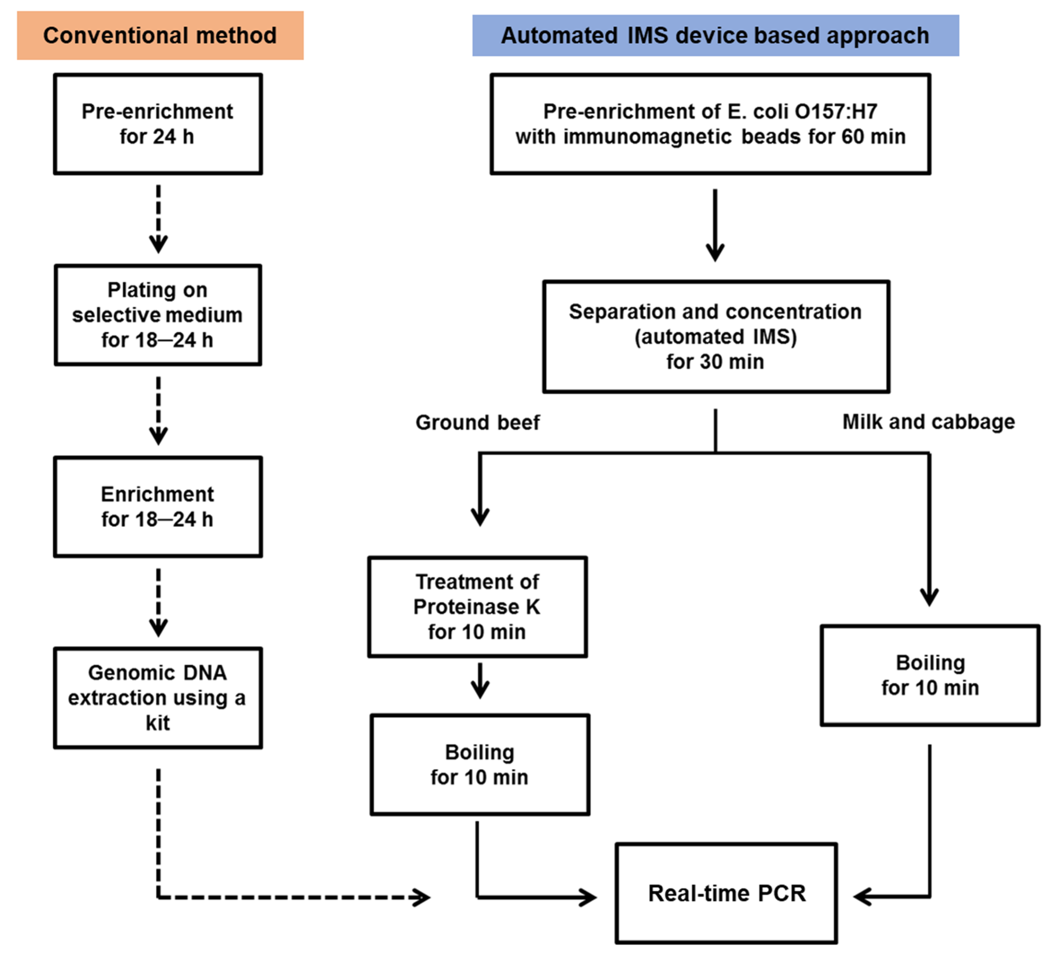
2.3. Automated IMS Process

The principle of the automated IMS operation and the performance of the fabricated automated IMS device have been described in our previous studies [26,27]. In brief, the automated IMS device used consisted of a peristaltic-pump-based fluidics control component and an external magnetic field-based bacterial recovery component. A 100-mL incubated food sample with target-specific immunomagnetic beads was injected automatically into the device, and 150 mL of BPWT flowed into the collection tube. The 250 mL solution was then equally divided into 8 collection tubes. Subsequently, the cylindrical glass-covered magnetic bars were used to isolate the immunomagnetic beads along with the target bacteria from the sample solution. After completion of the sample injection, the immunomagnetic beads bound with bacteria were separated automatically and concentrated into a single recovery tube containing 2 mL of PBST (PBS buffer with 0.5% Tween-20). The recovery process was repeated twice to obtain the remaining immunomagnetic beads, and the entire process was completed automatically in 30 min. Finally, the bacteria that adhered to the immunomagnetic beads were collected in 2 mL of recovery solution and concentrated into 100 μL of elution buffer by manual magnetic separation.

2.4. Real-Time PCR

Real-time PCR was performed by adding SYBR Green PCR Master Mix (2×, 10 μL), each primer (10 pmol, 1 μL), boiled cell lysate (2 μL), and deionized water (6 μL) into a 100 μL PCR tube. The sequence-targeted eaeA gene of E. coli O157:H7 was amplified using the following primers: forward primer 5′-GTA AGT TAC ACT ATA AAA GCA CCG TCG-3′ and reverse primer 5′-TCT GTG TGG ATG GTA ATA AAT TTT TG-3′ [11]. The bacteria recovered by the automated IMS were heated at 95 °C for 5 min, followed by 45 cycles of denaturation at 95 °C for 15 s, annealing at 60 °C for 20 s, and extension at 72 °C for 20 s. Subsequently, the temperature was gradually increased for the melting curve analysis and then reduced to 4 °C. Fluorescence intensity was monitored during amplification and melting curve analysis, and the collected data were analyzed using MyGo-mini software version 3.2 (Ecogen, Barcelona, Spain).

2.5. Recovery Monitoring and Data Analysis

The conventional method was used to confirm recovery of E. coli O157:H7 and to obtain a standard curve to assess the correlation between quantification cycle (Cq) and concentration of genomic DNA (Figure 1). This method involved culture-based enrichment for 18–24 h and growth on the selective agar medium that supported the growth of the target bacteria. A single colony was picked and enriched for 18–24 h, and the genomic DNA of the target bacteria at 109 CFU/mL was then extracted using the QIAamp DNA Mini Kit. Next, the eluted DNA was serially diluted, and each decimal dilution was amplified by real-time PCR under the same conditions mentioned above. The target E. coli O157:H7 cell lysates (101–105 CFU in 100 μL elution buffer) that had been prepared by heating were quantitated by real-time PCR. In addition, the number of cells was confirmed by counting the colonies grown on selective agar medium. Negative-control experiments were performed using genomic DNA extracted from nontarget S. enterica and S. aureus at 109 CFU/mL by real-time PCR using E. coli O157:H7 eaeA gene-specific primers.
In the case of the ground beef sample, the recovered bacteria conjugated with immunomagnetic beads were treated with proteinase K at 65 °C for 10 min to eliminate protein-related PCR inhibitors and subsequently heated at 85 °C for 10 min to extract genomic DNA. In the case of milk and cabbage samples, the recovered bacterial cells were directly boiled at 85 °C for 10 min in elution buffer without additional treatment. At this point, the immunomagnetic beads were immediately separated from the solution by using an external magnetic field. Subsequently, real-time PCR analysis was performed using the recovered 2 µL of bacterial lysate under the same operating conditions mentioned above.

3. Results and Discussion

3.1. Establishment of a Standard Curve

A standard curve was generated using genomic DNA from E. coli O157:H7 to confirm the efficiency of the PCR primers used. Eluted genomic DNA from E. coli O157:H7 was prepared as a 10-fold dilution series. The Cq values of real-time PCR were plotted on a logarithmic scale versus the DNA concentration (inset graph of Figure 2). The slope (–3.5) of the standard curve indicated that the efficiency of the primer for the target gene was 93.07%, based on Equation (1).
Efficiency (%) = (10−1/slope − 1) × 100
It has been proposed in previous literature that the efficiency should be between 90% and 110% in order for it to be considered suitable for quantitative detection [28]. Furthermore, the Cq values were generated from the E. coli O157:H7 cell lysate and compared with the concentration of recovered bacteria in the food samples (Figure 2). These results were obtained by real-time PCR under the same operating conditions used for the recovered sample. Thus, it was possible to quantify the cells in the recovered sample and to theoretically evaluate the initial concentration of E. coli O157:H7 present in the foods [27].

3.2. Detection of E. coli O157:H7 by Automated IMS Combined with Real-Time PCR

The automated IMS system required only 90 min to complete the entire process, including enrichment, separation, and concentration of target bacteria from food samples. The tested food samples were artificially contaminated with a set number of bacteria (101–103 CFU in 25 mL or mg) and homogenized with the BPWT solution. The pre-enrichment step was implemented for 60 min, and then the sample was injected into the automated IMS device along with additional BPWT solution. Finally, the separated and collected immunomagnetic beads with the target bacteria bound to their surfaces were reconcentrated in the elution buffer (Figure 3a). In order to confirm the efficacy of the IMS system compared with the real-time PCR analysis, 1/10 of the recovered solution containing the target bacteria was subjected to cell counting. The performance and efficiency of the automated IMS device developed in our study have been confirmed by previous studies [26,27].
As an additional pretreatment step, proteinase K was added to the recovered ground beef sample and then boiled for 10 min. Depending on the sample matrix, treatment with proteinase K is required for enhancing the efficiency of real-time PCR. However, proteinase K not only helps to denature proteins, which are major PCR inhibitors, but also destroys DNA polymerase [16]. Thus, it is important to boil the sample after treatment with proteinase K. However, the milk and cabbage food samples were only boiled after finishing the automated IMS process. Following real-time PCR analysis, the immunomagnetic beads in the recovered sample solution were removed using an external magnetic field, which also acted as an inhibitor. Finally, the eluted solution was subjected to real-time PCR-based analysis (Figure 3b).
Milk contains fat globules and proteins, such as casein, which may interrupt the interaction between immunomagnetic beads and target bacteria. To enhance sample homogenization and reduce nonspecific reactions of antibodies and unbound food moieties, the surfactant Tween-20 was used as a blocking agent for immunodetection and as a solubilizing agent. As shown in Table 1 and Figure 4a, the target E. coli O157:H7 could be detected at 101 CFU in 25 mL of milk (Cq = 40) and amplified five out of five times, with a standard deviation of <0.6. Thus, it appears that the IMS device could eliminate the PCR inhibitors in milk by magnetic separation. This is critical for obtaining PCR products from milk samples containing target bacteria [29]. The number of colonies counted on the selective agar medium was compared with that of the recovered samples from the ground beef and cabbage (data not shown). As shown in Figure 4b, the Cq value based on the initial concentration in 25 mL of food sample corresponded to the Cq value from the generated standard curve. As a result, the number of bacteria recovered by the automated IMS process could be calculated from the standard curve (black line). Thus, the number of bacteria calculated from Cq value was comparable to that derived from counting the colonies on the selective agar medium.
It is important to consider the composition of beef, as it may influence the efficiency of real-time PCR. Beef is mainly composed of fat and protein, both of which can hinder the detection of bacteria by real-time PCR. In particular, beef proteins may cause major problems owing to coagulation during the amplification reaction [13]. Because a small number of ground beef samples were also separated with immunomagnetic beads during the IMS process, proteinase K was used for protein breakdown before performing real-time PCR analysis. As shown in Figure 4a, the Cq value for the ground beef sample had a larger standard deviation than that for the milk sample. This result indicates that the beef sample matrix may have influenced the efficiency of the real-time PCR reaction. Nevertheless, the limit of detection for the ground beef sample was 101 CFU in 25 mg (Cq = 38) as the initial concentration. Furthermore, the Cq value for the beef sample was the same as or lower than that for the standard curve (black line), as shown in Figure 4b.
The concentration of E. coli O157:H7 recovered from the cabbage sample was lower than that from other foods, and the limit of detection of real-time PCR was 102 CFU in 25 mg. Because cabbage only has a small amount of nutrients available for bacterial growth during the pre-enrichment step, the number of concentrated cells with the immunomagnetic beads was not sufficiently increased. Thus, the release of DNA polymerase inhibitors in plant tissues reduces the efficiency of real-time PCR [30]. The conditions for obtaining high-quality target DNA, sufficient enrichment of the target pathogen, and low presence of PCR inhibitors need to be optimized by simple modifications with a short pretreatment time.
However, the application of a long enrichment time has been shown to be ineffective in preventing foodborne bacteria from entering the food supply chain [31,32]. To enhance the detection limit of real-time PCR, enrichment with immunomagnetic beads was extended from 60 to 120 min. The subsequent steps were the same as those used for the conditions described above. As shown in Figure 5 and Table 2, the food samples in this study were used to investigate the amplification of target bacteria at low initial concentrations, as first estimated theoretically. In the milk samples, the target gene was amplified three out of five times, and the standard deviation of the Cq value was <1. In the ground beef samples, the target gene was detected two out of five times, and the standard deviation of the Cq value was >1. In contrast, no fluorescence signal was observed when the same number of bacteria was spiked into the cabbage sample, indicating that the cabbage sample required an additional pretreatment step, including longer enrichment (Figure 5a). Despite its limitations, this automated IMS device is speculated to be effective in rapidly detecting pathogenic bacteria [33].

4. Conclusions

We applied an automated IMS device combined with real-time PCR to detect pathogenic bacteria by effective pretreatment of a large volume of food samples. The IMS-based method provides specificity based on antibody-conjugated magnetic beads, which enable the rapid retrieval of target bacteria from a heterogeneous food matrix. Furthermore, the bacteria recovered using immunomagnetic beads could be easily processed to extract genomic DNA for real-time PCR-based detection. Therefore, this automated system can improve the limit of detection and reduce the time required for this process. In this study, real-time PCR conditions were determined depending on the food matrix. The entire process for the detection of the contaminated target bacteria (101 CFU in 25 mL or g), including food pretreatment and IMS steps, was completed within 3 h. Thus, a potentially single bacterium in 25 mL or 25 g of food sample could be detected within 4 h by performing additional enrichment. The theoretical initial concentration of target bacteria in the food samples was predicted based on the standard curve of cultured E. coli O157:H7.

Author Contributions

Conceptualization, J.Y.P. and M.-C.L.; methodology, J.Y.P. and K.P.; validation, G.O. and H.-J.C.; data curation, T.J.P. and N.L.; writing—original draft preparation, J.Y.P. and M.-C.L.; writing—review and editing, S.-W.C. All authors have read and agreed to the published version of the manuscript.

Funding

This research was funded by the Main Research Program (E0193120-02) of the Korea Food Research Institute (KFRI) funded by the Ministry of Science and ICT.

Conflicts of Interest

The authors declare no conflict of interest.

References

  1. Doyle, M.P.; Schoeni, J.L. Isolation of Escherichia coli O157: H7 from retail fresh meats and poultry. Appl. Environ. Microbiol. 1987, 53, 2394–2396. [Google Scholar] [CrossRef] [Green Version]
  2. Coia, J.E.; Johnston, Y.; Steers, N.J.; Hanson, M.F. A survey of the prevalence of Escherichia coli O157 in raw meats, raw cow’s milk and raw-milk cheeses in south-east Scotland. Int. J. Food Microbiol. 2001, 66, 63–69. [Google Scholar] [CrossRef]
  3. Oliver, S.P.; Jayarao, B.M.; Almeida, R.A. Disease. Foodborne pathogens in milk and the dairy farm environment: Food safety and public health implications. Foodborne Pathog. Dis. 2005, 2, 115–129. [Google Scholar] [CrossRef] [Green Version]
  4. Wells, J.G.; Shipman, L.D.; Greene, K.D.; Sowers, E.G.; Green, J.H.; Cameron, D.N.; Downes, F.P.; Martin, M.L.; Griffin, P.M.; Ostroff, S.M. Isolation of Escherichia coli serotype O157: H7 and other Shiga-like-toxin-producing E. coli from dairy cattle. J. Clin. Microbiol. 1991, 29, 985–989. [Google Scholar] [CrossRef] [Green Version]
  5. Jett, B.D.; Hatter, K.L.; Huycke, M.M.; Gilmore, M.S. Simplified agar plate method for quantifying viable bacteria. Biotechniques 1997, 23, 648–650. [Google Scholar] [CrossRef] [PubMed]
  6. Lee, N.; Kwon, K.Y.; Oh, S.K.; Chang, H.-J.; Chun, H.S.; Choi, S.-W. A multiplex PCR assay for simultaneous detection of Escherichia coli O157: H7, Bacillus cereus, Vibrio parahaemolyticus, Salmonella spp., Listeria monocytogenes, and Staphylococcus aureus in Korean ready-to-eat food. Foodborne Pathog. Dis. 2014, 11, 574–580. [Google Scholar] [CrossRef] [PubMed]
  7. Wang, R.F.; Cao, W.W.; Cerniglia, C.E. A universal protocol for PCR detection of 13 species of foodborne pathogens in foods. J. Appl. Microbiol. 1997, 83, 727–736. [Google Scholar] [CrossRef]
  8. Kim, J.H.; Jung, S.; Oh, S.W. Combination of bacteria concentration and DNA concentration for rapid detection of E. coli O157: H7, L. monocytogenes, and S. Typhimurium without microbial enrichment. LWT 2020, 117, 108609. [Google Scholar] [CrossRef]
  9. Alía, A.; Andrade, M.J.; Córdoba, J.J.; Martín, I.; Rodríguez, A. Development of a multiplex real-time PCR to differentiate the four major Listeria monocytogenes serotypes in isolates from meat processing plants. Food Microbiol. 2020, 87, 103367. [Google Scholar] [CrossRef] [PubMed]
  10. Kim, J.H.; Oh, S.W. Rapid and sensitive detection of E. coli O157: H7 and S. Typhimurium in iceberg lettuce and cabbage using filtration, DNA concentration, and qPCR without enrichment. Food Chem. 2020, 327, 127036. [Google Scholar] [CrossRef] [PubMed]
  11. Sharma, V.K.; Dean-Nystrom, E.A. Detection of enterohemorrhagic Escherichia coli O157: H7 by using a multiplex real-time PCR assay for genes encoding intimin and Shiga toxins. Vet. Microbiol. 2003, 93, 247–260. [Google Scholar] [CrossRef]
  12. D’Urso, O.F.; Poltronieri, P.; Marsigliante, S.; Storelli, C.; Hernández, M.; Rodríguez-Lázaro, D. A filtration-based real-time PCR method for the quantitative detection of viable Salmonella enterica and Listeria monocytogenes in food samples. Food Microbiol. 2009, 26, 311–316. [Google Scholar] [CrossRef] [PubMed]
  13. Lantz, P.-G.; Knutsson, R.; Blixt, Y.; Al-Soud, W.A.; Borch, E.; Rådström, P. Detection of pathogenic Yersinia enterocolitica in enrichment media and pork by a multiplex PCR: A study of sample preparation and PCR-inhibitory components. Int. J. Food Microbiol. 1998, 45, 93–105. [Google Scholar] [CrossRef]
  14. Nogva, H.K.; Rudi, K.; Naterstad, K.; Holck, A.; Lillehaug, D. Application of 5′-nuclease PCR for quantitative detection of Listeria monocytogenes in pure cultures, water, skim milk, and unpasteurized whole milk. Appl. Environ. Microbiol. 2000, 66, 4266–4271. [Google Scholar] [CrossRef] [PubMed] [Green Version]
  15. Powell, H.A.; Gooding, C.M.; Garrett, S.D.; Lund, B.M.; McKee, R.A. Proteinase inhibition of the detection of Listeria monocytogenes in milk using the polymerase chain reaction. Lett. Appl. Microbiol. 1994, 18, 59–61. [Google Scholar] [CrossRef]
  16. Rossen, L.; Nørskov, P.; Holmstrøm, K.; Rasmussen, O.F. Inhibition of PCR by components of food samples, microbial diagnostic assays and DNA-extraction solutions. Int. J. Food Microbiol. 1992, 17, 37–45. [Google Scholar] [CrossRef]
  17. Demeke, T.; Jenkins, G.R. Influence of DNA extraction methods, PCR inhibitors and quantification methods on real-time PCR assay of biotechnology-derived traits. Anal. Bioanal. Chem. 2010, 396, 1977–1990. [Google Scholar] [CrossRef] [PubMed]
  18. Maher, N.; Dillon, H.K.; Vermund, S.H.; Unnasch, T.R. Magnetic bead capture eliminates PCR inhibitors in samples collected from the airborne environment, permitting detection of Pneumocystis carinii DNA. Appl. Environ. Microbiol. 2001, 67, 449–452. [Google Scholar] [CrossRef] [Green Version]
  19. Stals, A.; Baert, L.; Van Coillie, E.; Uyttendaele, M. Extraction of food-borne viruses from food samples: A review. Int. J. Food Microbiol. 2012, 153, 1–9. [Google Scholar] [CrossRef]
  20. Fu, Z.; Rogelj, S.; Kieft, T.L. Rapid detection of Escherichia coli O157: H7 by immunomagnetic separation and real-time PCR. Int. J. Food Microbiol. 2005, 99, 47–57. [Google Scholar] [CrossRef]
  21. Sharma, H.; Mutharasan, R. Review of biosensors for foodborne pathogens and toxins. Sensors Actuat B Chem. 2013, 183, 535–549. [Google Scholar] [CrossRef]
  22. Shukla, S.; Lee, G.; Song, X.; Park, S.; Kim, M. Immunoliposome-based immunomagnetic concentration and separation assay for rapid detection of Cronobacter sakazakii. Biosens Bioelectron. 2016, 77, 986–994. [Google Scholar] [CrossRef] [PubMed]
  23. Varshney, M.; Yang, L.; Su, X.-L.; Li, Y. Magnetic nanoparticle-antibody conjugates for the separation of Escherichia coli O157: H7 in ground beef. J. Food Prot. 2005, 68, 1804–1811. [Google Scholar] [CrossRef] [PubMed]
  24. Fedio, W.M.; Jinneman, K.C.; Yoshitomi, K.J.; Zapata, R.; Wendakoon, C.N.; Browning, P.; Weagant, S.D. Detection of E. coli O157: H7 in raw ground beef by Pathatrix™ immunomagnetic-separation, real-time PCR and cultural methods. Int. J. Food Microbiol. 2011, 148, 87–92. [Google Scholar] [CrossRef] [PubMed]
  25. Mullane, N.R.; Murray, J.; Drudy, D.; Prentice, N.; Whyte, P.; Wall, P.; Parton, A.; Fanning, S. Detection of Enterobacter sakazakii in dried infant milk formula by cationic-magnetic-bead capture. Appl. Environ. Microbiol. 2006, 72, 6325–6330. [Google Scholar] [CrossRef] [Green Version]
  26. Lim, M.-C.; Park, J.Y.; Park, K.; Ok, G.; Jang, H.-J.; Choi, S.-W. An automated system for separation and concentration of food-borne pathogens using immunomagnetic separation. Food Control 2017, 73, 1541–1547. [Google Scholar] [CrossRef]
  27. Park, J.Y.; Park, K.; Ok, G.; Chang, H.-J.; Park, T.J.; Choi, S.-W.; Lim, M.-C. Detection of Escherichia coli O157: H7 using automated immunomagnetic separation and enzyme-based colorimetric assay. Sensors 2020, 20, 1395. [Google Scholar] [CrossRef] [Green Version]
  28. Bustin, S.A.; Benes, V.; Garson, J.A.; Hellemans, J.; Huggett, J.; Kubista, M.; Mueller, R.; Nolan, T.; Pfaffl, M.W.; Shipley, G.L. The MIQE Guidelines: Minimum Information for Publication of Quantitative Real-Time PCR Experiments. Clin. Chem. 2009, 55, 611–622. [Google Scholar] [CrossRef] [Green Version]
  29. Lim, M.-C.; Lee, G.-H.; Huynh, D.T.N.; Hong, C.-E.; Park, S.-Y.; Jung, J.-Y.; Park, C.-S.; Ko, S.; Kim, Y.-R. Biological preparation of highly effective immunomagnetic beads for the separation, concentration, and detection of pathogenic bacteria in milk. Colloids Surf. B Biointerfaces 2016, 145, 854–861. [Google Scholar] [CrossRef]
  30. Liming, S.H.; Bhagwat, A.A. Application of a molecular beacon—Real-time PCR technology to detect Salmonella species contaminating fruits and vegetables. Int. J. Food Microbiol. 2004, 95, 177–187. [Google Scholar] [CrossRef]
  31. Vidic, J.; Vizzini, P.; Manzano, M.; Kavanaugh, D.; Ramarao, N.; Zivkovic, M.; Radonic, V.; Knezevic, N.; Giouroudi, I.; Gadjanski, I. Point-of-need DNA testing for detection of foodborne pathogenic bacteria. Sensors 2019, 19, 1100. [Google Scholar] [CrossRef] [PubMed] [Green Version]
  32. Mir, S.A.; Shah, M.A.; Mir, M.M.; Dar, B.N.; Greiner, R.; Roohinejad, S. Microbiological contamination of ready-to-eat vegetable salads in developing countries and potential solutions in the supply chain to control microbial pathogens. Food Control 2018, 85, 235–244. [Google Scholar] [CrossRef]
  33. Li, J.; Liu, Q.; Wan, Y.; Wu, X.; Yang, Y.; Zhao, R.; Chen, E.; Cheng, X.; Du, M. Rapid detection of trace Salmonella in milk and chicken by immunomagnetic separation in combination with a chemiluminescence microparticle immunoassay. Anal. Bioanal. Chem. 2019, 411, 6067–6080. [Google Scholar] [CrossRef] [PubMed]
Figure 1. Procedure for the detection of target Escherichia coli O157:H7 in food samples. Contaminated food samples were subjected to homogenization and incubation on selective agar medium for biochemical analysis using either the conventional method or the approach proposed in this study using the automated immunomagnetic separation (IMS) device for the pretreatment step.
Processes 08 00908 g001
Figure 2. Standard curves for heat-killed E. coli O157:H7 and for eluted genomic DNA (inset). The curves display a linear correlation between quantification cycle (Cq) and the quantity of cell lysate or dilution factor of genomic DNA.
Processes 08 00908 g002
Figure 3. Schematic illustration of the detection method for E. coli O157:H7 using the automated IMS device combined with real-time polymerase chain reaction (PCR): (a) The IMS process was used for recovering target bacteria from food samples, in which the target bacteria found in a large volume of food were separated and concentrated into a small-volume chamber; (b) the recovered bacteria conjugated with immunomagnetic beads were lysed by heating. After separating the magnetic beads using an external magnetic field, the desired target genomic DNA was obtained, amplified, and detected by real-time PCR.
Processes 08 00908 g003
Figure 4. Detection of E. coli O157:H7 in milk, ground beef, and cabbage using the automated IMS device and analysis of the recovered bacteria by real-time PCR. (a) The bars indicate quantification of E. coli O157:H7 by real-time PCR for various initial concentrations of bacteria (101–103 CFU) in foods (25 mL or mg). Error bars represent standard deviation. (b) The average Cq values are indicated with dots, and the black line represents the standard curve. Error bars represent standard deviation.
Processes 08 00908 g004
Figure 5. Detection of E. coli O157:H7 in 3 different food samples using a single colony. The automated IMS system was used with an incubation time of 120 min, and the recovered bacteria were analyzed by real-time PCR. (a) Fluorescence intensity of the signal depending on the amplification cycle for bacterial samples recovered from milk (red) and beef (green). (b) The average Cq values for milk and ground beef samples. Error bars represent standard deviation.
Processes 08 00908 g005
Table 1. Cq analysis with standard deviations and number of repetitions/detections (R/D) of the real-time PCR assay using the samples recovered by the automated IMS process from E. coli O157:H7-contaminated food samples.
Initial Concentration (CFU in 25 mL or g)MilkGround BeefCabbage
Repetition Number (R/D)CqRepetition Number (R/D)CqRepetition Number (R/D)Cq
1015/540 ± 0.55/538 ± 0.45/0None
1025/537 ± 0.45/536 ± 15/537 ± 0.6
1035/533 ± 0.65/532 ± 1.55/534 ± 0.7
Table 2. Cq value analysis with standard deviations and number of repetitions/detections (R/D) of the real-time PCR assay using the samples recovered from E. coli O157:H7-contaminated food samples by the automated IMS process.
Initial ConcentrationMilkGround BeefCabbage
Repetition Number (R/D)CqRepetition Number (R/D)CqRepetition Number (R/D)Cq
<10 colonies in 25 mL or g5/343 ± 0.75/241 ± 1.55/0None

