Article

Optimum Conditions for Microwave Assisted Extraction for Recovery of Phenolic Compounds and Antioxidant Capacity from Macadamia (Macadamia tetraphylla) Skin Waste Using Water

School of Environmental and Life Sciences, University of Newcastle, Newcastle, NSW 2258, Australia
*
Author to whom correspondence should be addressed.
These authors contributed equally to this work.
Processes 2016, 4(1), 2; https://doi.org/10.3390/pr4010002
Submission received: 2 November 2015 / Accepted: 29 December 2015 / Published: 31 December 2015

Abstract

:
This study aimed to develop optimal microwave assisted extraction conditions for recovery of phenolic compounds and antioxidant properties from the macadamia skin, an abundant waste source from the macadamia industry. Water, a safe, accessible, and inexpensive solvent, was used as the extraction solvent and Response Surface Methodology (RSM) was applied to design and analyse the conditions for microwave-assisted extraction (MAE). The results showed that RSM models were reliable for the prediction of extraction of phenolic compounds and antioxidant properties. Within the tested ranges, MAE radiation time and power, as well as the sample-to-solvent ratio, affected the extraction efficiency of phenolic compounds, flavonoids, proanthocyanidins, and antioxidant properties of the macadamia skin; however, the impact of these variables was varied. The optimal MAE conditions for maximum recovery of TPC, flavonoids, proanthocyanidins and antioxidant properties from the macadamia skin were MAE time of 4.5 min, power of 30% (360 W) and sample-to-water ratio of 5 g/100 mL. Under these conditions, an extract could be prepared with TPC of 45 mg/g, flavonoids of 29 mg RUE/g of dried macadamia skin.

1. Introduction

Waste generated throughout the cycle of food production is known as a major problem of the food industry as it not only has adverse effects on the environment and human health, but is also associated with high costs for treatment [1,2]. Many attempts have been made to retrieve, recycle, or utilise wasted by-products in order to reduce the negative effects and/or to add more value for the food industry [2]. The macadamia is known as a native plant of Australia with two more popular species, the Macadamia integrifolia (smooth shelled) and the Macadamia tetraphylla (rough shelled) (Figure 1) [3]. Approximately 8300 tons of macadamia kernel alone were produced in 2012 with a value of more than $120 million [4].
As the kernel itself only accounts for approximately 20% of the total weight of the plant, while the skin and husk total approximately 80% of the fruit weight, it can be estimated that about 18,000 tons of skin, and a similar amount of husk, are generated. However, only small portions of this waste have been utilized to produce activated carbon material [5], to make furniture panels [6], to use as a renewable fuel source for energy production and to prepare garden mulch [3]. It should also be noted that the global production of macadamia has been projected to increase about 10% annually, resulting in more waste being generated from the macadamia industry [4]. Therefore, it is necessary to develop methods to utilize the large quantities of waste from the macadamia industry.
Figure 1. Macadamia tetraphylla nuts.
Processes 04 00002 g001
Phenolic compounds are generally found in vegetables, fruits, and many food sources that commonly form a large portion of the human diet [7]. In the early 1960s, phenolic compounds were considered as a metabolic waste product stored in the plant vacuole [8]. Today they are known as one of the most concentrated and therapeutically useful bioactive substances [7]. Besides plant materials, phenolic compounds can be abundant in agro-industrial wastes and by-products [9]. Phenolic compounds have attracted great attention for their potential use in the food industry and therapeutic effects as a health promoter [10]. Therefore, it is worthy to recover the phenolic compounds and antioxidants from macadamia skin.
Microwave-assisted extraction (MAE) has been widely applied for the recovery of bioactive compounds and it is considered one of the dominant trends in the “green chemistry” movement [11,12,13]. Application of MAE not only reduces the extraction time and amount of solvent required, but also increases the extraction yield with less degradation of bioactive compounds [12,14,15]. Therefore, this study employed MAE for the recovery of phenolic compounds and antioxidant capacity from macadamia skin. Water, which is a safe, accessible, and cheap solvent, was used as the extraction solvent, and Response Surface Methodology (RSM) was applied for designing experimental conditions and analyzing the experimental data to reduce the number of experiments and to determine the relationships between different variables on the response variables [16]. Therefore, the aim of this study was to apply RSM for development of the optimal microwave assisted extraction conditions for recovery of phenolic compounds and antioxidant properties from the macadamia skin using water for further isolation and utilization.

2. Materials and Methods
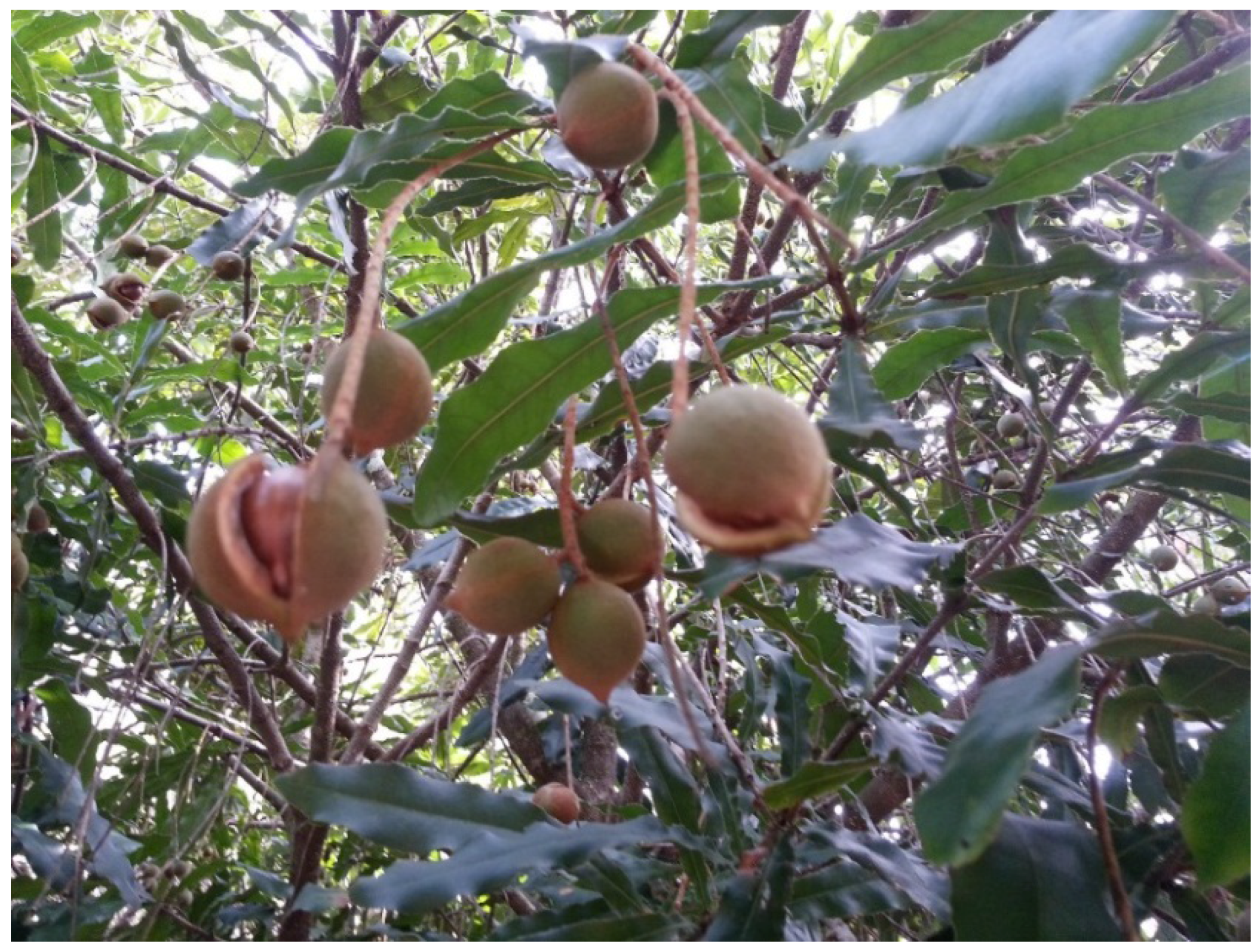
2.1. Materials

Macadamia (Macadamia tetraphylla) nuts were collected from the Central Coast region, New South Wales, Australia (latitude of 33.4° S, longitude of 151.4° E) in July of 2014. The skin of the nuts was separated from the harvested nuts and then frozen in liquid nitrogen immediately and freeze dried (FD3 freeze dryer, Thomas Australia Pty. Ltd., Seven Hills, NSW, Australia) in order to minimise oxidation or degradation of phenolic compounds. The dried skin was then ground into small particle sizes using a commercial blender (John Morris Scientific, Chatswood, NSW, Australia) and then sieved using a steel mesh sieve (1.4 mm EFL 2000; Endecotts Ltd., London, UK). The dried ground skin was kept in a sealed and labelled container at 5 °C until further analysed.

2.2. Chemicals

All chemicals used in this study were analytical grade. Methanol and potassium persulfate were purchased from Merck (Damstadt, Germany). Folin-ciocalteau phenol regent, anhydrous sodium carbonate, sodium nitrile, ferric chloride, gallic acid, rutin, catechin, neocuproine, 2,4,6-Tris(2-pyridyl)-s-triazine, (±)-6-hydroxy-2,5,7,8-tetramethylchromane-2-carboxylic acid (trolox) and 2,2-Diphenyl-1-picrylhydrazyl (DPPH) were purchased from Sigma-Aldrich Co. (Castle Hill, New South Wales (NSW), Australia). Sodium acetate trihydrate was purchased from Government Stores Department (Sydney, NSW, Australia). Aluminium chloride was obtained from Ajax Chem. (Sydney, NSW, Australia) and hydrochloric acid was obtained from Lab-scan Ltd. (South Australia, Australia).

2.3. Microwave-Assisted Extraction (MAE)

Water was chosen as the extraction solvent as it is a safe and inexpensive solvent, and it is easily accessible when compared to other organic solvents [17]. The microwave extraction was conducted using a household microwave equipped with inverter technology (1200 W, Frequency 2450 MHz, Sharp Carousel, Abeno-ku, Osaka, Japan) at the pre-determined conditions that were designed by the Response Surface Methodology program for time (minutes), power (%, W), and sample-to-solvent ratio (g/100 mL). When the extraction was completed, the vessels were then immediately placed into an ice bath to cool to room temperature. The extracts were then filtered using filter paper (Lomb Scientific, Taren Point, NSW, Australia) and diluted for quantitative analysis.

2.4. Response Surface Methodology

Response Surface Methodology (RSM) software was used to design experiments and analyse results, via JMP software (Version 11) with a Box-Behnken design with three central point replicates. The optimum range of the microwave variables was preliminarily identified and the range for microwave time was 2.5–5.5 min, power was 30%–70% (360–840 W) and sample-to-solvent ratio was 2–8 g/100 mL. The independent variables and their code variable levels are shown in Table 1. The JMP software was also used to develop the model equation, to graph 3D plots and 2D contour plots of the responses, as well as predicting the optimum conditions of the independent variables.
Table 1. Box-Behnken design and the observed responses.
RunExperimental ConditionsExperimental Results
X1X2X3TPCFlavon-oidsProantho-cyanidinsABTSDPPHCUPRACFRAP
12.550263.7113.0716.34172.56200.21426.4258.39
24302111.4321.1037.63544.67479.79726.33119.07
3470269.4319.8810.38343.02396.25683.0164.89
45.550268.3523.3117.09289.65348.96606.2991.89
52.530535.766.2211.18147.32143.60287.3948.04
62.570548.2016.289.42308.87216.62360.8647.49
7450532.8826.2222.18352.19331.00583.1191.22
8450540.4635.0917.59319.28283.93475.9386.21
9450532.4036.6217.03298.35340.53718.1271.35
105.530544.1026.4118.35390.75287.67459.8386.24
115.570518.233.657.9846.6043.3399.8817.99
122.550830.2113.1717.43123.23138.00320.2042.74
13430833.2516.7422.32254.67240.19472.7268.69
14470843.0615.1434.97217.87258.69544.9575.21
155.550833.3617.0223.52190.58193.72526.64101.28
X1 (time, min), X2 (power, %, W) and X3 (sample-to-solvent ratio, g/100 mL). TPC (mg GAE/g of dried weight), Flavonoids (mg RUE/g of dried weight), Proanthocyanidins (mg CE/g of dried weight), ABTS (µM TE/g of dried weight), DPPH (µM TE/g of dried weight), CUPRAC (µM TE/g of dried weight) and FRAP (µM TE/g of dried weight).
To express the amount of phenolic compounds and the level of antioxidant properties as a function of the independent variables, a second-order polynomial equation must be employed [17]:
Y   =   β o + i = 1 k β i Χ i + ​  i = 1 i < j k 1 j = 2 k β i j Χ i Χ j ​  + ​  i = 1 k β i i Χ i 2
where various Xi values are independent variables affecting the responses Y; β0, βi, βii and βij are the regression coefficients for intercept, linear, quadratic, and interaction terms, respectively; and k is the number of variables.
The three independent variables were assigned as: X1 (time, min), X2 (power, %, W), and X3 (sample-to-solvent ratio, g/100 mL). Thus, the function containing these three independent variables is expressed as follows:
Y =   β 0 + β 1 X 1 + β 2 X 2 + β 3 X 3 + β 12 X 1 X 2 + β 13 X 1 X 3 + β 23 X 2 X 3 + β 11 X 1 2 + β 22 X 2 2 + β 33 X 3 2

2.5. Methods for the Determination of Chemical Properties

2.5.1. Total Phenolic Content (TPC)

The total phenolic content (TPC) was determined as described by Vuong et al. [18]. 1 mL of diluted sample was added with 5 mL of 10% (v/v) Folin-Ciocalteu reagent, followed by the addition of 4 mL of 7.5% (w/v) Na2CO3, then combined well on a vortex mixer and incubated in the dark at room temperature for one hour before the absorbance was measured at 760 nm using a UV spectrophotometer (Varian Australia Pty. Ltd., Mulgrave, Victoria, Australia). A standard curve was created using gallic acid and the results were then specified in mg of gallic acid equivalents per g of sample (mg GAE/g).

2.5.2. Total Flavonoids

The total flavonoid content was measured as described by Zhishen et al. [19]. 0.5 mL of diluted sample was added with 2 mL of H2O and 0.15 mL of 5% (w/v) NaNO2 and left at room temperature for 6 min. Next 0.15 mL of 10% (w/v) AlCl3 was added and left at room temperature for a further 6 min. Lastly 2 mL 4% (w/v) NaOH and 0.7 mL of H2O were added, and the final solution was mixed well and left at room temperature for a further 15 min before the absorbance was measured at 510 nm using a UV spectrophotometer. A standard curve was designed using rutin and the results were then specified in mg of rutin equivalents per gram of sample (mg RUE/g).

2.5.3. Proanthocyanidins

The amount of proanthocyanidins was determined as described by Li et al. [20]. 0.5 mL of diluted sample was added to 3 mL of 4% (w/v) of vanillin and then 1.5 mL of concentrated HCl was added and left at room temperature for 15 min before measurement of the absorbance at 500 nm using a UV spectrophotometer. A standard curve was designed through the use of catechin and the results were expressed as mg of catechin equivalents per gram of sample (mg CE/g).

2.6. Methods for the Determination of Antioxidant Properties

2.6.1. ABTS Radical Scavenging Capacity

ABTS radical scavenging capacity was determined according to the methods described by Thaipong et al. [21] and Kamonwannasit, et al. [22] with some modifications. A stock solution was prepared by adding 10 mL of 7.4 mM ABTS solution to 10 mL of 2.6 mM K2S2O8 and left at room temperature in the dark for 15 h, and then stored at −20 °C until required. The working solution was freshly prepared by mixing 1 mL of stock solution with 60 mL of methanol to obtain an absorbance value of 1.1 ± 0.02 at 734 nm. 0.15 mL of sample was added with 2.85 mL of the working solution and mixed, then left in the dark at room temperature for 2 h before its absorbance was measured at 734 nm using a UV-VIS spectrophotometer (Cary 50 Bio, Varian Australia Pty. Ltd.). A standard curve was designed through the application of trolox and the results were expressed as μMoles trolox equivalents per gram of dried sample (μM TE/g).

2.6.2. DPPH Radical Scavenging Activity

The DPPH radical scavenging activity was measured based on the method described by Thaipong et al. [21], with some modifications. A stock solution was prepared by dissolving 24 mg DPPH in 100 mL methanol and then stored at −20 °C until required. The fresh working solution was prepared by mixing 10 mL stock solution in 45 mL methanol to obtain an absorbance at 515 nm of 1.1 ± 0.02. 0.15 mL of sample was mixed with 2.85 mL of working solution and then left in the dark, at room temperature for 3 h before measuring the absorbance at 515 nm using the UV spectrophotometer. A standard curve was designed through the use of trolox and the results were expressed as μMoles of trolox equivalents per g of sample (μM TE/g), as seen with ABTS radical scavenging activity.

2.6.3. Cupric Reducing Antioxidant Capacity (CUPRAC)

CUPRAC was determined as described by Apak et al. [23] with a minor adjustment. 1 mL of CuCl2, 1 mL of neocuproine and 1 mL of NH4Ac were added and then 1.1 mL of diluted sample was added. After combining well, the mixture was incubated at room temperature for 1.5 h before measuring the absorbance at 450 nm using the UV spectrophotometer. A standard curve was designed through the use of trolox and the results were expressed as μMoles of trolox equivalents per g of sample (μM TE/g).

2.6.4. Ferric Reducing Antioxidant Power (FRAP)

FRAP was measured as described by Thaipong et al. [21] and Kamonwannasit et al. [22]. A working FRAP solution was prepared by mixing 300 mM Acetate buffer, 10 mM TPTZ in 40 mM HCl and 20 mM FeCl3 in the ratio of 10:1:1 and warmed at 37 °C in a water bath (Ratek Instruments Pty. Ltd., Boronia, Victoria, Australia) before using. To 0.15 mL of sample, 2.85 mL of the working FRAP solution was added and incubated at room temperature in the dark for 30 min, after which its absorbance was read at 593 nm. A standard curve was designed through the use of trolox and the results were expressed as μMoles trolox equivalents per gram of dried sample (μM TE/g).

2.7. Statistical Analyses

The statistical design program JMP (Version 11, SAS, Cary, NC, USA) was used to design the experiments and all the experiments were performed in triplicate. The program was used to create the model equation, to graph the 3D and 2D contour plots of the responses and to predict the optimum values for the independent variables. The Student’s t-test from SPSS software (Version 20, IBM, Armonk, NY, USA) was applied to compare the sample means. The differences between the sample means were chosen at the significance level of p < 0.05.

3. Results and Discussion

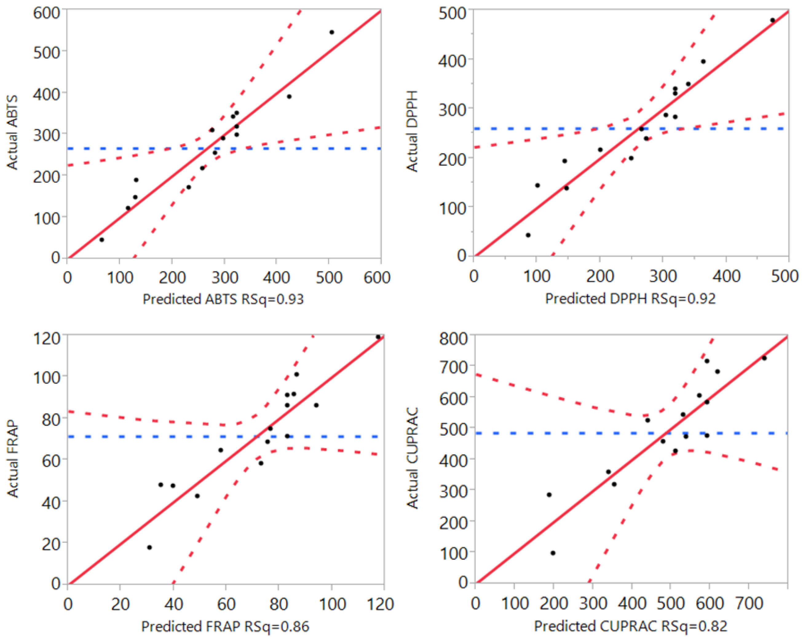
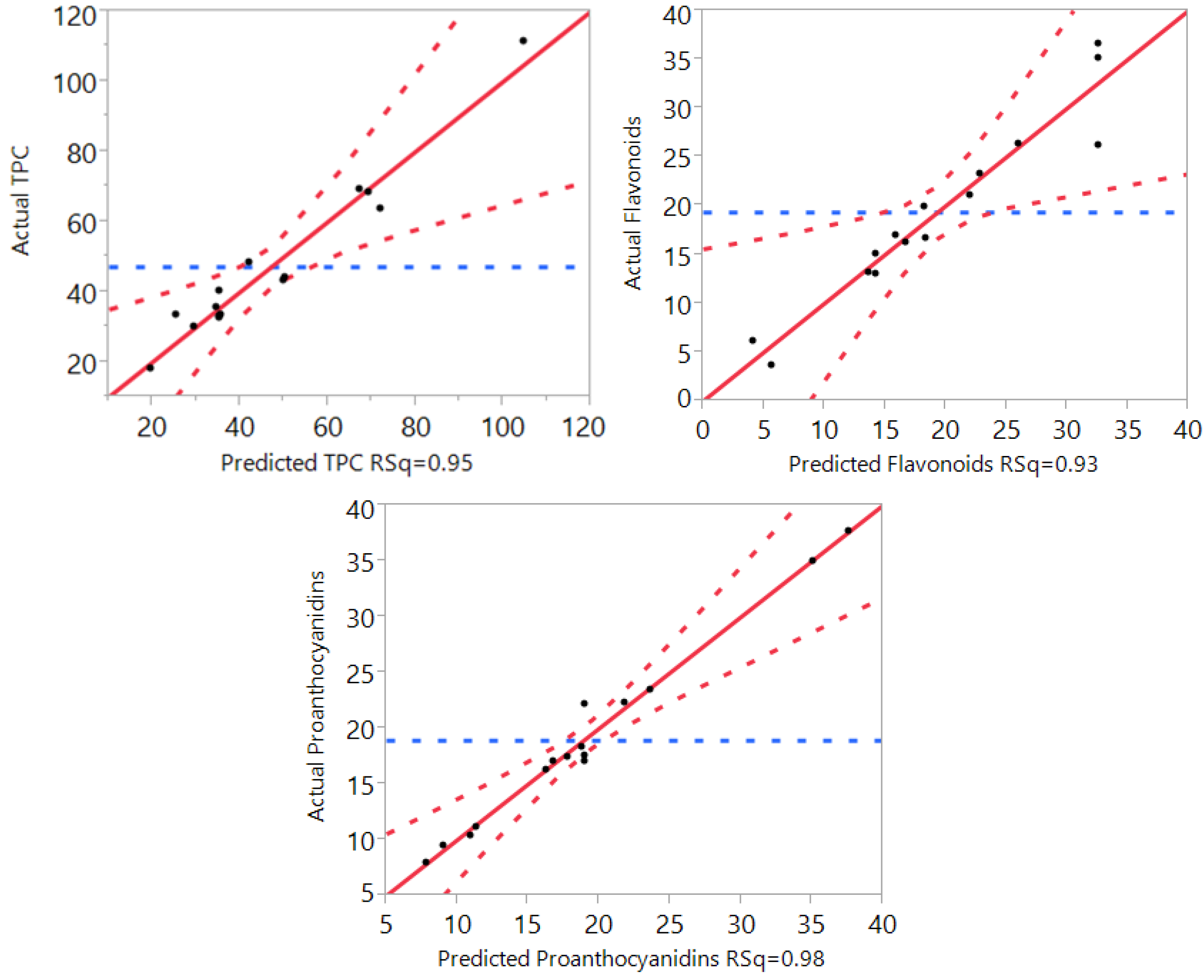
3.1. Statistical Analysis and Fitting of the Model

In order to ensure that the RSM mathematical models are reliable in the prediction of MAE conditions for TPC, flavonoids, proanthocyanidins, and the antioxidant capacity from the skin of the macadamia, different statistical analyses of variation including “lack of fit”, R squared, Predicted Residual Sum of Square (PRESS), F ratio, and Prob > F were identified and examined and the results are shown in Table 2. The “lack of fit” is able to calculate whether the model has the expected impact, and the R squared value is able to assess the proportion of variation that occurs in the response that is able to be accounted for by the model, rather than by random error, therefore an R squared value that nears 1 indicates that the model is a strong predictor of the response [24]. Results (Table 2) showed that “lack of fit” for phenolic compounds, flavonoids, proanthocyanidins, and four antioxidant assays were significantly higher than 0.05, meaning that the models for phenolic compounds and antioxidant properties were fitted and reliable for prediction of the actual values. Furthermore, R squared values for phenolic compounds and antioxidant properties were higher than 0.82, indicating that at least 82% of the predicted values could be matched with the actual values.
Table 2. Analysis of variance for the determination of model fitting.
TPCFlavon-oidsProantho-cyanidinsAntioxidant Capacity
ABTSDPPHCUPRACFRAP
Lack of fit0.1670.8920.9790.1360.9890.5250.239
R20.950.930.980.930.920.820.86
Adjusted R20.870.810.950.790.790.490.62
PRESS512943957234,529186,966800,86517,548
F ratio of Model11.137.6031.986.856.702.513.54
Prob > F0.010.020.0010.020.020.160.09
The PRESS value shows how well the predictive model fits each point in the design. The F Ratio is the test statistic for a test of whether the model differs significantly from a model where all predicted values are the response mean. Lastly, the Prob > F is able to measure the probability of actually obtaining an F ratio that is as high as the one that is being observed, in the case where all parameters are zero, except for the intercept. Smaller Prob > F values specify that the observed F ratio is highly unlikely [24]. The results (Table 2) showed that the PRESS, the F ratio and “Prob > F” for phenolic compounds, flavonoids, proanthocyanidins, and antioxidant properties all supported that the mathematical models for these responses are reliable for prediction of the values of these responses.
The results (Figure 2) further showed the correlation between the predicted values and the actual values. As seen from Figure 2, the predicted values for phenolic compounds, flavonoids and proathocyanidins were linear to their actual values, indicating a close relationship and further supporting that the mathematical models were reliable predictors for these responses.
Figure 2. The correlation between the predicted and the actual values for TPC, flavonoids, and proanthocyanidins.
Processes 04 00002 g002
The values (Y) for phenolic compounds, flavonoids and proanthocyanidins from the macadamia skin could be fitted to the below second-order polynomial Equations (3)–(5):
Y T P C = 35.244467 1.7289 X 1 5.700763 X 2 21.63091 X 3 9.576575 X 1 X 2 0.373125 X 1 X 3 + 12.95065 X 2 X 3 7.029333 X 1 2 + 8.3548417 X 2 2 + 20.692542 X 3 2
Y F l a v o n o i d s = 32.642667 + 2.7052375 X 1 1.941475 X 2 1.912388 X 3 8.205 X 1 X 2 1.597925 X 1 X 3 0.0948 X 2 X 3 10.53715 X 1 2 8.966521 X 2 2 5.463846 X 3 2
Y P r o a n t h o c y a n i d i n s = 18.933333 + 1.5706125 X 1 3.341063 X 2 + 2.10115 X 3 2.1531 X 1 X 2 + 1.335525 X 1 X 3 + 9.976425 X 2 X 3 7.465317 X 1 2 + 0.2666333 X 2 2 + 7.1262583 X 3 2
Figure 3 further illustrated the correlation between the predicted values and the actual values for the four types of antioxidant assays including DPPH, ABTS, FRAP, and CUPRAC. The predicted values were found to be linear with the actual values, with the R squared value for DPPH, ABTS, FRAP, and CUPRAC, of 0.93, 0.92, 0.86, and 0.82, respectively. These results further supported that the mathematical models were also appropriate for the prediction of the antioxidant values in the current study.
Figure 3. Correlation between the predicted and the actual values for ABTS total antioxidant capacity, DPPH free radical scavenging capacity, cupric reducing antioxidant power (CUPRAC), and ferric reducing antioxidant power (FRAP).
Processes 04 00002 g003
The models could be fitted to the following second-order polynomial Equations (6)–(9):
Y A B T S = 323.27111 + 20.698194 X 1 52.63111 X 2 70.44375 X 3 126.4244 X 1 X 2 12.4375 X 1 X 3 + 41.211111 X 2 X 3 122.969 X 1 2 + 23.082361 X 2 2 6.297917 X 3 2
Y D P P H = 318.48887 + 21.90625 X 1 29.54325 X 2 74.32553 X 3 79.3389 X 1 X 2 23.25695 X 1 X 3 + 25.51215 X 2 X 3 14.596 X 1 2 11.08731 X 2 2 + 36.329342 X 3 2
Y C U P R A C = 222.0527 + 10.75407 X 1 + 55.64071 X 2 67.3052 X 3 59.8078 X 1 + 8.335271 X 1 X 3 + 124.7403 X 2 X 3 + 10.0905 X 1 2 + 9.952907 X 2 2 + 104.4285 X 3 2
Y F R A P = 230.6576 + 49.23343 X 1 + 13.09773 X 2 36.3954 X 3 + 22.71136 X 1 X 2 4.04451 X 1 X 3 + 48.2822 X 2 X 3 + 57.01089 X 1 2 + 31.90199 X 2 2 + 6.542708 X 3 2

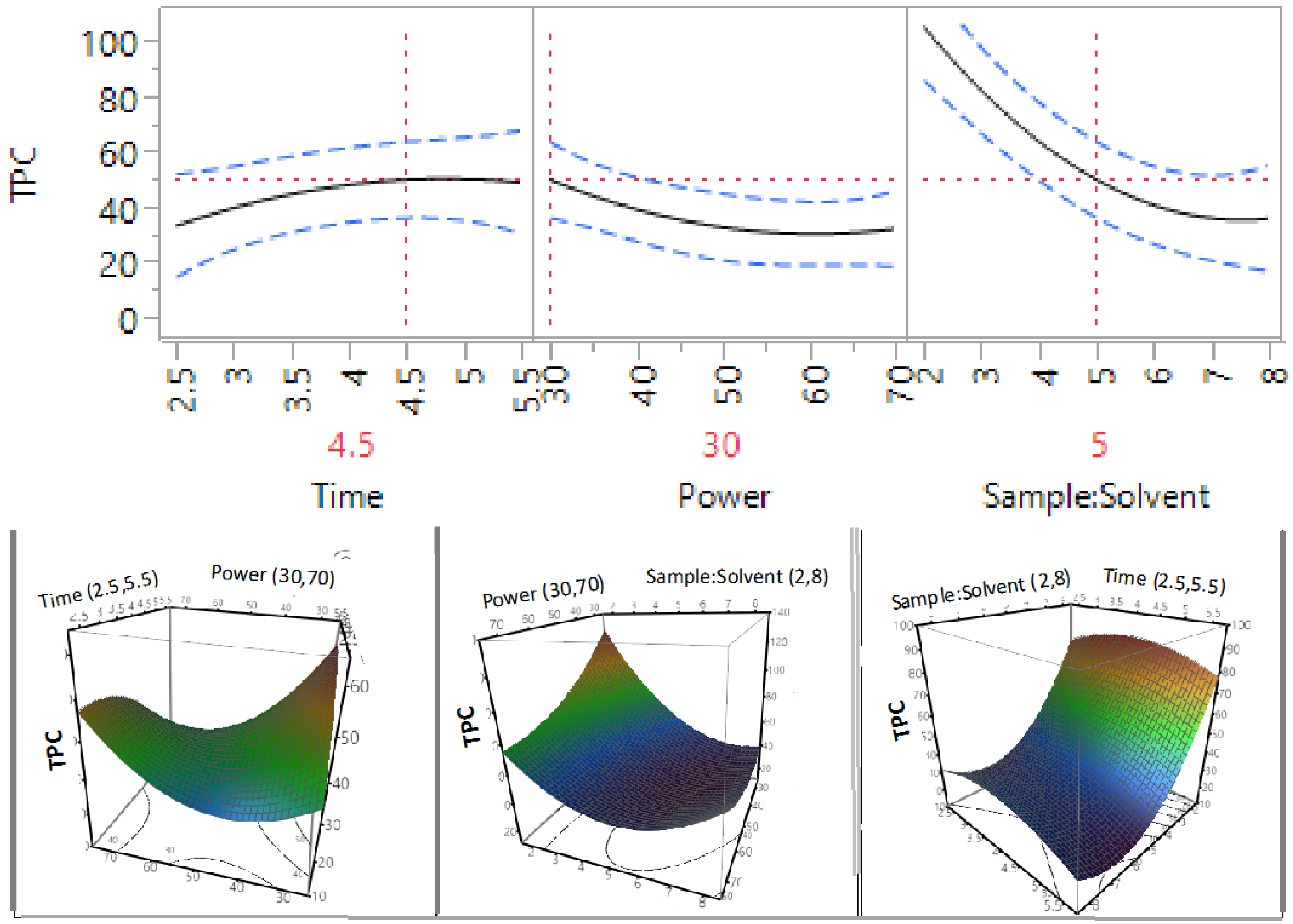
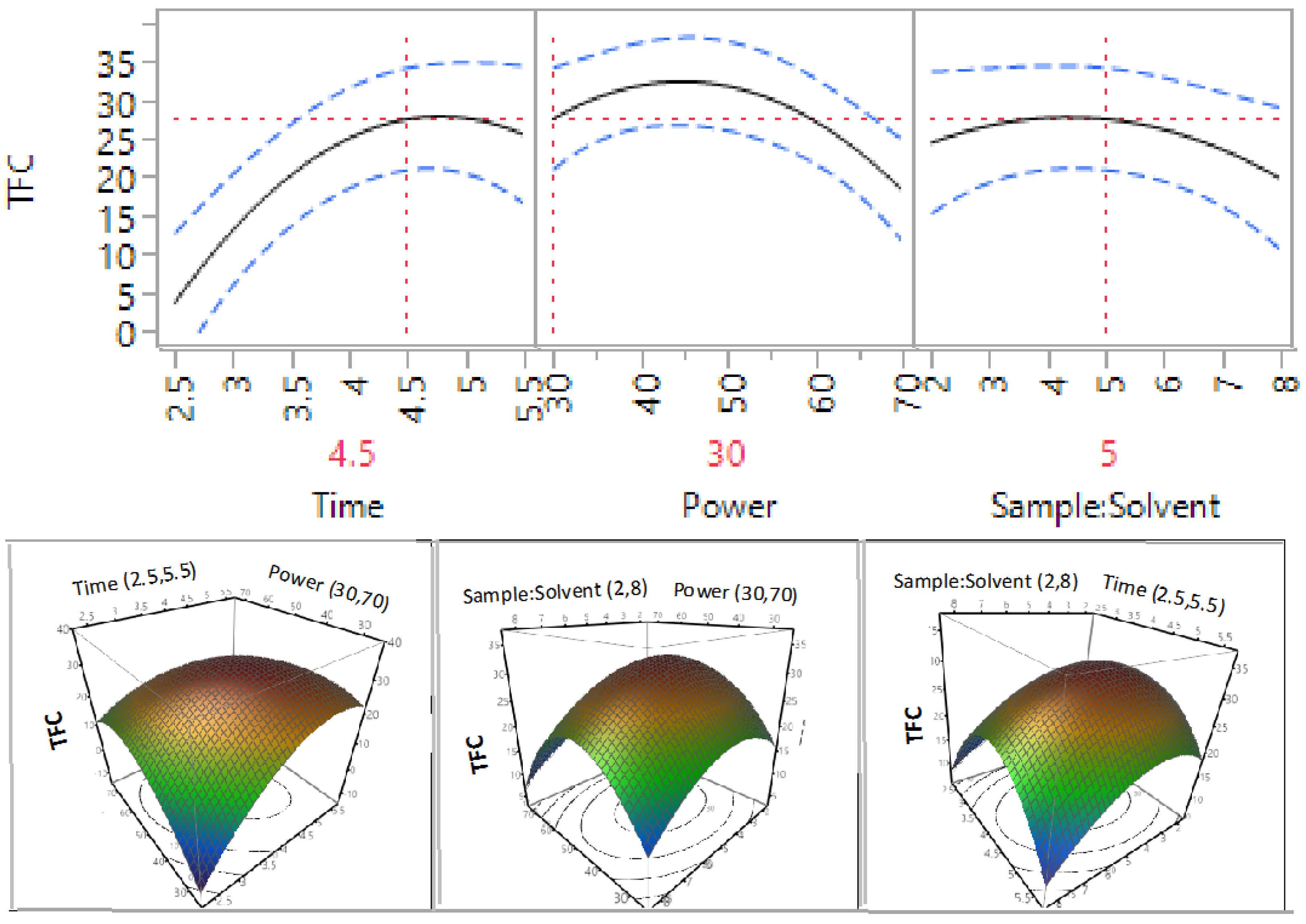
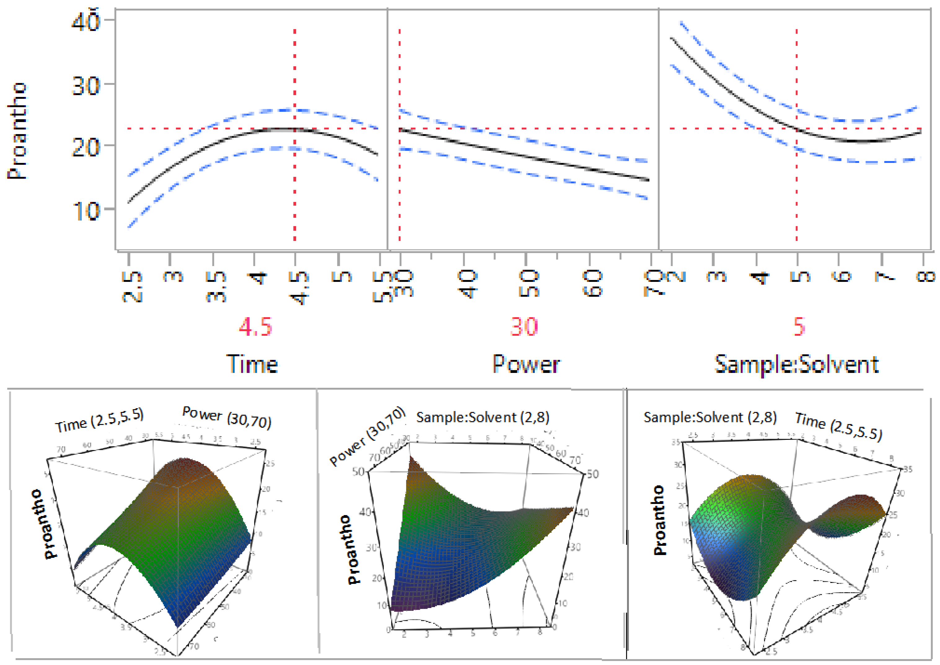
3.2. Effect of Extraction Independent Variables on Phenolic Compounds, Flavonoids, and Proanthocyanidins

The impact of MAE radiation time, power and sample-to-solvent ratio on the extraction of phenolic compounds (TPC) is shown in Figure 4 and Table 3.
Table 3. Analysis of variance for the experimental results on TPC, flavonoids, and proanthocyanidins.
ParameterDFTPCFlavonoidsProanthocyanidins
EstimateProb > |t|EstimateProb > |t|EstimateProb > |t|
β0135.240.0008 *32.64<0.0001 *18.93<0.0001 *
β11−1.730.58722.710.11681.570.0624
β21−5.700.1141−1.940.2322−3.340.0038 *
β31−21.630.0008 *−1.910.23832.100.0241 *
β121−9.580.0724−8.210.0097 *−2.150.0683
β131−0.370.9329−1.600.46481.340.2102
β23112.950.0278−0.090.96449.980.0001 *
β111−7.030.1702−10.540.0041 *−7.470.0006 *
β2218.350.1153−8.970.008 *0.270.7939
β33120.690.0053 *−5.460.0484 *7.130.0007 *
* Significantly different at p < 0.05; β0: Intercept; β1, β2, and β3: Linear regression coefficients for time, power and sample-to-solvent ratio; β12, β13, and β23: Regression coefficients for interaction between time x power, time x sample-to-solvent ratio and power x sample-to-solvent ratio; β11, β22, and β33: Quadratic regression coefficients for time x time, power X power and sample-to-solvent ratio x sample-to-solvent ratio.
Figure 4. Impact of time (2.5–5.5 min), power (30%–70%, 360–840 W) and sample-to-solvent ratio (2–8 g/100 mL) on TPC (mg GAE/g).
Processes 04 00002 g004
The results showed that microwave radiation time and power in the tested ranges did not significantly affect the extraction efficiency of TPC; however, the sample-to-solvent ratio was found to have a statistically significant impact on the extraction efficiency of TPC (p < 0.05). The TPC extraction efficiency decreased when a higher sample-to-solvent ratio was applied. These findings were supported by the previous studies on prune [25], apple pomace [26], Melissa officinali [27], and Eucalyptus robusta [12]. The impact of sample-to-solvent ratio on the extraction yield of TPC can be explained by the increase in the density of the sample in the solvent, which resulted in lower extraction efficiency [12].
The impact of MAE radiation time, power, and sample-to-solvent ratio on the extraction efficiency of flavonoids from macadamia skin is illustrated in Table 3 and Figure 5. The results indicated that all three extraction parameters, within the tested ranges, did not have a significant impact on the extraction efficiency of flavonoids, but the interaction between time and power significantly affected extraction efficiency of flavonoids (p < 0.05). Previous studies also found that MAE power and sample-to-solvent ratio did not significantly affect the extraction efficiency of flavonoids, but reported that the MAE time did have a significant impact [12]. The difference can be explained by the narrow range of the tested conditions; this narrow time range was not long enough to give a significant difference.
Figure 5. Impact of time (2.5–5.5 min), power (30%–70%, 360–840 W) and sample-to-solvent ratio (2–8 g/100 mL) on flavonoids (mg RE/g).
Processes 04 00002 g005
The impact of MAE radiation time, power and sample-to-solvent ratio on the extraction efficiency of proanthocyanidins is outlined in Table 3 and Figure 6. It can be seen from Table 3 that MAE time did not have a significant impact, but the power and the sample-to-solvent ratio had a significant impact on the level of extracted proanthocyanidins (p < 0.05). The higher the power or the sample-to-solvent ratio applied, the lower the extraction efficiency of proanthocyanidins was achieved (Figure 6). These findings were also in agreement with the previous study on Eucalyptus robusta [12]. The increase of power resulted in a lowering of the extraction efficiency that can be explained by the degradation of proanthocyanidins at higher temperatures caused by the higher power.
Figure 6. Impact of time (2.5–5.5 min), power (30%–70%, 360–840 W) and sample-to-solvent ratio (2–8 g/100 mL) on proanthocyanidins (mg CAE/g).
Processes 04 00002 g006

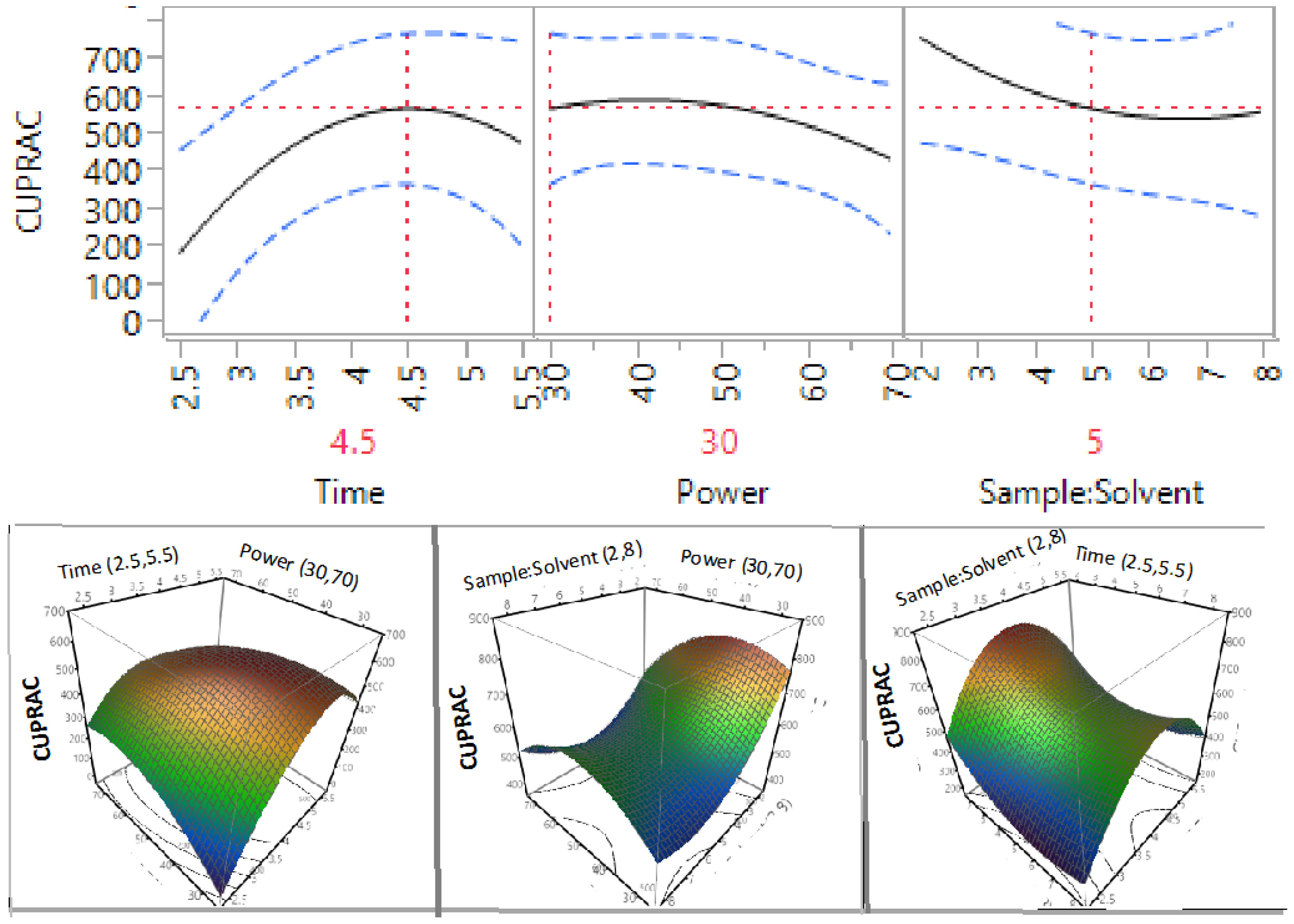
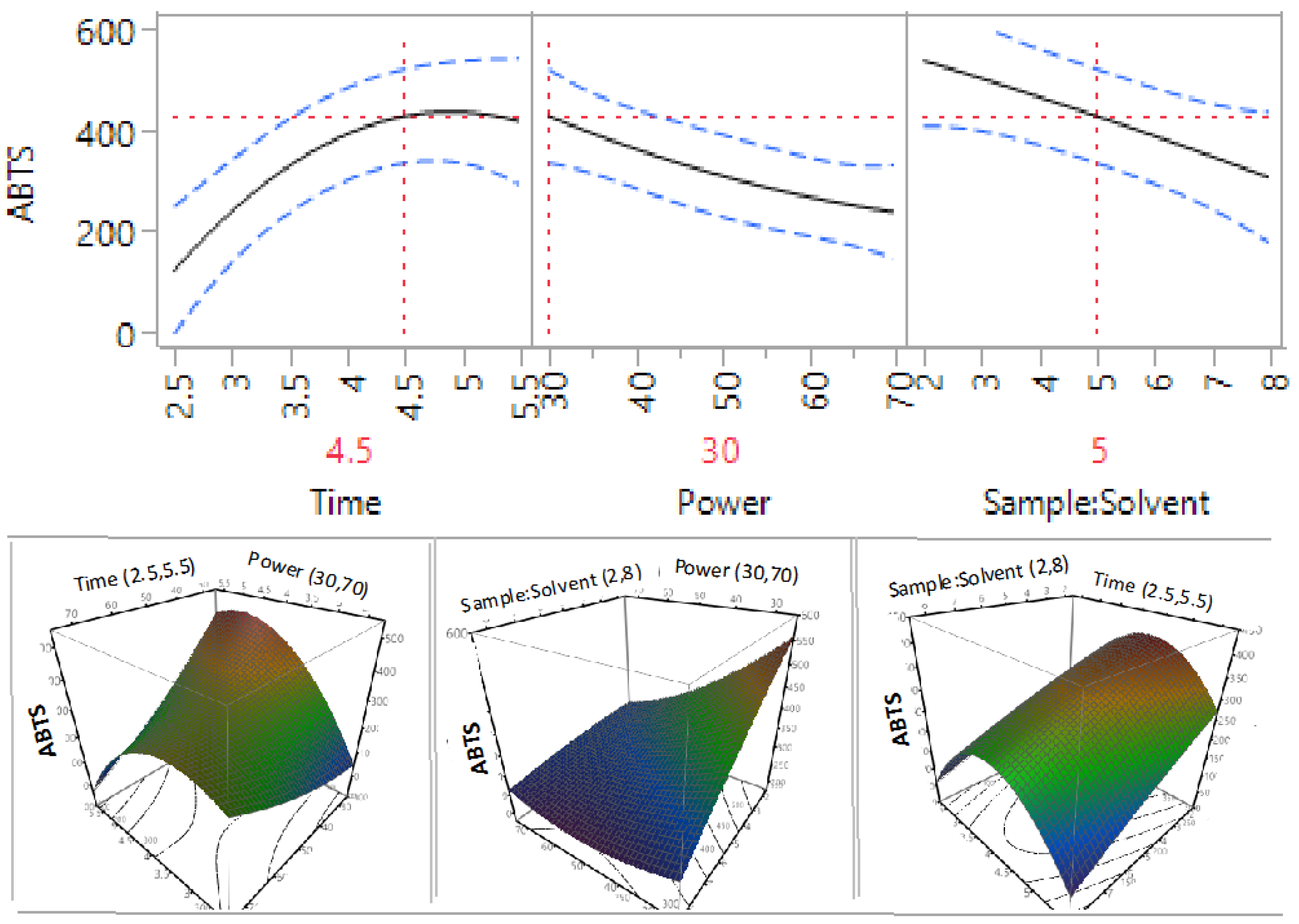
3.3. Effect of Extraction Independent Variables on the Antioxidant Capacity of Macadamia Tetraphylla Skin

Four antioxidant assays were used in this study to determine the antioxidant capacity of the extracts prepared under a variety of extraction conditions from the skin of the macadamia. This is because each antioxidant assay has its own advantages and limitations [28]. For example, ABTS antioxidant assay can be used over a large pH range, with a variety of solvents. Whereas, many antioxidants that typically react with peroxyl radicals may react slower, or not at all, with DPPH [29]. In addition, FRAP antioxidant assay only measures the reducing capability of the sample, based upon the ferric ion [29]. Therefore, more than one antioxidant assays were applied to obtain a better estimation for the antioxidant capacity.
Table 4. Analysis of variance for the experimental results on antioxidant capacity.
ParameterDFABTSDPPHCUPRACFRAP
EstimateProb > |t|EstimateProb > |t|EstimateProb > |t|EstimateProb>|t|
β01323.270.0002 *318.490.0001 *592.380.0004 *82.930.0003*
β1120.700.347121.910.282837.220.430312.590.0765
β21−52.630.0461 *−29.540.1656−32.200.4916−14.560.0498 *
β31−70.440.0167 *−74.330.0095 *−72.190.1572−5.790.3531
β121−126.420.0065 *−79.340.0274 *−108.360.1378−16.930.088
β131−12.440.6778−23.260.40796.640.91816.260.4695
β23141.210.20425.510.367328.890.657815.180.1163
β111−122.970.0086 *−134.600.004 *−213.630.0205 *−20.690.0555
β22123.080.4675−11.090.6963−76.770.2834−12.300.1997
β331−6.300.838736.330.233391.130.213111.340.2316
* Significantly different at p < 0.05; β0: Intercept; β1, β2, and β3: Linear regression coefficients for time, power and sample-to-solvent ratio; β12, β13, and β23: Regression coefficients for interaction between time x power, time x sample-to-solvent ratio and power x sample-to-solvent ratio; β11, β22, and β33: Quadratic regression coefficients for time x time, power X power, and sample-to-solvent ratio x sample-to-solvent ratio.
The impact of MAE radiation time, power, and sample-to-solvent ratio on the ABTS antioxidant capacity of the macadamia skin is represented in Table 4 and Figure 7. The results showed that the MAE radiation time did not have a significant impact, but the MAE power and the sample-to-solvent ratio did have a significant impact on the ABTS antioxidant capacity of the macadamia skin extract (p < 0.05). The higher the MAE power and sample-to-solvent ratio that were applied, the lower the antioxidant capacity obtained was. The results also showed that the interaction between time x sample-to-solvent ratio, and power x sample-to-solvent ratio did not have a significant impact, but the interaction between MAE time x power had a significant impact on ABTS antioxidant capacity of the macadamia skin extract.
Figure 7. Impact of time (2.5–5.5 min), power (30%–70%, 360–840 W) and sample-to-solvent ratio (2–8 g/100 mL) on ABTS antioxidant capacity (µM TE/g).
Processes 04 00002 g007
Figure 8. Impact of time (2.5–5.5 min), power (30%–70%, 360–840 W) and sample-to-solvent ratio (2–8 g/100 mL) on DPPH antioxidant capacity (µM TE/g).
Processes 04 00002 g008
The results (Table 4 and Figure 8) illustrated the impact of MAE radiation time, power, and sample-to-solvent ratio on the DPPH free radical scavenging capacity of the macadamia skin extract. MAE radiation time and power were found not to significantly affect the DPPH, but the sample-to-solvent ratio did significantly affect the DPPH free radical scavenging capacity of the macadamia skin extract (p < 0.05). As seen in the ABTS assay, the interaction between time x sample-to-solvent ratio, and power x sample-to-solvent ratio did not have a significant impact, but the interaction between MAE time x power had a significant impact on ABTS antioxidant capacity of the macadamia skin extract (p < 0.05).
Figure 9. Impact of time (2.5–5.5 min), power (30%–70%, 360–840 W) and sample-to-solvent ratio (2–8 g/100 mL) on CUPRAC (µM TE/g).
Processes 04 00002 g009
Figure 10. Impact of time (2.5–5.5 min), power (30%–70%, 360–840 W) and sample-to-solvent ratio (2–8 g/100 mL) on FRAP antioxidant capacity (µM TE/g).
Processes 04 00002 g010
The influence of MAE radiation time, power and sample-to-solvent ratio on the cupric ion reducing antioxidant capacity (CUPRAC) of the macadamia skin is represented in Table 4 and Figure 9. The results (Table 4) revealed that MAE radiation time, power, and sample-to-solvent ratio did not have a significant impact on the CUPRAC of the macadamia skin (p > 0.05). Similarly, there was no significant impact between the interaction of MAE time x power, time x sample-to-solvent ratio, and power x sample-to-solvent ratio of the macadamia skin extract (p > 0.05).
Finally, the results (Table 4 and Figure 10) indicated the impact of MAE radiation time, power, and sample-to-solvent ratio on the FRAP of the macadamia skin extract. MAE power was found to have a significant impact on the FRAP of the macadamia skin extract; whereas, MAE radiation time and sample-to-solvent ratio did not have a significant effect. The results (Table 4) also showed that there was no significant impact between the interaction of MAE time x power, time x sample-to-solvent ratio, and power x sample-to-solvent ratio on FRAP of the macadamia skin extract (p > 0.05).

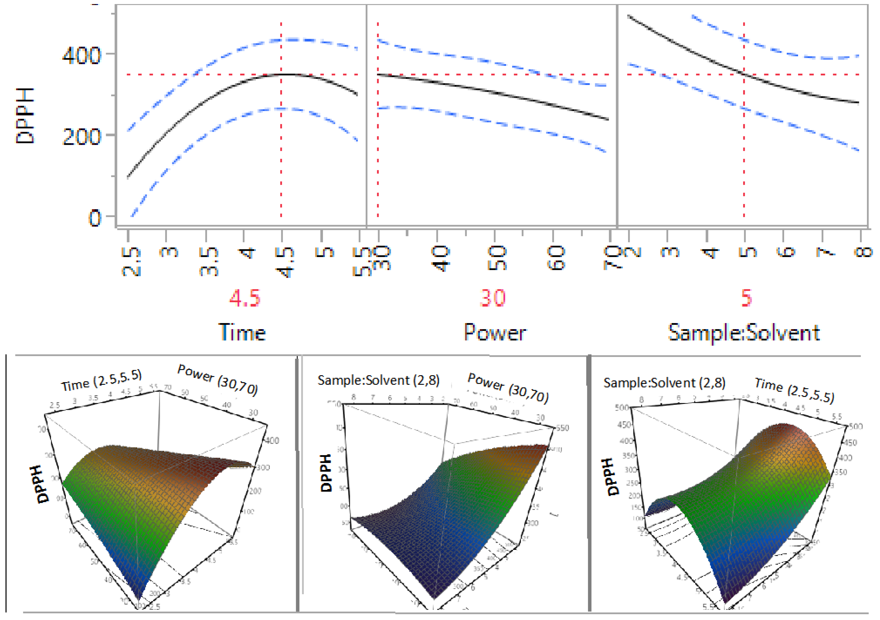
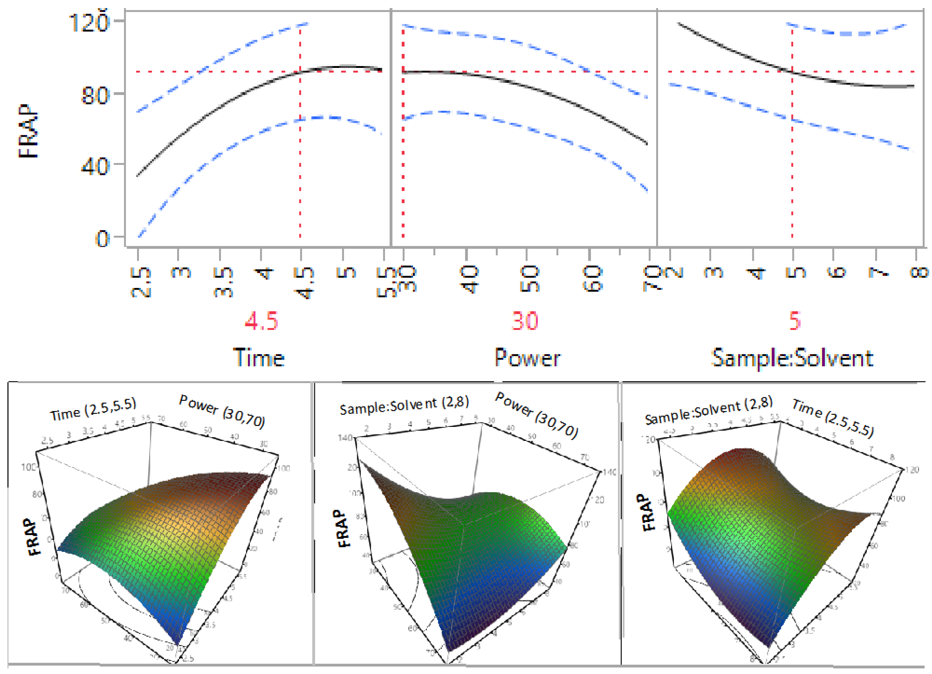
3.4. Optimisation and Validation of Microwave Extraction Conditions

The predicted mathematical models of this study indicated that the optimal extraction conditions for the highest level of TPC, flavonoids, proanthocyanidins, and antioxidant properties were MAE time of 4.5 min, power of 30% (360 W), and a sample-to-solvent ratio of 5 g/100 mL. To ensure that the results of the predicted conditions were matched with the results when these conditions were applied in reality, the sample of macadamia skin was extracted under the recommended conditions in triplicates. The actual results and the predicted results are shown in Table 5. As can be seen from the Table 5, all the experimental values for TPC, flavonoid, proanthocyanidin, and antioxidant properties were not significantly different to their predicted values (p > 0.05), indicating that these predicted conditions were valid and could be applied for maximum recovery of phenolic compounds and antioxidant properties from the macadamia skin.
Table 5. Validation of the predicted values for TPC, flavonoids, proanthocyanidins, and antioxidant potential.
Values
PredictedExperimental (n = 3)
TPC (mg GAE/g)51.13 ± 13.86 a44.75 ± 2.34 a
Flavonoids (mg RE/g)28.08 ± 6.64 a29.10 ± 1.04 a
Proanthocyanidins (mg GAE/g)22.95 ± 3.05 a33.60 ± 0.48 b
ABTS (µM TE/g)434.36 ± 92.71 a361.60 ± 14.22 a
DPPH (µM TE/g)355.74 ± 84.61 a292.78 ± 17.63 a
CUPRAC (µM TE/g)572.60 ± 201.70 a459.80 ± 51.75 a
FRAP (µM TE/g)92.73 ± 26.29 b297.03 ± 24.74 b
All the values are means ± standard deviations and those in the same row not sharing the same superscript letter (a or b) are significantly different from each other (p < 0.05).
Under these extraction conditions, approximately 45 mg of TPC, 29 mg of flavonoids and 33 mg of proanthocyanidins could be extracted from one gram of dried macadamia skin. Alasalvar and Shahidi [30] reported that one gram of macadamia kernel contained 1.56 mg of the phenolic compounds, meaning that the level of phenolic compounds in the macadamia skin is significantly higher than that in the kernel. In addition, Yang [31] reported the flavonoid content of the macadamia kernel was 1.379 mg/g, which was less than 5% of the flavonoids available in the skin of the macadamia, revealing that macadamia skin is the waste, but it is a rich source of phenolic compounds. Furthermore, Alasalvar and Shahidi [30] also reported a FRAP antioxidant value for macadamia kernel was 0.42 mM/100 g or 4.2 µM TE/g, which is also significantly lower than the FRAP values found in macadamia skin in the current study. Therefore, these findings further confirmed that macadamia skin is a rich source of phenolic content and is also a potent source of antioxidants in comparison with its kernel.
In comparison with the conventional extraction method at optimal conditions of 90 °C, 20 min, and sample-to-solvent ratio of 5 g/100 mL, which could extract 96 mg of TPC, 24 mg of flavonoids, and 97 mg of proanthocyanidins from a dried gram of macadamia skin [32], MAE method under these optimal conditions gave significant lower levels of TPC, flavonoids and proanthocyanidins. The reason for the low recovery yields of bioactive compounds when using MAE method can be explained by the heat, which was generated during extraction process. As the bioactive compounds from the macadamia skin were sensitive to the high temperature, thus they were partially degraded during the extraction process.

4. Conclusions

The results showed that MAE time, MAE power, and sample-to-solvent ratio could impact on the extraction efficiency of TPC, flavonoids, proathocyanidins, and antioxidant properties of the skin of the macadamia tetraphylla, however, the degree of effect was varied. Within the tested ranges, sample-to-solvent ratio had a significant impact on the extraction efficiency of TPC and proanthocyanidins, while MAE power had a significant impact on the extraction of proanthocyanidins. Both power and sample-to-solvent ratio were found to have a significant impact on ABTS antioxidant capacity, while only sample-to-solvent ratio was found to have a significant effect on DPPH, and power had a significant influence on FRAP. The optimal MAE conditions for maximum recovery of TPC, flavonoids, proanthocyanidins, and antioxidant levels from the skin of the macadamia using water were MAE time of 4.5 min, power of 30% (360 W) and sample-to-water ratio of 5 g/100 mL. At these conditions, approximately 45 mg of TPC, 29 mg of flavonoids, and 33 mg of proanthocyanidins could be extracted from one gram of dried macadamia skin.

Acknowledgments

The authors would like to thank the Faculty of Science and IT of the University of Newcastle for financing this project.

Author Contributions

Q.V.V. and A.D. designed experiments. A.D. conducted experiments. Q.V.V. and A.D. analysed data and developed this manuscript.

Conflicts of Interest

The authors declare no conflict of interest.

References

  1. Baiano, A. Recovery of biomolecules from food wastes—A review. Molecules 2014, 19, 14821–14842. [Google Scholar] [CrossRef] [PubMed]
  2. Laufenberg, G.; Kunz, B.; Nystroem, M. Transformation of vegetable waste into value added products: (A) the upgrading concept; (B) practical implementations. Bioresour. Technol. 2003, 87, 167–198. [Google Scholar] [CrossRef]
  3. Munro, I.A.; Garg, M.L. Nutrient composition and health beneficial effects of macadamia nuts. In Tree Nuts; CRC Press: Boca Raton, FL, USA, 2008. [Google Scholar]
  4. Australian-Macadamia-Society. The Macadamia Industry. Available online: http://www.australian-macadamias.org/industry/about-aussie-macadamias/the-macadamia-industry?lang=en (accessed on 11 November 2015).
  5. Poinern, G.E.J.; Senanayake, G.; Shah, N.; Thi-Le, X.N.; Parkinson, G.M.; Fawcett, D. Adsorption of the aurocyanide, complex on granular activated carbons derived from macadamia nut shells—A preliminary study. Miner. Eng. 2011, 24, 1694–1702. [Google Scholar] [CrossRef]
  6. Wechsler, A.; Ramirez, M.; Crosky, A.; Zaharia, M.; Jones, H.; Ballerini, A.; Nunez, M.; Sahajwalla, V. Physcial properties of furniture panels from macadamia shells. In Proceedings of the 18th International Conferences on Composite Materials, Jeju Island, Korea, 21–26 August 2011.
  7. Apak, R.; Güçlü, K.; Demirata, B.; Özyürek, M.; Çelik, S.; Bektaşoğlu, B.; Berker, K.; Özyurt, D. Comparative evaluation of various total antioxidant capacity assays applied to phenolic compounds with the cuprac assay. Molecules 2007, 12, 1496–1547. [Google Scholar] [CrossRef] [PubMed]
  8. Bernal, J.; Mendiola, J.A.; Ibáñez, E.; Cifuentes, A. Advanced analysis of nutraceuticals. J. Pharm. Biomed. Anal. 2011, 55, 758–774. [Google Scholar] [CrossRef] [PubMed]
  9. Arcan, I.; Yemenicioğlu, A. Incorporating phenolic compounds opens a new perspective to use zein films as flexible bioactive packaging materials. Food Res. Int. 2011, 44, 550–556. [Google Scholar] [CrossRef]
  10. Huang, W.Y.; Cai, Y.Z.; Zhang, Y. Natural phenolic compounds from medicinal herbs and dietary plants: Potential use for cancer prevention. Nutr. Cancer 2009, 62, 1–20. [Google Scholar] [CrossRef] [PubMed]
  11. Kammerer, D.R.; Kammerer, J.; Valet, R.; Carle, R. Recovery of polyphenols from the by-products of plant food processing and application as valuable food ingredients. Food Res. Int. 2014, 65, 2–12. [Google Scholar] [CrossRef]
  12. Bhuyan, D.J.; van Vuong, Q.; Chalmers, A.C.; van Altena, I.A.; Bowyer, M.C.; Scarlett, C.J. Microwave-assisted extraction of eucalyptus robusta leaf for the optimal yield of total phenolic compounds. Ind. Crops Prod. 2015, 69, 290–299. [Google Scholar] [CrossRef]
  13. Santana, C.M.; Ferrera, Z.S.; Padrón, M.E.T.; Rodríguez, J.J.S. Methodologies for the extraction of phenolic compounds from environmental samples: New approaches. Molecules 2009, 14, 298–320. [Google Scholar] [CrossRef] [PubMed]
  14. Karabegović, I.T.; Stojičević, S.S.; Veličković, D.T.; Nikolić, N.Č.; Lazić, M.L. Optimization of microwave-assisted extraction and characterization of phenolic compounds in cherry laurel (Prunus laurocerasus) leaves. Sep. Purif. Technol. 2013, 120, 429–436. [Google Scholar] [CrossRef]
  15. Qu, W.; Shi, S.; Li, P.; Pan, Z.; Venkitasamy, C. Extraction kinetics and properties of proanthocyanidins from pomegranate peel. Int. J. Food Eng. 2014, 10, 683–695. [Google Scholar] [CrossRef]
  16. Meyers, R.H.; Montgomery, D.C. Response Surface Methodology: Process and Product Optimization Using Designed Experiments, 2nd ed.; Wiley: New York, NY, USA, 2002. [Google Scholar]
  17. Vuong, Q.V.; Stathopoulos, C.E.; Golding, J.B.; Nguyen, M.H.; Roach, P.D. Optimum conditions for the water extraction of l-theanine from green tea. J. Sep. Sci. 2011, 34, 2468–2474. [Google Scholar] [CrossRef] [PubMed]
  18. Vuong, Q.; Goldsmith, C.; Dang, T.; Nguyen, V.; Bhuyan, D.; Sadeqzadeh, E.; Scarlett, C.; Bowyer, M. Optimisation of ultrasound-assisted extraction conditions for phenolic content and antioxidant capacity from euphorbia tirucalli using response surface methodology. Antioxidants 2014, 3, 604–617. [Google Scholar] [CrossRef]
  19. Zhishen, J.; Mengcheng, T.; Jianming, W. The determination of flavonoid contents in mulberry and their scavenging effects on superoxide radicals. Food Chem. 1999, 64, 555–559. [Google Scholar] [CrossRef]
  20. Li, Y.; Guo, C.; Yang, J.; Wei, J.; Xu, J.; Cheng, S. Evaluation of antioxidant properties of pomegranate peel extract in comparison with pomegranate pulp extract. Food Chem. 2006, 96, 254–260. [Google Scholar] [CrossRef]
  21. Thaipong, K.; Boonprakob, U.; Crosby, K.; Cisneros-Zevallos, L.; Hawkins Byrne, D. Comparison of ABTS, DPPH, FRAP, and ORAC assays for estimating antioxidant activity from guava fruit extracts. J. Food Compos. Anal. 2006, 19, 669–675. [Google Scholar] [CrossRef]
  22. Kamonwannasit, S.; Nantapong, N.; Kumkrai, P.; Luecha, P.; Kupittayanant, S.; Chudapongse, N. Antibacterial activity of Aquilaria crassna leaf extract against Staphylococcus epidermidis by disruption of cell wall. Ann. Clin. Microbiol. Antimicrob. 2013, 12, 20. [Google Scholar] [CrossRef] [PubMed]
  23. Apak, R.; Güçlü, K.; Özyürek, M.; Karademir, S.E. Novel total antioxidant capacity index for dietary polyphenols and vitamins C and E, using their cupric ion reducing capability in the presence of neocuproine: CUPRAC method. J. Agric. Food Chem. 2004, 52, 7970–7981. [Google Scholar] [CrossRef] [PubMed]
  24. JMP. Regression Reports. Available online: http://www.jmp.com/support/help/Regression_Reports.shtml (accessed on 28 October 2015).
  25. Haddadi-Guemghar, H.; Janel, N.; Dairou, J.; Remini, H.; Madani, K. Optimisation of microwave-assisted extraction of prune (Prunus domestica) antioxidants by response surface methodology. Int. J. Food Sci. Technol. 2014, 49, 2158–2166. [Google Scholar] [CrossRef]
  26. Bai, X.-L.; Yue, T.-L.; Yuan, Y.-H.; Zhang, H.-W. Optimization of microwave-assisted extraction of polyphenols from apple pomace using response surface methodology and HPLC analysis. J. Sep. Sci. 2010, 33, 3751–3758. [Google Scholar] [CrossRef] [PubMed]
  27. Ince, A.E.; Şahin, S.; Şümnü, S.G. Extraction of phenolic compounds from melissa using microwave and ultrasound. Turk. J. Agric. For. 2013, 37, 69–75. [Google Scholar]
  28. Pisoschi, A.M.; Negulescu, G.P. Methods for total antioxidant activity determination: A review. Biochem. Anal. Biochem. 2011, 1, 1–5. [Google Scholar] [CrossRef]
  29. Prior, R.L.; Wu, X.; Schaich, K. Standardized methods for the determination of antioxidant capacity and phenolics in foods and dietary supplements. J. Agric. Food Chem. 2005, 53, 4290–4302. [Google Scholar] [CrossRef] [PubMed]
  30. Alasalvar, C.; Shahidi, F. Natural antioxidants in tree nuts. Eur. J. Lipid Sci. Technol. 2009, 111, 1056–1062. [Google Scholar] [CrossRef]
  31. Yang, J. Brazil nuts and associated health benefits: A review. LWT Food Sci. Technol. 2009, 42, 1573–1580. [Google Scholar] [CrossRef]
  32. Dailey, A.; Vuong, Q.V. Optimization of aqueous extraction conditions for recovery of phenolic content and antioxidant properties from macadamia (Macadamia tetraphylla) skin waste. Antioxidants 2015, 4, 699–718. [Google Scholar]

