
Article

Bioinspired Melittin-Derived Antimicrobial Peptides with Enhanced Selectivity Indexes

by
Lucas O. Rodrigues
1,†,
Letícia O. C. Nunes
1,2,†,
Ariani R. Aragão
1,
Amanda K. Surur
2,
Marcela N. Argentin
3,
Vitória T. Candido
3,
Leticia R. Casado
1,
Louise O. Fiametti
1,2,
Gabriel F. Hispagnol
1,
Ilana L. B. C. Camargo
3,
Carla R. Fontana
2,
Eduardo F. Vicente
4 and
Norival A. Santos-Filho
1,2,*
1
Institute of Chemistry, Sao Paulo State University, Araraquara 14800-060, Brazil
2
School of Pharmaceutical Sciences, Sao Paulo State University, Araraquara 14800-903, Brazil
3
Institute of Physics, University of Sao Paulo, Sao Carlos 13566-590, Brazil
4
School of Sciences and Engineering, Sao Paulo State University, Tupa 17602-496, Brazil
*
Author to whom correspondence should be addressed.
These authors contributed equally to this work.
Processes 2026, 14(10), 1630; https://doi.org/10.3390/pr14101630
Submission received: 26 March 2026 / Revised: 11 May 2026 / Accepted: 13 May 2026 / Published: 18 May 2026

Abstract

Antimicrobial peptides such as Melittin exhibit potent broad-spectrum activity but are limited by high cytotoxicity. The rational design of bioinspired Melittin-derived analogues represents a promising strategy to reduce toxicity while maintaining antimicrobial efficacy. In this study, Melittin and analogues (TT-1, FKW, and WKW) were synthesized using solid-phase peptide synthesis (SPPS) and characterized for biological and biophysical essays. Antimicrobial and hemolytic activity, serum stability, secondary structure, and membrane interaction were analysed. FKW and WKW exhibited broad-spectrum antimicrobial activity, with minimum inhibitory concentration (MIC) as low as 8 and 16 µg/mL against Staphylococcus aureus and Enterococcus faecium. Both analogues also showed improved activity against Klebsiella pneumoniae (32 µg/mL) and Pseudomonas aeruginosa (128 and 256 µg/mL for FKW and WKW, respectively) compared to Melittin (64 and 512 µg/mL). In terms of cytotoxicity, FKW and WKW showed significantly reduced hemolytic activity, with HC50 values of 264.8 µg/mL and 237.2 µg/mL, respectively, resulting in improved selectivity indexes relative to Melittin (HC50 of 7.9 µg/mL). In liposomes, both adopt α-helical structures and cause disruption via pore formation or detergent-like mechanisms. TT-1 showed minimal toxicity but weak antimicrobial activity (MIC > 256 µg/mL). Although FKW and WKW exhibited limited serum stability (half-lives of 2.2 and 1.5 h), their degradation may reduce systemic toxicity. Overall, these analogues demonstrate an improved balance between antimicrobial activity and safety.

1. Introduction

The World Health Organization (WHO) designates antimicrobial resistance as a major global health threat, associated with increased hospitalization rates, extended durations of hospital stay, escalated healthcare costs, and elevated mortality rates [1].
Antimicrobial resistance arises when microorganisms develop mechanisms that render them resistant to antimicrobial agents, including antibiotics routinely used to treat infections. It is estimated that by 2050, antimicrobial resistance will cause 10 million deaths and incur economic losses of over 100 trillion dollars, underscoring the need for novel and effective therapies [2,3,4,5,6].
WHO has categorized bacterial pathogens requiring urgent attention and necessitating novel therapeutic interventions, including ESKAPE pathogens (Enterococcus faecium, Staphylococcus aureus, Klebsiella pneumoniae, Acinetobacter baumannii, Pseudomonas aeruginosa and Enterobacter species), known for their specific susceptibility profiles [7,8]. This classification was established based on criteria, such as mortality, treatability, healthcare burden, prevalence of resistance, and other relevant parameters. Within this group, Gram-negative bacteria such as carbapenem-resistant A. baumannii, third-generation cephalosporin-resistant Enterobacterales, and carbapenem-resistant Enterobacterales, are classified as critical priority pathogens, because of their high mortality rates and the limited availability of therapeutic interventions [9,10]. These pathogenic bacteria can inactivate drugs, modify therapeutic targets, and reduce the accumulation of antibiotics in their membranes. In addition, their capacity to form biofilms contributes to clinical persistence, highlighting the urgent need for novel therapeutic strategies [11].
Promising alternatives to conventional treatments are antimicrobial peptides (AMPs), present in a wide range of organisms, including animals and plants. Antimicrobial peptides exhibit considerable variability in their characteristics, including mechanisms of action (from immunomodulatory to direct microbicidal effects), origin (natural or synthetic) and secondary structure adaptability depending on the environment. Notably, many AMPs also demonstrate activity against planktonic bacteria or biofilms [12,13,14,15,16,17,18].
An AMP extensively documented in the scientific literature is the 26-amino acid, α-helical antimicrobial peptide named Melittin (sequence: GIGAVLKVLTTGLPALISWIKRKRQQ-NH2). It has demonstrated with antiviral, antibacterial, antifungal, antiparasitic, antitumor and various other biological activities, highlighting its significant therapeutic potential [19,20,21]. Despite this potential, Melittin is also known for its cytotoxicity, primarily due to its ability to disrupt plasma membranes of eukaryotic cells through non-selective mechanisms, leading to cellular damage [22].
Given the challenges associated with Melittin’s therapeutic use, its analogues have emerged as promising alternatives to reduce cytotoxic effects, decrease hemolytic activity, and improve selectivity. Modifications in the peptide sequence can contribute to a more precise molecular targeting and improved efficacy, ultimately aiming to reduce the side effects associated with its potential application [23,24,25,26].
Highlighted among the developed analogues is the peptide TT-1 (sequence: KIKAVLKVLTT), which is a shortened, 11-residue that retains amphipathic properties. TT-1 was designed by shortening the peptide chain and substituting the glycine residues with lysine. Furthermore, TT-1 exhibited increased hydrophobicity and reduced net charge, suggesting improved stability and lower toxicity compared with Melittin. This analogue has shown inhibitory activity against the proliferation of thyroid cancer cells without significantly affecting normal thyroid follicular epithelial cells, thereby indicating selective cytotoxicity toward cancer [26].
Thus, this study aimed to systematically evaluate whether the TT-1 peptide and its rationally designed analogues could achieve an improved balance between antimicrobial efficacy and selectivity, with particular emphasis on reducing hemolytic effects. Based on the TT-1 scaffold, specific amino acid substitutions were strategically introduced to modulate key physicochemical parameters, such as hydrophobicity, amphipathicity, and charge distribution, thereby enhancing preferential interactions with bacterial membranes over eukaryotic counterparts. Ultimately, this article aimed to identify optimized peptide candidates with retained or improved antibacterial activity relative to Melittin, while minimizing toxicity, contributing to the rational development of selective antimicrobial agents.

2. Materials and Methods

2.1. Peptide Synthesis

Solid phase peptide synthesis (SPPS) was performed manually as previously described [27], using the Fmoc (9-fluorenylmethyloxycarbonyl, C15H11ClO2, CAS 28920-43-6) protocol on Rink-Amide resin. Coupling and deprotection were evaluated using ninhydrin test [28]. Cleavage from the resin and removal of the side-chain protecting groups were performed simultaneously with 95% trifluoroacetic acid (TFA—C2HF3O2, CAS 76-05-1), 2.5% water, and 2.5% triisopropylsilane (TIS—C9H21Si, CAS 6485-79-6) for 2 h. Crude peptide samples were purified by semi preparative HPLC on a Shimadzu system (Shimadzu Corporation, Kyoto, Japan). Peptide purification was performed by semi-preparative HPLC, using a Shimadzu system, equipped with a UV–Vis detector, monitoring absorbance at 220 nm. The degree of purity of the fractions was determined on a C18 reversed-phase HPLC using a Shimadzu chromatograph, an analytical column (Kromasil, Bohus, Sweden) with dimensions of 0.46 × 25 cm, gradient of 5–95% of solution B in 30 min and flow of 1 mL/min. The solutions employed were 0.045% TFA in ultrapure water (solution A) and 0.036% TFA in acetonitrile (solution B). Chromatograms were generated using OriginPro 9.0 (OriginLab Corporation, Northampton, MA, USA). Mass spectra were obtained on an LCQ FLEET mass spectrometer (Thermo Fisher Scientific Inc., Waltham, MA, USA) equipped with direct injection in positive detection mode to analyse the correct peptide synthesis.

2.2. Peptide Design and Prediction of Physicochemical Properties

Peptides rationally designed based on the N-terminal region of the primary structure of Melittin were selected for peptide synthesis and evaluation of their antibacterial activity. To reach this goal, a combination of three strategies was established. (1) Initially, the sequence of the peptide TT-1 was selected as a template, since this molecule had already been described as having lower toxicity than Melittin [24]. However, since the antitumoral activity of this molecule was relatively low and its antimicrobial activity had not yet been reported, we aimed, as a secondary objective, to identify peptides with enhanced antimicrobial activity compared to the TT-1 peptide (which had already been characterized by low toxicity). Then, strategy (2) was performed. The CAMPR4 server [29] was employed to predict potential regions within the TT-1 peptide that could be modified to enhance its antimicrobial activity. Subsequently, peptides incorporating the point mutations suggested by the CAMPR4 server were designed and submitted to helical wheel and physicochemical analyses (step (3)). Sequences with a more clearly defined α-helical and amphipathic structure, featuring distinct polar and apolar faces, along with those having the highest hydrophobicity index and hydrophobic moment, were selected for synthesis and further analysis of their antimicrobial and hemolytic activities.
Physicochemical characteristics of the parent peptide and its derivatives were predicted using the Expasy server through protparam analysis (https://web.expasy.org/protparam/, accessed on 27 May 2025). Antimicrobial activity and rational design predictions were performed using the CAMPR4 (Collection of Anti-Microbial Peptides) server [29]. Antimicrobial activity prediction and rational design were performed using the CAMPR4, employing the “Rational Design” module. The “Synthetic peptides” dataset was selected as reference, and three prediction algorithms were applied: Random Forest (RF), Support Vector Machine (SVM), and Artificial Neural Network (ANN). The input sequence used for prediction was the TT-1 peptide (KIKAVLKVLTT). All possible amino acid substitutions were computationally generated by the server, and each variant was evaluated based on its predicted antimicrobial probability score. Output scores ranged from 0 to 1, with higher values indicating a greater likelihood of antimicrobial activity. For each algorithm (RF, SVM, ANN), the top 10 highest-scoring variants were selected. Mutations that consistently appeared across at least two of the three prediction methods were considered significant and retained for further analysis. A selection threshold of ≥0.99 was applied to prioritize variants with high predicted antimicrobial potential (Table S1, Supplementary Materials). Additionally, substitutions were further filtered based on their impact on key physicochemical properties, including net charge, hydrophobicity, hydrophobic moment, and amphipathicity, as calculated using complementary tools Expasy ProtParam (ExPASy server, Swiss Institute of Bioinformatics, Lausanne, Switzerland) and HeliQuest (HeliQuest server, CNRS, Marseille, France).
Final candidate peptides were selected based on the combined criteria of (i) high prediction scores in CAMPR4, (ii) recurrence across multiple algorithms, and (iii) favorable structural features consistent with amphipathic α-helical antimicrobial peptides. Helical wheel projections of peptides were generated using HeliQuest (https://heliquest.ipmc.cnrs.fr/cgi-bin/ComputParams.py, accessed on 27 May 2025). Peptide structure prediction was conducted online using the PEP-FOLD 4 Server (de novo peptide structure prediction) by default simulation [30,31,32,33].

2.3. Circular Dichroism (CD) Spectroscopy

Circular dichroism (CD) spectra were recorded using a JASCO J-815 spectropolarimeter (JASCO, Tokyo, Japan), as previously described [34]. CD spectra were recorded in the 193–250 nm spectral range, with 1 mm pathlength quartz cuvettes at room temperature. To characterize the conformational changes in the peptides at 30 µM, the assay was performed in aqueous solution on Phosphate-buffered saline (PBS), in a secondary structure-inducing solvent trifluoroethanol 60% (TFE—C2H3F3O, CAS 75-89-8), and in membrane mimetic environment containing 3 mM of 100% 1-palmitoyl-2-oleoyl-sn-glycero-3-phosphocholine (POPC—C42H82NO8P, CAS 26853-31-6) and 3 mM of 80:20 POPC:POPG (POPG—1-Palmitoyl-2-oleoyl-sn-glycero-3-phosphoglycerol—C40H75NO10P, CAS 321863-21-2).

2.4. In Vitro Evaluation of Antibacterial Activity

Initially, preliminary screening was conducted using the strain Staphylococcus aureus (ATCC 25923) obtained from the National Institute of Quality Control in Health (INCQS) of the Oswaldo Cruz Foundation (FIOCRUZ—Manguinhos, RJ, Brazil) to assess the antimicrobial potential of the peptides. The strain was maintained in Tryptic Soy Broth (TSB—Kasvi®—Curitiba, PR, Brazil) plus 20% glycerol and frozen at −20 °C. Bacterial isolates were subcultured in Tryptic Soy Agar (TSA—Kasvi®—Curitiba, PR, Brazil) and incubated in aerobiosis at 37 °C for 24 h. Afterwards, the bacterial strain was transferred to Mueller Hinton Broth (MHB—Kasvi®—Curitiba, PR, Brazil) and diluted to a 0.5 McFarland scale (108 colony-forming units (CFUs)/mL). Subsequently, spectrophotometric analysis was performed to determine the optical density at 630 nm and confirm the concentration of bacteria.

2.5. Determination of the Minimum Inhibitory Concentration Used for the Initial Screening in S. aureus

Antibacterial activity and minimum inhibitory concentrations (MICs) were determined using the method described by the Clinical and Laboratory Standards Institute [35]. For MIC determination, all bacterial strains susceptible to the tested compounds were included. The peptides Melittin, TT-1, FKW and WKW were dissolved in dimethyl sulfoxide (DMSO; C2H6OS, CAS 67-68-5) to prepare a 100× concentrated stock solution. This stock was subsequently diluted 1:100 in cation-adjusted Mueller–Hinton broth (CAMHB; BD), following the CLSI guidelines.
Each peptide was tested at concentration ranging from 512 to 0.06 μg/mL in 1% DMSO. Two-fold serial dilutions (1:2) were prepared starting from the highest concentration (512 μg/mL). The plates were incubated at 37 °C under aerobic conditions, and bacterial growth inhibition was visually assessed after 24 h to determine the MIC—the lowest concentration at which visible microbial growth was inhibited.
For controls, CAMHB containing 1% DMSO was used. The positive control consisted of a bacterial suspension without the compound to confirm growth in CAMHB with 1% DMSO. The negative control included only CAMHB with 1% DMSO, without bacteria, to verify medium sterility. All assays were performed in triplicate. The replicate-level MIC values are provided in Table S2 of the Supplementary Materials.

2.6. Determination of the Minimum Inhibitory Concentration and Minimum Bactericidal Concentration for Multidrug-Resistant and ESKAPE Bacterial Strains

The bacterial suspension was used to determine the minimum inhibitory concentration (MIC), as described by the Clinical and Laboratory Standards Institute [35]. The Minimum Inhibitory Concentration of the peptides Melittin, TT-1, FKW and WKW was determined against nine well-characterized reference strains representing the ESKAPE pathogens group, including Gram-positive (S. epidermidis ATCC 35984, S. aureus ATCC 25923, S. aureus ATCC 8095, E. faecalis ATCC 29212, E. faecium ATCC 700221), and Gram-negative species (K. pneumoniae ATCC 700603, E. coli ATCC 25922, A. baumannii ATCC 19606, P. aeruginosa ATCC 27853. Each compound was initially dissolved in DMSO to prepare a 100-fold concentrated stock solution, which was subsequently diluted 1:100 in Cation-Adjusted Mueller-Hinton Broth (CAMHB) (BD), following the guidelines established by CLSI (2013) with slight modifications. Serial two-fold (1:2) dilutions were then prepared in % DMSO to obtain final concentrations ranging from 512 to 0.06 μg/mL.
Incubation was carried out at 35 °C, and a visual assessment of bacterial growth inhibition was performed after 24 h to determine the minimal inhibitory concentration (MIC), the lowest concentration at which the compound effectively inhibited microbial growth. CAMHB containing 1% DMSO was used as the negative control to confirm the sterility of the medium, and bacterial suspension without the compound was added to assess microbial growth in CAMHB with 1% DMSO as the positive control. All assays were conducted in triplicate.
The minimum bactericidal concentration (MBC) assay was conducted as an extension of the MIC test. Following visual determination of the MIC, 100 μL from the well corresponding to the MIC, as well as one dilution below and all dilutions above, were inoculated onto MHCA-agar plates using a microdrop technique, without streaking. The plates were incubated at 37 °C for 24 h, after which visual inspection was performed to determine the lowest concentration at which no bacterial growth was observed.
To classify the antimicrobial activity as either bactericidal or bacteriostatic, the compounds were evaluated based on the MBC/MIC ratio. Compounds with an MBC/MIC ratio less than or equal to four were considered bactericidal, whereas those with a ratio greater than four were classified as bacteriostatic [36].

2.7. Hemolytic Assay

The toxicity assay on human erythrocytes was adapted from Santos-Filho and co-workers [37]. A solution of erythrocytes was prepared at a 1:25 ratio in PBS buffer (pH 7.4) and incubated at 37 °C. Stock solutions of the peptides Melittin, TT-1, FKW and WKW were prepared at 1 mg/2 mL in pH 7.4 PBS buffer. Triton X-100 (Sigma-Aldrich, St. Louis, MO, USA) at 1% (v/v in PBS) was used as positive control, and PBS alone as negative control.
Serial dilutions of the peptides, ranging from 512 μg/mL to 1 μg/mL, were prepared in microtubes. Subsequently, 100 μL of erythrocyte solution was added to each dilution and incubated at 37 °C for 1 h. Microtubes were then centrifuged at 3000 rpm for 5 min and the supernatant was transferred to a 96-well microplate, including positive and negative controls. Assays were conducted in duplicate, and absorbance was measured at 540 nm using a microplate reader. Results are presented as mean +/− SD.
Hemolysis percentage was calculated using the following equation:
H e m o l y s i s ( % ) : ( s a m p l e   a b s .     n e g a t i v e   c o n t r o l   a b s . ) ( p o s i t i v e   c o n t r o l     n e g a t i v e   c o n t r o l ) × 100
The HC50 values (concentration required to induce 50% hemolysis) were determined using nonlinear regression based on a four-parameter logistic (4PL) model, fitting log-transformed peptide concentrations versus percentage hemolysis. The analyses were performed using GraphPad Prism 10 (GraphPad Software, San Diego, CA, USA) with a least-squares fitting approach. The HC50 values were derived from the EC50 parameter of the fitted curves, and 95% confidence intervals (95% CI) were calculated to assess the precision of the estimates. Selectivity Index (S.I.) was determined by calculating the ratio between the concentration that induces 50% hemolysis (HC50) and the Minimum Inhibitory Concentration (MIC) of each peptide.

2.8. Serum Stability Assay

Stability of the peptides in blood serum was evaluated by adapting the protocol of Santos-Filho et al. [37], with minor modifications. A blood serum solution was prepared (25% serum in buffer solution pH 7.4). Stock solutions of the peptides Melittin, TT-1, FKW and WKW (1 mg/mL PBS buffer solution pH 7.4) and 96% ethanol were used to precipitate serum proteins. The chromatographic profile of albumin was determined by preparing a serum control solution, which consisted of 100 µL of serum solution and 100 µL of 96% ethanol centrifuged at 3000 rpm for 2 min was analysed using Shimadzu HPLC, in analytical mode, on a C18 reverse phase column (Sepax GP-C18 4.6 × 250 mm 5 µm), in a gradient of 5 to 95% solvent B over 30 min.
Stock solutions of the peptides were used as controls. Aliquots containing 100 µL of peptide stock and 100 µL of serum were prepared and incubated at 37 °C for 1, 4, 12, and 24 h. After incubation, proteins were precipitated using 96% ethanol, followed by centrifugation, and the samples were analyzed by HPLC. The percentage of degradation was calculated based on the peak area corresponding to the peptide, with the peptide alone at 1 mg/mL considered 100% intact (0% degradation). The half-life (t½) of the peptides in serum was estimated based on the percentage of intact peptide over time, as determined by HPLC using peak area. Initially, the data were expressed as percentages relative to the initial time point (t0). The half-life was determined by identifying the time interval in which the peptide percentage decreased from above 50% to below 50%. Linear interpolation was then applied within this interval, taking into account the percentage change between the two experimental points. The fraction of the decrease required to reach 50% relative to the total decrease within the interval was calculated and applied proportionally to the corresponding time span. For peptide TT-1, however, this estimation was not feasible due to pronounced degradation already observed at the initial time point, thereby preventing identification of the 50% threshold. Chromatographic profiles were processed and plotted using OriginPro 9.0 and are provided as Supplementary Materials.

2.9. Permeabilization of Vesicles

Lipid films were prepared at a concentration of 15 mM of POPC (100%) and 15 mM of 80:20% of POPC:POPG [34]. The lipid film was dissolved in a chloroform/methanol solution (4:1/v:v) and 250 μL of 50 mmol/L carboxyfluorescein (CF—C21H12O7, CAS 72088-94-9) solution in 0.01 mol/L PBS buffer was added. The solution was heated to 60 °C, sonicated and stirred for 10 min at equal intervals to form vesicles encapsulated with CF.
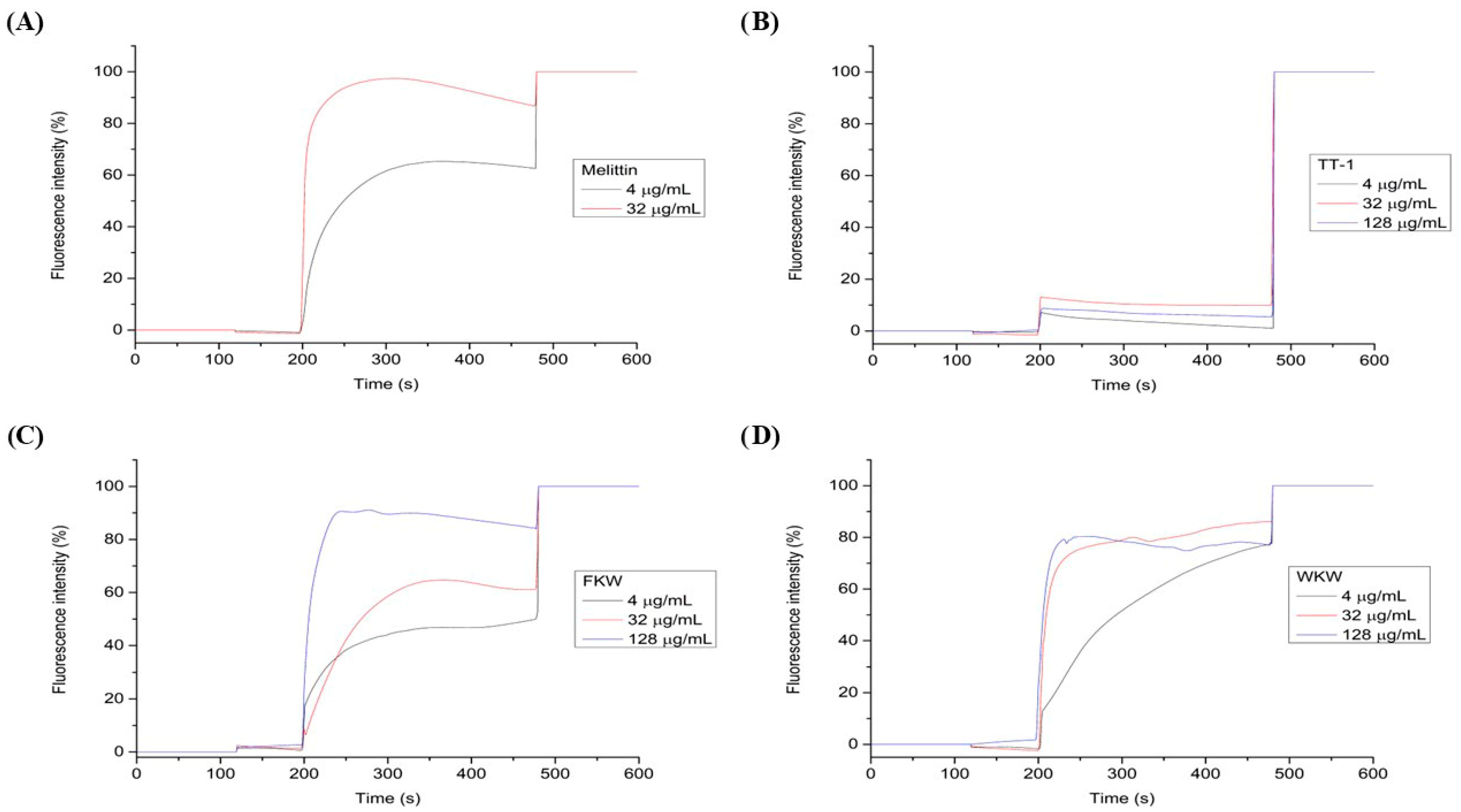
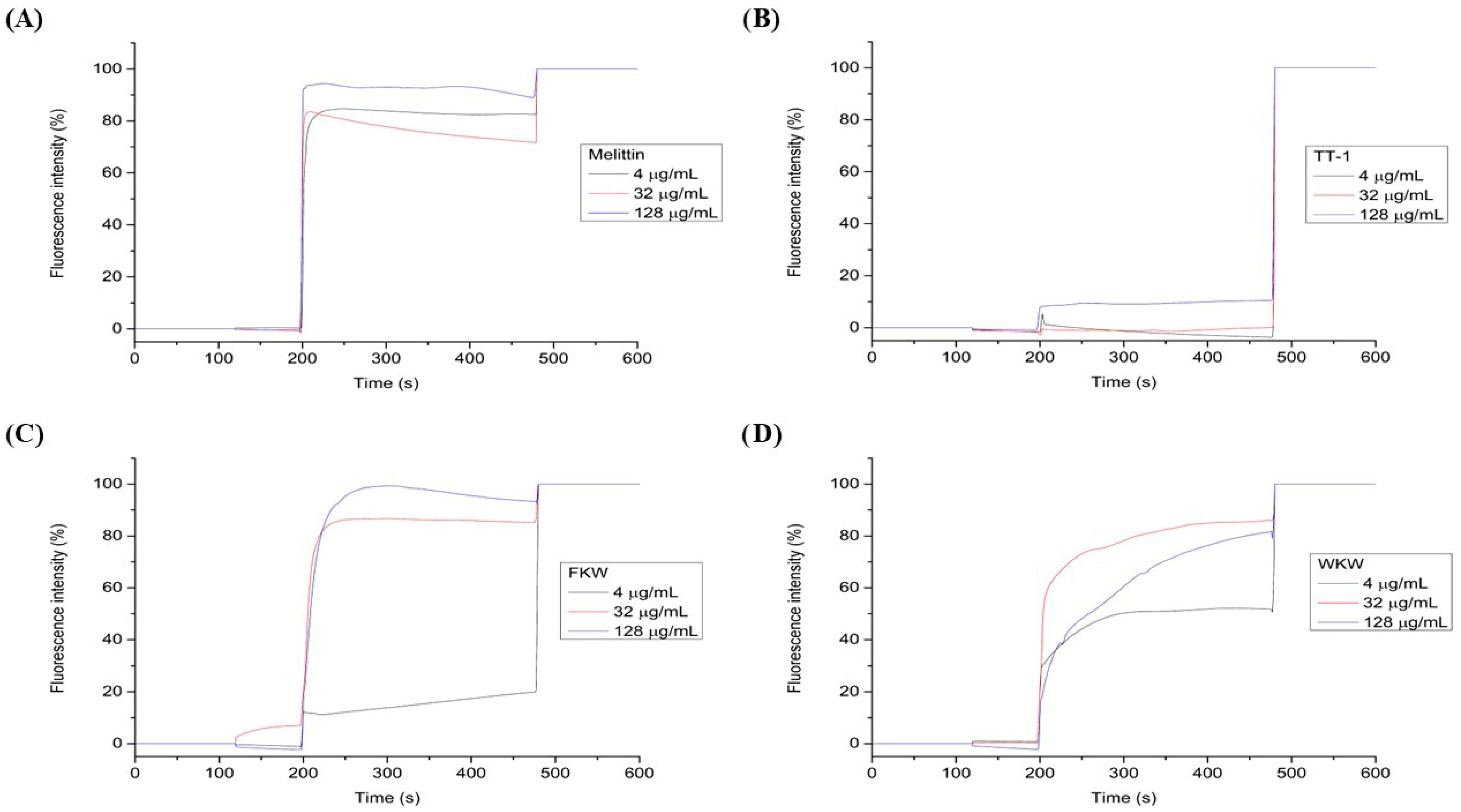
Fluorescence intensity was quantified after the addition of the peptide at concentrations of 4 µg/mL, 32 µg/mL and 128 µg/mL after 195 s, and the addition of 1% Triton solution after 475 s for complete rupture (100%) of the vesicle. Multilamellar vesicles (MLVs) were extruded using an Avanti Polar Lipids extruder (Avanti Polar Lipids, Alabaster, AL, USA) with a 100 nm pore polycarbonate filter to obtain large unilamellar vesicles (LUVs). LUVs were added to a size exclusion column with Sephadex G25 resin.
Permeabilization of lipid vesicles containing carboxyfluorescein was carried out using a Jasco FP-8250 spectrofluorometer (JASCO, Tokyo, Japan), at wavelengths of 490 nm for excitation and 512 nm for emission, with a 600 s scan.
The following equation was used to calculate the CF release graphs:
R e l e a s e ( % ) = ( M   T r i t o n     M   S a m p l e ) ( M   T r i t o n     M   V e s i c l e s ) × 100
where M Triton is the average fluorescence value obtained after the addition of 1% Triton, M Vesicles is the average of the initial values of the run before the addition of the peptide, and M Sample are the fluorescence values obtained at each point after the addition of the peptide. The permeabilization graphs were analysed using OriginPro 9.0 (OriginLab Corporation, Northampton, MA, USA).

3. Results and Discussion

3.1. Peptide Design and Synthesis

Previous studies have described Melittin as a peptide with multiple biological activities, including antibacterial effects [38]. Despite its promising bioactivity, melittin exhibits strong non-specific cytolytic activity and high toxicity [39]. To address these limitations, analogue peptides have been designed to improve therapeutic efficacy while reducing toxicity. In this study, we hypothesized that short and potent AMPs with reduced or absent hemolytic activity could be developed by modifying active AMP sequences using a combination of rational design strategies and machine learning-based tools. Our goal was to preserve or enhance the antimicrobial activity of Melittin while generating shorter and more potent derivatives with reduced cytotoxicity.
Table 1 and Table 2 summarize the physicochemical and structural properties of melittin, TT-1, and the rationally designed peptides FKW and WKW, including net charge, hydrophobicity, hydrophobic moment, number of charged and aromatic residues, amino acid sequence, retention time in analytical RP-HPLC, and theoretical molecular mass. Melittin exhibits a higher net positive charge (+6) at physiological pH, compared to its analogues (+4), along with lower hydrophobicity and hydrophobic moment. The TT-1 analogue has a net charge of +4, at physiological pH, and hydrophobicity comparable to Melittin; however, it displays a higher hydrophobic moment. Some authors have suggested that increased hydrophobicity, hydrophobic moment, and a positively charged helical face significantly enhance hemolytic and Gram-positive antibacterial activities, while also reducing peptide specificity [40]. These physicochemical descriptors, including hydrophobicity and hydrophobic moment, were considered important parameters supporting the rational design strategy of the peptide analogues. Variations in these properties may contribute to differences in membrane interaction, amphipathicity, and consequently antimicrobial performance. However, the present dataset remains limited and does not allow definitive causal correlations between these descriptors and the observed biological activities. Therefore, the results should be interpreted as indicative trends that support the design rationale rather than broad generalizable relationships.
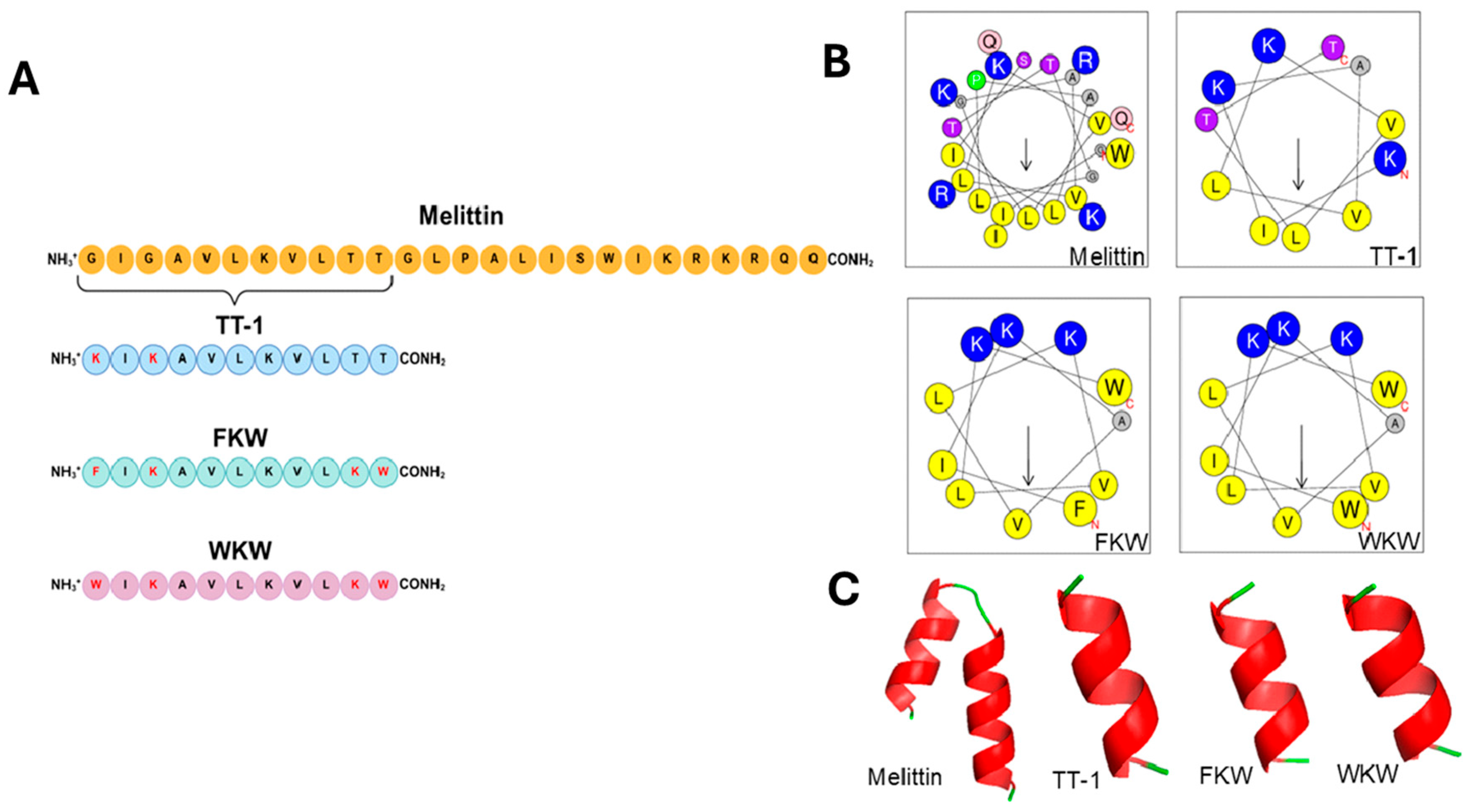
The rationale behind the modifications made to the FKW and WKW peptides was to investigate the effects of peptide chain length alteration, amino acid substitutions, and charge modifications (Figure 1). In the TT-1 peptide, glycine residues, which are known to disrupt helical conformation, were replaced with positively charged lysines, which are conformation-stabilizing amino acids [41]. The presence of these amino acids in hydrophobic chains has been shown to confer potent antimicrobial activity [42,43].
For the FKW peptide, derived from TT-1, three modifications were made: (1) substitution of lysine by phenylalanine, an aromatic amino acid, in position 1; (2) substitution of threonine by lysine, a cationic amino acid, at position 10; and (3) substitution of threonine by tryptophan, also an aromatic amino acid, in position 11. These changes improved the polar distribution within the α-helical structure and, consequently, enhanced the amphipathic character of the peptide.
The WKW peptide, in turn, was derived from FKW and modified by replacing phenylalanine at position 1 to tryptophan. This modification increases hydrophobicity, a crucial characteristic for antimicrobial peptides as it enhances interaction with the lipid bilayer of bacterial membranes [15]. Additionally, the hydrophobic amino acid tryptophan possesses a side chain with a large, flat aromatic ring, which significantly influences its preferred positioning within cellular membranes. This amino acid tends to localize at the interface between the hydrophilic and hydrophobic regions of the lipid bilayer [44].
It is important to emphasize that the design of potent antimicrobial peptides (AMPs) depends on achieving an optimal balance between cationic charge and hydrophobicity. The cationic nature of these molecules, commonly provided by amino acids such as lysine (K), arginine (R), and histidine (H), enables their preferential interaction with the negatively charged surfaces of bacterial membranes. In parallel, hydrophobic residues including tryptophan (W), leucine (L), isoleucine (I), and phenylalanine (F) contribute to membrane permeabilization by favoring insertion into lipid bilayers [45]. In particular, the indole moiety of tryptophan plays a key role in stabilizing peptide–membrane interactions through hydrophobic contacts at the membrane interface, thereby facilitating deeper membrane penetration [46]. Importantly, AMPs enriched in tryptophan and cationic residues have been associated with enhanced antimicrobial efficacy, as well as improved stability and reduced toxicity profiles [47,48,49].
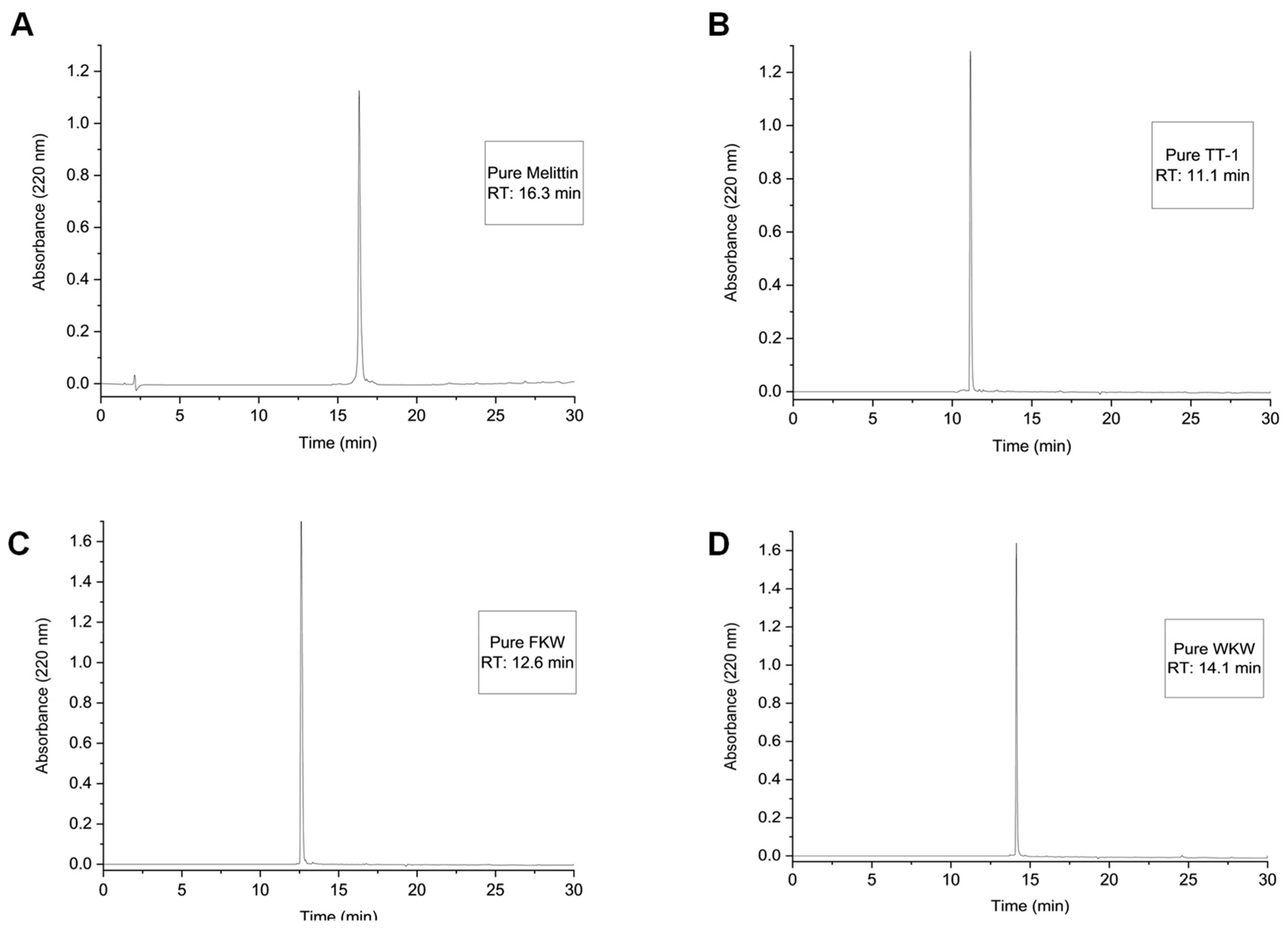
The peptides were synthesized by SPPS, obtained at high purity (>95%), confirmed by rp-HPLC (Figure 2), and their molecular identities verified by electrospray mass spectrometry. The WKW peptide showed a higher retention time and greater hydrophobicity compared to the other analogues but was slightly lower than Melittin on a C18 reversed-phase analytical HPLC column (Table 2).

3.2. Circular Dichroism Spectroscopy

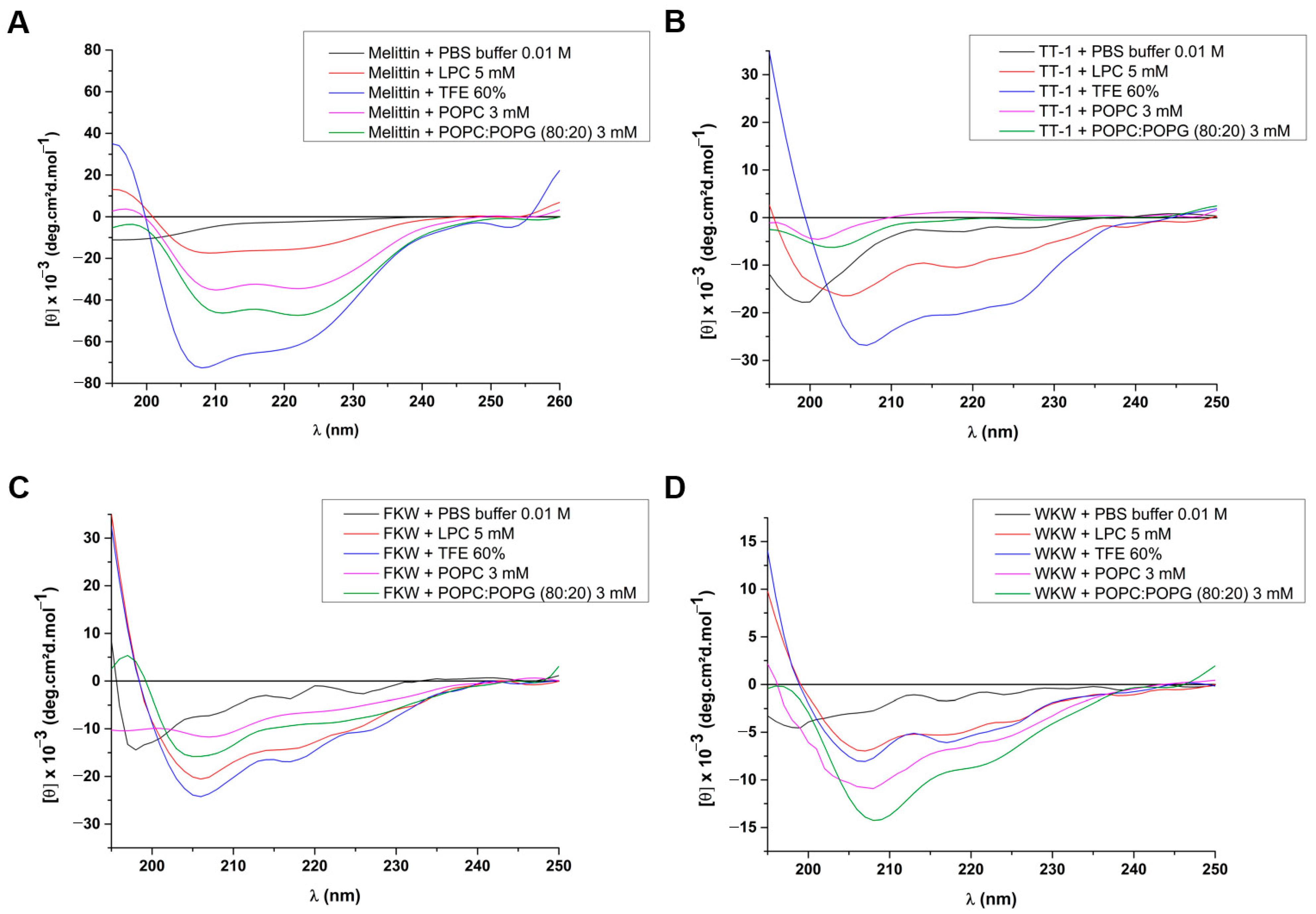
CD spectroscopy is primarily used to determine the secondary structure of proteins and peptides [50]. For aqueous solution using PBS buffer, all peptide exhibits a strong negative ellipticity near 200 nm and near-zero ellipticity above 215 nm, demonstrating as intrinsically disordered structures or unfolded random coils. Without the shielding effect of a hydrophobic environment, the peptides cannot stably form intramolecular hydrogen bonds. This is a common and frequent evolutionary trait for membrane-active peptides, ensuring they remain soluble and inactive while traversing aqueous biological fluids until reaching their target membrane [10].
TFE is a well-known structure-inducing solvent. This is due to a lower dielectric constant than water, which acts by displacing water molecules from the peptide backbone. This local dehydration forces the peptide to form intramolecular hydrogen bonds to stabilize its backbone, resulting in an α-helix. From Figure 3, the fact that the four peptides folded in TFE presumes a high intrinsic propensity to form α-helices when placed in a low-dielectric (hydrophobic) environment.
As expected, Melittin (Figure 3A) adopts strong α-helical conformations in all membrane mimics tested (LPC, POPC, and POPC:POPG). As a non-selective membrane-lytic peptide, the strong folding in both POPC and POPC:POPG indicates that its membrane binding is driven primarily by hydrophobic effects rather than electrostatic steering. These results are in accordance with the leakage essays and biological activities (see next section), explaining its high general toxicity/hemolytic activity by carpet-like mechanism [20].
Conversely, while TT-1 (Figure 3B) showed secondary structure in TFE, its response to actual lipid environments is extremely muted. In POPC, it remains almost completely unstructured and in LPC and POPC:POPG, it shows only minor conformational shifts. Thus, this data suggests that TT-1 lacks the necessary structural features—such as an optimal amphipathic face (see helical wheel, Figure 1) or a specific charge distribution—to successfully anchor into and partition into lipid bilayers. It is worthy highlighting here that TT-1 peptide does not content any aromatic residue at the C- or N-terminus, which can decrease the membrane partitioning in the primary interaction. Even though it can form a helix (as seen in TFE), the thermodynamic cost of inserting into the lipid bilayer is probably high, preventing the coil-to-helix transition at the membrane interface.
The FKW analogue (Figure 3C) showed transitions to an α-helix in all tested lipid environments. Interestingly, the negative signal at 222 nm and 208 nm is more intense in LPC micelles than POPC vesicles, and slightly shallower in POPC:POPG [51]. This indicates that FKW analogue possesses high partitioning in lipid interfaces. The strong signal in LPC suggests it favors highly curved, detergent-like surfaces, favoring smaller molecules. Its robust folding in POPC implies it can readily interact with zwitterionic eukaryotic membranes, meaning it may carry a risk of eukaryotic cytotoxicity (similar to Melittin, though the varying spectral depths, suggest different binding kinetics or aggregation states).
Finally, WKW shows a highly distinct pattern (Figure 3D). The most pronounced α-helical double minima among the lipid tests were found in the POPC:POPG (80:20) environment, even higher than TFE. However, in POPC and LPC, the structural induction is significantly stronger in the anionic membrane mimetics, suggesting an electrostatic combined with a hydrophobic selectivity. Therefore, this data strongly implies in a two-step electrostatic and hydrophobic mechanism. The initial interaction is driven by electrostatic attraction between positively charged residues on WKW and the negatively charged POPG headgroups. This electrostatic “steering” pulls a high concentration of the peptide to the membrane surface, changing its conformation. The presence of tryptophan aromatic residue neighboring the lysine facilitates a deeper insertion and a complete and more stabilized coil-to-helix transition compared to the neutral POPC membrane.

3.3. Biological Activity

Initial Screening

For an initial analysis of whether the peptides had antimicrobial potential, their activity against methicillin-sensitive Gram-positive S. aureus ATCC 25923 was determined.
In this study, Melittin demonstrated high antimicrobial activity, achieving complete (100%) inhibition of S. aureus growth at concentrations up to 8 µg/mL. The TT-1 analogue, however, did not exhibit complete bacterial growth inhibition at any of the tested concentrations (1–512 µg/mL). Notably, previous studies have reported the cytotoxic activity of TT-1 against tumor cells [24]; however, its antimicrobial potential had not been investigated prior to this work.
Regarding the new analogues proposed in the present study, which were designed through a rational design approach, the results indicate promising antimicrobial efficacy. The FKW analogue exhibited potent antibacterial activity, completely inhibiting S. aureus growth at concentrations up to 16 µg/mL (only one dilution step lower than Melittin). Even more impressive was the WKW analogue, which demonstrated complete bacterial inhibition at concentrations as low as 4 µg/mL, indicating superior activity compared to the parent peptide, Melittin. The minimum bactericidal concentration (MBC) analysis yielded results consistent with the observed inhibitory activity, with identical concentration thresholds. Table 3 summarizes the results.

3.4. Multidrug-Resistant and ESKAPE Bacterial Strains

It is well described that antimicrobial resistance is a chronic public health problem, and it is estimated that if no action is taken, approximately 10 million deaths worldwide will occur by 2050, becoming the leading cause of death worldwide [25]. Given the promising results obtained with the new Melittin analogues, further analyses were conducted using a larger number of bacterial strains.
The activity of the peptides against Gram-positive (S. epidermidis, S. aureus, S. aureus, E. faecalis, and E. faecium) and Gram-negative (K. pneumoniae, E. coli, A. baumannii, and P. aeruginosa) bacteria was evaluated.
Initially, the parental peptide, Melittin, was evaluated. As expected, Melittin exhibited strong antimicrobial activity against all tested strains, with low MIC and MBC values. As described in the literature, Melittin showed high antimicrobial activity [38] with better results for Gram-positive bacteria.
In line with our preliminary observations, the TT-1 peptide displayed low antimicrobial activity against most of the tested pathogens. However, it retained measurable activity against both Gram-positive and Gram-negative bacteria. The strains found to be susceptible to TT-1 included S. epidermidis (256 µg/mL), E. faecalis (512 µg/mL), E. faecium (512 µg/mL), and E. coli (512 µg/mL).
FKW and WKW analogues showed antimicrobial activity against all tested strains, with MIC values comparable to those observed for Melittin. In overall, both analogues maintained the antimicrobial activity, with the WKW analogue being more potent than FKW for strains S. aureus (ATCC 25923) (16 µg/mL), S. aureus (ATCC 8095) (8 µg/mL), E. faecium ATCC 700,221 (16 µg/mL) and A. baumannii (ATCC 19606) (32 µg/mL). Notably, both analogues increased the activity against K. pneumoniae (ATCC 700603) and P. aeruginosa (ATCC 27853) when compared to Melittin (Table 4).
Despite the promising broad-spectrum antimicrobial activity observed for the FKW and WKW analogues, some limitations should also be considered. Although both peptides demonstrated enhanced activity against K. pneumoniae compared to Melittin, their efficacy against E. faecalis remained relatively moderate, with higher MIC values compared to other susceptible strains. In addition, despite the relevant activity observed against P. aeruginosa, this pathogen continues to represent a particularly challenging target due to its highly restrictive outer membrane and intrinsic resistance mechanisms, which may limit peptide selectivity and effectiveness. These findings indicate that, although the designed analogues represent an important improvement over the parental peptide in several aspects, further structural optimization is still necessary to enhance potency and selectivity against more recalcitrant bacterial species.
It is important to note that, when comparing the results from initial screening with those from tests against multidrug-resistant and ESKAPE strains, the activity of FKW and WKW against S. aureus ATCC 25923 showed slight variation. This is because the assays were conducted in different laboratories. Although the same methodology was followed, minor discrepancies, such as the brand of culture media or the individual performing the test, can lead to small differences in the MIC values. As reported by several authors, variations in Minimum Inhibitory Concentration (MIC) values are commonly observed across laboratories, even when testing the same strain and compound. This variability may arise from methodological differences, and even with standardized protocols, inherent variability in microbial growth and antibiotic responses can result in subtle shifts in MIC values [52,53].
Peptide sequence modifications can enhance efficacy. Our results demonstrated that substituting specific amino acids to increase the hydrophobic moment, while preserving a helical structure with clearly defined polar and nonpolar faces, can maintain antimicrobial activity and, consequently, improve the peptide’s antibacterial potential. Furthermore, α-helical structuring in the presence of a bacterial membrane is one of the crucial characteristics of an membranolytic AMP [11].
As previously described, cationic charge and amphipathicity are critical determinants of the antimicrobial activity of peptides [11,16,54]. Furthermore, it is well established that tryptophan residues possess strong membrane-disruptive properties, which confer tryptophan-rich antimicrobial peptides with a unique ability to interact with bacterial membranes, potentially enhancing their antimicrobial efficacy [55].
The activity of the peptides was classified as bactericidal or bacteriostatic based on the MBC/MIC ratio, in accordance with Pankey and Sabath [36]. Except for TT-1, all peptides demonstrated bactericidal activity.

3.5. Hemolytic Assay

To evaluate the therapeutic potential of different peptide candidates, it is essential to assess their cytotoxicity against human erythrocytes, as one of the major limitations in applying antimicrobial peptides is the significant toxicity frequently exhibited by this class of compounds [35].
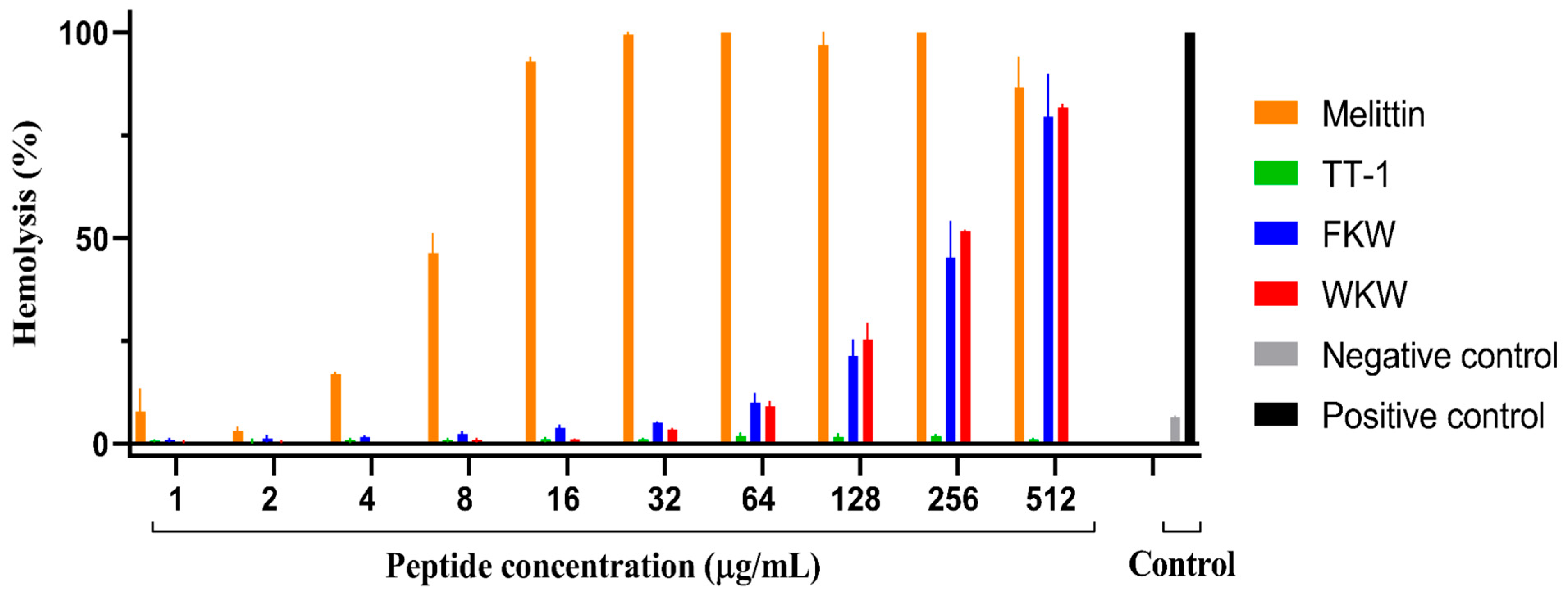
The results (Figure 4 and Table 5) showed that Melittin exhibited pronounced hemolytic activity, with an HC50 value of approximately 7.9 µg/mL. This finding is consistent with previous reports in the literature, where Melittin’s lytic effect is attributed to its ability to disrupt cellular membranes, primarily due to its cationic amphipathic structure [56,57].
In contrast, the Melittin analogue TT-1 exhibited markedly reduced hemolytic activity, even at the highest concentration tested (512 µg/mL). This observation suggests that specific amino acid substitutions, in conjunction with an altered net charge and hydrophobicity, contribute significantly to the reduced cytotoxic profile of this peptide [56,57].
The FKW analogue, derived from TT-1, displayed hemolytic activity comparable to that of Melittin at 512 µg/mL. However, at lower concentrations, a marked reduction in hemolytic activity was observed. The HC50 for FKW was determined to be 264.8 µg/mL, indicating that although the peptide demonstrates notable toxicity at high concentrations, its hemolytic potential diminishes significantly at sub-toxic doses. It is important to highlight that, when evaluating the antimicrobial activity against multidrug-resistant and ESKAPE strains, this molecule exhibited MIC and MBC values at or below 32 µg/mL for most strains (10-fold lower concentration than the HC50). Even for the strains in which the peptide showed MIC/MBC at the highest concentration (128 µg/mL), this activity was still observed at concentrations below the HC50.
The substitution of lysine by phenylalanine at the N-terminal, and threonine by tryptophan at the C-terminal, contributed to an increase in the hydrophobicity of FKW relative to TT-1, which may explain the observed increase in hemolytic activity [58]. These findings indicate that such structural modifications influenced the peptide’s bioactivity and suggested that rational design effectively balanced the enhanced antimicrobial efficacy with moderate hemolytic potential.
The WKW analogue exhibited a dose-dependent hemolytic profile, showing increased toxicity at higher concentrations and a significant decrease in hemolytic activity at lower concentrations. As observed for FKW, despite replacing phenylalanine with tryptophan, the N-terminal region of WKW retained a high degree of hydrophobicity, which appeared to correlate with its hemolytic behavior. The HC50 value of WKW was calculated as 237.2 µg/mL, which was slightly lower than that of FKW, reinforcing the link between hydrophobicity and cytotoxicity. In addition to the FKW analogue, the HC50 should be considered in relation to antimicrobial activity, particularly against multidrug-resistant and ESKAPE strains. Accordingly, the WKW peptide also demonstrated MIC and MBC values at or below 32 µg/mL for most strains—concentrations lower than the HC50, except for Pseudomonas aeruginosa ATCC 27853, where antimicrobial activity was observed at concentrations close to the HC50.
The modifications introduced into the Melittin sequence in these analogues clearly demonstrated that specific structural changes could alter both the functional and toxicological properties of the peptides. Additionally, C-terminal amidation has been previously associated with enhanced antimicrobial activity and reduced erythrocyte toxicity of various peptides [59].
Based on our findings, the rational design of the FKW and WKW analogues proved effective. Although the increased hydrophobicity of these peptides resulted in higher hemolytic activity than TT-1, these modifications led to a significant improvement in antimicrobial performance. When comparing FKW and WKW directly with Melittin, it can be concluded that both analogues maintained antimicrobial activity at similar concentrations or slightly lower dilutions. Importantly, their HC50 values indicated that they were substantially less cytotoxic than Melittin.

3.6. Selectivity Index

The Selectivity Index (SI)—or therapeutic index—is a widely adopted parameter to evaluate the specificity of antimicrobial compounds. It is determined by calculating the ratio between the concentration that induces 50% hemolysis (HC50) and the Minimum Inhibitory Concentration (MIC) of each peptide [60]. A higher SI value indicates greater antimicrobial specificity [61].
As shown in Table 6, Melittin, a peptide well-established for its cytolytic properties, exhibited Selectivity Index values ranging from 2.0 to 0.02, with higher values observed against Gram-positive bacteria.
For FKW, the SI was 33.1 against Staphylococcus epidermidis (ATCC 35984), 16.6 against S. aureus (ATCC 8095), and 8.3 against S. aureus (ATCC 25923), Enterococcus faecium (ATCC 700221), Klebsiella pneumoniae (ATCC 700603), and Escherichia coli (ATCC 25922). Against Enterococcus faecalis (ATCC 29212), Acinetobacter baumannii (ATCC 19606), and Pseudomonas aeruginosa (ATCC 27853), FKW presented lower SI values; however, these still indicated higher selectivity compared to Melittin.
Similarly, WKW exhibited prominent potential for biotechnological applications, with SI of 29.6 against S. epidermidis (ATCC 35984) and S. aureus (ATCC 8095); 14.8 against S. aureus (ATCC 25923) and E. faecium (ATCC 700221); and 7.4 against K. pneumoniae (ATCC 700603), E. coli (ATCC 25922), and A. baumannii (ATCC 19606).
Among the analogues evaluated in this study, TT-1 exhibited the lowest selectivity indices, whereas FKW and WKW showed markedly high SI values, highlighting their significant potential for biotechnological applications.

3.7. Serum Stability Assay

Predicting the pharmacokinetics of a peptide presents a significant challenge due to the various factors that determine its in vivo behavior, such as absorption rate, first-pass metabolism, renal and hepatic clearance, cellular binding and uptake, and circulatory peptidases [62]. The evaluation of peptide stability and half-life in the development of peptides for in vivo use is a critical step, as unstable peptides with short half-lives in the presence of serum proteases may be disqualified due to their limited bioavailability [63].
The enzymatic stability of peptides depends on various factors, particularly the type and sequence of amino acids, overall size, flexibility, and conformation. There are several ways to circumvent this proteolytic effect, one of which is the modification of the peptide’s structure in a way that is not recognized by proteases, even modest changes in which the peptide can be cleaved by an enzyme can lead to significant alterations [63].
The serum stability profiles of the synthesized peptides were evaluated, and their apparent half-lives (t½) were estimated based on the degradation kinetics observed by HPLC analysis (Table 7). Among the evaluated peptides, Melittin exhibited the greatest stability, with an estimated half-life of approximately 9.6 h, substantially higher than those observed for the analogues FKW and WKW, which presented half-lives of 2.2 h and 1.5 h, respectively. In contrast, the TT-1 analogue underwent extensive degradation at the earliest evaluated time point, preventing reliable determination of its half-life. These findings quantitatively confirm the distinct stability profiles among the peptides and demonstrate that the structural modifications introduced into the analogues significantly affected their susceptibility to serum proteases.
The superior stability of Melittin may be associated with its longer sequence length and its well-characterized amphipathic α-helical conformation, which may confer partial protection against enzymatic cleavage. Conversely, the shorter half-lives observed for FKW and WKW suggest that the amino acid substitutions introduced into these analogues increased their accessibility to proteolytic enzymes. Additionally, metabolic fragments derived from the peptide analogues were detected throughout the incubation period, reinforcing the occurrence of progressive enzymatic processing. After 12 and 24 h of incubation, all analogues were extensively degraded.
Although enhanced stability is often desirable to improve systemic exposure, complete resistance to proteolytic degradation is not necessarily advantageous for antimicrobial peptides. Molecules that persist for prolonged periods under sub-inhibitory concentrations may contribute to selective pressure and favor the emergence of antimicrobial resistance. Furthermore, highly persistent bioactive peptides may accumulate in the environment if improperly discarded. Therefore, from a drug development perspective, achieving an optimal balance between biological activity and controlled degradability is likely more desirable than maximizing peptide stability alone.

3.8. Permeabilization of Vesicles

To understand the effect of these molecules on the lipid membrane, a permeabilization test was carried out on vesicles containing POPC 100% (Figure 5) to resemble the lipid composition of mammalian eukaryotic cells and POPC + POPG (80:20%) (Figure 6) to approximate the lipid composition of bacterial membranes, with the presence of a negative charge [12].
Carboxyfluorescein release, or vesicle permeabilization assays, are commonly employed to investigate the mechanisms of peptide action. By using large unilamellar vesicles (LUVs), interactions between the peptides and the vesicle membranes can be analysed, providing valuable insights into the structure–function relationships of the peptides. Typically, a gradual increase in fluorescence intensity is indicative of pore formation, serving as a strong clue to the peptide’s mechanism of action [10]. In contrast, an abrupt increase followed by rapid stabilization of fluorescence suggests that the peptide may act in a detergent-like manner.
To better understand the membrane interactions of the peptides, permeabilization assays were performed using vesicles composed of POPC (100%), representing zwitterionic eukaryotic membranes, and POPC:POPG (80:20%), which mimics the negatively charged surface of bacterial membranes. The leakage profiles observed in Figure 5 and Figure 6 were strongly associated with the structural behavior identified by CD spectroscopy (Figure 3) and with the biological activities described in the antimicrobial and hemolytic assays (Figure 4 and Table 4, Table 5 and Table 6).
Melittin exhibited an immediate and complete release of carboxyfluorescein in both POPC and POPC:POPG vesicles, even at lower concentrations, indicating rapid membrane disruption through a detergent-like mechanism. These findings are fully consistent with the CD data, where Melittin adopted strong α-helical conformations in all membrane-mimetic environments, independent of membrane charge (Figure 3A). This non-selective membrane interaction explains both its potent antimicrobial activity and its pronounced hemolytic effect, reflected by the very low HC50 value observed in Figure 4 and Table 5.
In contrast, TT-1 produced minimal permeabilization in both vesicle systems, with fluorescence remaining low and nearly linear throughout the assay. This behavior correlates well with its weak conformational response in POPC and POPC:POPG observed by CD spectroscopy (Figure 3B), suggesting inefficient membrane partitioning and limited insertion into lipid bilayers. Consequently, TT-1 displayed markedly reduced hemolytic activity, but also poor antimicrobial efficacy against most bacterial strains (Figure 4 and Table 4). The FKW and WKW analogues showed intermediate membrane-disruptive behavior compared with Melittin and TT-1. FKW induced substantial permeabilization in both POPC and POPC:POPG vesicles, especially at higher concentrations, indicating a strong membrane affinity with lower selectivity between zwitterionic and anionic membranes. This observation agrees with the CD spectra, where FKW adopted α-helical conformations in all lipid environments tested, including POPC vesicles (Figure 3C). Such behavior is consistent with its improved antimicrobial activity, but also explains the moderate hemolytic activity observed in Figure 4.
Interestingly, WKW displayed a more selective permeabilization profile, with stronger leakage effects in POPC:POPG vesicles than in POPC vesicles. This result closely parallels the CD data, in which WKW exhibited the most pronounced α-helical folding in the anionic POPC:POPG environment (Figure 3D). The preferential interaction with negatively charged membranes supports the hypothesis of an electrostatically guided membrane-binding mechanism, followed by hydrophobic insertion mediated by tryptophan residues. This selective interaction pattern may explain why WKW maintained potent antimicrobial activity while exhibiting substantially reduced hemolytic activity compared with Melittin (Figure 4 and Table 4, Table 5 and Table 6).
Overall, the vesicle permeabilization assays reinforced the structure–activity relationships observed throughout the study. The results indicate that the rational introduction of aromatic and hydrophobic residues modulated peptide–membrane interactions, allowing FKW and especially WKW to preserve antimicrobial activity while partially reducing nonspecific disruption of eukaryotic-like membranes.
The detergent mechanism of action attributed to Melittin is due to the molecule’s ability to interact with the lipid bilayer and induce the rupture of this bilayer [64,65]. In terms of detailed molecular mechanisms, several overlapping mechanisms of AMP activity probably exist, and it may not be possible to define a single mode. For vesicles composed of 100% POPC, at the lowest concentration tested (4 µg/mL), Melittin apparently acts through a pore-forming mechanism. However, at higher concentrations (32–128 µg/mL), 100% CF release was observed immediately after peptide addition. The results obtained at the highest concentration tested (128 µg/mL) were not shown because fluorescence rapidly reached the plateau (100%) and remained constant until the end of the experiment.
However, the detergent capacity of Melittin, the shortening of its sequence and the replacement of the glycine residues that make up the N-terminal region of the molecule with two lysines showed a modification of the mechanism of action, suggesting an inactivation of its activity in the membrane. This property may be related to the overall composition of Melittin, as the residues that make up the C-terminal region of the molecule are also associated with the ability to interact with cell membranes [66].
In addition, the substitution of lysine with phenylalanine suggests a greater interaction with the membrane due to the increased capacity of phenylalanine-containing peptides to permeabilize the membrane, as well as their position in the peptide sequence, since the N-terminal region of the molecule plays a crucial role in the activity of the molecule [67,68].
On the other hand, the modification of phenylalanine by tryptophan in the N-terminal region observed in the WKW sequence was sufficient to interfere with the mode of action in the membrane, since tryptophan increases the peptide’s capacity for aggregation and induces the membrane pore formation [59,69].

4. Conclusions

This study demonstrates that rational design effectively balances the antimicrobial efficacy and cytotoxicity of Melittin derivatives. The FKW and WKW analogues maintained broad-spectrum activity against clinically relevant pathogens, including ESKAPE strains, at levels comparable to or occasionally exceeding the parental peptide, Melittin. Both analogues exhibited significantly reduced hemolytic activity and improved selectivity indices, indicating a wider therapeutic window. Mechanistically, structural modifications preserved antimicrobial function while shifting membrane interactions from indiscriminate disruption to controlled pore formation. Although the analogues are susceptible to proteolytic degradation in serum, this instability may mitigate long-term toxicity.
Overall, the findings reinforce the potential of rationally designed Melittin-derived peptides as promising scaffolds for the development of new antimicrobial agents with improved therapeutic windows. However, further optimization and validation, including expanded testing against clinically resistant isolates, advanced membrane selectivity studies, protease-resistance strategies, and in vivo toxicity and efficacy assays, will be essential before considering future translational applications.

Supplementary Materials

The following supporting information can be downloaded at: https://www.mdpi.com/article/10.3390/pr14101630/s1, Figure S1: Serum degradation assay of peptide Melittin (A), TT-1 (B) and analogues FKW (C) and WKW (D) observed on analytical HPLC in 5–95% of solvent B gradient after incubation in human serum at zero time, 1 h, 4 h, 12 h, and 24 h in a Shimadzu chromatography; Table S1: CAMPR4 prediction scores and selection criteria for TT-1-derived peptide variants; Table S2: Replicate-level MIC values (µg/mL) of Melittin and its analogues against Staphylococcus aureus.

Author Contributions

Conceptualization, L.O.R. and N.A.S.-F.; methodology, I.L.B.C.C., C.R.F. and N.A.S.-F.; validation, L.O.R., L.O.C.N., A.R.A., A.K.S., G.F.H., M.N.A. and V.T.C.; formal analysis, G.F.H., M.N.A. and V.T.C.; investigation, G.F.H., M.N.A. and V.T.C.; resources, I.L.B.C.C., C.R.F. and N.A.S.-F.; data curation, I.L.B.C.C., C.R.F. and N.A.S.-F.; writing—original draft preparation, L.O.R., L.O.C.N., A.R.A., L.O.F., L.R.C. and N.A.S.-F.; writing—review and editing, L.O.R., L.O.C.N., A.R.A., A.K.S., M.N.A., V.T.C., L.O.F., L.R.C., G.F.H., C.R.F., I.L.B.C.C., E.F.V. and N.A.S.-F.; visualization, I.L.B.C.C., C.R.F., E.F.V. and N.A.S.-F.; supervision, I.L.B.C.C., C.R.F. and N.A.S.-F.; project administration, L.O.R. and N.A.S.-F.; funding acquisition, I.L.B.C.C., C.R.F. and N.A.S.-F. All authors have read and agreed to the published version of the manuscript.

Funding

This research was funded by the São Paulo Research Foundation (FAPESP grant # 2023/03857-1; grant # 2022/05787-8), the Center for Research and Innovation in Biodiversity and New Drugs (CIBFar-FAPESP grant # 2013/07600-3), the Coordination for the Improvement of Higher Education Personnel (CAPES) and the National Counsel of Technological and Scientific Development (CNPq).

Data Availability Statement

Data supporting the conclusions of this study can be made available upon request from the corresponding author.

Conflicts of Interest

The authors declare no conflicts of interest.

References

  1. Ferdinand, A.S.; McEwan, C.; Lin, C.; Betham, K.; Kandan, K.; Tamolsaian, G.; Pugeva, B.; McKenzie, J.; Browning, G.; Gilkerson, J.; et al. Development of a cross-sectoral antimicrobial resistance capability assessment framework. BMJ Glob. Health 2024, 9, e013280. [Google Scholar] [CrossRef]
  2. Ahmed, S.K.; Hussein, S.; Qurbani, K.; Ibrahim, R.H.; Fareeq, A.; Mahmood, K.A.; Mohamed, M.G. Antimicrobial resistance: Impacts, challenges, and future prospects. J. Med. Surg. Public Health 2024, 2, 100081. [Google Scholar] [CrossRef]
  3. CDC. Centers for Disease Control and Prevention. Antibiotic/Antimicrobial Resistance (AR/AMR)—About Antibiotic Resistance; CDC: Atlanta, GA, USA, 2022. [Google Scholar]
  4. Morrison, L.; Zembower, T.R. Antimicrobial Resistance. Gastrointest. Endosc. Clin. North. Am. 2020, 30, 619–635. [Google Scholar] [CrossRef]
  5. O’Neill, J. Tackling Drug-Resistant Infections Globally; Final Report and Recommendations the Review on Antimicrobial Resistance Chaired by Jim O’Neill; Wellcome Trust: London, UK, 2016. [Google Scholar]
  6. Christaki, E.; Marcou, M.; Tofarides, A. Antimicrobial Resistance in Bacteria: Mechanisms, Evolution, and Persistence. J. Mol. Evol. 2020, 88, 26–40. [Google Scholar] [CrossRef] [PubMed]
  7. Roque-Borda, C.A.; da Silva, P.B.; Rodrigues, M.C.; Azevedo, R.B.; Di Filippo, L.; Duarte, J.L.; Chorilli, M.; Festozo Vicente, E.; Pavan, F.R. Challenge in the Discovery of New Drugs: Antimicrobial Peptides against WHO-List of Critical and High-Priority Bacteria. Pharmaceutics 2021, 13, 773. [Google Scholar] [CrossRef]
  8. Roque-Borda, C.A.; Bento da Silva, P.; Rodrigues, M.C.; Di Filippo, L.D.; Duarte, J.L.; Chorilli, M.; Vicente, E.F.; Garrido, S.S.; Rogério Pavan, F. Pharmaceutical Nanotechnology: Antimicrobial Peptides as Potential New Drugs against WHO List of Critical, High, and Medium Priority Bacteria. Eur. J. Med. Chem. 2022, 241, 114640. [Google Scholar] [CrossRef]
  9. WHO—World Health Organization. WHO Publishes List of Bacteria for Which New Antibiotics are Urgently Needed; WHO: Geneva, Switzerland, 2017. [Google Scholar]
  10. Sati, H.; Carrara, E.; Savoldi, A.; Hansen, P.; Garlasco, J.; Campagnaro, E.; Boccia, S.; Castillo-Polo, J.A.; Magrini, E.; Garcia-Vello, P.; et al. WHO Bacterial Priority Pathogens List Advisory Group. The WHO Bacterial Priority Pathogens List 2024: A prioritisation study to guide research, development, and public health strategies against antimicrobial resistance. Lancet Infect. Dis. 2025, 25, 1033–1043. [Google Scholar] [CrossRef] [PubMed]
  11. Oliveira DMPde Forde, B.M.; Kidd, T.J.; Harris, P.N.A.; Schembri, M.A.; Beatson, S.A.; Paterson, D.L.; Walker, M.J. Antimicrobial Resistance in ESKAPE Pathogens. Clin. Microbiol. Rev. 2020, 33, 10–1128. [Google Scholar] [CrossRef]
  12. Bicho, G.F.H.; Nunes, L.O.C.; Fiametti, L.O.; Argentin, M.N.; Candido, V.T.; Camargo, I.L.B.C.; Cilli, E.M.; Santos-Filho, N.A. Synthesis, Characterization, and Study of the Antimicrobial Potential of Dimeric Peptides Derived from the C-Terminal Region of Lys49 Phospholipase A2 Homologs. Toxins 2024, 16, 308. [Google Scholar] [CrossRef] [PubMed] [PubMed Central]
  13. Sebold, B.C.; Li, J.; Ni, G.; Fu, Q.; Li, H.; Liu, X.; Wang, T. Going Beyond Host Defence Peptides: Horizons of Chemically Engineered Peptides for Multidrug-Resistant Bacteria. BioDrugs 2023, 37, 607–623. [Google Scholar] [CrossRef]
  14. Sarkar, T.; Chetia, M.; Chatterjee, S. Antimicrobial Peptides and Proteins: From Nature’s Reservoir to the Laboratory and Beyond. Front. Chem. 2021, 9, 691532. [Google Scholar] [CrossRef] [PubMed]
  15. Torres, M.D.T.; Sothiselvam, S.; Lu, T.K.; de la Fuente-Nunez, C. Peptide Design Principles for Antimicrobial Applications. J. Mol. Biol. 2019, 431, 3547–3567. [Google Scholar] [CrossRef] [PubMed]
  16. Mahlapuu, M.; Björn, C.; Ekblom, J. Antimicrobial peptides as therapeutic agents: Opportunities and challenges. Crit. Rev. Biotechnol. 2020, 40, 978–992. [Google Scholar] [CrossRef]
  17. Kumar, P.; Kizhakkedathu, J.N.; Straus, S.K. Antimicrobial peptides: Diversity, mechanism of action and strategies to improve the activity and biocompatibility in vivo. Biomolecules 2018, 8, 4. [Google Scholar] [CrossRef]
  18. Dijksteel, G.S.; Ulrich, M.M.W.; Middelkoop, E.; Boekema, B.K.H.L. Lessons Learned from Clinical Trials Using Antimicrobial Peptides (AMPs). Front. Microbiol. 2021, 12, 616979. [Google Scholar] [CrossRef]
  19. Habermann, E.; Jentsch, J. Sequenzanalyse des Melittins aus den tryptischen und peptischen Spaltstücken. Spaltstücken 1967, 348, 37–50. [Google Scholar] [CrossRef]
  20. Memariani, H.; Memariani, M.; Moravvej, H.; Shahidi-Dadras, M. Melittin: A venom-derived peptide with promising anti-viral properties. Eur. J. Clin. Microbiol. Infect. Dis. 2020, 39, 5–17. [Google Scholar] [CrossRef]
  21. Hong, J.; Lu, X.; Deng, Z.; Xiao, S.; Yuan, B.; Yang, K. How Melittin inserts into cell membrane: Conformational changes, inter-peptide cooperation, and disturbance on the membrane. Molecules 2019, 249, 1775. [Google Scholar] [CrossRef]
  22. Guha, S.; Ferrie, R.P.; Ghimire, J.; Ventura, C.R.; Wu, E.; Sun, L.; Kim, S.Y.; Wiedman, G.R.; Hristova, K.; Wimley, W.C. Applications and evolution of Melittin, the quintessential membrane active peptide. Biochem. Pharmacol. 2021, 193, 114769. [Google Scholar] [CrossRef] [PubMed]
  23. Jamasbi, E.; Mularski, A.; Separovic, F. Model Membrane and Cell Studies of Antimicrobial Activity of Melittin Analogues. Curr. Top. Med. Chem. 2016, 16, 40–45. [Google Scholar] [CrossRef] [PubMed]
  24. Subbalakshmi, C.; Nagaraj, R.; Sitaram, N. Biological activities of C-terminal 15-residue synthetic fragment of Melittin: Design of an analog with improved antibacterial activity. FEBS Lett. 1999, 4481, 62–66. [Google Scholar] [CrossRef]
  25. Matthyssen, T.; Li, W.; Holden, J.A.; Lenzo, J.C.; Hadjigol, S.; O’Brien-Simpson, N.M. Dimerization and lysine substitution of melittin have differing effects on bacteria. Front. Pharmacol. 2024, 15, 1443497. [Google Scholar] [CrossRef] [PubMed] [PubMed Central]
  26. Wan, L.-L.; Zhang, D.-Q.; Zhang, J.-N.; Ren, L.-Q. Anti-hepatocarcinoma activity of TT-1, an analog of Melittin, combined with interferon-α via promoting the interaction of NKG2D and MICA. J. Zhejiang Univ. Sci. B 2017, 186, 522–531. [Google Scholar] [CrossRef]
  27. Merrifield, R.B. Solid phase peptide synthesis. I. The synthesis of a tetrapeptide. J. Am. Chem. Soc. 1963, 85, 2149–2154. [Google Scholar] [CrossRef]
  28. Kaiser, E.; Colescott, R.; Bossinger, C.; Cook, P. Color test for detection of free terminal amino groups in the solid-phase synthesis of peptides. Anal. Biochem. 1970, 2, 595–598. [Google Scholar] [CrossRef]
  29. Gawde, U.; Chakraborty, S.; Waghu, F.H.; Barai, R.S.; Khanderkar, A.; Indraguru, R.; Shirsat, T.; Idicula-Thomas, S. CAMPR4: A database of natural and synthetic antimicrobial peptides. Nucleic Acids Res. 2023, 51, D377–D383. [Google Scholar] [CrossRef] [PubMed]
  30. Rey, J.; Murail, S.; de Vries, S.; Derreumaux, P.; Tufféry, P. PEP-FOLD4: A pH-dependent force field for peptide structure prediction in aqueous solution. Nucleic Acids Res. 2023, 51, W432–W437. [Google Scholar] [CrossRef]
  31. Tufféry, P.; Derreumaux, P. A refined pH-dependent coarse-grained model for peptide structure prediction in aqueous solution. Front. Bioinform. 2023, 3, 1113928. [Google Scholar] [CrossRef]
  32. Binette, V.; Mousseau, N.; Tufféry, P. A Generalized Attraction-Repulsion Potential and Revisited Fragment Library Improves PEP-FOLD Peptide Structure Prediction. J. Chem. Theory Comput. 2022, 18, 2720–2736. [Google Scholar] [CrossRef] [PubMed]
  33. Lamiable, A.; Thévenet, P.; Rey, J.; Vavrusa, M.; Derreumaux, P.; Tufféry, P. PEP-FOLD3: Faster de novo structure prediction for linear peptides in solution and in complex. Nucleic Acids Res. 2016, 44, W449–W454. [Google Scholar] [CrossRef] [PubMed]
  34. Santos-Filho, N.A.; Lorenzon, E.N.; Ramos, M.A.; Santos, C.T.; Piccoli, J.P.; Bauab, T.M.; Fusco-Almeida, A.M.; Cilli, E.M. Synthesis and characterization of an antibacterial and nontoxic dimeric peptide derived from the C-terminal region of Bothropstoxin-I. Toxicon 2015, 103, 160–168. [Google Scholar] [CrossRef]
  35. Clinical and Laboratory Standards Institute. Performance Standards for Antimicrobial Susceptibility Testing; 23th Informational Supplement; CLSI document M100-S23; Clinical and Laboratory Standards Institute: Wayne, PA, USA, 2013. [Google Scholar]
  36. Pankey, G.A.; Sabath, L.D. Clinical Relevance of Bacteriostatic versus Bactericidal Mechanisms of Action in the Treatment of Gram-Positive Bacterial Infections. Clin. Infect. Dis. 2004, 38, 864–870. [Google Scholar] [CrossRef]
  37. Santos-Filho, N.A.; Fernandes, R.S.; Sgardioli, B.F.; Ramos, M.A.S.; Piccoli, J.P.; Camargo, I.L.B.C.; Bauab, T.M.; Cilli, E.M. Antibacterial activity of the non-cytotoxic peptide (p-BthTX-I)2 and its serum degradation product against multidrug-resistant bacteria. Molecules 2017, 22, 1898. [Google Scholar] [CrossRef]
  38. El-Seedi, H.; El-Wahed, A.A.; Yosri, N.; Musharraf, S.G.; Chen, L.; Moustafa, M.; Zou, X.; Al-Mousawi, S.; Guo, Z.; Khatib, A.; et al. Antimicrobial properties of apis mellifera’s bee venom. Toxins 2020, 12, 451. [Google Scholar] [CrossRef]
  39. Papo, N.; Shai, Y. Can we predict biological activity of antimicrobial peptides from their interactions with model phospholipid membranes? Peptides 2003, 24, 1693–1703. [Google Scholar] [CrossRef]
  40. Dathe, M.; Wieprecht, T.; Nikolenko, H.; Handel, L.; Maloy, W.L.; MacDonald, D.L.; Beyermann, M.; Bienert, M. Hydrophobicity, hydrophobic moment and angle subtended by charged residues modulate antibacterial and haemolytic activity of amphipathic helical peptides. FEBS Lett. 1997, 403, 208–212. [Google Scholar] [CrossRef] [PubMed]
  41. Ahmad, N.N.; Ahmad Kamarudin, N.H.; Leow, A.T.C.; Rahman, R.N.Z.R.A. The Role of Surface Exposed Lysine in Conformational Stability and Functional Properties of Lipase from Staphylococcus Family. Molecules 2020, 25, 3858. [Google Scholar] [CrossRef] [PubMed] [PubMed Central]
  42. Yang, A.-S.; Honig, B. Free Energy Determinants of Secondary Structure Formation: II. Antiparallel b-Sheets. J. Mol. Biol. 1995, 252, 366–376. [Google Scholar] [CrossRef]
  43. Park, H.J.; Kang, H.K.; Park, E.; Kim, M.K.; Park, Y. Bactericidal activities and action mechanism of the novel antimicrobial peptide Hylin a1 and its analog peptides against Acinetobacter baumannii infection. Eur. J. Pharm. Sci. 2022, 175, 106205. [Google Scholar] [CrossRef] [PubMed]
  44. Straus, S.K. Tryptophan- and arginine-rich antimicrobial peptides: Anti-infectives with great potential. Biochim. Biophys. Acta (BBA) —Biomembr. 2024, 1866, 184260. [Google Scholar] [CrossRef]
  45. Zou, J.; Wang, J.; Gao, L.; Zhu, J.; Xue, W.; Xu, N.; Yu, Q.; Gou, S.; Zhang, Y.; Liu, H.; et al. An antimicrobial peptide based on tryptophan/arginine minimalist design effectively combating multidrug-resistant bacterial infections via dual mechanisms of action. Chem. Eng. J. 2026, 537, 176476. [Google Scholar] [CrossRef]
  46. Santamaria, C.; Larios, S.; Ângulo, Y.; Pizarro-Cerda, J.; Gorvel, J.; Moreno, E. Antimicrobial activity of myotoxic phospholipases A2 from crotalid snake venoms and synthetic peptide variants derived from their C-terminal region. Toxicon 2005, 45, 807–815. [Google Scholar] [CrossRef]
  47. Zhong, C.; Zhang, F.; Yao, J.; Zhu, Y.; Zhu, N.; Zhang, Y.; Liu, H.; Gou, S.; Ni, J. Antimicrobial peptides with symmetric structures against multidrug-resistant bacteria while alleviating antimicrobial resistance. Biochem. Pharmacol. 2021, 186, 114470. [Google Scholar] [CrossRef] [PubMed]
  48. Yang, S.T.; Shin, S.Y.; Hahm, K.S.; Kim, J.I. Design of perfectly symmetric Trp-rich peptides with potent and broad-spectrum antimicrobial activities. Int. J. Antimicrob. Agents 27 2006, 4, 325–330. [Google Scholar] [CrossRef]
  49. Zhang, F.; Yang, P.; Mao, W.; Zhong, C.; Zhang, J.; Chang, L.; Wu, X.; Liu, H.; Zhang, Y.; Gou, S.; et al. Short, mirror-symmetric antimicrobial peptides centered on “RRR” have broad-spectrum antibacterial activity with low drug resistance and toxicity. Acta Biomater. 2022, 154, 145–167. [Google Scholar] [CrossRef]
  50. Fiametti, L.O.; Franco, C.A.; Nunes, L.O.C.; de Castro, L.M.; Santos-Filho, N.A. Study of Intracellular Peptides of the Central Nervous System of Zebrafish (Danio rerio) in a Parkinson’s Disease Model. Int. J. Mol. Sci. 2025, 26, 2017. [Google Scholar] [CrossRef] [PubMed] [PubMed Central]
  51. Avitabile, C.; D’Andrea, L.; Romanelli, A. Circular Dichroism studies on the interactions of antimicrobial peptides with bacterial cells. Sci. Rep. 2014, 4, 4293. [Google Scholar] [CrossRef]
  52. Schuurmans, J.M.; Hayali, A.S.N.; Koenders, B.B.; ter Kuile, B.H. Variations in MIC value caused by differences in experimental protocol. J. Microbiol. Methods 2009, 79, 44–47. [Google Scholar] [CrossRef]
  53. Mouton, J.W.; Meletiadis, J.; Voss, A.; Turnidge, J. Variation of MIC measurements: The contribution of strain and laboratory variability to measurement precision. J. Antimicrob. Chemother. 2018, 73, 2374–2379. [Google Scholar] [CrossRef]
  54. Lewies, A.; Wentzel, J.F.; Jacobs, G.; du Plessis, L.H. The potential use of natural and structural analogues of antimicrobial peptides in the fight against neglected tropical diseases. Molecules 2015, 20, 15392–15433. [Google Scholar] [CrossRef] [PubMed]
  55. Bi, X.; Wang, C.; Dong, W.; Zhu, W.; Shang, D. Antimicrobial properties and interaction of two Trp-substituted cationic antimicrobial peptides with a lipid bilayer. J. Antibiot. 2014, 67, 361–368. [Google Scholar] [CrossRef] [PubMed]
  56. Pandidan, S.; Mechler, A. Nano-viscosimetry analysis of the membrane disrupting action of the bee venom peptide Melittin. Sci. Rep. 2019, 9, 10841. [Google Scholar] [CrossRef]
  57. Zhang, H.Q.; Sun, C.; Xu, N.; Liu, W. The current landscape of the antimicrobial peptide Melittin and its therapeutic potential. Front. Immunol. 2024, 15, 1326033. [Google Scholar] [CrossRef]
  58. Polyansky, A.A.; Vassilevski, A.A.; Volynsky, P.E.; Vorontsova, O.v.; Samsonova, O.v.; Egorova, N.S.; Krylov, N.A.; Feofanov Av Arseniev, A.S.; Grishin, E.v.; Efremov, R.G. N-terminal amphipathic helix as a trigger of hemolytic activity in antimicrobial peptides: A case study in latarcins. FEBS Lett. 2009, 583, 2425–2428. [Google Scholar] [CrossRef] [PubMed]
  59. Strandberg, E.; Tiltak, D.; Ieronimo, M.; Kanithasen, N.; Wadhwani, P.; Ulrich, A.S. Influence of C-terminal amidation on the antimicrobial and hemolytic activities of cationic α-helical peptides. Pure Appl. Chem. 2007, 79, 717–728. [Google Scholar] [CrossRef]
  60. Sosiangdi, S.; Taemaitree, L.; Tankrathok, A.; Daduang, S.; Boonlue, S.; Klaynongsruang, S.; Jangpromma, N. Rational design and characterization of cell-selective antimicrobial peptides based on a bioactive peptide from Crocodylus siamensis hemoglobin. Sci. Rep. 2023, 13, 16096. [Google Scholar] [CrossRef] [PubMed]
  61. Chen, Y.; Mant, C.T.; Farmer, S.W.; Hancock, R.E.; Vasil, M.L.; Hodges, R.S. Rational design of alpha-helical antimicrobial peptides with enhanced activities and specificity/therapeutic index. J. Biol. Chem. 2005, 280, 12316–12329. [Google Scholar] [CrossRef]
  62. Boöttger, R.; Hoffmann, R.; Knappe, D. Differential stability of therapeutic peptides with different proteolytic cleavage sites in blood, plasma and serum. PLoS ONE 2017, 12, e0178943. [Google Scholar] [CrossRef] [PubMed]
  63. Gentilucci, L.; de Marco, R.; Cerisoli, L. Chemical Modifications Designed to Improve Peptide Stability: Incorporation of Non-Natural Amino Acids, Pseudo-Peptide Bonds, and Cyclization. Curr. Pharm. Des. 2010, 16, 3185–3203. [Google Scholar] [CrossRef]
  64. Miyazaki, Y.; Shinoda, W. Cooperative antimicrobial action of Melittin on lipid membranes: A coarse-grained molecular dynamics study. Biochim. Biophys. Acta—Biomembr. 2022, 1864, 183955. [Google Scholar] [CrossRef]
  65. Chen, E.H.L.; Wang, C.H.; Liao, Y.T.; Chan, F.Y.; Kanaoka, Y.; Uchihashi, T.; Kato, K.; Lai, L.; Chang, Y.W.; Ho, M.C.; et al. Visualizing the membrane disruption action of antimicrobial peptides by cryo-electron tomography. Nat. Commun. 2023, 14, 5464. [Google Scholar] [CrossRef]
  66. Hall, K.; Lee, T.H.; Aguilar, M.I. The role of electrostatic interactions in the membrane binding of Melittin. J. Mol. Recognit. 2011, 24, 108–118. [Google Scholar] [CrossRef] [PubMed]
  67. Chiangjong, W.; Chutipongtanate, S.; Hongeng, S. Anticancer peptide: Physicochemical property, functional aspect and trend in clinical application (Review). Int. J. Oncol. 2020, 57, 678–696. [Google Scholar] [CrossRef] [PubMed]
  68. Santos-Filho, N.A.; Righetto, G.M.; Pereira, M.R.; Piccoli, J.P.; Almeida, L.M.T.; Leal, T.C.; Camargo, I.L.B.C.; Cilli, E.M. Effect of C-terminal and N-terminal dimerization and alanine scanning on antibacterial activity of the analogues of the peptide p-BthTX-I. Pept. Sci. 2021, 114, e24243. [Google Scholar] [CrossRef]
  69. Ferreira, A.R.; Teixeira, C.; Sousa, C.F.; Bessa, L.J.; Gomes, P.; Gameiro, P. How insertion of a single tryptophan in the N-terminus of a cecropin a-Melittin hybrid peptide changes its antimicrobial and biophysical profile. Membranes 2021, 11, 48. [Google Scholar] [CrossRef] [PubMed]
Figure 1. Rational design of the peptides Melittin, TT-1 and the analogues FKW and WKW. (A) Primary structures of the peptides. Amino acid residues highlighted in red indicate the regions where sequence substitutions occur. (B) Helical wheel projections of the peptides, which were generated using HeliQuest. Hydrophobic residues are shown in yellow, serine and threonine in purple, basic residues in dark blue, proline in green and glutamine in pink. Arrows represent the direction and magnitude of the hydrophobic moment. The residue marked with ‘N’ denotes the N-terminal end of the predicted amphipathic helix, while the residue marked with ‘C’ represents the C-terminal end. (C) Predicted peptide structures generated using the PEP-FOLD4 server. Helical regions are indicated in red, while potentially disordered regions are shown in green.
Processes 14 01630 g001
Figure 2. Chromatograms of pure peptides (A) Melittin, (B) TT-1, (C) FKW and (D) WKW.
Processes 14 01630 g002
Figure 3. CD spectra of the peptides (A) Melittin, (B) TT-1, (C) FKW and (D) WKW in aqueous solution (PBS), in solution containing micelles (LPC 5 mM), in structuring solution (TFE 60%) and POPC 100% and POPC:POPG (80:20%) vesicles.
Processes 14 01630 g003
Figure 4. Hemolytic assay of the peptide Melittin, TT-1, and analogues FKW and WKW at concentrations ranging from 1 µg/mL to 512 µg/mL. Triton at 1% was used as a positive control, and PBS was used as a negative control. The supernatants were pipetted into a microplate and analyzed using a plate reader at 540 nm. All assays were performed in duplicate and presented as mean +/− SD.
Processes 14 01630 g004
Figure 5. Carboxyfluorescein release promoted by peptides from large unilamellar vesicles (LUV) containing 15 mM POPC. Fluorescence intensity for the peptides Melittin (A), TT-1 (B) and analogues FKW (C) and WKW (D) at the concentrations of 4 µg/mL, 32 µg/mL and 128 µg/mL. The measurements were performed in a Jasco FP-8250 spectrofluorometer with wavelengths of 490 nm for excitation and 512 nm for emission, with a 600 s scan.
Processes 14 01630 g005
Figure 6. Carboxyfluorescein release promoted by peptides from large unilamellar vesicles (LUV) containing 15 mM POPC:POPG (80:20%). Fluorescence intensity for the peptides Melittin (A), TT-1 (B) and analogues FKW (C) and WKW (D) at the concentrations of 4 µg/mL, 32 µg/mL and 128 µg/mL. The measurements were performed in a Jasco FP-8250 spectrofluorometer with wavelengths of 490 nm for excitation and 512 nm for emission, with a 600 s scan.
Processes 14 01630 g006
Table 1. Physicochemical properties of the peptides Melittin, TT-1 and analogues FKW and WKW.
PeptideHydrophobicityHydrophobic
Moment
Net Charge *Aromatic
Residues
Theoretical pI
Melittin0.5110.394+6112.02
TT-10.5000.518+4010.30
FKW0.8200.599+4210.30
WKW0.8620.635+4210.30
* Charges were calculated at physiological pH. Physicochemical characteristics of the parent peptide and its derivatives were predicted using the Expasy server through protparam analysis.
Table 2. Characteristics of the peptide Melittin, TT-1 and analogues FKW and WKW in relation to retention time and molecular mass.
PeptideSequenceRT * (min)Theoretical Molar Mass (g/mol) **
MelittinGIGAVLKVLTTGLPALISWIKRKRQQ16.32847.49
TT-1KIKAVLKVLTT11.11212.57
FKWFIKAVLKVLKW12.61343.74
WKWWIKAVLKVLKW14.11382.79
* RT: Retention time. ** Theoretical mass was predicted using the Expasy server through protparam analysis.
Table 3. MIC and MBC of the peptides Melittin, TT-1, FKW and WKW against S. aureus ATCC 25923.
PeptideMIC (µg/mL)MBC (µg/mL)
Melittin88
TT-1>512>512
FKW1616
WKW44
MIC: Minimum inhibitory concentration. MBC: Minimum bactericidal concentration.
Table 4. Minimum inhibitory concentration (MIC) and minimum bactericidal concentration (MBC) for the peptides Melittin, TT-1, FKW and WKW.
Bacterial StrainsMIC (µg/mL)MBC (µg/mL)
MelittinTT-1FKWWKWMelittinTT-1FKWWKW
S. epidermidis ATCC 35984 *42568882563216
S. aureus ATCC 25923 *8N.R.32168N.R.12816
S. aureus ATCC 8095 *4N.R.1684N.R.328
E. faecalis ATCC 29212 *45121281288>512128128
E. faecium ATCC 700221 *451232164>5126432
K. pneumoniae ATCC 700603 **64N.R.323264N.R.6464
E. coli ATCC 25922 **16512323232>5123232
A. baumannii ATCC 19606 **8N.R.64324N.R.6432
P. aeruginosa ATCC 27853 **512N.R.128256512N.R.256256
N.R.: This test was not carried out because the compound showed no activity against the strains. Activity defined by the MBC/MIC ratio. For results ≤4 the activity was considered bactericidal, according to Pankey and Sabath [36]. * Gram-positive bacteria. ** Gram-negative bacteria.
Table 5. HC50 values of the peptides evaluated in the hemolytic activity assay.
PeptideHC50 * (µg/mL)95% CI (µg/mL)
Melittin7.96.96 to 8.95
TT-1>512-
FKW264.8234 to 300.3
WKW237.2228.2 to 246.5
* HC50 values of the peptides evaluated in the hemolytic activity assay were determined by nonlinear regression using a four-parameter logistic (4PL) model, considering the relationship between log-transformed concentration and percentage hemolysis, with least-squares fitting in GraphPad Prism (version 10). Values are presented as HC50 in µg/mL, along with their corresponding 95% confidence intervals (95% CI).
Table 6. Selectivity index (SI) for the peptides Melittin, TT-1, FKW and WKW.
Selectivity Index (SI) #
Bacterial StrainsMelittinTT-1FKWWKW
S. epidermidis ATCC 35984 *2.0233.129.6
S. aureus ATCC 25923 *1.0-8.314.8
S. aureus ATCC 8095 *2.0-16.629.6
E. faecalis ATCC 29212 *2.012.11.85
E. faecium ATCC 700221 *2.018.314.8
K. pneumoniae ATCC 700603 **0.1-8.37.4
E. coli ATCC 25922 **0.518.37.4
A. baumannii ATCC 19606 **1.0-4.17.4
P. aeruginosa ATCC 27853 **0.02-1.00.9
# SI’s were calculated from the ratio of the peptide concentrations that caused 50% lysis of erythrocyte (HC50) to the minimum inhibitory concentration (MIC). The formula used was SI  =  HC50/MIC. When no detectable hemolytic activity was observed, a value of 512 μg/mL was used to calculate SI. * Gram-positive bacteria. ** Gram-negative bacteria.
Table 7. Degradation profile for the peptides Melittin, TT-1, FKW and WKW.
Degradation Profile (%)
PeptideStock0 min1 h4 h12 h24 hHalf-Life (h) *
Melittin016.7216.7258.1667.1479.89.6
TT-1082.4187.5695.1999.83100-
FKW0033.8173.131001002.2
WKW010.0642.4487.3197.0897.691.5
* Half-life (t½) of the peptides Melittin, FKW and WKW in serum were estimated by linear interpolation between experimental time points bracketing 50% degradation, based on the remaining fraction determined by HPLC. For the peptide TT-1, half-life could not be determined due to pronounced degradation at the initial time point, precluding reliable kinetic analysis.
Disclaimer/Publisher’s Note: The statements, opinions and data contained in all publications are solely those of the individual author(s) and contributor(s) and not of MDPI and/or the editor(s). MDPI and/or the editor(s) disclaim responsibility for any injury to people or property resulting from any ideas, methods, instructions or products referred to in the content.