Article

Comparative Removal of Hexavalent Chromium from Aqueous Solution Using Plant-Derived and Industrial Zirconia Nanoparticles

1
Center for Eco-Environment Restoration of Hainan Province, School of Ecology, Hainan University, Haikou 570228, China
2
School of Topical Agriculture and Forestry, Hainan University, Haikou 570228, China
3
Hainan Provincial Ecological and Environmental Monitoring Center, Haikou 571126, China
*
Authors to whom correspondence should be addressed.
These authors contributed equally to this work.
Processes 2025, 13(9), 2794; https://doi.org/10.3390/pr13092794
Submission received: 11 July 2025 / Revised: 23 August 2025 / Accepted: 27 August 2025 / Published: 1 September 2025

Abstract

This study presents a plant-fabricated nanoparticle system of zirconia (ZrO2) using Sonchus asper plant extract, compared with conventionally synthesized ZrO2, for their efficacy in Cr(VI) removal from aqueous solutions. The nanoparticles were characterized using X-ray diffraction (XRD), transmission electron microscopy (TEM), scanning electron microscopy (SEM) coupled with energy-dispersive X-ray spectroscopy (EDS) for elemental composition, Fourier-transform infrared spectroscopy (FTIR), and Brunauer–Emmett–Teller (BET) analysis. The plant-fabricated ZrO2 exhibited mesoporosity and enhanced surface functionality, attributed to bioactive compounds from Sonchus asper, which improved adsorption performance via increased surface area and residual organic functional groups. Batch adsorption experiments showed that Cr(VI) removal was optimized at 100 mg/L Cr(VI), 300 mg/L adsorbent dosage, pH 5, and 30 min reaction time at 25 °C. Adsorption followed the Langmuir isotherm and pseudo-second-order kinetics models. According to Langmuir model fitting, the maximum adsorption capacity (qmax) reached 142.24 mg/g for PF-ZrO2 NPs and 133.11 mg/g for conventional ZrO2 NPs, indicating the superior adsorption performance of the green-synthesized material. This work highlights the sustainable potential of plant-fabricated ZrO2 nanoparticles as cost-effective and environmentally friendly nano-adsorbents for heavy metal remediation, contributing to the achievement of UN SDG No. 6 by providing clean water solutions.

1. Introduction

Industrialization in many developing countries, including China, has led to a surge in environmental pollutants such as heavy metal ions, toxic organic compounds, and hazardous chemicals [1]. Among these, heavy metals like chromium (Cr), lead (Pb), copper (Cu), cadmium (Cd), mercury (Hg), cobalt (Co), and nickel (Ni) are especially persistent and bioaccumulative, contributing significantly to water pollution [2]. Notably, hexavalent chromium (Cr(VI)) stands out as one of the most hazardous contaminants due to its high solubility, mobility, and toxicity in aqueous environments [3].
A major source of Cr(VI) pollution is the leather tanning industry, where chromium-based tanning agents are widely used [4]. Approximately 80–90% of global leather production relies on chromium tanning, using Cr salts equivalent to ~2% of leather weight [5]. However, only 60–70% of this chromium is retained during the process; the remainder is discharged into wastewater, contributing to serious environmental contamination. Additional sources include textile dyeing and pigment manufacturing [6]. Improper management of wastewater and solid waste from tanneries can contaminate soil and water resources [7]. Cr(VI) contamination poses substantial risks to both ecological and human health [8,9]. It is toxic to aquatic organisms, soil microbes, plants, animals, and humans [10]. Acute exposure can result in poisoning, while chronic exposure—particularly through drinking water—has been linked to respiratory issues, gastrointestinal disorders, and increased incidence of lung cancer [11]. Moreover, Cr(VI) adversely affects soil health by altering pH, inhibiting enzymatic activity, and disrupting microbial balance, ultimately leading to impaired plant growth and diminished crop yields [12]. Therefore, the development of cost-effective and environmentally friendly remediation technologies for Cr(VI) removal from water is an urgent priority [13].
Traditional methods for Cr(VI) removal, such as the use of activated carbon, metal oxides, and synthetic polymer-based adsorbents, often face several limitations. These include high production costs, secondary pollution risks, low regeneration efficiency, and insufficient surface functional groups for effective metal ion binding. Moreover, the synthesis of many conventional adsorbents involves toxic chemicals and energy-intensive procedures, which are not environmentally sustainable.
Among available strategies, nanomaterials offer significant potential for wastewater pollutant removal owing to their exceptionally high surface area, customizable surface chemistry, and chemical stability [14,15,16]. Among various treatment approaches, adsorption using nanomaterials stands out as an efficient and cost-effective strategy for purifying contaminated water [17,18]. Among these, zirconium oxide (ZrO2) nanoparticles are particularly appealing due to their excellent thermal stability, corrosion resistance, mechanical robustness, and wide applicability in photocatalysis, antimicrobial coatings, and adsorption systems [19,20]. Despite their advantages, the conventional synthesis of ZrO2 nanoparticles often involves toxic precursors, high energy inputs, and the generation of hazardous byproducts, limiting their sustainability and scalability [21]. Alternatively, green synthesis has emerged as a cleaner, safer, and more environmentally responsible approach, typically utilizing plant extracts or microorganisms as reducing and capping agents [22]. Green synthesis requires milder reaction conditions, generates fewer toxic residues, and is compatible with large-scale production [23]. Plant-based synthesis is especially attractive due to the abundance, cost-effectiveness, and renewable nature of plant materials, which often contain bioactive compounds (e.g., phenolics, flavonoids, and alkaloids) capable of reducing metal ions [24].
In this study, Sonchus asper (L.) Hill, a widespread herbaceous plant belonging to the Asteraceae family, is employed for the first time in the green synthesis of ZrO2 nanoparticles. Sonchus asper, known as prickly sow thistle, is widely distributed across China and recognized for its antioxidant, anti-inflammatory, and bioactive phytochemical properties [25]. Its extracts are rich in vitamin C, carotenoids, and phenolic compounds [26,27], which are hypothesized to facilitate the bio-reduction and stabilization of metal nanoparticles.
The main objectives of this study were (1) to synthesize ZrO2 nanoparticles using Sonchus asper leaf extracts via a green synthesis approach; (2) to characterize and compare green-synthesized and commercially available (chemically synthesized) ZrO2 nanoparticles; and (3) to evaluate their Cr(VI) adsorption performance in aqueous media. Advanced characterization techniques, including X-ray diffraction (XRD), energy-dispersive X-ray spectroscopy (EDS), transmission electron microscopy (TEM), scanning electron microscopy (SEM), and Fourier-transform infrared spectroscopy (FTIR), were employed to analyze the structure, morphology, and surface chemistry of the nanoparticles. This research provides novel insights into the potential of eco-friendly ZrO2 nanomaterials for sustainable Cr(VI) remediation and lays a foundation for the use of underutilized plants like Sonchus asper in green nanotechnology.

2. Materials and Methods

2.1. Chemicals and Materials

Zirconium oxychloride octahydrate (ZrOCl2·8H2O, ≥98.99%), potassium dichromate (K2Cr2O7), sulfuric acid (H2SO4), and sodium hydroxide (NaOH), all of analytical grade, were purchased from Sinopharm Chemical Reagent Co., Ltd. (Shanghai, China). These reagents were used without further purification. Sonchus asper plants used for green synthesis were collected from farmland in Hubei Province, China. All aqueous solutions were prepared using ultrapure water (resistivity ≥ 18.2 MΩ·cm) obtained from a Milli-Q system (Merck Millipore, Darmstadt, Germany).

2.2. Synthesis of Plant Fabricated Zirconium Oxychloride Nanoparticles (PF-ZrO2 NPs) via Sonchus Asper Aqueous Extract

Sonchus asper was collected from the field between October and February and washed thoroughly with distilled water to remove residual soil and decayed leaves. The plant material was air-dried and then pulverized into fine powder using a grinder (JJ-2 Plant Grinder, Yongguang Medical Instrument Co., Ltd., Shanghai, China) and sieved to pass through a 200-mesh screen (Xinxiang Xianchen Vibration Machinery Co., Ltd., Xinxiang, China). Approximately 10 g of the powder was mixed with 200 mL of ultrapure water and stirred at 80 ± 2 °C in a water bath (HH-S, Xinchen Experimental Equipment Co., Ltd., Shanghai, China) for 30 min. After cooling to room temperature, the mixture was filtered (Whatman No.1 filter paper, Cytiva, Buckinghamshire, UK) to obtain a brown extract, which was used as a reducing and capping agent. Zirconium oxychloride octahydrate (0.1 M, Sinopharm Chemical Reagent Co., Ltd., Shanghai, China) was mixed with the extract in a 1:1 (v/v) ratio and heated at 70 °C for 6 h. The appearance of a white precipitate indicated the formation of plant-fabricated (PF)-ZrO2 NPs [28]. The mixture was then centrifuged at 5000 rpm for 10 min (TD-5A Centrifuge, Xiangyi Centrifuge Instrument Co., Ltd., Changsha, China) to obtain a brown precipitate, which was dried in an oven at 70 °C (DHG-9070A, Yiheng Scientific Instrument Co., Ltd., Shanghai, China). The dried sample was subsequently heated in a muffle furnace at 500 °C for 4 h (SX2-4-10, Tianjin Zhonghuan Laboratory Electric Furnace Co., Ltd., Tianjin, China) to produce pure PF-ZrO2 NPs.

2.3. Characterization of NPs

A comprehensive characterization of both PF-ZrO2 and chemically synthesized ZrO2 nanoparticles was conducted using multiple advanced analytical instruments. X-ray diffraction (XRD) was performed using a SmartLab X-ray diffractometer (Rigaku Corporation, Tokyo, Japan) equipped with Cu Kα radiation (λ = 1.5406 Å), operating at 40 kV and 40 mA, to determine the crystalline phase composition and assess the structural integrity of the materials. Surface morphology and particle aggregation behavior were analyzed using a Verios G4 UC scanning electron microscope (Thermo Fisher Scientific, Brno, Czech Republic), while the internal nanostructure and dispersion were further examined using a Talos F200X G2 transmission electron microscope (Thermo Fisher Scientific, Brno, Czech Republic). Elemental composition and distribution were investigated through energy-dispersive X-ray spectroscopy (EDS) integrated within the SEM and TEM systems. Fourier-transform infrared (FTIR) spectra were recorded using a Nicolet iS50 FTIR spectrometer (Thermo Fisher Scientific, Waltham, MA, USA) to identify surface functional groups and verify the presence of organic residues from green synthesis. Specific surface area and porosity were analyzed using nitrogen adsorption–desorption isotherms on a Micromeritics ASAP 2460 surface area and porosity analyzer (Micromeritics Instrument Corporation, Norcross, GA, USA). The Brunauer–Emmett–Teller (BET) method was applied to calculate surface area parameters. Total pore volume (V_total) was obtained at relative pressure P/P0 ≈ 0.99, and BJH pore-size distributions were derived from the desorption branch of the isotherm.
All spectral and structural data were processed and visualized using Origin 2024 software (OriginLab Corporation, Northampton, MA, USA).

2.4. Determination of Cr(VI) Concentration

The concentration of hexavalent chromium (Cr(VI)) in aqueous solution was determined using a UV–visible spectrophotometer (Shimadzu UV-2600, Kyoto, Japan). Cr(VI) exhibits a characteristic absorption peak at approximately 370–372 nm, which was used for direct quantification. All experimental samples were measured in quartz cuvettes with a path length of 1 cm. To ensure that the absorbance values remained within the linear range, the samples were diluted 100-fold prior to measurement. The initial Cr(VI) concentration in the test solution was 100 mg/L. A series of standard K2Cr2O7 solutions (0–1.0 mg/L after dilution) was prepared to construct the calibration curve, which showed excellent linearity with a correlation coefficient (R2) > 0.999. Each sample was measured in triplicate, and ultrapure water was used as the blank to correct for background absorbance.

2.5. Adsorption Studies

Batch adsorption experiments were conducted to evaluate the Cr(VI) removal performance of both plant-fabricated and chemically synthesized ZrO2 nanoparticles. A fixed adsorbent dosage of 300 mg/L was applied to 100 mL aqueous Cr(VI) solutions with an initial concentration of 100 mg/L. To investigate the influence of pH, the solution pH was adjusted to 2, 3, 4, 5, 6, 7, and 8 using 1 M H2SO4 or NaOH prior to nanoparticle addition. For kinetic studies, the contact times were set at 0, 0.1, 0.2, 0.3, 0.4, 0.5, 1.0, 1.5, 2.0, 2.5, and 3h, allowing detailed monitoring of adsorption within the first 30 min. After reaction, suspensions were centrifuged at 6000 rpm for 10 min, and the supernatants were analyzed using UV–vis spectrophotometry at 200–600 nm. A strong and characteristic absorption peak was observed at approximately 372 nm, which corresponds to the electronic transitions of Cr(VI) species. This wavelength was subsequently used for all quantitative analyses, as it provides maximum sensitivity and specificity for Cr(VI) detection.
To investigate the adsorption kinetics of Cr(VI) onto PF-ZrO2 and ZrO2 nanoparticles, two widely accepted kinetic models were applied, the pseudo-first-order and pseudo-second-order models. These models were used to analyze the time-dependent adsorption data and to elucidate the controlling mechanisms of the adsorption process.
The pseudo-first-order kinetic model is expressed as follows:
l n ( q e q t ) = l n   q e k 1 t
where
q t (mg·g−1) is the amount of Cr(VI) adsorbed at time (min),
q e (mg·g−1) is the equilibrium adsorption capacity,
k 1 (min−1) is the rate constant of the pseudo-first-order model.
The pseudo-second-order kinetic model is given by the following:
t q t = 1 k 2 q e 2 + t q e
where
k 2 (g·mg−1·min−1) is the rate constant of the pseudo-second-order model,
q t ,   q e : same as above,
t : contact time (min).
A plot of t :/ q t versus t determination of q e and k 2 from the slope and intercept.
To describe the equilibrium behavior of Cr(VI) adsorption onto PF-ZrO2 and ZrO2 nanoparticles, the experimental data were fitted using the following classical isotherm models: the Langmuir isotherm model, the Freundlich isotherm model, and the Temkin isotherm model. These models provide insight into the adsorption mechanism and surface characteristics of the adsorbents.
q e = q m K L C e 1 + K L C e
q e = K F C e 1 n
q e = B ln K T C e
In the Langmuir isotherm model, the constants are K L and qm, where K L represents the Langmuir equilibrium constant and qm indicates the adsorption capacity, reflecting the material’s surface heterogeneity. For the Freundlich isotherm model, the constants are K F and n, where K F is the Freundlich constant representing adsorption capacity, and n denotes adsorption intensity, also indicating surface heterogeneity. In the Temkin model, the constants B and K T correspond to the heat of adsorption and the equilibrium binding constant, respectively. Here, B reflects the trend of heat change with increasing adsorption, while K T represents the equilibrium binding constant of the Temkin isotherm.
In these models, q t (mg·g−1) represents the amount of adsorbate on the adsorbent at time, q e is the adsorption capacity at equilibrium, C0 is the initial concentration of solute in solution before adsorption, and C e is the solute concentration after adsorption.

2.6. Desorption Studies

Desorption experiments were performed to evaluate the potential release of Cr(VI) from the ZrO2 nanoparticles after adsorption. Following the adsorption tests, 0.5 g of Cr-loaded plant-fabricated and industrially synthesized zirconia nanoparticles each was transferred into 50 mL of NaOH solutions at two concentrations (0.10 M and 0.50 M). The suspensions were agitated at 200 rpm for 24 h at room temperature using a mechanical shaker. After incubation, the supernatants were collected and analyzed to determine the concentration of Cr(VI) desorbed into the solution.

3. Results and Discussion

3.1. Characterization of Nanoparticles

X-ray diffraction (XRD) patterns of PF-ZrO2 and chemically synthesized ZrO2 nanoparticles are shown in Figure 1A,B. Measurements were performed using Cu Kα radiation (λ = 1.5406 Å) over a 2θ range of 10–80°. The PF-ZrO2 sample displayed prominent peaks at 2θ ≈ 30.20°, 35.30°, 50.30°, and 60.10°, indexed to the (111), (200), (220), and (311) planes of cubic zirconia (fluorite-type structure; JCPDS No. 27-0997). The chemically synthesized ZrO2 showed peaks at 2θ ≈ 28.24°, 31.50°, 50.18°, and 60.04°, corresponding to the (111), (200), (220), and (311) planes of cubic zirconia (JCPDS No. 37-1484). Calculated d-spacings and lattice parameters (a ≈ 5.108 Å for PF-ZrO2; a ≈ 5.122 Å for ZrO2) are in good agreement with reference cubic zirconia values. No peaks corresponding to monoclinic or tetragonal phases were detected, confirming that both materials crystallized in the cubic phase. Broader peaks for PF-ZrO2 indicate a smaller crystallite size and lower crystallinity compared to the chemically synthesized counterpart, likely due to organic components in the plant extract limiting crystal growth during synthesis.
Figure 2 presents the FTIR spectra of PF-ZrO2 NPs and ZrO2 NPs. The absorption peaks in the 500–800 cm−1 range correspond to the characteristic vibrational bands of Zr–O bonds, confirming the formation of the zirconia crystalline phase [29]. A broad band around 3400–3500 cm−1 is attributed to O–H stretching vibrations, suggesting the presence of surface hydroxyl groups [30]. PF-ZrO2 NPs also exhibited distinct absorption peaks at ~2920 cm−1 (C–H) and ~1380 cm−1 (C–N), which are indicative of organic residues derived from the plant extract. Notably, the absence of a strong carbonyl (C=O) stretching band near 1720 cm−1 indicates that most carboxyl-containing phytochemicals were decomposed during the high-temperature calcination step (500 °C for 4 h). These results suggest that while heat-labile compounds are lost, thermally stable organic moieties remain on the PF-ZrO2 surface and may provide additional active sites for Cr(VI) adsorption through hydrogen bonding or electrostatic interactions. The corresponding functional group assignments for each observed peak are summarized in Table 1.
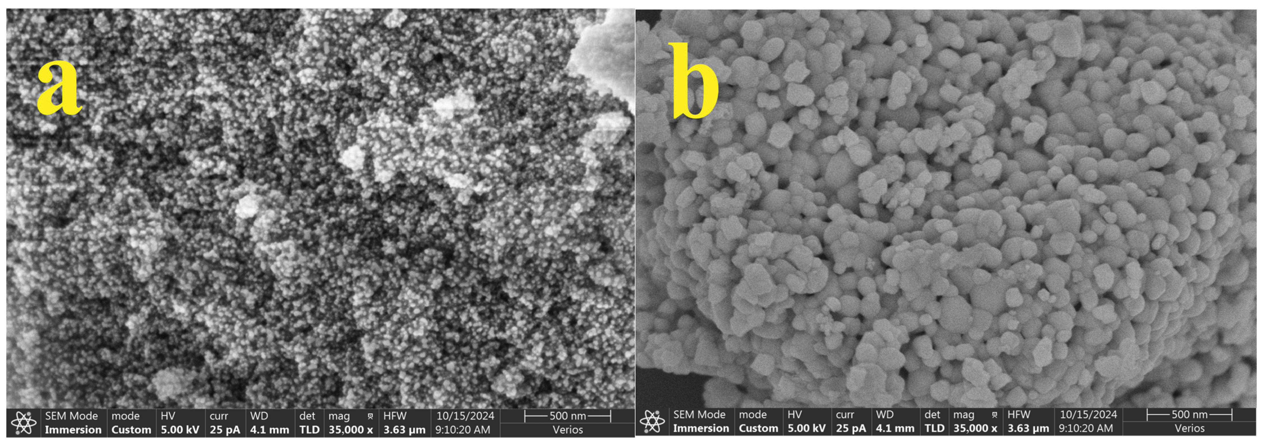
Figure 3 displays the scanning electron microscopy (SEM) images of ZrO2 nanoparticles synthesized via green and chemical methods: PF-ZrO2 NPs using Sonchus asper extract (a) and commercially synthesized ZrO2 NPs (b), both observed at 35,000× magnification. The green-synthesized PF-ZrO2 NPs (Figure 3a) reveal a heterogeneous surface structure with a dispersed, loosely aggregated morphology composed of fine-grained, irregular nanoparticles [31]. These particles display pronounced surface roughness and interparticle voids, likely resulting from the interaction of plant-derived biomolecules, such as flavonoids, phenolic acids, and tannins, with zirconium ions during the reduction and nucleation process [31]. Such a structure implies a higher specific surface area and abundant surface-active sites, which are particularly beneficial for the adsorption of heavy metals like Cr(VI) due to enhanced surface accessibility and reactive functional groups [32]. In contrast, the chemically synthesized ZrO2 NPs (Figure 3b) appear more densely packed, spherical, and uniformly distributed, forming compact aggregates with relatively smoother surfaces. This morphology is indicative of a high-temperature, template-free synthesis process that favors crystal growth and yields nanoparticles with lower surface reactivity but higher structural integrity and phase uniformity [33,34]. The more condensed surface of chemically synthesized ZrO2 may result in fewer active sites for adsorption, while the bio-template-directed assembly of PF-ZrO2 potentially retains biogenic ligands or organic residues on the surface that can facilitate metal binding via complexation or electrostatic interactions [35]. These morphological contrasts suggest that PF-ZrO2 NPs, with their rougher texture, nanoscale porosity, and biocompatible surface chemistry, may offer superior performance in environmental remediation applications—particularly in Cr(VI) adsorption—compared to their commercial counterparts [36]. Moreover, the environmentally benign and energy-efficient synthesis of PF-ZrO2 further underscores the promise of plant-based nanotechnology in designing functional materials for sustainable water treatment systems.
Figure S1 illustrates the SEM-EDS spectra and elemental distribution maps for both plant-fabricated ZrO2 nanoparticles (PF-ZrO2 NPs) and commercially synthesized ZrO2 NPs, providing insight into their elemental composition and surface homogeneity. The EDS spectrum for PF-ZrO2 (Figure S1c) shows three major peaks corresponding to zirconium (Zr), oxygen (O), and carbon (C), with their respective atomic percentages being 17.6%, 53.5%, and 28.9%. The notable presence of carbon confirms the successful incorporation of phytochemicals from Sonchus asper onto the nanoparticle surface, likely resulting from residual organic molecules acting as capping agents during the green synthesis process [37]. In contrast, the EDS spectrum of chemically synthesized ZrO2 (Figure S1d) reveals a higher zirconium content (33.4%) and lower carbon content (11.5%), suggesting a purer, more crystalline material with minimal organic residues. Elemental mapping (Figure S1a for PF-ZrO2 and Figure S1b for ZrO2) further supports these findings: in PF-ZrO2, the Zr (green), C (red), and O (blue) maps show a uniform and homogeneously distributed signal across the nanoparticle surface, indicating consistent elemental dispersion and the presence of surface-bound organic matter. Conversely, the mapping for commercial ZrO2 shows a strong Zr and O signal but a much weaker C distribution, corroborating the EDS quantitative data. These observations confirm that the green synthesis route not only maintains elemental uniformity but also introduces functionalized organic moieties, which could play a significant role in enhancing surface reactivity and metal ion adsorption [38]. This added functionality, along with a balanced Zr:O ratio, highlights the green-synthesized ZrO2 as a promising candidate for environmental remediation, particularly in adsorption-based removal of toxic heavy metals such as Cr(VI).
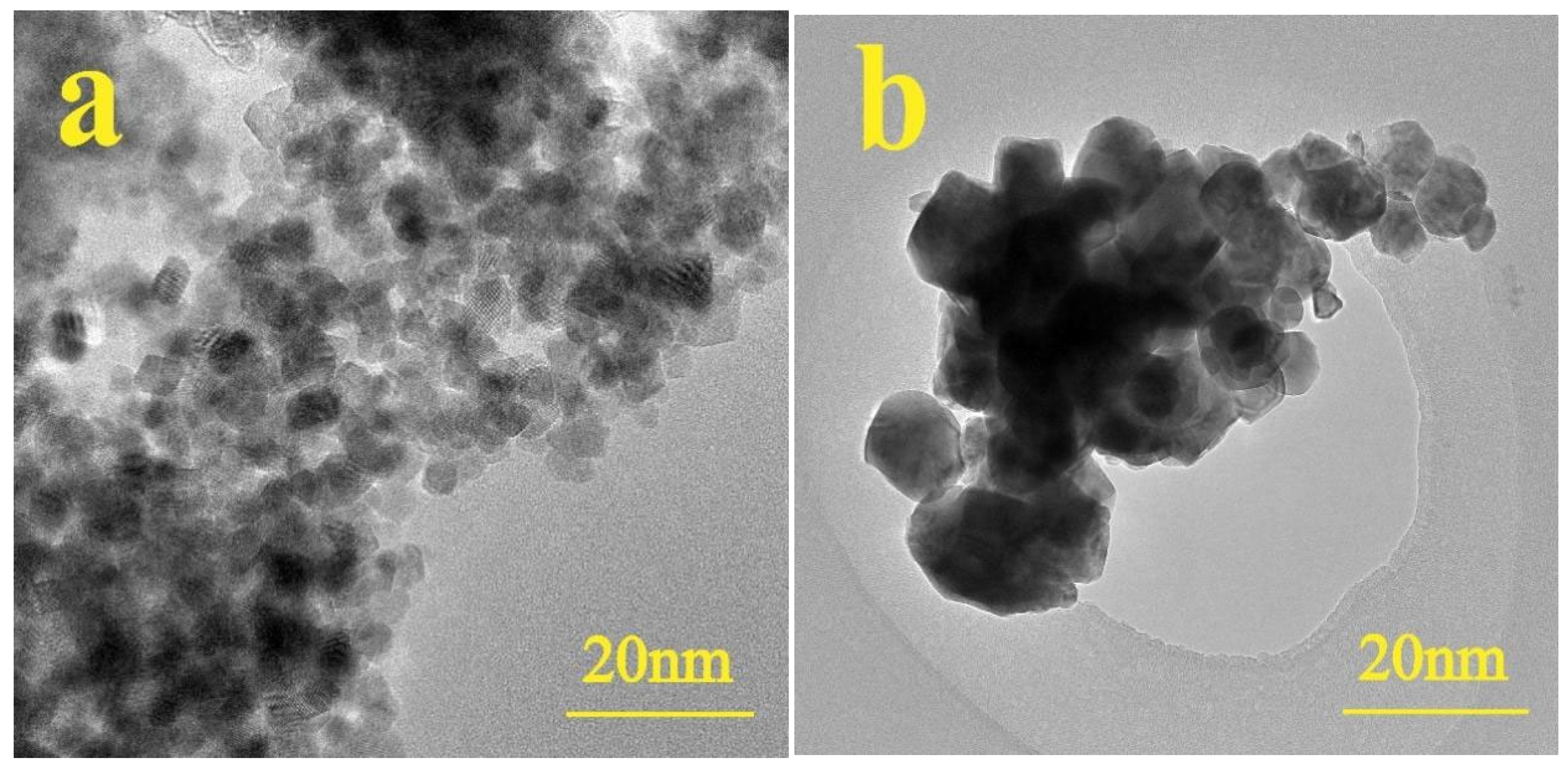
Figure 4 presents transmission electron microscopy (TEM) images of PF-ZrO2 nanoparticles synthesized using Sonchus asper extract (a) and chemically synthesized ZrO2 nanoparticles (b), both imaged at a 20 nm scale to examine particle morphology and nanostructure. The PF-ZrO2 NPs (Figure 4a) appear as discrete, uniformly dispersed nanoparticles with irregular but relatively consistent shapes. The particles exhibit a loosely aggregated structure with visible interparticle boundaries, suggesting minimal agglomeration and a high degree of individualization [36]. The particle size of PF-ZrO2 NPs and chemically synthesized ZrO2 NPs was analyzed using TEM images with appropriate scale bars (20 nm). As shown in Figure 4. The average particle size was estimated by analyzing TEM images using ImageJ software (version 1.54d, National Institutes of Health, Bethesda, MD, USA). A scale was set using the internal scale bar provided in the images. Over 50 nanoparticles were manually selected and measured using the built-in measurement tool, and the diameter was recorded. The particle size distribution was plotted based on these measurements, and the average size and standard deviation were calculated (Figure S2). PF-ZrO2 NPs exhibited a much smaller and more uniform size distribution, with an average diameter of 7.8 ± 13.9 nm, while chemically synthesized ZrO2 NPs showed a broader size distribution and a significantly larger average particle size of 29.8 ± 35.7 nm. These findings indicate that green synthesis facilitates the formation of ultra-small ZrO2 nanoparticles with reduced aggregation and narrower size distribution. This morphology is likely due to the stabilizing influence of phytochemicals in the plant extract, which caps the nanoparticles and prevents excessive clustering [36]. Additionally, the slight contrast differences and varying degrees of electron density indicate the presence of surface organic layers, possibly derived from phenolics, flavonoids, or other secondary metabolites [39]. In comparison, the chemically synthesized ZrO2 NPs display a denser, more agglomerated morphology with larger clusters of fused nanoparticles, some forming polycrystalline domains with indistinct boundaries. The compact appearance of these particles reflects the absence of natural capping agents, resulting in higher particle-particle fusion during thermal synthesis [40]. Despite the differences in agglomeration behavior, both samples fall within the nanometric range, suggesting comparable primary particle sizes. However, the PF-ZrO2 shows superior dispersity and structural openness, which are advantageous for surface-mediated applications such as adsorption, catalysis, and ion exchange. These findings underscore the influence of the synthesis route on particle morphology: the green method yields more surface-accessible, well-dispersed nanoparticles, whereas the chemical method favors denser aggregates with potentially reduced active surface area. The TEM analysis, therefore, reinforces the suitability of PF-ZrO2 for applications in environmental remediation, where high surface exposure and active binding sites are critical for performance.
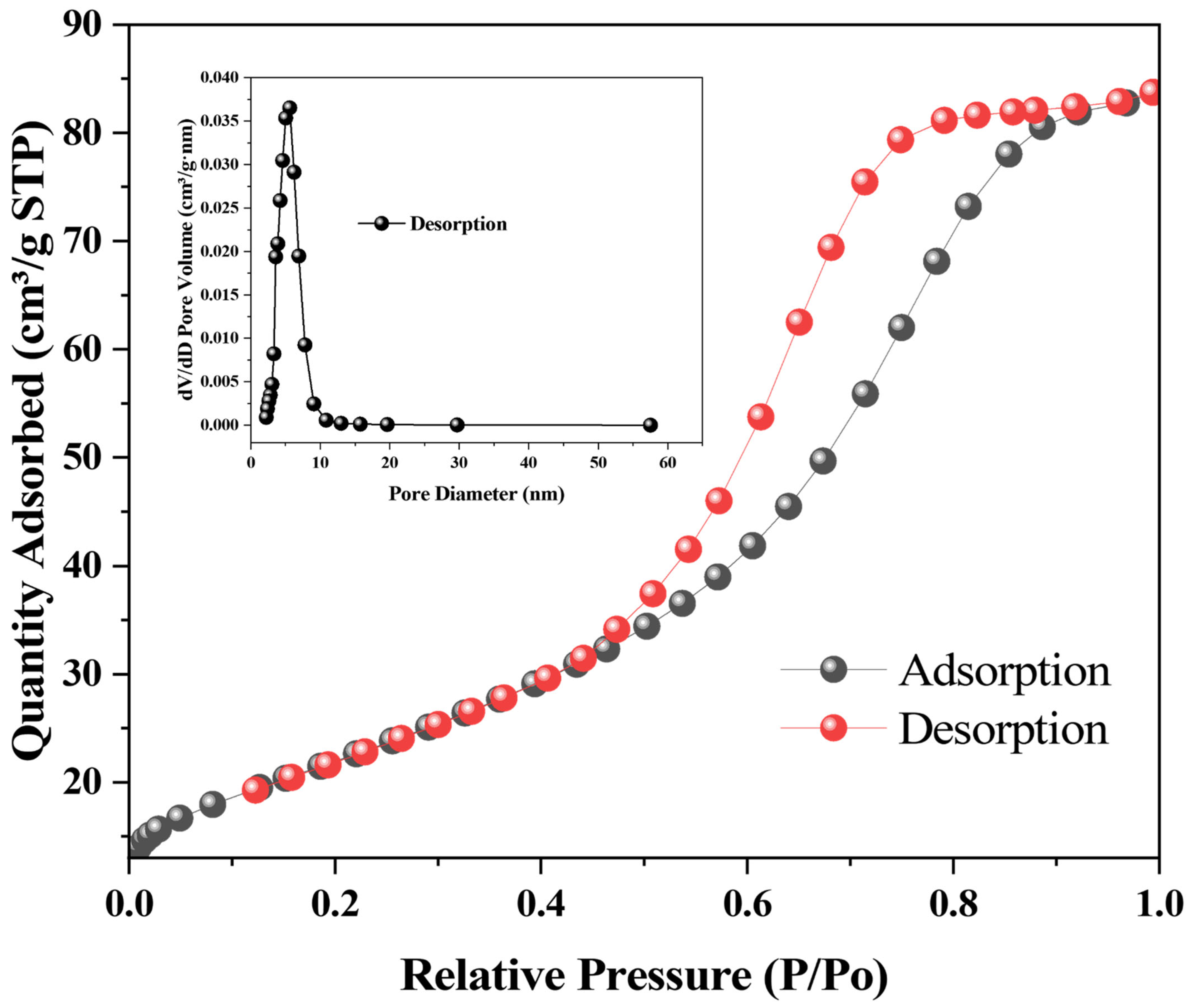
Figure 5 presents the nitrogen adsorption-desorption isotherm of PF-ZrO2 NPs, which exhibits a typical type IV isotherm with an H1-H2 type hysteresis loop, indicating the presence of mesoporous characteristics. The sharp uptake at relative pressures between 0.5 and 0.9 suggests capillary condensation in mesopores, while the hysteresis loop implies a combination of uniform pore channels and bottleneck effects. The average pore diameter was determined to be in the range of 7.8 ± 13.9 nm, and the material demonstrates a relatively narrow pore size distribution. These features support efficient mass transport and are beneficial for adsorption processes. Notably, the adsorption and desorption branches of the isotherm are nearly overlapping, suggesting a high degree of reversibility in the adsorption process. While such behavior is often indicative of physisorption, this does not exclude the occurrence of chemisorption. In fact, subsequent kinetic and isotherm modeling results confirmed a pseudo-second-order model and strong Langmuir fitting, both of which support a chemisorption-dominated mechanism. The apparent contradiction can be reconciled by considering that Cr(VI) interacts with surface functional groups on PF-ZrO2 NPs via reversible coordination or weak complexation. These interactions may be sufficiently labile to allow easy desorption under alkaline conditions, thereby producing a near-symmetrical isotherm curve. Although BET measurements were only conducted for PF-ZrO2NPs, the data sufficiently demonstrate their porous nature and suitability for heavy metal removal applications.

3.2. Batch Adsorption Experiment

3.2.1. Effect of pH

To evaluate the influence of pH on Cr(VI) adsorption performance, batch experiments were conducted at pH values ranging from 2 to 8 (Figure S3A). The pH of the solution is a critical factor that governs both the surface charge of the adsorbent and the speciation of Cr(VI) in aqueous media [41]. Under acidic conditions (pH 2–5), the surfaces of PF-ZrO2 and ZrO2 NPs are predominantly protonated, resulting in a net positive surface charge that promotes electrostatic attraction with anionic Cr(VI) species such as HCrO4 and Cr2O72− [42,43]. Notably, the maximum adsorption efficiency was observed at pH 5, where PF-ZrO2 and ZrO2 achieved removal rates of 93.2% and 92.7%, respectively.
Although pH values lower than 5 theoretically strengthen electrostatic interactions, the slight decline in removal efficiency below this point can be attributed to two key factors. First, the high concentration of protons (H+) at lower pH competes with Cr(VI) species for the limited active binding sites, thereby reducing adsorption. Second, under strongly acidic conditions (especially at pH < 4), Cr(VI) partially exists as H2CrO4, a neutral molecular form that exhibits much lower affinity for surface interaction via electrostatic or ligand-exchange mechanisms [44].
At pH values above 5, the surface of the adsorbents becomes increasingly negatively charged, leading to electrostatic repulsion with CrO42−—the dominant Cr(VI) species under near-neutral and alkaline conditions. Consequently, adsorption efficiency declines substantially. The overall trend confirms that pH 5 represents an optimal balance between favorable surface charge, species distribution, and minimal proton competition, leading to the highest Cr(VI) removal.

3.2.2. Effect of Metal Ion Concentration

Figure S3B illustrates the adsorption rate trends of PF-ZrO2 and ZrO2 NPs across different Cr(VI) concentrations. Both materials exhibit a similar pattern, where the adsorption rate initially increases with Cr(VI) concentration and then declines. At lower Cr(VI) concentrations, the adsorption rate rises with concentration due to the mass transfer effect driven by the concentration gradient; higher solution concentrations accelerate the diffusion rate of chromium ions to the adsorbent surface, thereby enhancing adsorption efficiency [45]. The peak adsorption rates were observed within the concentration range of 50–70 mg/L, reaching 98.2% for PF-ZrO2 NPs and 87.7% for ZrO2 NPs. However, as the Cr(VI) concentration increased beyond this range, the adsorption rate began to decline, particularly when concentrations exceeded 80 mg/L. Notably, PF-ZrO2 NPs exhibited a significantly higher peak adsorption efficiency (98.2%) compared to chemically synthesized ZrO2 NPs (87.7%), likely due to their smaller particle size and more porous morphology.
This decrease may result from the saturation of active sites on the adsorbent, with intensified competition among chromium ions [46]. Additionally, higher Cr(VI) concentrations can increase diffusion resistance, reducing adsorption efficiency. Furthermore, excessive chromium ion concentrations may lead to multilayer adsorption or particle aggregation, which reduces the effective surface area of the adsorbent, further diminishing the adsorption rate [47]. Notably, PF-ZrO2 NPs demonstrated better adaptability to varying Cr(VI) concentrations compared to ZrO2 NPs, which could be due to their looser structure and greater availability of adsorption sites, allowing them to maintain higher adsorption capacities at elevated concentrations [48]. In contrast, the more compact surface structure of ZrO2 NPs, with fewer available adsorption sites, results in faster saturation at higher concentrations. This explains why, despite both materials exhibiting saturation at high Cr(VI) concentrations, PF-ZrO2 NPs maintain a slightly higher overall adsorption rate than ZrO2 NPs.

3.2.3. Effect of Dose

Figure S4 depicts the influence of adsorbent dosage on the removal efficiency of hexavalent chromium (Cr(VI)) by PF-ZrO2 (a) and chemically synthesized ZrO2 (b) nanoparticles at a constant temperature (25 ± 2 °C), contact time (30 min), and pH (5), with an initial Cr(VI) concentration of 100 mg L−1. Both adsorbents exhibited a rapid increase in Cr(VI) removal with rising dosage from 0.1 to 0.5 g, achieving over 80% removal efficiency at low adsorbent loadings. This sharp rise can be attributed to the increased number of available active sites for Cr(VI) binding on the nanoparticle surfaces [49]. Beyond 0.5 g, the removal efficiency plateaued, indicating saturation of adsorption sites and possibly particle agglomeration, which reduces the effective surface area available for adsorption. Notably, PF-ZrO2 showed marginally higher removal efficiency across all doses, suggesting that the green-synthesized nanoparticles possess a greater number of accessible binding sites, potentially due to the surface functionalization conferred by phytochemicals from Sonchus asper. The slightly better performance of PF-ZrO2 may also be linked to its more porous and dispersed morphology, as supported by SEM and TEM results, which enhances the diffusion of Cr(VI) ions toward active sites. In contrast, the chemically synthesized ZrO2 showed similar but slightly lower adsorption trends, potentially due to denser packing and reduced surface functionality. These results affirm that nanoparticle dosage plays a critical role in determining adsorption performance, and that green-synthesized PF-ZrO2 can achieve high removal efficiency even at lower doses, underscoring its potential as an eco-friendly and cost-effective adsorbent for Cr(VI) remediation in aqueous environments.

3.2.4. Effect of Contact Time

The effect of contact time on the adsorption efficiency of Cr(VI) (100 mg L−1) using PF-ZrO2 (a) and chemically synthesized ZrO2 (b) nanoparticles under identical conditions (25 ± 2 °C, pH 5, and fixed adsorbent dosage of 100 mg). Both adsorbents exhibited a rapid increase in Cr(VI) removal within the first 30 min, reaching over 85% removal efficiency. This sharp initial uptake can be attributed to the abundance of available active sites on the nanoparticle surfaces during the early stages of adsorption [50,51]. After this initial phase, the adsorption capacity plateaued, indicating that equilibrium had been achieved and the remaining unoccupied sites were either less accessible or already saturated with Cr(VI) ions. Notably, both green-synthesized PF-ZrO2 and chemically synthesized ZrO2 showed similar trends in equilibrium kinetics; however, PF-ZrO2 maintained a slightly higher and more stable removal efficiency throughout the observed time frame. This suggests that the PF-ZrO2 nanoparticles, owing to their porous structure and possible presence of bio-derived surface functional groups, may offer a more accessible and reactive surface for Cr(VI) binding. The stabilization of removal rates beyond 30 min implies that longer contact times may not significantly enhance adsorption performance under these conditions. These results highlight the fast adsorption kinetics and surface efficiency of PF-ZrO2, affirming its potential as a rapid and effective adsorbent for Cr(VI) remediation in wastewater systems.

3.2.5. Adsorption Kinetics

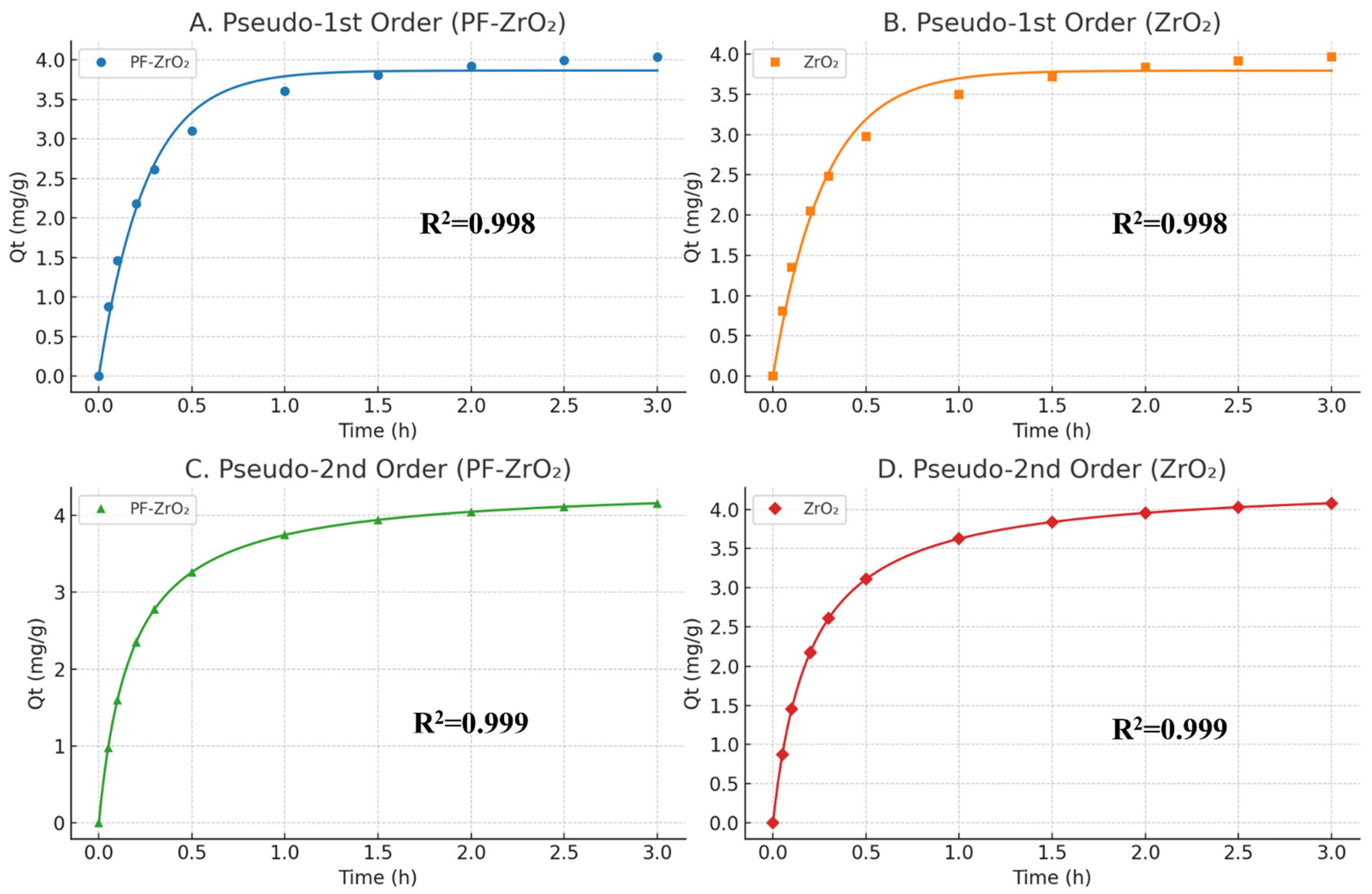
Pseudo-first-order and pseudo-second-order models are commonly used to examine the kinetics of metal ion adsorption onto adsorbents, largely determining the feasibility of the adsorption system and the potential applications of the adsorbent [52]. The experimental adsorption data and the fitting results of the pseudo-first-order and pseudo-second-order models are presented in Table 2. Figure 6 presents the kinetic modelling of Cr(VI) adsorption onto PF-ZrO2 nanoparticles using pseudo-first-order and pseudo-second-order models. The adsorption process showed a rapid initial uptake within the first hour, which gradually plateaued, indicating the attainment of equilibrium [53]. Both kinetic models were fitted to the experimental data, with the pseudo-second-order model exhibiting a better alignment with the observed values throughout the time course, especially during the equilibrium phase. This suggests that chemisorption is the dominant rate-controlling mechanism, involving valence forces through electron sharing or exchange between Cr(VI) and active functional groups on the PF-ZrO2 surface [53]. The higher correlation of the pseudo-second-order model supports the hypothesis that adsorption is not purely physical but likely involves surface complexation or chemical bonding interactions. The strong fit of both models in the early stages also highlights the high reactivity and availability of adsorption sites in the PF-ZrO2 structure, further validating its efficiency as a sustainable adsorbent for Cr(VI) remediation. This dual fitting behavior can be attributed to the multi-step nature of the adsorption process. In the initial stage, rapid physical interactions such as electrostatic attraction and surface diffusion may dominate, aligning with the assumptions of the pseudo-first-order model. As the process continues, chemical bonding and surface complexation become more pronounced, better described by the pseudo-second-order model. The high surface area and abundant functional groups on PF-ZrO2 facilitate both fast physisorption and slower chemisorption, leading to effective Cr(VI) uptake over time. It is worth noting that kinetic analysis was conducted at a single Cr(VI) concentration (50 mg/L), selected based on isotherm optimization. While this provides meaningful insight into adsorption behavior, future studies incorporating a range of concentrations are warranted to validate the kinetic parameters more comprehensively.

3.2.6. Adsorption Isotherms

The adsorption data of PF-ZrO2 NPs were fitted to both the Langmuir and the Freundlich models to determine which model better describes the adsorption behavior (Figure 7A,B). Equilibrium isotherms (20–100 mg L−1, pH 5, 25 °C, 30 min, 300 mg L−1 dose) were used for fitting. Based on the goodness-of-fit, the Langmuir model achieved a higher correlation coefficient (R2 = 0.997; χ2 = 0.961) for PF-ZrO2, while the Freundlich model also showed an excellent fit (R2 = 0.994). For ZrO2, the Langmuir model still provided a reasonable correlation (R2 = 0.845). The maximum adsorption capacities (qₘₐₓ) obtained from the Langmuir model were 142.24 mg g−1 for PF-ZrO2 and 133.11 mg g−1 for ZrO2, reflecting the monolayer adsorption potential of both materials.
Although both models fitted the data well, the Langmuir model provided a slightly better representation, suggesting that the adsorption process tends toward a monolayer mechanism with homogeneous surface adsorption sites. However, the relatively good fit to the Freundlich model implies that surface heterogeneity and multilayer adsorption effects may also be involved. This phenomenon—where both models yield satisfactory fits—can be attributed to the complex surface properties of the material, such as coexisting mesopores, variable binding sites, and physicochemical heterogeneity.
Such dual-model fitting has also been reported in other nanomaterial-based adsorbents, indicating that real-world adsorption often involves a hybrid mechanism. The Freundlich fit suggests that at different local regions of the sorbent surface, non-uniform interactions or multilayer adsorption might occur, especially at higher concentrations. Therefore, the process can be reasonably explained as a combination of dominant monolayer adsorption with auxiliary multilayer or site-heterogeneous interactions, especially given the porous nature and surface complexity of the PF-ZrO2 NPs.
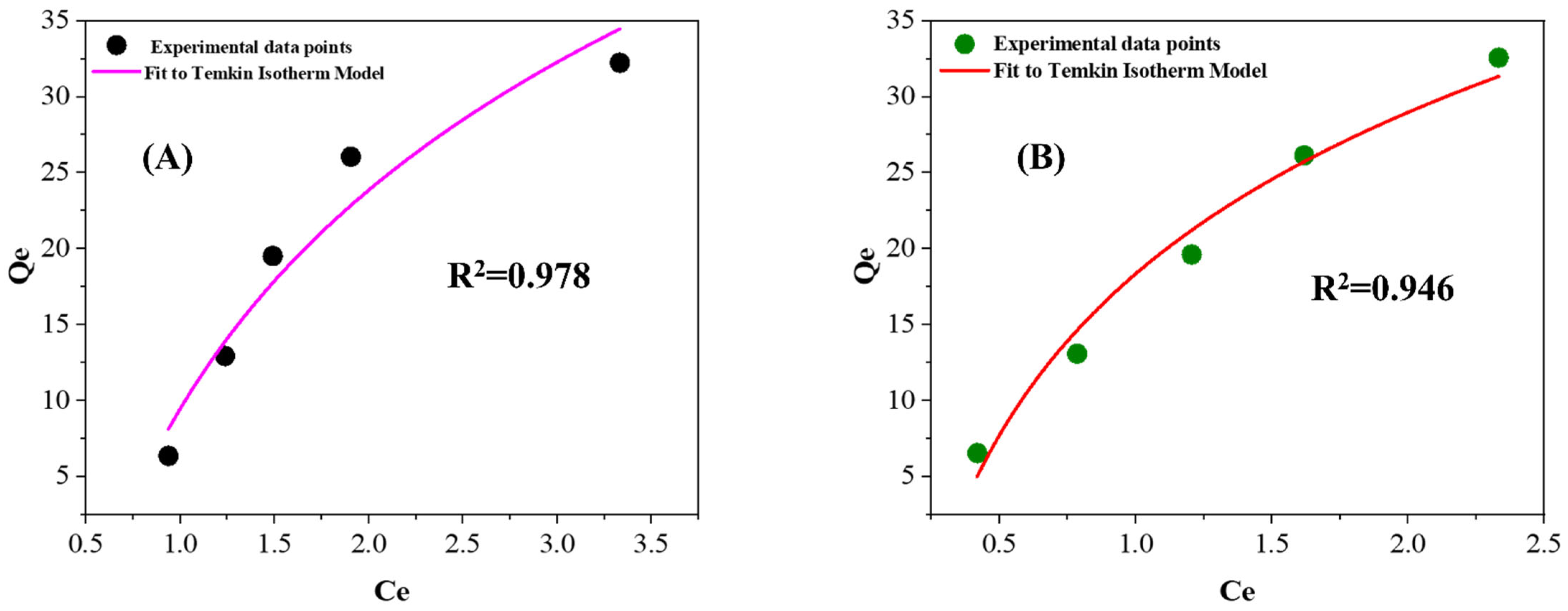
The Temkin adsorption isotherm model assumes that the binding energies on the solid surface of the adsorbent are uniformly distributed up to a certain maximum binding energy [54]. Fitting the experimental data using the Temkin isotherm model yielded R2 values of 0.946 and 0.978 for PF-ZrO2 NPs (Figure 8A) and ZrO2 NPs (Figure 8B), respectively. Compared to the Langmuir and Freundlich models, the Temkin model showed a significantly lower fitting degree, indicating that it is not suitable for describing the adsorption behavior of Cr(VI) on PF-ZrO2 NPs and ZrO2 NPs. The poorer fit for PF-ZrO2 NPs suggests a higher degree of heterogeneity in its adsorption process, implying that the adsorption is not purely physical [55]. Based on the aforementioned models, it can be inferred that the adsorption of Cr(VI) on PF-ZrO2 NPs and ZrO2 NPs is a monolayer process occurring on a heterogeneous surface.

3.3. Desorption Efficiency of Nanoparticles

Figure S5 illustrates the desorption efficiency of Cr(VI) from both green-synthesized and chemically synthesized ZrO2 nanoparticles at two different NaOH concentrations (0.1 M and 0.5 M). A clear trend is observed wherein increasing the NaOH concentration enhances the desorption efficiency for both nanoparticle types. At 0.1 M NaOH, chemical ZrO2 NPs achieved a desorption efficiency of approximately 29%, compared to 22% for green ZrO2 NPs. When the NaOH concentration was increased to 0.5 M, the desorption efficiency rose markedly, reaching 55% for chemical ZrO2 and 45% for green ZrO2 NPs. This suggests that stronger alkaline conditions facilitate the release of Cr(VI) ions from the nanoparticle surfaces, likely due to the competition between hydroxide ions (OH) and Cr(VI) species for adsorption sites. The higher desorption observed in chemically synthesized ZrO2 may be attributed to its more crystalline surface and fewer surface functional groups compared to the green ZrO2 NPs, which may bind Cr(VI) more strongly due to the presence of residual biomolecules and oxygen-containing groups from plant extract residues [56]. PF-ZrO2 NPs exhibited slightly lower desorption; this implies a stronger binding affinity for Cr(VI), which is advantageous in long-term remediation applications where leaching is a concern [57]. Conversely, the higher desorption capacity of chemical ZrO2 suggests it may be more suitable for regenerable systems requiring multiple adsorption-desorption cycles. These results highlight the importance of tailoring nanoparticle properties based on the intended reuse or environmental stability of the adsorbent.

3.4. Adsorption Mechanism

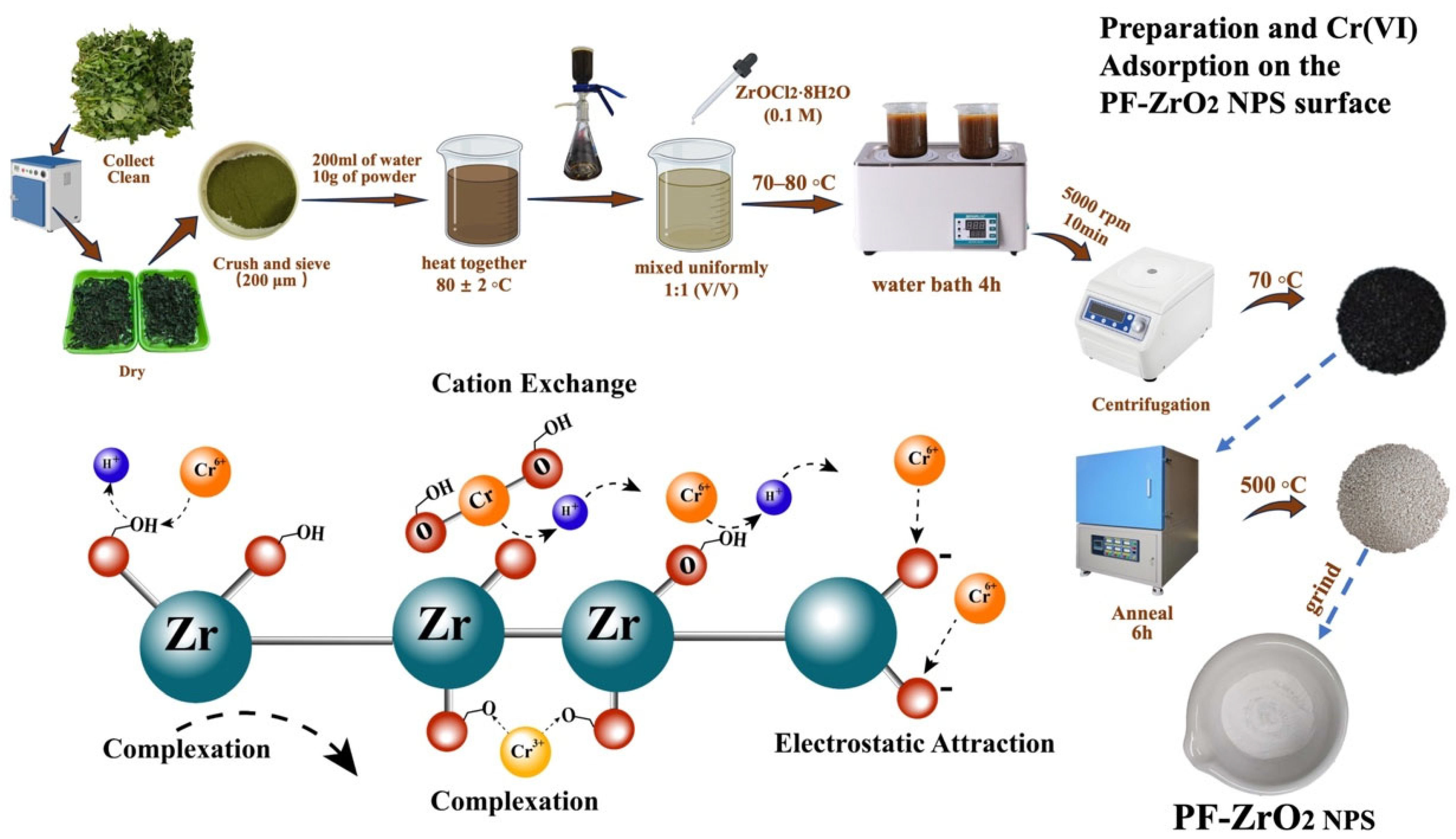
The Cr(VI) removal by PF-ZrO2 nanoparticles synthesized via the green method is likely driven by a combination of electrostatic attraction, cation exchange, and surface complexation (Figure 9) [58]. Under mildly acidic to neutral conditions, surface hydroxyl groups (–OH) on PF-ZrO2 are protonated, generating positively charged sites that attract negatively charged chromate species such as CrO42− or HCrO4 [59]. Residual thermally stable organic groups-such as C–H and C–N-introduced by the plant extract during synthesis may also participate in adsorption, enhancing surface heterogeneity and providing additional binding interactions. While the original heat-labile bioactive molecules are largely decomposed during annealing, these stable residues may still contribute to adsorption through hydrogen bonding or weak electrostatic forces. The overall removal process may also involve partial reduction of Cr(VI) to Cr(III), followed by inner-sphere complexation with oxygen-containing surface groups (e.g., Zr–O–Cr) [60].

4. Future Perspectives

Plant-fabricated nanoparticles, particularly zirconia, hold significant environmental advantages and exhibit material performance comparable to traditional chemical synthesis methods [61]. Analysis indicates that plant-fabricated nanoparticles show notably lower aggregation than their chemically synthesized counterparts, implying superior dispersibility. This enhanced dispersibility offers a larger specific surface area, which is crucial for applications in catalysis and adsorption, ultimately improving efficiency [62]. In the adsorption of pollutants such as Cr(VI) from water, smaller particles can facilitate faster and more effective removal, thereby boosting the overall performance of adsorption-based systems [63].
In addition, plant-based synthesis not only optimizes the physical characteristics of nanoparticles but also allows for tunable control over particle size and morphology by adjusting the type and concentration of plant extracts used [64]. This strategy opens up new possibilities in nanotechnology, offering a method that is both environmentally friendly and capable of enhancing material performance. Features such as low aggregation, high dispersibility, and nanoscale dimensions provide opportunities for applications in environmental remediation, catalysis, medicine, and energy-related fields [65].
However, the transition from laboratory-scale plant-based nanoparticle synthesis to industrial-scale production still faces several short-term challenges. These include difficulties in standardizing plant extract compositions due to seasonal and regional variability, limited reaction yields, and inconsistency in nanoparticle size and morphology when scaling up. Additionally, current green synthesis methods often involve lengthy preparation times and energy-intensive post-treatments (e.g., calcination), reducing overall process efficiency. Overcoming these barriers will require process optimization, development of scalable reactor systems, stabilization of extract composition, and enhanced reproducibility through better control of synthesis parameters.
Despite these challenges, the further development and integration of plant-fabricated nanomaterials into industrial applications hold substantial promise. With continued innovation in green chemistry and nanomanufacturing, this approach can meet environmental sustainability goals while providing cost-effective and high-performance alternatives to conventional nanomaterials. This transition strongly supports the broader objectives of sustainable development, particularly in achieving clean water technologies aligned with UN SDG No. 6.

5. Conclusions

In this study, Sonchus asper plant extract was utilized as a reducing agent in the plant fabrication (PF) of zirconia (ZrO2) nanoparticles (NPs). A comparative analysis was conducted with industrially produced ZrO2 nanoparticles to evaluate various parameters. XRD analysis confirmed that both PF-ZrO2 and chemically synthesized ZrO2 NPs adopt a cubic fluorite-type structure, with peak positions and calculated lattice parameters matching JCPDS reference values. Broader peaks in PF-ZrO2 indicate smaller crystallites, consistent with green synthesis effects. TEM and XRD analyses showed that PF-ZrO2 NPs have an average particle size of 7.8 ± 13.9 nm, in contrast to 29.8 ± 35.7 nm for ZrO2 NPs, with PF-ZrO2 NPs displaying a smaller size, which generally correlates with a higher specific surface area. SEM imaging illustrated that PF-ZrO2 NPs exhibit greater variability in spatial dimensions, with some particles featuring grooved structures, rough surfaces, and abundant voids, resulting in a loose particle distribution. Based on Langmuir isotherm analysis (Section 3.2.5; Figure 9; Table 2), the maximum capacities were 142.24 mg g−1 (PF-ZrO2) and 133.11 mg g−1 (ZrO2). BET/BJH analyses corroborate a mesoporous texture for both samples (type-IV, H1–H2 hysteresis), with PF-ZrO2 exhibiting higher S_BET and pore volume, supporting its superior adsorption performance. Thus, ZrO2 nanoparticles synthesized using Sonchus asper extract show great potential as eco-friendly and effective adsorbents.

Supplementary Materials

The following supporting information can be downloaded at: https://www.mdpi.com/article/10.3390/pr13092794/s1, Figure S1: SEM-EDX spectra of PF-ZrO2 NPs (a) and ZrO2 NPs (b), and elemental mapping showing the uniform distribution of zirconium (Zr), oxygen (O), and carbon (C) in PF-ZrO2 NPs (c) and ZrO2 NPs (d); Figure S2: Particle size distribution of PF-ZrO2 and chemically synthesized ZrO2 nanoparticles analyzed from TEM images; Figure S3: Effect of solution pH (A) and initial Cr(VI) concentration (B) on the adsorption efficiency of PF-ZrO2 NPs and ZrO2 NPs. Adsorption experiments were carried out at 25 °C ± 2, with a contact time of 30 min and a nanoparticle dosage of 100 mg L−1. The pH values were adjusted to 2, 3, 4, 5, 6, 7, and 8, while initial Cr(VI) concentrations were set at 20, 30, 40, 50, 60, 70, 80, 90,and 100 mg L−1 to evaluate their individual effects on removal performance. Error bars represent ± standard deviation (n = 3); Figure S4: Effect of adsorbent dosage on the removal efficiency of Cr(VI) by PF-ZrO2 and ZrO2 nanoparticles at 25 ± 2 °C, pH 6, and an initial Cr(VI) concentration of 100 mg/L. Error bars represent ± standard deviation (n = 3); Figure S5: Desorption of Cr(VI) from ZrO2 and PF-ZrO2 NPs. Error bars represent ± standard deviation (n = 3).

Author Contributions

G.W. conducted the experiment, collected and treated the data, created figures, and drafted the manuscript. W.L. contributed to experiment performance, investigation, analysis, and writing. F.Q. provided review, editing, and proofreading support. M.D. conducted data analyses, contributed to writing, and made corrections. S.Y. conducted data analyses, contributed to writing, and made corrections. J.W. and S.M. contributed to writing, reviewing, correcting, editing, and proofreading the manuscript, as well as corresponding with collaborators. All authors have read and agreed to the published version of the manuscript.

Funding

This study was supported by The National Natural Science Foundation of China (NSFC-31860728), Launch Fund of Hainan University High level Talent (RZ2100003226), the research fund of The Innovation Platform for Academicians of Hainan Province (YSPTZX20212), Key R and D projects in Hainan Province (No. ZDYF2021XDNY185), and Hainan Province Science and Technology Special Fund (ZDYF2021SHFZ071).

Data Availability Statement

This paper and its Supplementary Information contain all relevant data.

Conflicts of Interest

The authors declare no conflict of interest.

References

  1. Jaffari, Z.H.; Hong, J.; Park, K.Y. A Systematic Review of Innovations in Tannery Solid Waste Treatment: A Viable Solution for the Circular Economy. Sci. Total Environ. 2024, 948, 174848. [Google Scholar] [CrossRef] [PubMed]
  2. Vincent, J.B. Chromium. In Encyclopedia of Human Nutrition, 4th ed.; Academic Press: New York, NY, USA, 2023; Volumes 1–4, ISBN 9780323908160. [Google Scholar]
  3. Sharma, A.; Kapoor, D.; Wang, J.; Shahzad, B.; Kumar, V.; Bali, A.S.; Jasrotia, S.; Zheng, B.; Yuan, H.; Yan, D. Chromium Bioaccumulation and Its Impacts on Plants: An Overview. Plants 2020, 9, 100. [Google Scholar] [CrossRef] [PubMed]
  4. Sirajuddin; Kakakhel, L.; Lutfullah, G.; Bhanger, M.I.; Shah, A.; Niaz, A. Electrolytic Recovery of Chromium Salts from Tannery Wastewater. J. Hazard. Mater. 2007, 148, 560–565. [Google Scholar] [CrossRef] [PubMed]
  5. Yusuf, M.A.; Agustina, L. The Potential Application of Photocatalytic Processes in the Processing of Wastewater in the Leather Industry: A Review. IOP Conf. Ser. Earth Environ. Sci. 2023, 1253, 012025. [Google Scholar] [CrossRef]
  6. Nur-E-Alam, M.; Mia, M.A.S.; Ahmad, F.; Rahman, M.M. An Overview of Chromium Removal Techniques from Tannery Effluent. Appl. Water Sci. 2020, 10, 205. [Google Scholar] [CrossRef]
  7. Anjum, A.; Mazari, S.A.; Hashmi, Z.; Jatoi, A.S.; Abro, R.; Bhutto, A.W.; Mubarak, N.M.; Dehghani, M.H.; Karri, R.R.; Mahvi, A.H.; et al. A Review of Novel Green Adsorbents as a Sustainable Alternative for the Remediation of Chromium (VI) from Water Environments. Heliyon 2023, 9, e15575. [Google Scholar] [CrossRef]
  8. Das, P.K. Toxicological Implications of Hexavalent Chromium in Humans. Adv. Clin. Toxicol. 2022, 7, 1–4. [Google Scholar] [CrossRef]
  9. Munir, N.; Jahangeer, M.; Bouyahya, A.; El Omari, N.; Ghchime, R.; Balahbib, A.; Aboulaghras, S.; Mahmood, Z.; Akram, M.; Shah, S.M.A.; et al. Heavy Metal Contamination of Natural Foods Is a Serious Health Issue: A Review. Sustainability 2022, 14, 161. [Google Scholar] [CrossRef]
  10. Kamila, S.; Shaw, P.; Islam, S.; Chattopadhyay, A. Ecotoxicology of Hexavalent Chromium in Fish: An Updated Review. Sci. Total Environ. 2023, 890, 164395. [Google Scholar] [CrossRef]
  11. Assefa, H.; Singh, S.; Olu, F.E.; Dhanjal, D.S.; Mani, D.; Khan, N.A.; Singh, J.; Ramamurthy, P.C. Advances in Adsorption Technologies for Hexavalent Chromium Removal: Mechanisms, Materials, and Optimization Strategies. Desalin. Water Treat. 2024, 319, 100576. [Google Scholar] [CrossRef]
  12. Kushwaha, B.K.; Singh, V.P. Glutathione and Hydrogen Sulfide Are Required for Sulfur-Mediated Mitigation of Cr(VI) Toxicity in Tomato, Pea and Brinjal Seedlings. Physiol. Plant. 2020, 168, 406–421. [Google Scholar] [CrossRef] [PubMed]
  13. Tamez, C.; Hernandez, R.; Parsons, J.G. Removal of Cu (II) and Pb (II) from Aqueous Solution Using Engineered Iron Oxide Nanoparticles. Microchem. J. 2016, 125, 97–104. [Google Scholar] [CrossRef] [PubMed]
  14. Chua, S.F.; Nouri, A.; Ang, W.L.; Mahmoudi, E.; Mohammad, A.W.; Benamor, A.; Ba-Abbad, M. The Emergence of Multifunctional Adsorbents and Their Role in Environmental Remediation. J. Environ. Chem. Eng. 2021, 9, 104793. [Google Scholar] [CrossRef]
  15. Tamayo-Meza, P.A.; Silva-Rivera, U.S.; Flores-Herrera, L.A.; Rodríguez-Alvarado, L.W.; Pérez-Cruz, J.H.; Rivera-López, J.E. Analysis of the Extreme Equilibrium Conditions of an Internal Cavity Located inside a Flat Metal Plate Subjected to an Internal Pressure P. Materials 2020, 13, 2043. [Google Scholar] [CrossRef]
  16. Qin, H.; Kuang, T.; Li, Q.; Yue, X.; Gao, H.; Liu, F.; Yi, Y. Stress Concentration Induced by the Crystal Orientation in the Transient-Liquid-Phase Bonded Joint of Single-Crystalline Ni3Al. Materials 2019, 12, 2765. [Google Scholar] [CrossRef]
  17. Sun, Y.; Zhang, X.; Zhang, M.; Ge, M.; Wang, J.; Tang, Y.; Zhang, Y.; Mi, J.; Cai, W.; Lai, Y.; et al. Rational Design of Electrospun Nanofibers for Gas Purification: Principles, Opportunities, and Challenges. Chem. Eng. J. 2022, 446, 137099. [Google Scholar] [CrossRef]
  18. Wiśniewski, P.; Jasiński, J.; Mazurak, A.; Stonio, B.; Majkusiak, B. Investigation of Electrical Properties of the Al/SiO2/N++-Si Resistive Switching Structures by Means of Static, Admittance, and Impedance Spectroscopy Measurements. Materials 2021, 14, 6042. [Google Scholar] [CrossRef]
  19. Chakravarty, R.; Shukla, R.; Ram, R.; Tyagi, A.K.; Dash, A.; Venkatesh, M. Practicality of Tetragonal Nano-Zirconia as a Prospective Sorbent in the Preparation of 99Mo/99mTc Generator for Biomedical Applications. Chromatographia 2010, 72, 875–884. [Google Scholar] [CrossRef]
  20. Fathima, J.B.; Pugazhendhi, A.; Venis, R. Synthesis and Characterization of ZrO2 Nanoparticles-Antimicrobial Activity and Their Prospective Role in Dental Care. Microb. Pathog. 2017, 110, 245–251. [Google Scholar] [CrossRef]
  21. Jamkhande, P.G.; Ghule, N.W.; Bamer, A.H.; Kalaskar, M.G. Metal Nanoparticles Synthesis: An Overview on Methods of Preparation, Advantages and Disadvantages, and Applications. J. Drug Deliv. Sci. Technol. 2019, 53, 101174. [Google Scholar] [CrossRef]
  22. Ying, S.; Guan, Z.; Ofoegbu, P.C.; Clubb, P.; Rico, C.; He, F.; Hong, J. Green Synthesis of Nanoparticles: Current Developments and Limitations. Environ. Technol. Innov. 2022, 26, 102336. [Google Scholar] [CrossRef]
  23. Sathishkumar, M.; Sneha, K.; Yun, Y.S. Green Fabrication of Zirconia Nano-Chains Using Novel Curcuma longa Tuber Extract. Mater. Lett. 2013, 98, 242–245. [Google Scholar] [CrossRef]
  24. da Silva, A.F.V.; Fagundes, A.P.; Macuvele, D.L.P.; de Carvalho, E.F.U.; Durazzo, M.; Padoin, N.; Soares, C.; Riella, H.G. Green Synthesis of Zirconia Nanoparticles Based on Euclea natalensis Plant Extract: Optimization of Reaction Conditions and Evaluation of Adsorptive Properties. Colloids Surfaces A Physicochem. Eng. Asp. 2019, 583, 123915. [Google Scholar] [CrossRef]
  25. Cho, M.S.; Kim, J.H.; Kim, C.S.; Mejías, J.A.; Kim, S.C. Sow Thistle Chloroplast Genomes: Insights into the Plastome Evolution and Relationship of Two Weedy Species, Sonchus asper and Sonchus oleraceus (Asteraceae). Genes 2019, 10, 881. [Google Scholar] [CrossRef] [PubMed]
  26. Guarrera, P.M.; Salerno, G.; Caneva, G. Food, Flavouring and Feed Plant Traditions in the Tyrrhenian Sector of Basilicata, Italy. J. Ethnobiol. Ethnomed. 2006, 2, 37. [Google Scholar] [CrossRef]
  27. Khan, R.A.; Khan, M.R.; Sahreen, S. Protective Effect of Sonchus asper Extracts against Experimentally Induced Lung Injuries in Rats: A Novel Study. Exp. Toxicol. Pathol. 2012, 64, 725–731. [Google Scholar] [CrossRef]
  28. Al-nayili, A.; Idan, A.H. Environmentally Friendly Production, Characterization, and Utilization of ZrO2 Nanoparticles for the Adsorption of Amoxicillin in Water Solutions. J. Mol. Liq. 2023, 389, 122875. [Google Scholar] [CrossRef]
  29. Hao, Y.; Li, J.; Yang, X.; Wang, X.; Lu, L. Preparation of ZrO2-Al2O3 Composite Membranes by Sol-Gel Process and Their Characterization. Mater. Sci. Eng. A 2004, 367, 243–247. [Google Scholar] [CrossRef]
  30. Chen, S.; Yin, Y.; Wang, D.; Liu, Y.; Wang, X. Structures, Growth Modes and Spectroscopic Properties of Small Zirconia Clusters. J. Cryst. Growth 2005, 282, 498–505. [Google Scholar] [CrossRef]
  31. Amin, F.; Fozia; Khattak, B.; Alotaibi, A.; Qasim, M.; Ahmad, I.; Ullah, R.; Bourhia, M.; Gul, A.; Zahoor, S.; et al. Green Synthesis of Copper Oxide Nanoparticles Using Aerva javanica Leaf Extract and Their Characterization and Investigation of In Vitro Antimicrobial Potential and Cytotoxic Activities. Evid.-Based Complement. Altern. Med. 2021, 2021, 5589703. [Google Scholar] [CrossRef]
  32. Pantò, F.; Dahrouch, Z.; Saha, A.; Patanè, S.; Santangelo, S.; Triolo, C. Photocatalytic Degradation of Methylene Blue Dye by Porous Zinc Oxide Nanofibers Prepared via Electrospinning: When Defects Become Merits. Appl. Surf. Sci. 2021, 557, 149830. [Google Scholar] [CrossRef]
  33. Cha, T.G.; Baker, B.A.; Salgado, J.; Bates, C.J.; Chen, K.H.; Chang, A.C.; Akatay, M.C.; Han, J.H.; Strano, M.S.; Choi, J.H. Understanding Oligonucleotide-Templated Nanocrystals: Growth Mechanisms and Surface Properties. ACS Nano 2012, 6, 8136–8143. [Google Scholar] [CrossRef]
  34. Bumajdad, A.; Nazeer, A.A.; Al Sagheer, F.; Nahar, S.; Zaki, M.I. Controlled Synthesis of ZrO2 Nanoparticles with Tailored Size, Morphology and Crystal Phases via Organic/Inorganic Hybrid Films. Sci. Rep. 2018, 8, 3695. [Google Scholar] [CrossRef]
  35. Madkikar, P.; Wang, X.; Mittermeier, T.; Monteverde Videla, A.H.A.; Denk, C.; Specchia, S.; Gasteiger, H.A.; Piana, M. Synthesis Optimization of Carbon-Supported ZrO2 Nanoparticles from Different Organometallic Precursors. J. Nanostruct. Chem. 2017, 7, 133–147. [Google Scholar] [CrossRef]
  36. Febriantini, D.; Purnamasari; Liandi, A.R.; Usman; Yulizar, Y. A Comprehensive Study on the Influence of Single and Multiple Phytochemicals in Facilitating Green Synthesis of ZrO2 Nanoparticles. Nano-Struct. Nano-Objects 2024, 39, 101303. [Google Scholar] [CrossRef]
  37. Luo, M.; Jiang, X.; Liu, Y.; Liu, Y.; Yu, H.; Niu, Y.; Meng, X.; Wang, L.; Niu, Y. Enhanced Adsorption Complexation of Biochar by Nitrogen-Containing Functional Groups. J. Environ. Chem. Eng. 2023, 11, 111194. [Google Scholar] [CrossRef]
  38. Zeng, G.; Li, G.; Wu, Y.; Tang, C.; Yuan, W.; Zhong, H.; Deng, N.; Liu, Q.; Liu, J.; Ouyang, K.; et al. Preparation of High Specific Surface Area Zn/Al/Zr LDH@HTCC for Enrichment and Recovery of Phosphorus from Water. Surf. Interfaces 2023, 42, 103330. [Google Scholar] [CrossRef]
  39. Hijazi, B.U.; Faraj, M.; Mhanna, R.; El-Dakdouki, M.H. Biosynthesis of Silver Nanoparticles as a Reliable Alternative for the Catalytic Degradation of Organic Dyes and Antibacterial Applications. Curr. Res. Green Sustain. Chem. 2024, 8, 100408. [Google Scholar] [CrossRef]
  40. Daulay, A.; Andriayani; Marpongahtun; Gea, S. Synthesis and Application of Silicon Nanoparticles Prepared from Rice Husk for Lithium-Ion Batteries. Case Stud. Chem. Environ. Eng. 2022, 6, 100256. [Google Scholar] [CrossRef]
  41. Awual, M.R.; Hasan, M.M.; Islam, A.; Rahman, M.M.; Asiri, A.M.; Khaleque, M.A.; Sheikh, M.C. Introducing an Amine Functionalized Novel Conjugate Material for Toxic Nitrite Detection and Adsorption from Wastewater. J. Clean. Prod. 2019, 228, 778–785. [Google Scholar] [CrossRef]
  42. Yao, Y.; Mi, N.; He, C.; Zhang, Y.; Yin, L.; Li, J.; Wang, W.; Yang, S.; He, H.; Li, S.; et al. A Novel Colloid Composited with Polyacrylate and Nano Ferrous Sulfide and Its Efficiency and Mechanism of Removal of Cr(VI) from Water. J. Hazard. Mater. 2020, 399, 123082. [Google Scholar] [CrossRef]
  43. Silva, B.; Figueiredo, H.; Quintelas, C.; Neves, I.C.; Tavares, T. Zeolites as Supports for the Biorecovery of Hexavalent and Trivalent Chromium. Microporous Mesoporous Mater. 2008, 116, 555–560. [Google Scholar] [CrossRef]
  44. Zhang, H.; Peng, L.; Chen, A.; Shang, C.; Lei, M.; He, K.; Luo, S.; Shao, J.; Zeng, Q. Chitosan-Stabilized FeS Magnetic Composites for Chromium Removal: Characterization, Performance, Mechanism, and Stability. Carbohydr. Polym. 2019, 214, 276–285. [Google Scholar] [CrossRef]
  45. Qi, Z.; Zhong, S.; Huang, X.; Xu, Y.; Zhang, H.; Shi, B. Concentration Division for Adsorption Coefficient Prediction Using Machine Learning with Abraham Descriptors: Data-Splitting Approach Comparison and Critical Factors Identification. Carbon 2024, 230, 100949. [Google Scholar] [CrossRef]
  46. Qi, L.; Qin, W. Unveiling the Fast Adsorption and Desorption of Heavy Metals on/off Nanoplastics by Real-Time in-Situ Potentiometric Sensing. Sci. Total Environ. 2024, 943, 173789. [Google Scholar] [CrossRef]
  47. Ehrampoush, M.H.; Miria, M.; Salmani, M.H.; Mahvi, A.H. Cadmium Removal from Aqueous Solution by Green Synthesis Iron Oxide Nanoparticles with Tangerine Peel Extract. J. Environ. Health Sci. Eng. 2015, 13, 84. [Google Scholar] [CrossRef]
  48. Luo, M.; Lin, H.; Li, B.; Dong, Y.; He, Y.; Wang, L. A Novel Modification of Lignin on Corncob-Based Biochar to Enhance Removal of Cadmium from Water. Bioresour. Technol. 2018, 259, 312–318. [Google Scholar] [CrossRef]
  49. Rahmawati, I.; Pratama, A.W.; Amalia, R.; Wahab, I.A.; Qolbi, N.S.; Putri, B.A.; Fachri, B.A.; Palupi, B.; Fitriana, M.R.; Reza, M.; et al. Solvothermal Synthesis of Sugarcane Bagasse-Bentonite-Magnetite Nanocomposite for Efficient Cr(VI) Removal from Electroplating Wastewater: Kinetics and Isotherm Investigations. Case Stud. Chem. Environ. Eng. 2024, 10, 100949. [Google Scholar] [CrossRef]
  50. Yang, M.; Cai, X.; Chen, X.; Guan, S.; Yan, K.; An, L.; Xing, J. Shape Recovery Aerogels from Wheat Straw-Based Cellulose Nanofibrils for Dynamic Removal of Cr(VI). Cellulose 2023, 30, 5777–5793. [Google Scholar] [CrossRef]
  51. El-Aassar, M.R.; Mohamed, F.M.; Alsohaimi, I.H.; Khalifa, R.E. Fabrication of Novel Valorized Ecofriendly Olive Seed Residue/Anthracite/Chitosan Composite for Removal of Cr(VI): Kinetics, Isotherms and Thermodynamics Modeling. Cellulose 2021, 28, 7165–7183. [Google Scholar] [CrossRef]
  52. Chaudhry, S.A.; Khan, T.A.; Ali, I. Zirconium Oxide-Coated Sand Based Batch and Column Adsorptive Removal of Arsenic from Water: Isotherm, Kinetic and Thermodynamic Studies. Egypt. J. Pet. 2017, 26, 553–563. [Google Scholar] [CrossRef]
  53. Rodrigues, L.A.; Maschio, L.J.; da Silva, R.E.; da Silva, M.L.C.P. Adsorption of Cr(VI) from Aqueous Solution by Hydrous Zirconium Oxide. J. Hazard. Mater. 2010, 173, 630–636. [Google Scholar] [CrossRef] [PubMed]
  54. Sivashankar, R.; Sathya, A.B.; Krishnakumar, U.; Sivasubramanian, V. Synthesis of Magnetic Biocomposite for Efficient Adsorption of Azo Dye from Aqueous Solution. Ecotoxicol. Environ. Saf. 2015, 121, 149–153. [Google Scholar] [CrossRef] [PubMed]
  55. Hung, D.Q.; Dinh, L.X.; Van Tung, N.; Huong, L.T.M.; Lien, N.T.; Minh, P.T.; Le, T.H. The Adsorption Kinetic and Isotherm Studies of Metal Ions (Co2+, Sr2+, Cs+) on Fe3O4 Nanoparticle of Radioactive Importance. Results Chem. 2023, 6, 101095. [Google Scholar] [CrossRef]
  56. Duan, J.; Chen, B.; Zhang, Y.; Cai, P.; Wang, F. Enhanced Adsorption of Cr(VI) from Aqueous Solutions by CTAB-Modified Schwertmannite: Adsorption Performance and Mechanism. Chem. Eng. Res. Des. 2024, 208, 464–474. [Google Scholar] [CrossRef]
  57. Lu, J.; Wu, C.; Tian, C.; Wang, L.; Li, W.; Liu, B. A Multifunctional Fertilizer for Long-Term Release and Remediation of Cr(VI)-Polluted Soil: Dialdehyde CMC Urea-Formaldehyde Grafted on NZVI@MOF(Mg)-74. Surf. Interfaces 2024, 46, 104039. [Google Scholar] [CrossRef]
  58. Younis, A.M.; Saleh, S.M.; Albadri, A.E.A.E.; Elkady, E.M. Enteromorpha compressa Macroalgal Biomass Nanoparticles as Eco-Friendly Biosorbents for the Efficient Removal of Harmful Metals from Aqueous Solutions. Analytica 2024, 5, 322–342. [Google Scholar] [CrossRef]
  59. Haruna, M.S.; Oladapo, B.H.; Mustapha, S.; Scholartica, C.E.; Tijani, J.O.; Abdulkareem, A.S. A Review of Iron-Tungstate Nanomaterials: Synthesis Methods, Physicochemical Properties, Environmental Fate and Application for Wastewater Treatment. Sustain. Chem. Environ. 2024, 5, 100074. [Google Scholar] [CrossRef]
  60. Premachandra, A.; McKay, Y.; McClure, M.; Sarkar, I.; Lutes, K.; Rollings-Scattergood, S.; Latulippe, D. High-Throughput Screening to Evaluate Optimum Coagulation Conditions via Colloidal Stability Analysis. Chemosphere 2023, 341, 139798. [Google Scholar] [CrossRef]
  61. Van Tran, T.; Nguyen, D.T.C.; Kumar, P.S.; Din, A.T.M.; Jalil, A.A.; Vo, D.V.N. Green Synthesis of ZrO2 Nanoparticles and Nanocomposites for Biomedical and Environmental Applications: A Review. Environ. Chem. Lett. 2022, 20, 1309–1331. [Google Scholar] [CrossRef]
  62. Vijayaraghavan, K.; Ashokkumar, T. Plant-Mediated Biosynthesis of Metallic Nanoparticles: A Review of Literature, Factors Affecting Synthesis, Characterization Techniques and Applications. J. Environ. Chem. Eng. 2017, 5, 4866–4883. [Google Scholar] [CrossRef]
  63. Chen, X.; Li, X.; Xu, D.; Yang, W.; Bai, S. Application of Nanoscale Zero-Valent Iron in Hexavalent Chromium-Contaminated Soil: A Review. Nanotechnol. Rev. 2020, 9, 736–750. [Google Scholar] [CrossRef]
  64. Thatyana, M.; Dube, N.P.; Kemboi, D.; Manicum, A.L.E.; Mokgalaka-Fleischmann, N.S.; Tembu, J.V. Advances in Phytonanotechnology: A Plant-Mediated Green Synthesis of Metal Nanoparticles Using Phyllanthus Plant Extracts and Their Antimicrobial and Anticancer Applications. Nanomaterials 2023, 13, 2616. [Google Scholar] [CrossRef]
  65. Dheyab, M.A.; Oladzadabbasabadi, N.; Aziz, A.A.; Khaniabadi, P.M.; Al-ouqaili, M.T.S.; Jameel, M.S.; Braim, F.S.; Mehrdel, B.; Ghasemlou, M. Recent Advances of Plant-Mediated Metal Nanoparticles: Synthesis, Properties, and Emerging Applications for Wastewater Treatment. J. Environ. Chem. Eng. 2024, 12, 112345. [Google Scholar] [CrossRef]
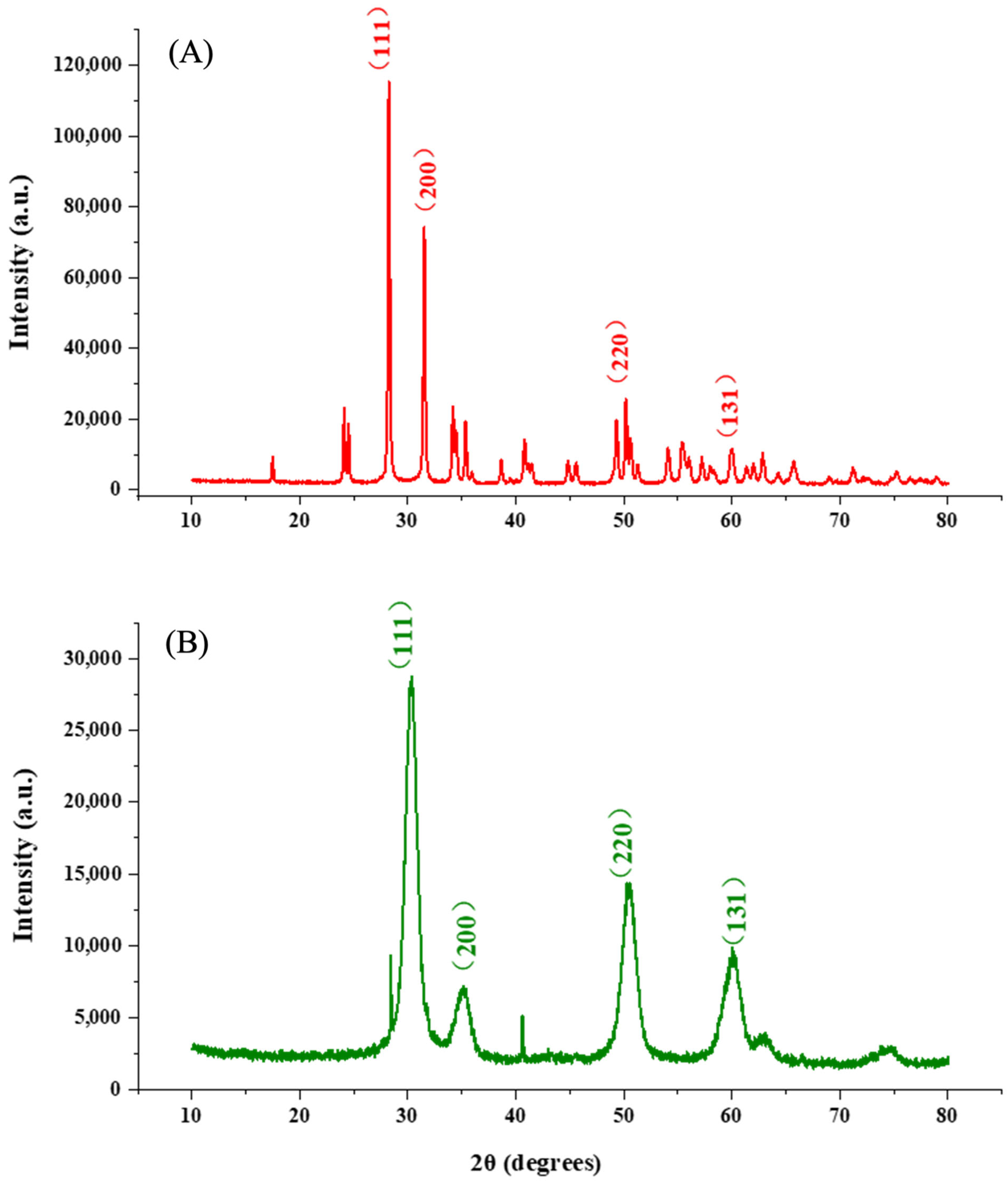
Figure 1. XRD patterns of (A) ZrO2 and (B) PF-ZrO2 NPs.
Processes 13 02794 g001
Figure 2. FTIR spectra of (A) PF-ZrO2 and (B) ZrO2 NPs.
Processes 13 02794 g002
Figure 3. SEM images of PF-ZrO2 NPs (a) and ZrO2 NPs (b), showing similarities in surface structure between the two samples.
Processes 13 02794 g003
Figure 4. TEM images of PF-ZrO2 NPs (a) and ZrO2 NPs (b), showing similarities in surface structure between the two samples.
Processes 13 02794 g004
Figure 5. Nitrogen adsorption-desorption isotherms and corresponding pore size distribution (inset) of PF-ZrO2 nanoparticles synthesized via the green method.
Processes 13 02794 g005
Figure 6. Adsorption kinetics of Cr(VI) onto ZrO2 nanoparticles fitted with pseudo-first-order and pseudo-second-order models. (A) Pseudo-first-order kinetics model for PF-ZrO2 NPs synthesized using plant extract; (B) Pseudo-first-order kinetics model for chemically synthesized ZrO2 NPs; (C) Pseudo-second-order kinetics model for PF-ZrO2 NPs; and (D) Pseudo-second-order kinetics model for chemically synthesized ZrO2 NPs.
Processes 13 02794 g006
Figure 7. (A) Linearized Langmuir isotherm plot (1/qe vs 1/Ce) and (B) linearized Freundlich isotherm plot (log qe vs log Ce) for the adsorption of Cr(VI) onto PF-ZrO2 and ZrO2 NPs. Experimental data were fitted to evaluate the adsorption mechanism and surface characteristics of the adsorbents under equilibrium conditions (initial Cr(VI) concentration: 20–100 mg L−1, pH 5, 25 °C, dosage: 300 mg L−1, contact time: 30 min).
Processes 13 02794 g007
Figure 8. Temkin isotherm model fitting for the adsorption of Cr(VI) onto ZrO2 nanoparticles. (A) represents the chemically synthesized ZrO2 NPs, and (B) represents the PF-ZrO2 NPs synthesized using plant extract. The experimental data points are shown as stars (A) and circles (B), while the solid lines represent the model fitting.
Processes 13 02794 g008
Figure 9. Proposed adsorption mechanism of Cr(VI) removal by PF-ZrO2 nanoparticles synthesized via Sonchus asper extract. The mechanism involves (i) electrostatic attraction between protonated surface hydroxyl groups (–OH2+) and negatively charged chromate species (HCrO4, CrO42−), (ii) cation exchange of surface-bound protons or other exchangeable cations with Cr(VI) species, and (iii) surface complexation with oxygen atoms from Zr–O groups. Residual thermally stable organic functional groups (e.g., C–H, C–N) derived from the plant extract may enhance surface heterogeneity and contribute additional binding interactions, while heat-labile carboxyl-containing compounds are largely decomposed during calcination. Partial reduction of Cr(VI) to Cr(III) and subsequent complexation may also occur under the experimental conditions.
Processes 13 02794 g009
Table 1. FTIR peak assignments of functional groups in PF-ZrO2 and ZrO2 NPs.
Wavenumber (cm−1)AssignmentObserved in
~500–800Zr–OBoth
~1380C–NPF-ZrO2
~1600–1800C=OPF-ZrO2
~2920C–HPF-ZrO2
~3400–3500O–HBoth
Table 2. Experimentally determined kinetic, Langmuir, Freundlich, and Temkin parameters for Cr(VI) adsorption by PF-ZrO2 NPs synthesized using plant extract and chemically synthesized ZrO2 NPs.
PFO PSO
k1 (min−1) q e (mg g−1)R2k2 (g mg−1 min) q e (mg g−1)R2
PF-ZrO2NPs1.73 × 1084.22950.9988.10 × 10184.22950.999
ZrO2NPs14.384374.20420.998426.48254.20560.999
R2 = 0.994
Langmuir Freundlich
q m (mg g−1) K L (L mg−1)R2 K F (L mg−1)1/ n R2
PF-ZrO2NPs142.240.14220.99716.2960.85300.994
ZrO2NPs133.110.09310.84511.9630.87370.834
Temkin
K T (L g−1) B R2
PF-ZrO2NPs1.572620.7890.946
ZrO2NPs3.299315.3440.978
Disclaimer/Publisher’s Note: The statements, opinions and data contained in all publications are solely those of the individual author(s) and contributor(s) and not of MDPI and/or the editor(s). MDPI and/or the editor(s) disclaim responsibility for any injury to people or property resulting from any ideas, methods, instructions or products referred to in the content.