Article

A Novel Fluorescent Probe AP for Highly Selective and Sensitive Detection of Hg2+ and Its Application in Environmental Monitoring

1
Faculty of Environmental Science and Engineering, Kunming University of Science and Technology, Kunming 650500, China
2
National and Local Joint Engineering Research Center for Green Preparation Technology of Biobased Materials, Yunnan Minzu University, Kunming 650500, China
*
Authors to whom correspondence should be addressed.
Processes 2025, 13(7), 2306; https://doi.org/10.3390/pr13072306
Submission received: 29 May 2025 / Revised: 6 July 2025 / Accepted: 17 July 2025 / Published: 19 July 2025
(This article belongs to the Section Environmental and Green Processes)

Abstract

Mercury is a highly toxic heavy metal that poses serious threats to human health and environmental safety, highlighting the critical importance of accurate Hg2+ detection. In this study, a novel fluorescent probe AP was synthesized by conjugating fluorescein, serving as the luminescent group, with pyridine-2-carboxaldehyde to enable selective Hg2+ detection. Hg2+ binds to AP in a 1:2 stoichiometric ratio, inducing the opening of the spiro-lactam ring and resulting in a significant fluorescence enhancement. The probe exhibited excellent selectivity and sensitivity toward Hg2+. A strong linear correlation was observed between its fluorescence intensity and Hg2+ concentration (R2 = 0.99952), with a detection limit of as low as 9.75 × 10⁻⁸ mol/L. The average recoveries of Hg2+ across various water matrices ranged from 95.23% to 103.40%, with relative standard deviations (RSDs) below 3.07%. These results indicate that the probe performs effectively in real water-sample testing.

1. Introduction

Mercury (Hg), a recalcitrant and highly toxic heavy metal, presents a significant threat to both human health and environmental integrity [1,2,3]. Its ability to bind with minerals and organic compounds enables its transformation into methylmercury under anoxic conditions, significantly amplifying its environmental toxicity [4,5,6,7]. In aquatic ecosystems, mercury bioaccumulates in fish and shellfish, entering the human food chain [8,9,10]. Chronic exposure to Hg2+ can lead to severe health issues, including neurocognitive deficits, motor dysfunction, irreversible neurological damage, and Minamata disease [11,12,13,14,15]. Given mercury’s persistence, bioaccumulation, and biomagnification potential, robust Hg2+ monitoring frameworks are essential to prevent ecological collapse and guide effective remediation strategies.
Currently, the gold-standard methods for quantifying Hg2+ contamination include atomic absorption and emission spectroscopy [16,17], inductively coupled plasma mass spectrometry (ICP-MS) [18,19,20], and high-performance liquid chromatography (HPLC) [21]. However, these techniques are hampered by complex sample preparation and high equipment costs, limiting their use in resource-limited areas and large-scale environmental monitoring [22,23]. To address these challenges, fluorescent chemosensors have emerged as a promising alternative. They offer sensitivity, selectivity, and cost-effectiveness, making them a leading choice for innovative Hg2+ detection research [24,25,26].
The design of fluorescent probes hinges on three critical components: a fluorophore, a recognition motif, and a linker. The fluorophore, which converts molecular recognition into measurable fluorescence signals, is central to the design of small-molecule probes, influencing both performance and applicability [27,28,29]. Numerous Hg2+-detecting fluorescent probes have been developed using various fluorophores. For instance, in 2018, Zhang et al. [30] synthesized an isocyanate-functionalized 1,8-naphthalenediformimide-based probe (M2) that selectively responded to Hg2+ in a THF–water system (VTHF:Vwater = 3:7, 5 mM PBS buffer solution, pH = 7.4). Duygu Aydin et al. [31] developed a benzothiazole-based probe (NETBZ) that exhibited a fluorescence “turn-off” upon Hg2+ binding, transitioning from blue fluorescence to colorless under ultraviolet light upon interaction with Hg2+. Wen et al. [32] designed a BODIPY–Rhodamine ratiometric probe (BR) where the BODIPY serves as the energy donor and the Rhodamine acts as the energy acceptor. Upon binding with Hg2+, the spirolactam ring of Rhodamine opens, leveraging fluorescence resonance energy transfer for Hg2+ detection. Elsayed Elbayoumy et al. [33] reported a triazole-pyridine-based sensor (TOHC) for Hg2+ detection in real water samples. However, the synthesis of these fluorophores is often complex. This study aimed to develop simpler Hg2+-detecting fluorescent probes using fluorescein derivatives, which offer excellent aqueous solubility, high quantum yields, and ease of derivatization [34,35,36,37]. We synthesized a novel Hg2+-responsive probe (AP) via Schiff base condensation using fluorescein, hydrazine hydrate, and pyridine-2-carboxaldehyde; optimized the probe’s performance; and demonstrated its effectiveness in detecting Hg2+ in real water samples.

2. Materials and Methods

2.1. Materials and Instruments

Fluorescein, pyridine-2-carboxaldehyde, HEPES buffer, and all ionic salts were procured from Shanghai Macklin Biochemical Technology Co., Ltd. (Shanghai, China) Hydrazine hydrate (80%), glacial acetic acid, absolute ethanol, dimethyl sulfoxide (DMSO), acetonitrile, tetrahydrofuran (THF), and N, N-dimethylformamide (DMF) were obtained from Chengdu Kelong Chemical Co., Ltd. (Chengdu, China). All chemical reagents were of analytical grade and used without further purification.
High-resolution mass spectra (HRMS) were acquired using a BRUKER ESI-Q-TOF mass spectrometer (Bruker, Germany). Nuclear magnetic resonance (NMR) spectra were recorded on a BRUKER AV III-400 spectrometer (Bruker, Germany), with tetramethylsilane (TMS) as the internal standard. The Fourier transform infrared (FT-IR) spectra were obtained using a NICOLET iS10 spectrometer (Thermo Fisher Scientific, America). The fluorescence spectra were measured with a Cary Eclipse fluorescence spectrophotometer (Agilent Technologies, America).

2.2. Synthesis of Probe AP

The synthesis of the probe AP was accomplished through a two-step reaction process, as depicted in Scheme 1. In the first step, fluorescein was reacted with hydrazine hydrate to form an intermediate hydrazone compound. This intermediate was subsequently condensed with pyridine-2-carboxaldehyde via a Schiff base reaction to yield the final fluorescent probe AP. The reaction progress was monitored using thin-layer chromatography (TLC), and the intermediate and final products were purified by column chromatography on silica gel. The structures of the synthesized compounds were fully characterized by HRMS, NMR, and FT-IR spectroscopy to verify their identity and purity.

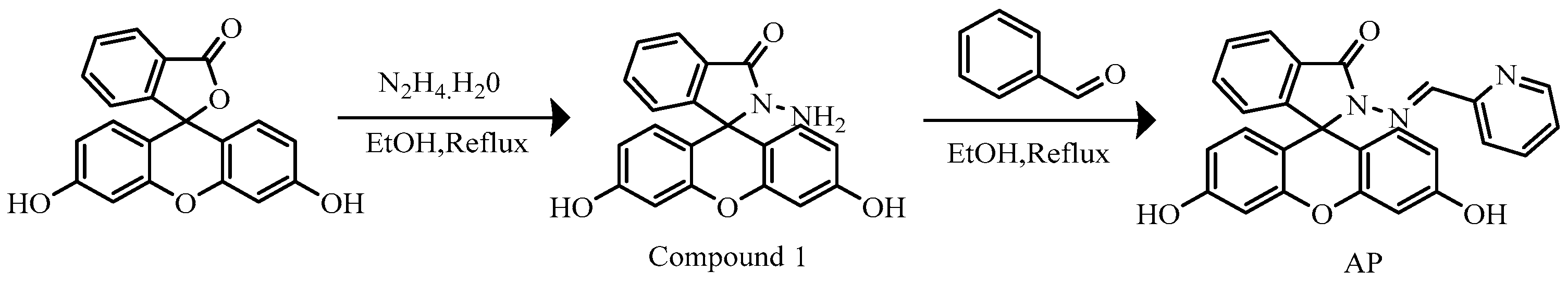
2.2.1. Synthesis of Compound 1

Compound 1 was synthesized according to the procedure described in reference [38]. Fluorescein (1.66 g, 5 mmol) was weighed accurately and dissolved in anhydrous ethanol (40 mL) in a 100 mL round-bottom flask. The solution was stirred until complete dissolution was achieved. A reflux condenser was attached to the flask to prevent solvent loss during heating. Hydrazine hydrate (2.5 mL, in excess) was added dropwise to the solution under continuous stirring. The reaction mixture was heated to 80 °C and maintained at this temperature with constant stirring for 6 h, during which the solution turned into a dark red, transparent liquid.
After cooling, the reaction mixture was concentrated using a rotary evaporator until solid formation began. The concentrate was then poured into a large volume of deionized water to induce precipitation. The mixture was allowed to stand undisturbed to ensure complete precipitation. The precipitate was collected by vacuum filtration, washed thoroughly with deionized water until the filtrate became colorless, and further purified by washing with anhydrous ethanol. The crude product was recrystallized from anhydrous ethanol to enhance its purity. The purified product was dried under a vacuum to yield fluorescein hydrazide (Compound 1) with a yield of 67.05% (1.16 g).
The structure of Compound 1 was confirmed by 1H NMR and 13C NMR spectroscopy. The 1H NMR spectrum (400 MHz, DMSO-d6) showed the following chemical shifts: δ 9.79 (s, 2H), 7.79–7.75 (m, 1H), 7.51–7.47 (m, 2H), 7.00–6.96 (m, 1H), 6.59 (d, J = 2.4 Hz, 2H), 6.47–6.36 (m, 4H), and 4.37 (s, 2H). The 13C NMR spectrum (101 MHz, DMSO-d6) displayed the following carbon resonances: δ 165.94, 158.66, 152.88, 151.99, 133.06, 129.81, 128.87, 128.42, 123.89, 122.82, 112.46, 110.43, 102.83, and 65.08. These data are consistent with the expected structure of fluorescein hydrazide.

2.2.2. Preparation of Probe AP

The probe AP was synthesized according to the procedure reported in reference [39]. A mixture of fluorescein hydrazide (Compound 1, 0.69 g, 2 mmol) and pyridine-2-carboxaldehyde (0.21 g, 2 mmol) was prepared in anhydrous ethanol (40 mL) in a round-bottom flask equipped with a reflux condenser. The solution was heated to 80 °C under reflux. Glacial acetic acid (692 µL) was added as a catalyst, and the reaction was stirred vigorously for 2 h. After cooling to room temperature, the precipitate was collected by vacuum filtration and washed thoroughly with cold ethanol (3 × 10 mL) to remove any unreacted starting materials and impurities. The crude product was further purified by recrystallization from anhydrous ethanol. The purified product was dried in a vacuum oven at 40 °C for 6 h, yielding the probe AP as a pale orange solid (0.72 g, 83.21% yield).
The structure of the probe AP was fully characterized by NMR spectroscopy and HRMS. The 1H NMR spectrum (400 MHz, DMSO-d6) showed the following signals: δ 9.94 (s, 2H), 8.55 (s, 1H), 8.48 (d, J = 5.0 Hz, 1H), 7.95 (d, J = 7.4 Hz, 1H), 7.79 (t, J = 7.8 Hz, 1H), 7.68–7.62 (m, 2H), 7.59 (t, J = 7.3 Hz, 1H), 7.36–7.30 (m, 1H), 7.12 (d, J = 7.4 Hz, 1H), 6.67 (s, 2H), 6.53 (d, J = 8.6 Hz, 2H), and 6.46 (d, J = 8.8 Hz, 2H). The 13C NMR spectrum (101 MHz, DMSO-d6) revealed the following carbon resonances: δ 164.07, 158.75, 153.10, 151.84, 151.08, 149.40, 146.48, 136.97, 134.44, 129.14, 127.85, 124.49, 123.72, 123.41, 119.11, 112.53, 109.46, 102.62, and 64.90. HRMS (ESI) analysis confirmed the molecular ion peak at m/z 458.1144 [M+Na]+, which matched the calculated value of 458.1111 for C26H17N3O4.

2.3. Preparation of Solutions for Spectroscopic Testing

2.3.1. HEPES Buffer Solution Preparation

A HEPES buffer solution was prepared by dissolving 4.77 g of HEPES in 900 mL of water. The pH was adjusted to 7.40 using 0.1 mol/L NaOH, and the solution was then diluted to a final volume of 1000 mL with water. The buffer solution was stored at 4 °C.

2.3.2. Preparation of Stock Solutions

Stock solutions of the following metal ions were prepared at a concentration of 1 × 10⁻² mol/L: Ag+, Al3+, Ba2+, Ca2+, Cd2+, Co2+, Cr3+, Cu2+, Fe2+, Fe3+, K+, Mg2+, Mn2+, Hg2+, Na+, Ni2+, Pb2+, and Zn2+. For the probe stock solution, the compound was dissolved in DMSO to yield a 1.0 × 10⁻³ mol/L solution at room temperature. Spectral testing was conducted with an excitation wavelength of 252 nm and slit widths of 5 nm for both excitation and emission.

2.4. Detection Limit Calculation

The detection limit (LOD) of the probe AP for Hg2+ was calculated using the equation
LOD = 3σ/k
where σ is the standard deviation of the fluorescence intensity of the probe blank solution and k is the slope of the linear regression plot of the fluorescence intensity versus the Hg2+ concentration. During the LOD calculation, we performed 20 blank measurements. The standard deviation of these fluorescence intensity values was determined to be 0.0775, and this data has been included in Table S1 of the Supplementary Information.

2.5. Detection in Real Water Samples

The practical applicability of the probe AP was evaluated by analyzing the Hg2+ levels in environmental water samples, including tap water, river water (Laoyu River), and lake water (Dianchi Lake). Each sample was first filtered through a 0.22 µm membrane to remove particulates. No Hg2+ was detected in the unspiked samples. A standard addition method was then applied by spiking the samples with known Hg2+ concentrations of 30, 60, and 90 µM. The measured concentrations and recovery rates were calculated to assess the accuracy and reliability of the method.

3. Results

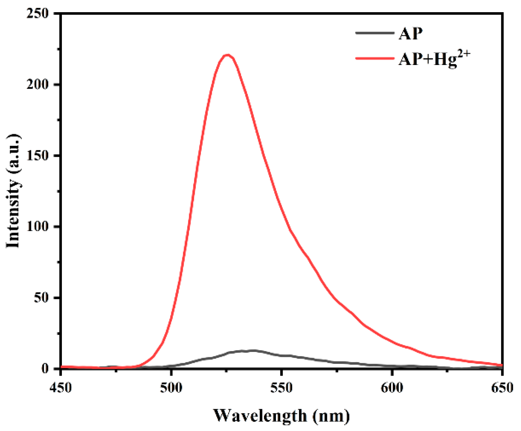
3.1. Feasibility Study of Probe AP

To determine whether probe AP could be used for detecting Hg2+, experiments were conducted using fluorescence spectroscopy. The probe concentration was set at 10 µM in a DMSO/HEPES mixture (1:9, v/v; 20 mM HEPES, pH = 7.4). As shown in Figure 1, the blank probe solution exhibited minimal fluorescence intensity. However, upon the addition of Hg2+, a significant enhancement in the fluorescence intensity was observed at the emission peak at 525 nm. This marked increase in the fluorescence intensity confirms that probe AP is capable of effectively detecting Hg2+.

3.2. Optimization of Detection Conditions

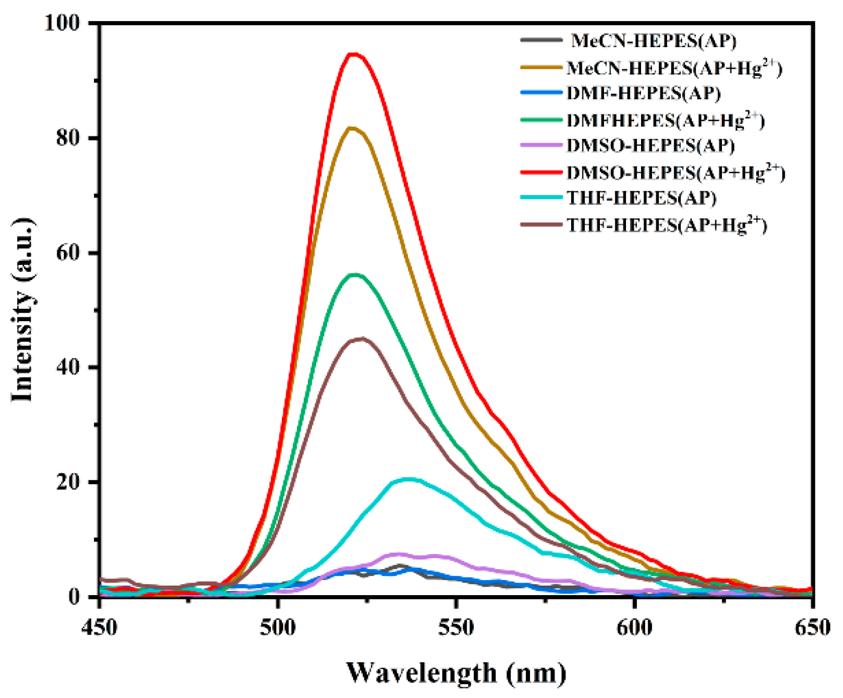
3.2.1. Solvent Type Effects on Fluorescence Properties of Probe AP

The fluorescence properties of probe AP are highly dependent on the solvent environment, which significantly influences its interaction with metal ions. To maximize the detection efficiency, the fluorescence response of probe AP to Hg2+ was evaluated in four different solvents: acetonitrile (MeCN), tetrahydrofuran (THF), N,N-dimethylformamide (DMF), and dimethyl sulfoxide (DMSO).
Probe AP was dissolved in each solvent to prepare a 1.0 × 10⁻³ mol/L stock solution. Then, 40 µL of each stock solution was placed in a 5 mL centrifuge tube and mixed with 20 µL of a 1.0 × 10⁻² mol/L Hg2+ solution. The volume was adjusted to 4 mL with a HEPES buffer and the corresponding solvent, maintaining a 1:9 organic solvent-to-buffer ratio. After sufficient reaction time, fluorescence measurements were taken.
As shown in Figure 2, the probe AP exhibited a fluorescence response to Hg2+ in all tested solvents, with the intensity increasing with the solvent polarity. The highest fluorescence intensity was observed in the DMSO/HEPES mixture. Therefore, DMSO was selected as the solvent for subsequent experiments to optimize the probe performance.

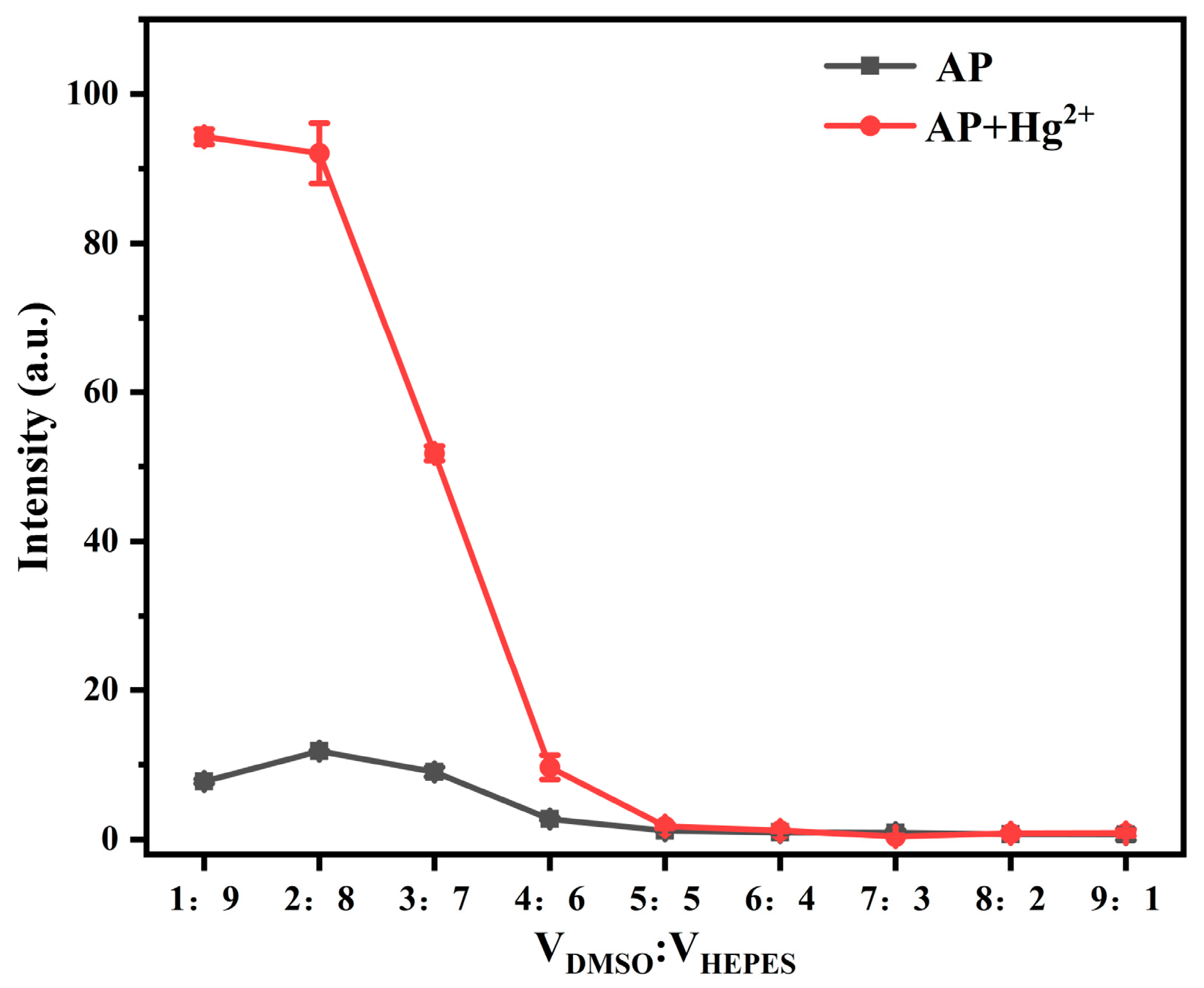
3.2.2. Effect of Solvent Ratio on Fluorescence Properties of Probe AP

The ratio of DMSO to HEPES in the test system significantly affected the probe’s ability to recognize metal ions. To determine the optimal solvent composition, the fluorescence response of the probe AP to Hg2+ was evaluated across various DMSO/HEPES ratios. As shown in Figure 3, the maximum fluorescence intensity was achieved at a DMSO/HEPES volume ratio of 1:9. In this formulation, the 10% DMSO facilitated the initial dissolution of the probe without interfering with the aqueous-phase reaction, while the 90% HEPES buffer solution ensured that the probe was well-suited for detecting Hg2+ in environmental samples. Therefore, all subsequent experiments were conducted using a DMSO/HEPES ratio of 1:9 (v/v).

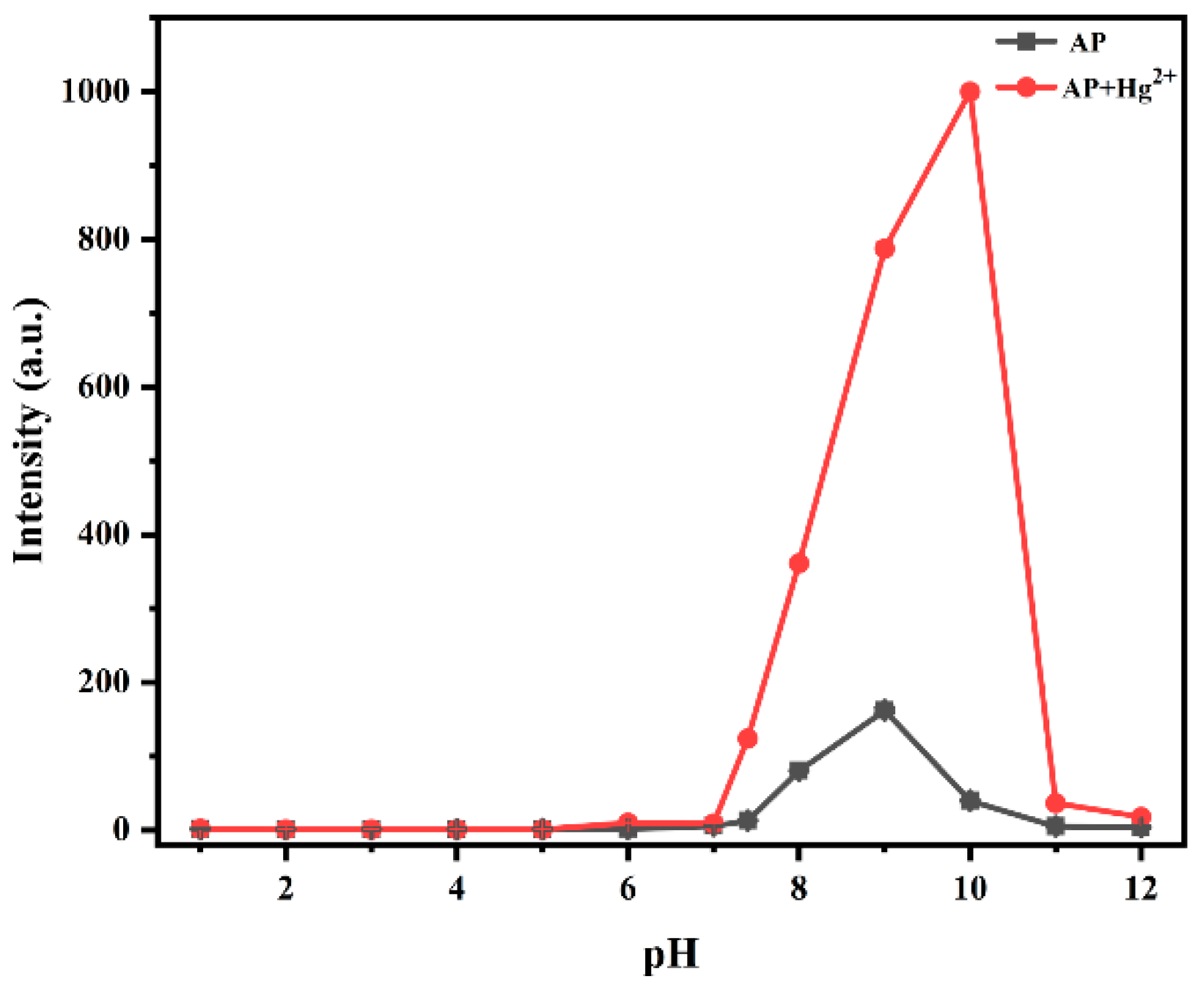
3.2.3. Effect of pH on Fluorescence Properties of Probe AP

To explore how the acidity or alkalinity of the solution affected the fluorescence detection performance of the probe AP, HEPES buffer solutions with varying pH values (1–12) were prepared. Under the conditions of a probe concentration of 1.0 × 10⁻⁵ mol/L and a DMSO/HEPES ratio of 1:9 (v/v, 20 mM HEPES, pH = 7.4), 20 µL of a 1.0 × 10⁻² mol/L Hg2+ stock solution was added to examine the fluorescence changes at different pH values. As shown in Figure 4, in the absence of Hg2+, the probe exhibited minimal fluorescence at 525 nm within the pH range of 1–7.4. When the pH was >7.4, the fluorescence intensity gradually increased, likely due to an alkaline-promoted spirolactam ring opening and hydroxyl deprotonation, reaching maximum intensity at pH 10. After the Hg2+ was added, the fluorescence intensity under the acidic conditions showed minimal change compared to the free probe, indicating that the probe remained in the closed-ring state. Between pH 7 and 10, the Hg2+ induced the formation of open-ring complexes, with the fluorescence intensity increasing with the alkalinity. Considering that some metal ions may form precipitates in an alkaline environment, pH 7.4 was selected for subsequent experiments to ensure optimal detection conditions.

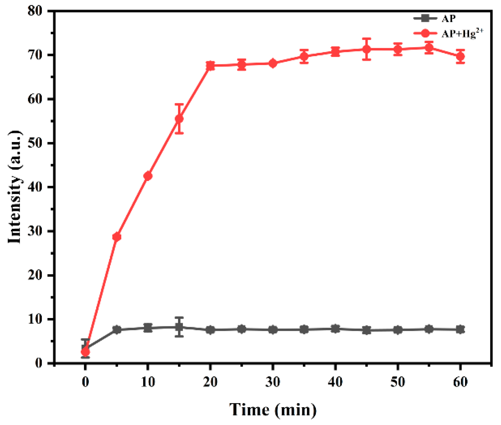
3.2.4. Response Time of Probe AP Toward Hg2+

To assess the interaction kinetics between probe AP and Hg2+, time-dependent fluorescence changes were monitored at 525 nm. The experimental system consisted of probe AP (1.0 × 10⁻⁵ mol/L) and Hg2+ (5.0 × 10⁻⁵ mol/L) in a DMSO/HEPES solvent system (1:9 v/v, 20 mM HEPES, pH 7.4). The fluorescence intensity was recorded at various time intervals, with time plotted on the x-axis and fluorescence intensity on the y-axis.
As shown in Figure 5, the fluorescence intensity of the probe–Hg2+ system increased progressively over time and reached a plateau within 20 min of mixing. Therefore, all fluorescence measurements were performed exactly 20 min post-reaction to ensure consistent and comparable conditions.

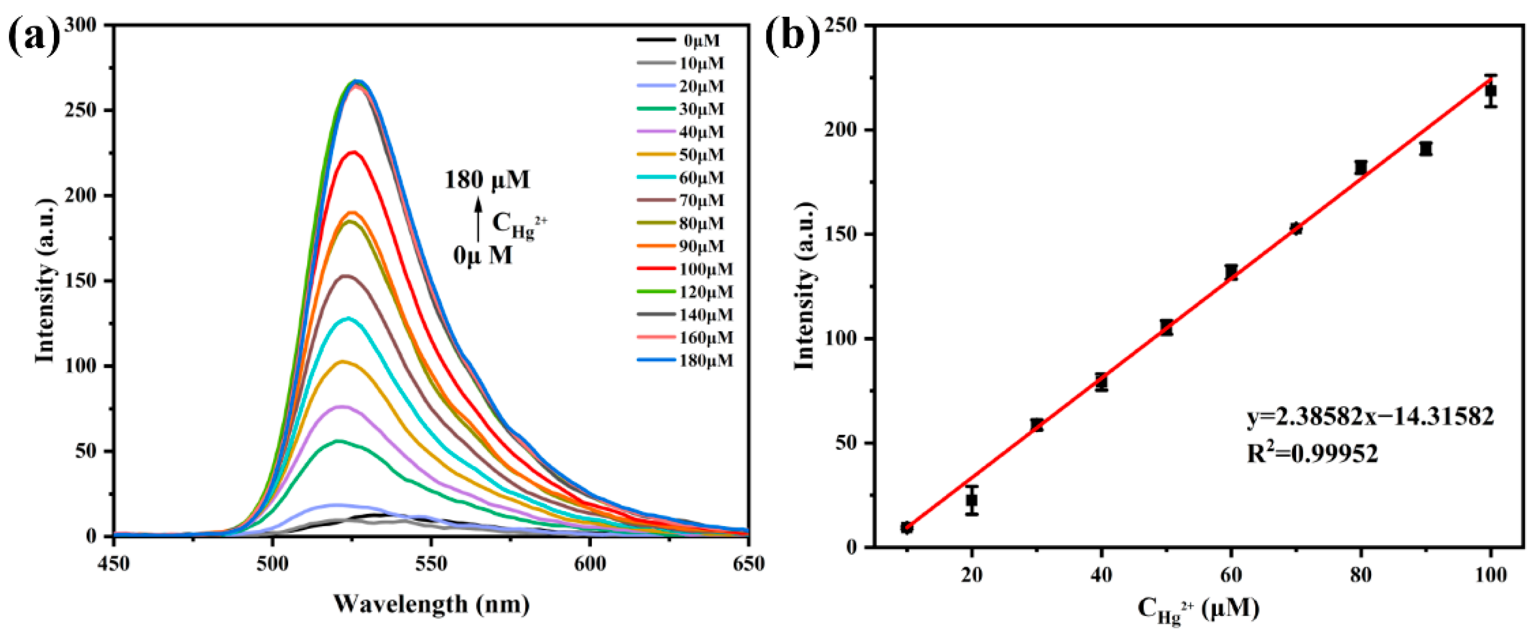
3.3. Fluorescence Titration Experiment of Probe AP with Hg2+

To investigate the effect of Hg2+ concentrations on the fluorescence intensity of probe AP, fluorescence emission spectra were measured at various Hg2+ concentrations. Several 5 mL centrifuge tubes containing testing solutions were prepared, each receiving 40 µL of a 1 × 10⁻³ mol/L probe AP stock solution. Varying volumes of a 1 × 10⁻² mol/L Hg2+ stock solution were added to each tube according to a specific concentration gradient, ensuring that the Hg2+ concentration ranged from 0 to 18 equivalents relative to the probe concentration. The mixtures were diluted to 4 mL with a DMSO–HEPES buffer solution (DMSO/HEPES = 1:9, v/v, 20 mM HEPES, pH = 7.4). After the solutions were allowed to stabilize, their fluorescence spectra were measured.
As shown in Figure 6, the maximum emission wavelength of the system was observed at 525 nm, and the fluorescence intensity at this wavelength increased with the rising Hg2+ concentration. When the Hg2+ concentration reached 100 µM, the fluorescence intensity plateaued. Within the Hg2+ concentration range of 10–100 µM, the probe exhibited a good linear response (R2 = 0.99952) for Hg2+ quantification. The linear equation was y = 2.38582x − 14.31582. Based on the detection limit formula, LOD = 3σ/k (σ = 0.0775), the probe’s detection limit for Hg2+ was calculated to be as low as 9.75 × 10⁻⁸ mol/L. Furthermore, a comparative analysis was conducted between this probe and several previously reported fluorescent probes for Hg2+ detection. The detailed results are summarized in Table 1. The comparison shows that the probe AP has lower consumption of organic solvents, a larger Stokes shift, and a lower detection limit, demonstrating superior performance.

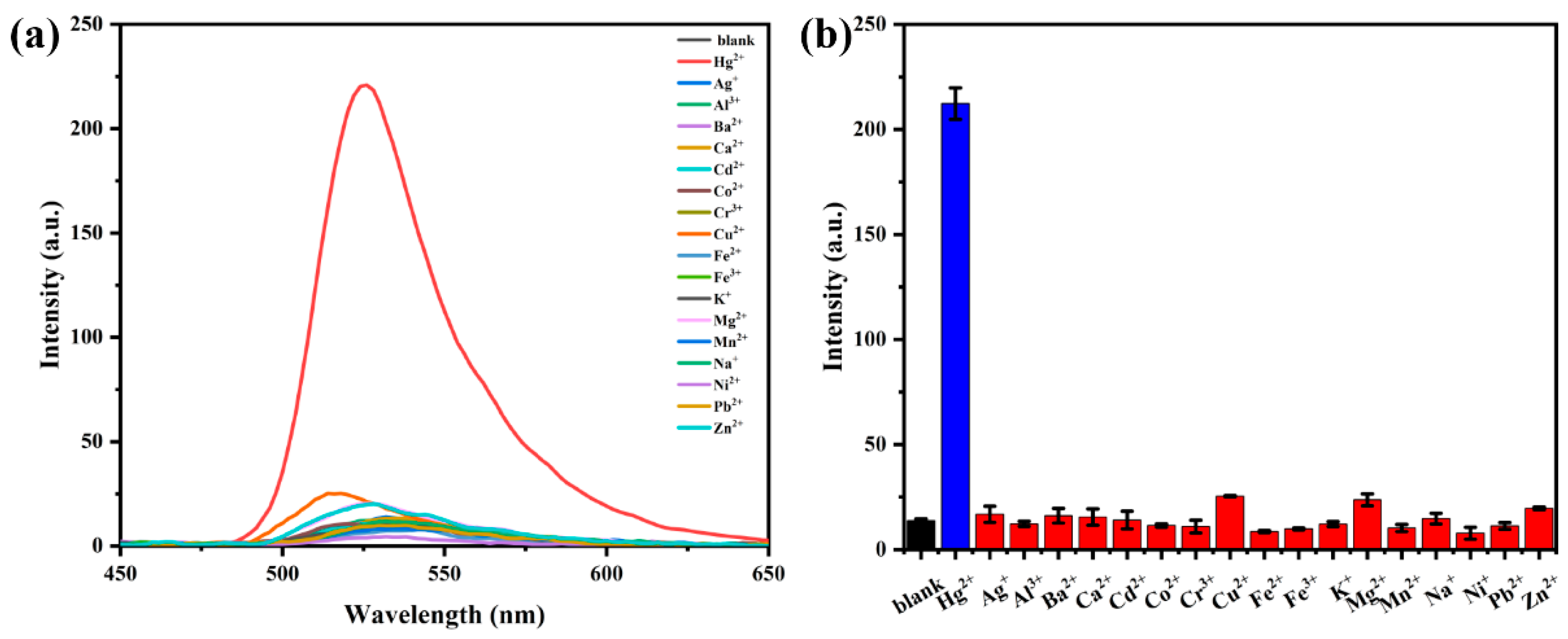
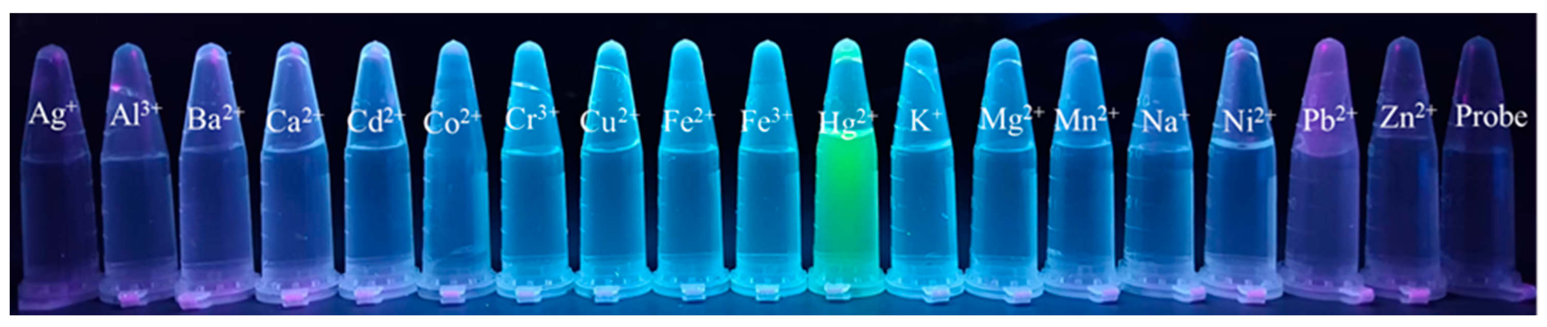
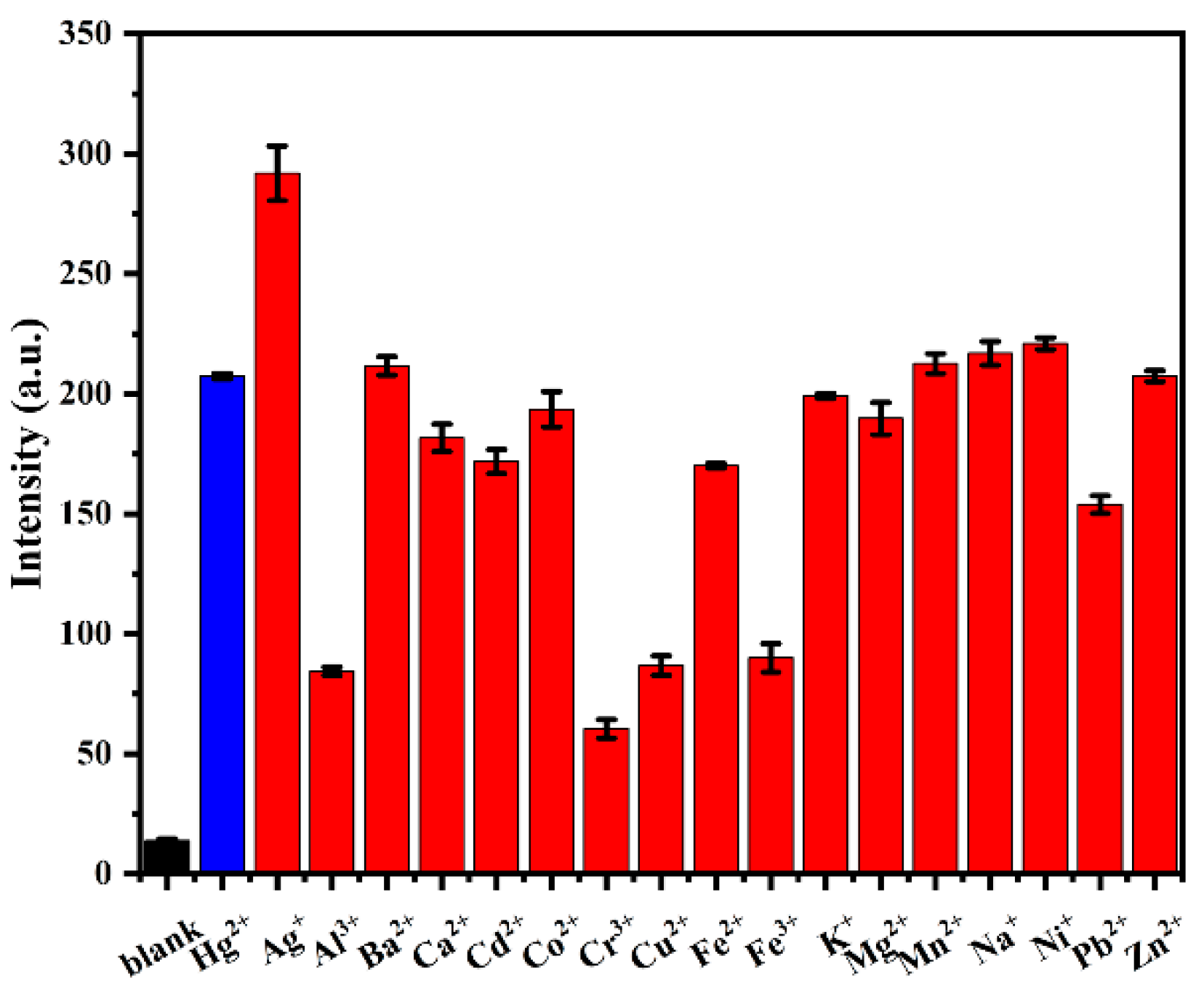
3.4. Selectivity Study of Probe AP for Hg2+

To assess the specificity of probe AP for Hg2+ detection, selectivity experiments were conducted using a variety of common metal cations. Multiple 5 mL centrifuge tubes containing testing solutions were prepared, each with 40 µL of a 1 × 10⁻³ mol/L stock solution of probe AP. Subsequently, 40 µL of a 1 × 10⁻² mol/L stock solution of Hg2+ or other metal cations (Ag+, Al3+, Ba2+, Ca2+, Cd2+, Co2+, Cr3+, Cu2+, Fe2+, Fe3+, K+, Mg2+, Mn2+, Na+, Ni2+, Pb2+, Zn2+) was added to each tube. The mixtures were diluted to 4 mL with a DMSO–HEPES buffer solution (DMSO/HEPES = 1:9, v/v, 20 mM HEPES, pH = 7.4). For comparison, a blank solution containing only 1 × 10⁻⁵ mol/L probe AP was also prepared. The fluorescence spectra of the probe in the presence of different metal ions were then investigated.
As shown in Figure 7a,b, the probe alone exhibited minimal fluorescence. However, a significant increase in fluorescence intensity at 525 nm was observed upon the addition of the Hg2+. Furthermore, Figure 8 illustrates that under 365 nm UV light, the probe solution only emitted bright green fluorescence in the presence of Hg2+, with no significant fluorescence changes observed in the presence of the other metal ions. These results confirm that the reaction between the Hg2+ and the probe led to fluorescence activation, demonstrating that probe AP has a high specificity for Hg2+ detection.

3.5. Interference Study of Probe AP for Hg2+ Detection

To assess the anti-interference capability of probe AP in complex systems, an experiment was designed as follows: Several 5 mL centrifuge tubes containing testing solutions were prepared. One tube received 40 µL of a 10 mM Hg2+ solution. The remaining tubes were each spiked with 40 µL of a mixed solution containing 10 mM Hg2+ along with various metal ions (Ag+, Al3+, Ba2+, Ca2+, Cd2+, Co2+, Cr3+, Cu2+, Fe2+, Fe3+, K+, Mg2+, Mn2+, Na+, Ni2+, Pb2+, Zn2+), each at 10 mM. Subsequently, 40 µL of a 1 mM probe stock solution was added to each tube, and the final volume was adjusted to 4 mL with a DMSO/HEPES buffer solution (1:9 v/v). A blank control containing only the probe solution without any metal ions was also prepared.
Fluorescence spectral changes were monitored for all solutions. As shown in Figure 9, the probe AP demonstrated significant fluorescence enhancement toward Hg2+ even in the presence of other metal ions. Although minor fluorescence quenching was observed in the systems containing Al3+, Cr3+, Cu2+, and Fe3+, the fluorescence intensity remained predominantly enhanced. This slight interference may be attributed to the precipitation of these metal ions under weakly alkaline conditions. Overall, these results highlight that probe AP retains a high selectivity for Hg2+ with a robust anti-interference capability, underscoring its potential for Hg2+ detection in intricate environmental matrices.

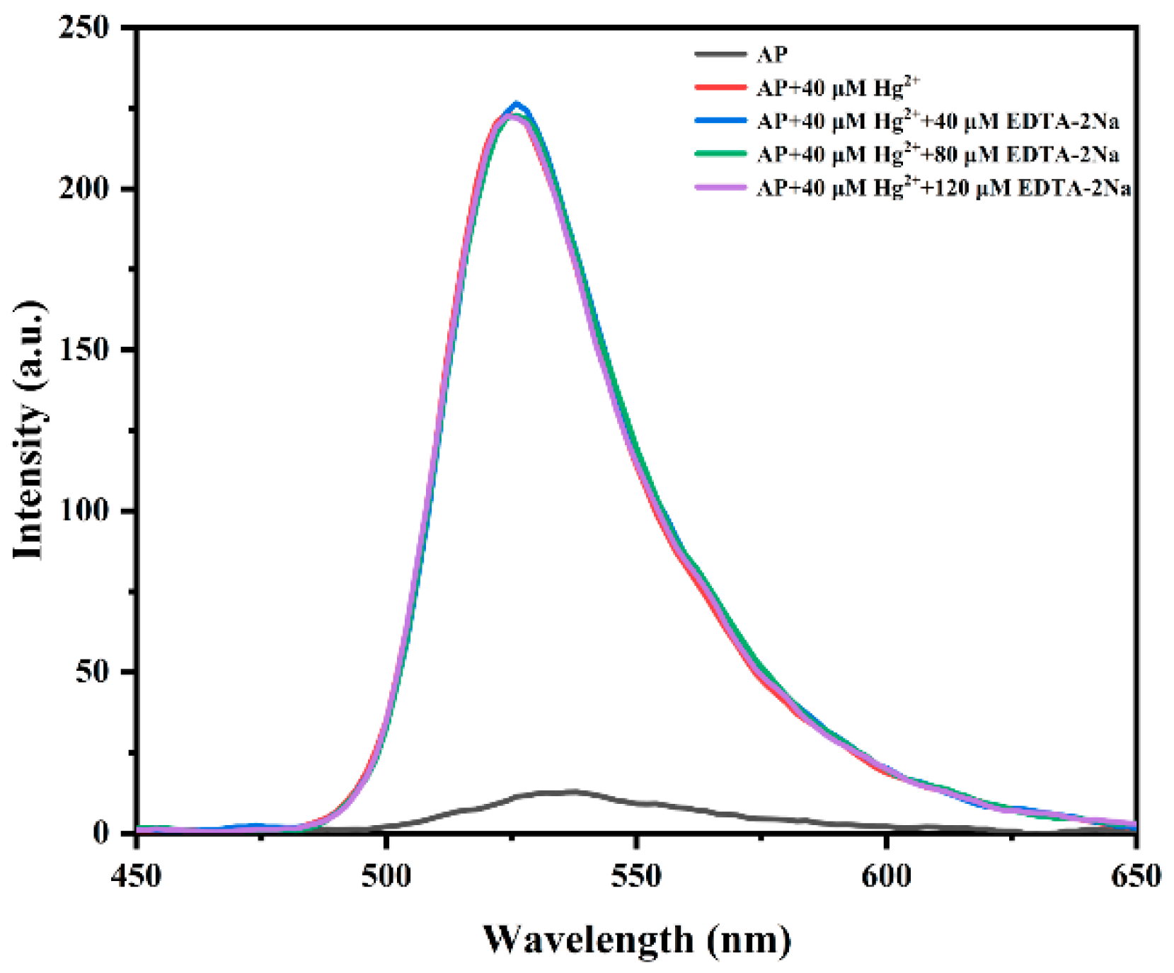
3.6. Reversibility Investigation of Probe AP

To investigate the reversibility of the reaction between probe AP and Hg2+, a titration experiment was conducted using the chelating agent ethylenediaminetetraacetic acid disodium salt (EDTA-2Na) in the AP-Hg2+ system. This experiment aimed to determine whether the fluorescence response of probe AP to Hg2+ could be reversed by the addition of EDTA-2Na. As shown in Figure 10, the fluorescence intensity of the AP–Hg2+ system remained essentially unchanged upon the addition of moderate to excessive amounts of EDTA-2Na. This result indicates that the recognition process between probe AP and Hg2+ is irreversible. The lack of fluorescence quenching or significant changes in intensity suggests that the binding of Hg2+ to probe AP is highly stable and not easily disrupted by EDTA-2Na, a common chelating agent.

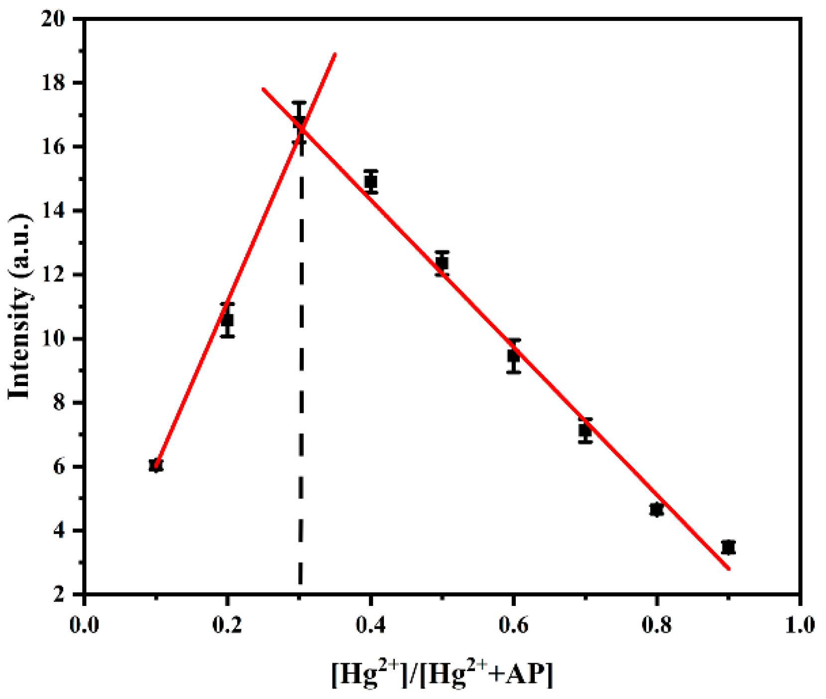
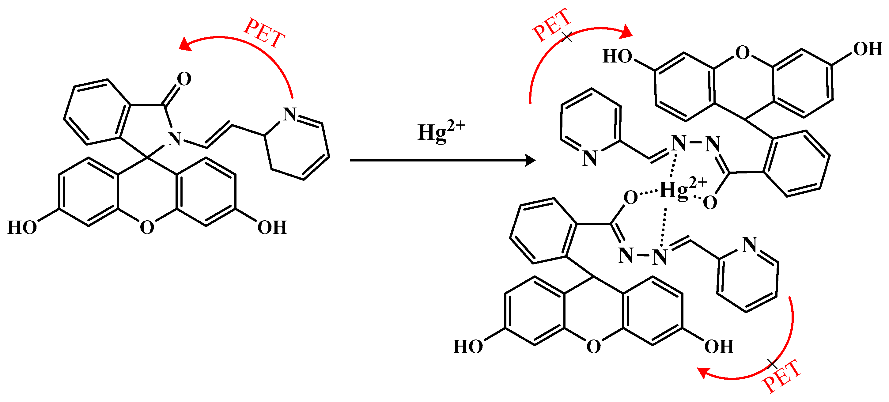
3.7. Mechanism Study of Probe AP for Hg2+ Recognition

To elucidate the reaction mechanism between probe AP and Hg2+, the coordination stoichiometry was determined using Job’s plot method. The total concentrations of probe AP and Hg2+ were maintained at 20 µM. The volume ratio of the AP to the Hg2+ solutions was varied to obtain Hg2+ molar fractions ranging from 0.1 to 0.9. The fluorescence intensity at 525 nm for each sample was measured. A Job plot was constructed by plotting the fluorescence intensity at 525 nm (y-axis) against the molar fraction of the Hg2+ (x-axis). As shown in Figure 11, the inflection point of the curve occurred at a molar fraction of 0.3, indicating a 2:1 binding stoichiometry between the AP and Hg2+. This suggests that two molecules of AP will coordinate with one Hg2+ ion to form a stable complex, leading to the observed fluorescence enhancement.
Based on the analysis of the Job’s plot results and the known complexation mechanisms between traditional spirolactam structures and metal ions, we propose the following binding mechanism for probe AP with Hg2+: The blank solution of probe AP exhibits minimal fluorescence, likely due to the photoinduced electron transfer (PET) effect. Upon the addition of Hg2+, the probe and Hg2+ undergo complexation, resulting in the formation of a ring-opened compound. This interaction induces the opening of the spirolactam ring of the fluorophore and disrupts the isomerization of the carbon–nitrogen double bond. Consequently, the electron transfer within the probe molecule is altered, the PET process is inhibited, and fluorescence emission is observed. The proposed complexation mechanism is illustrated in Figure 12.
To further validate the binding mechanism between probe AP and Hg2+, a 1H NMR titration experiment was conducted in deuterated DMSO. The 1H NMR spectra of probe AP before and after the addition of Hg2+ were compared, as shown in Figure 13. Before complexation, the signal peaks of the hydroxyl group and the hydrogen on the carbon–nitrogen double bond appeared at 9.94 ppm and 8.55 ppm, respectively. After complexation, these peaks experienced a downfield shift to 9.99 ppm and an upfield shift to 8.36 ppm, respectively. Additionally, the chemical shifts of the aromatic hydrogens also changed, indicating structural alterations in the aromatic ring of the probe AP. These observations are consistent with the hypothesis of ring opening upon complexation between probe AP and Hg2+.

3.8. Detection in Real Samples

To assess the practical applicability of probe AP for Hg2+ detection in real-world scenarios, spike-and-recovery experiments were conducted using different environmental water samples, including tap water, river water (Laoyu River), and lake water (Dianchi Lake). Prior to testing, all samples were filtered through a 0.22 µm membrane to remove large particulates, and the background Hg2+ levels were confirmed to be negligible. Known concentrations of Hg2+ were added to the samples using the standard addition method, and the recovery rates were calculated. As summarized in Table 2, the average recoveries of the Hg2+ in different water matrices ranged from 95.23% to 103.40%, with relative standard deviations (RSDs) of below 3.07%. These results demonstrate that probe AP exhibits high sensitivity and accuracy in real-sample analysis, highlighting its potential for practical environmental monitoring applications.

4. Conclusions

In this study, we successfully synthesized a novel fluorescent probe AP via a Schiff base reaction using fluorescein hydrazide and pyridine-2-carboxaldehyde as precursors. The probe exhibited remarkable selectivity for Hg2+, with a highly linear response (R2 = 0.99952) across the concentration range of 10–100 μM. Its detection limit for Hg2+ was as low as 9.75 × 10⁻⁸ mol/L. A Job’s plot analysis confirmed a 2:1 binding stoichiometry between probe AP and Hg2+. Additionally, spike-and-recovery experiments in real water samples demonstrated its practical applicability, achieving average recovery rates of 95.23–103.40% with relative standard deviations (RSDs) below 3.07%. These results collectively highlight probe AP’s significant potential for practical Hg2+ detection and environmental monitoring applications.

Supplementary Materials

The following supporting information can be downloaded at: https://www.mdpi.com/article/10.3390/pr13072306/s1, Figure S1: 1H-NMR of compound 1; Figure S2: 13C-NMR of compound 1; Figure S3: IR spectrum of compound 1; Figure S4: 1H-NMR of probe AP; Figure S5: 13C-NMR of probe AP; Figure S6: IR spectrum of probe AP; Figure S7: HRMS spectrum of probe AP; Figure S8: Calibration curve of the CVAAS signal response for Hg2+; Table S1: The standard deviation of fluorescence intensity values to calculate the LOD; Table S2: Comparison of fluorescence detection using probe AP and CVAAS the analyzing Hg2+ in wastewater from flower cake production.

Author Contributions

Z.Y.: conceptualization, investigation, formal analysis, writing—original draft. C.L.: conceptualization, investigation, data curation. Q.W.: resources, methodology, validation. Y.H.: investigation, resources, formal analysis. S.T.: resources, supervision, validation, methodology, project administration, writing—review and editing. All authors have read and agreed to the published version of the manuscript.

Funding

The authors acknowledge the financial supports from the Yunnan Fundamental Research Projects (Grant No. 202501AT070292).

Data Availability Statement

The raw data supporting the conclusions of this article will be made available by the authors on request.

Conflicts of Interest

The authors declare no conflicts of interest.

References

  1. Gauthama, B.U.; Narayana, B.; Sarojini, B.K.; Suresh, N.K.; Sangappa, Y.; Kudva, A.K.; Satyanarayana, G.; Raghu, S.V. Colorimetric “off–on” fluorescent probe for selective detection of toxic Hg2+ based on rhodamine and its application for in-vivo bioimaging. Microchem. J. 2021, 166, 106233. [Google Scholar] [CrossRef]
  2. Ren, Y.; Yang, W.; Tan, Z.; Zhang, L.; Pan, R. A rapid response optical fiber sensor with highly selective probe for trace detection of mercury (II) ions. Microchem. J. 2024, 206, 111534. [Google Scholar] [CrossRef]
  3. Isaad, J.; Malek, F.; Achari, A.E. Colorimetric and fluorescent probe based on coumarin/ thiophene derivative for sequential detection of mercury (II) and cyanide ions in an aqueous medium. J. Mol. Struct. 2022, 1270, 133838. [Google Scholar] [CrossRef]
  4. Li, Y.; Zhang, Y.; Wang, M.; Wang, D.; Chen, K.; Lin, P.; Ge, Y.; Liu, W.; Wu, J. Highly selective fluorescence probe with peptide backbone for imaging mercury ions in living cells based on aggregation-induced emission effect. J. Hazard. Mater. 2021, 415, 125712. [Google Scholar] [CrossRef] [PubMed]
  5. Ramki, K.; Thiruppathi, G.; Ramasamy, S.K.; Sundararaj, P.; Sakthivel, P. An aggregation-induced emission-based ratiometric fluorescent chemosensor for Hg (II) and its application in Caenorhabditis elegans imaging. Methods 2024, 221, 1–11. [Google Scholar] [CrossRef] [PubMed]
  6. Tamizhselvi, R.; Gangatharan Vinoth Kumar, G.; Anbarasu, G.; Arumugam Napoleon, A. Dual-channel fluorescent probe utilizing hydrazone for Zn2+ and Hg2+ detection: Exploring distinct signaling mechanisms and applications in bioimaging and latent fingerprint analysis. J. Photochem. Photobiol. A Chem. 2024, 452, 115609. [Google Scholar] [CrossRef]
  7. Patthana, P.; Zhong, H.-C.; Wu, Q.; Ren, T.-B.; Yuan, L. Engineering a far-red fluorescent probe for rapid detection of Hg (II) ions in both cells and zebrafish. Spectrochim. Acta Part. A Mol. Biomol. Spectrosc. 2024, 318, 124469. [Google Scholar] [CrossRef] [PubMed]
  8. Kaewnok, N.; Kraithong, S.; Mahaveero, T.; Maitarad, P.; Sirirak, J.; Wanichacheva, N.; Swanglap, P. Silver nanoparticle incorporated colorimetric/fluorescence sensor for sub-ppb detection of mercury ion via plasmon-enhanced fluorescence strategy. J. Photochem. Photobiol. A Chem. 2022, 433, 114140. [Google Scholar] [CrossRef]
  9. Erdemir, E.; Suna, G.; Gunduz, S.; Şahin, M.; Eğlence-Bakır, S.; Karakuş, E. Rapid, ultrasensitive, highly selective detection of toxic Hg (II) ions in seabass, swordfish and water samples. Food Chem. 2022, 371, 131309. [Google Scholar] [CrossRef] [PubMed]
  10. Ramki, K.; Selva Kumar, R.; Thiruppathi, G.; Sundararaj, P.; Sakthivel, P. Imidazole-based chromone Schiff base for highly selective detection of Hg (II) and bio-capability in Caenorhabditis elegans imaging. J. Photochem. Photobiol. A Chem. 2023, 444, 115022. [Google Scholar] [CrossRef]
  11. Zhu, Z.; Ding, H.; Wang, Y.; Fan, C.; Tu, Y.; Liu, G.; Pu, S. A ratiometric and colorimetric fluorescent probe for the detection of mercury ion based on rhodamine and quinoline–benzothiazole conjugated dyad. J. Photochem. Photobiol. A Chem. 2020, 400, 112657. [Google Scholar] [CrossRef]
  12. Chan, C.; Li, J.; Wu, J.; Zi, Y.; Xue, Z.; Ledwaba, M.; Mack, J.; Nyokong, T. An imidazole-based fluorescent probe for the Mercury (II) Ion with rapid response in vitro. Dye Pigment. 2023, 213, 111172. [Google Scholar] [CrossRef]
  13. Mohamed, M.B.I.; El-Sedik, M.S.; Youssef, Y.A.; Mohamed, N.A.; Aysha, T.S. New stilbene-biscarbothioamide based colorimetric chemosensor and turn on fluorescent probe for recognition of Hg2+ cation. J. Photochem. Photobiol. A Chem. 2022, 433, 114206. [Google Scholar] [CrossRef]
  14. Wang, G.; Xu, W.; Liu, Y.; Fu, N. Ultrasensitive detection and high-contrast bioimaging of Hg2+ using monothiosquaraine-based fluorescent probe via hydrogen bond promoted desulfurization. Microchem. J. 2022, 179, 107481. [Google Scholar] [CrossRef]
  15. Mishra, T.; Guria, S.; Sadhukhan, J.; Das, D.; Das, M.K.; Adhikari, S.S.; Maity, S.; Maiti, P. A naphthalimide appended rhodamine based biocompatible fluorescent probe: Chemosensor for selective detection of Hg2+ ion, live cell imaging and DFT study. J. Photochem. Photobiol. A Chem. 2024, 446, 115168. [Google Scholar] [CrossRef]
  16. Pan, J.; Ma, J.; Liu, L.; Li, D.; Huo, Y.; Liu, H. A novel carbazole-based highly sensitive and selective turn-on fluorescent probe for mercury (II) ions in aqueous THF. J. Photochem. Photobiol. A Chem. 2021, 416, 113322. [Google Scholar] [CrossRef]
  17. Giri, D.; Bankura, A.; Patra, S.K. Poly(benzodithieno-imidazole-alt-carbazole) based π-conjugated copolymers: Highly selective and sensitive turn-off fluorescent probes for Hg2+. Polymer 2018, 158, 338–353. [Google Scholar] [CrossRef]
  18. Wang, X.; Hu, C.; Wang, X.; Luo, Z.; Zhen, S.; Zhan, L.; Huang, C.; Li, Y. Facile synthesis of dual-ligand terbium-organic gels as ratiometric fluorescence probes for efficient mercury detection. J. Hazard. Mater. 2022, 436, 129080. [Google Scholar] [CrossRef] [PubMed]
  19. He, Y.; Chen, Y.-F.; Li, C.-C.; Wang, M.-H.; Li, L.; Chen, G.-Y.; Zhang, L.-M.; Wang, L.-Q.; Wang, J.-Y. A novel 1,3,5-Triazine-based fluorescent probe with large Stokes shift for the detection of Hg (II) and its application in cell, zebrafish and tobacco imaging. J. Photochem. Photobiol. A Chem. 2024, 447, 115171. [Google Scholar] [CrossRef]
  20. Zhang, K.; Peng, L.; Tian, X.; Guang, S.; Xu, H. Based on theoretical calculations designed a novel dual-channel chemo-sensor for Mg2+ and Zn2+ detection and bioimaging applications. Microchem. J. 2023, 189, 108328. [Google Scholar] [CrossRef]
  21. Wu, Y.; Wang, Z.; Yang, Y. A novel flavonol-based fluorescent probe for rapid detection of Hg2+ and its multi-functional applications. Dye Pigment. 2023, 216, 111353. [Google Scholar] [CrossRef]
  22. Zhu, M.; Sun, L.; Liu, X.; Pang, X.; Fan, F.; Yang, X.; Hua, R.; Wang, Y. A reversible CHEF-based NIR fluorescent probe for sensing Hg2+ and its multiple application in environmental media and biological systems. Sci. Total Environ. 2023, 874, 162460. [Google Scholar] [CrossRef] [PubMed]
  23. Wu, Y.; Guo, X.; Ma, X.; Zhu, Y.; Liu, Y.; Zeng, H. Novel near-infrared frequency up-conversion luminescence probe for monitoring biothiols in vitro and in vivo. Sens. Actuators B Chem. 2023, 385, 133705. [Google Scholar] [CrossRef]
  24. Chan, C.; Liu, H.; Xue, Z. Chromogenic and fluorescent probe for detection of mercury (II) ion based on mono-pyrrolyl substituted BODIPY. Microchem. J. 2021, 166, 106247. [Google Scholar] [CrossRef]
  25. Liu, H.; Xing, H.; Gao, Z.; You, M.; Li, B.; Feng, X.; Zhou, B.; Cong, Z.; Zhu, J.; Jin, M. A single-wavelength excited NIR fluorescence probe for distinguishing GSH/H2S and Cys/Hcy in living cells and zebrafish through separated dual-channels. Talanta 2023, 254, 124153. [Google Scholar] [CrossRef] [PubMed]
  26. Wen, S.; Wang, Q.; Guo, Z.; Chen, B.; Liu, Y.; Wang, P.; Yang, X.; An, Y. A rapid “on-off-on” peptide-based fluorescent probe for selective and consecutive detection of mercury and sulfide ions in aqueous systems and live cells. J. Photochem. Photobiol. A Chem. 2021, 417, 113354. [Google Scholar] [CrossRef]
  27. Khan, J. Optical Chemosensors Synthesis and Appplication for Trace Level Metal Ions Detection in Aqueous Media: A Review. J. Fluoresc. 2025, 35, 561–582. [Google Scholar] [CrossRef] [PubMed]
  28. Alhamami, M.A.M.; Algethami, J.S.; Khan, S. A Review on Thiazole Based Colorimetric and Fluorimetric Chemosensors for the Detection of Heavy Metal Ions. Crit. Rev. Anal. Chem. 2024, 54, 2689–2713. [Google Scholar] [CrossRef] [PubMed]
  29. Algethami, J.S.; Al-Saidic, H.M.; Alosaimi, E.H.; Yaser, A.A.; Mohammed, A.-A.K.; Khan, S. Recent Advancements in Fluorometric and Colorimetric Detection of Cd2+ Using Organic Chemosensors: A Review (2019–2024). Crit. Rev. Anal. Chem. 2024, 1–20. [Google Scholar] [CrossRef] [PubMed]
  30. Zhang, X.; Shi, W.; Chen, X.; Xie, Z. Isocyano-functionalized, 1,8-naphthalimide-based chromophore as efficient ratiometric fluorescence probe for Hg2+ in aqueous medium. Sens. Actuators B Chem. 2018, 255, 3074–3084. [Google Scholar] [CrossRef]
  31. Aydin, D.; Yilmaz, I. Simple synthesis and sensing applications of a new and low cost fluorescent chemosensor for selective recognition and facile removal of Hg2+. J. Photochem. Photobiol. A Chem. 2021, 414, 113280. [Google Scholar] [CrossRef]
  32. Wen, D.; Deng, X.; Xu, G.; Wu, H.; Yu, Y. A novel FRET fluorescent probe based on BODIPY-rhodamine system for Hg2+ imaging in living cells. J. Mol. Struct. 2021, 1236, 130323. [Google Scholar] [CrossRef]
  33. Elbayoumy, E.; Elhendawy, M.; Gaafar, M.M.; Moawed, E.A.; Aboelnga, M.M. Novel fluorescent sensor based on triazole-pyridine derivative for selective detection of mercury (II) ions in different real water samples: Experimental and DFT calculations. J. Mol. Liq. 2024, 401, 124589. [Google Scholar] [CrossRef]
  34. Tang, L.; Li, P.; Han, Y.; Yang, G.; Xin, H.; Zhao, S.; Guan, R.; Liu, Z.; Cao, D. A fluorescein-based fluorescent probe for real-time monitoring hypochlorite. J. Photochem. Photobiol. A Chem. 2023, 438, 114511. [Google Scholar] [CrossRef]
  35. Aruna; Verma, V.P.; Singh, A.P.; Shrivastava, R. Recent advancement in development of fluorescein based molecular probes for analytes sensing. J. Mol. Struct. 2024, 1295, 136549. [Google Scholar] [CrossRef]
  36. Li, G.; Zhang, B.; Song, X.; Xia, Y.; Yu, H.; Zhang, X.; Xiao, Y.; Song, Y. Ratiometric imaging of mitochondrial pH in living cells with a colorimetric fluorescent probe based on fluorescein derivative. Sens. Actuators B Chem. 2017, 253, 58–68. [Google Scholar] [CrossRef]
  37. Chen, H.; Zhou, B.; Ye, R.; Zhu, J.; Bao, X. Synthesis and evaluation of a new fluorescein and rhodamine B-based chemosensor for highly sensitive and selective detection of cysteine over other amino acids and its application in living cell imaging. Sens. Actuators B Chem. 2017, 251, 481–489. [Google Scholar] [CrossRef]
  38. Bao, X.; Cao, Q.; Wu, X.; Shu, H.; Zhou, B.; Geng, Y.; Zhu, J. Design and synthesis of a new selective fluorescent chemical sensor for Cu2+ based on a Pyrrole moiety and a Fluorescein conjugate. Tetrahedron Lett. 2016, 57, 942–948. [Google Scholar] [CrossRef]
  39. Yang, G.; Meng, X.; Fang, S.; Wang, L.; Wang, Z.; Wang, F.; Duan, H.; Hao, A. Two novel pyrazole-based chemosensors: “Naked-eye” colorimetric recognition of Ni2+ and Al3+ in alcohol and aqueous DMF media. New J. Chem. 2018, 42, 14630–14641. [Google Scholar] [CrossRef]
  40. Erdemir, S.; Yuksekogul, M.; Karakurt, S.; Kocyigit, O. Dual-channel fluorescent probe based on bisphenol A-rhodamine for Zn2+ and Hg2+ through different signaling mechanisms and its bioimaging studies. Sens. Actuators B Chem. 2017, 241, 230–238. [Google Scholar] [CrossRef]
  41. Joshi, S.; Kumari, S.; Sarmah, A.; Pant, D.D.; Sakhuja, R. Detection of Hg2+ ions in aqueous medium using an indole-based fluorescent probe: Experimental and theoretical investigations. J. Mol. Liq. 2017, 248, 668–677. [Google Scholar] [CrossRef]
  42. Chen, C.-G.; Vijay, N.; Thirumalaivasan, N.; Velmathi, S.; Wu, S.-P. Coumarin-based Hg2+ fluorescent probe: Fluorescence turn-on detection for Hg2+ bioimaging in living cells and zebrafish. Spectrochim. Acta Part A Mol. Biomol. Spectrosc. 2019, 219, 135–140. [Google Scholar] [CrossRef] [PubMed]
  43. Wang, L.; Ma, Y.; Lin, W. A coumarin-based fluorescent probe for highly selective detection of hazardous mercury ions in living organisms. J. Hazard. Mater. 2024, 461, 132604. [Google Scholar] [CrossRef] [PubMed]
  44. Qin, G.; Xu, K.; Gu, Y.; Meng, Z.; Gong, S.; Wang, Z.; Wang, S. A ratiometric fluorescent probe with a large Stokes shift for rapid and sensitive detection of Hg2+ in environmental water samples and its applications in living cells and zebrafish. Spectrochim. Acta Part A Mol. Biomol. Spectrosc. 2024, 319, 124522. [Google Scholar] [CrossRef] [PubMed]
Scheme 1. Synthetic route of probe AP.
Processes 13 02306 sch001
Figure 1. Fluorescence spectra of probe AP (10 μM) before and after the addition of Hg2+ (50 μM).
Processes 13 02306 g001
Figure 2. Fluorescence spectra of probe AP (10 μM) in different solvent systems before and after addition of Hg2+ (50 μM).
Processes 13 02306 g002
Figure 3. Fluorescence intensity (525 nm) of probe AP (10 μM) before and after addition of Hg2+ (50 μM) in DMSO/HEPES systems with different volume ratios.
Processes 13 02306 g003
Figure 4. Fluorescence intensity (525 nm) of probe AP (10 μM) before and after adding Hg2+ (50 μM) under different pH conditions.
Processes 13 02306 g004
Figure 5. Response time of probe AP (10 μM) to Hg2+ (50 μM).
Processes 13 02306 g005
Figure 6. (a) Fluorescence spectra of probe AP (10 μM) at different Hg2+ concentrations (0–180 μM); (b) linear relationship between the fluorescence intensity of probe AP (10 μM) and Hg2+ concentration (10–100 μM).
Processes 13 02306 g006
Figure 7. (a) Fluorescence spectra of probe AP (10 μM) before and after mixing with different metal ions (100 μM); (b) fluorescence intensity of probe AP (10 μM) at 525 nm before and after mixing with different metal ions (100 μM).
Processes 13 02306 g007
Figure 8. Color changes of probe AP under UV light (365 nm) after addition of different metal ions.
Processes 13 02306 g008
Figure 9. Fluorescence intensity (525 nm) of probe AP (10 μM) before and after mixing with different metal ions (100 μM) in the presence of Hg2+(100 μM).
Processes 13 02306 g009
Figure 10. Investigation on the reversibility of EDTA-2Na on the fluorescence spectra of probe AP (10 μM).
Processes 13 02306 g010
Figure 11. Job’s plot analysis for the binding stoichiometry between probe AP and Hg2+.
Processes 13 02306 g011
Figure 12. Recognition mechanism of fluorescent probe AP for Hg2+.
Processes 13 02306 g012
Figure 13. 1H NMR spectra of fluorescent probe AP and AP–Hg2+.
Processes 13 02306 g013
Table 1. Comparative analysis of probe AP and other reported probes.
ReferenceSolvent MediumStokes Shift (nm)LOD (mol/L)
[33]Not mentioned42.67.5 × 10−5
[40]MeCN/H2O (8:2)Not mentioned2.24 × 10−6
[41]DMF/H2O (3:7)1251.43 × 10−7
[42]MeCN/H2O (3:7)791.08 × 10⁻⁶
[43]DMF/PBS (1;9)1306.3 × 10−7
[44]MeOH/H2O (2:8)2002.36 × 10−7
This workDMSO/HEPES (1:9)2739.75 × 10−8
Table 2. Spike-and-recovery experiments of probe AP for Hg2+ in real water samples (n = 3).
Sample TypeSpiked (μM)Detected (μM)Recovery (%)RSD (%)
Tap water0-
30.028.569595.231.24
60.057.180895.302.19
90.093.0635103.401.31
River water0-
30.028.625495.423.07
60.057.583595.970.78
90.088.921098.801.22
Lake water0-
30.028.882796.281.31
60.058.415197.361.66
90.091.0609101.182.52
Disclaimer/Publisher’s Note: The statements, opinions and data contained in all publications are solely those of the individual author(s) and contributor(s) and not of MDPI and/or the editor(s). MDPI and/or the editor(s) disclaim responsibility for any injury to people or property resulting from any ideas, methods, instructions or products referred to in the content.

