Article

An Evaluation of the Antibacterial and Cytotoxic Activities of Essential Oils and Their Emulsions Against Nosocomial Pathogens

1
Department of Veterinary Pathobiology, Lithuanian University of Health Sciences, 44307 Kaunas, Lithuania
2
Department of Polymer Chemistry and Technology, Kaunas University of Technology, 44249 Kaunas, Lithuania
3
Institute of Natural and Applied Sciences, Department of Biotechnology, Çukurova University, 01330 Balcalı, Turkey
4
Faculty of Agriculture, Department of Horticulture, Çukurova University, 01330 Balcalı, Turkey
*
Author to whom correspondence should be addressed.
Processes 2025, 13(5), 1531; https://doi.org/10.3390/pr13051531
Submission received: 10 April 2025 / Revised: 7 May 2025 / Accepted: 14 May 2025 / Published: 16 May 2025

Abstract

Bacteria are the primary microorganisms responsible for nosocomial infections. This study investigates the antibacterial, biofilm-disrupting, and cytotoxic properties of essential oils and their emulsions for the treatment of nosocomial pathogens. The antibacterial activity of selected essential oils and their emulsions was evaluated against clinically relevant strains, including Staphylococcus aureus, methicillin-resistant Staphylococcus aureus, Staphylococcus epidermidis, Bacillus subtilis, Pseudomonas aeruginosa, and Salmonella Enteritidis. Among the tested compounds, cinnamaldehyde exhibited the most potent antibacterial activity, with minimum inhibitory concentrations ranging from 1.31 to 2.62 mg/mL against both Gram-positive and Gram-negative bacteria. Other essential oils, such as cinnamon, eucalyptus, and pine, also demonstrated antibacterial effects, although their efficacy against Pseudomonas aeruginosa was comparatively limited. In biofilm assays, cinnamaldehyde effectively disrupted biofilms formed by S. aureus, methicillin-resistant Staphylococcus aureus, and Pseudomonas aeruginosa, indicating its potential for treating biofilm-associated infections. Cytotoxicity testing revealed that while cinnamon essential oil and cinnamaldehyde exhibited cytotoxic effects at concentrations above 0.1%, other essential oils such as basil and eucalyptus were non-toxic at the tested concentrations. These findings suggest that cinnamaldehyde is a promising agent for managing nosocomial infections, combining effective antibacterial and biofilm-disrupting properties with acceptable safety for non-target cells at appropriate doses.

1. Introduction

Nosocomial infections, or hospital-acquired infections, are a significant global health concern. These infections are often caused by antibiotic-resistant pathogens, making them challenging to treat with conventional antibacterial agents. Several nosocomial pathogens, including S. aureus, methicillin-resistant S. aureus (MRSA), S. epidermidis, B. subtilis, P. aeruginosa, and Escherichia coli, are notorious for their ability to form biofilms [1,2,3,4,5,6]. Biofilms can form on medical devices and within human and animal tissues [7]. The development of these biofilms is associated with a range of factors, such as altered gene expression, a decrease in bacterial metabolism, and a shift in bacterial growth patterns, making these infections especially resilient to traditional antibiotic treatments [8,9,10,11,12,13,14,15,16].
The increasing resistance of these pathogens to traditional antibiotics has spurred the need for alternative therapeutic approaches, particularly those derived from natural products. Among these, essential oils (EOs) have gained considerable attention due to their broad-spectrum antibacterial properties [17,18,19,20,21,22,23]. EOs derived from various medicinal plants have been extensively studied for their antibacterial properties, including activity against key bacterial pathogens such as S. aureus, MRSA, and P. aeruginosa. For instance, it was reported that thyme and oregano essential oils were effective against S. aureus [24]. Although there have been significant advances, most studies still focus on a limited range of commonly used essential oils, often without detailed phytochemical analysis or evaluation under standardized conditions [20,21,23,25,26,27,28]. In contrast, our study investigates the antibacterial potential of essential oils extracted from plant species belonging to several families—Lauraceae, Lamiaceae, Myrtaceae, and Pinaceae. Furthermore, we integrate comprehensive GC-MS profiling to correlate chemical composition with bioactivity, offering deeper mechanistic insights. By targeting the same clinically relevant pathogens and applying a standardized comparative approach, our work not only confirms but also expands the current understanding of essential oil efficacy. This approach underscores the originality of our research and its potential applications in developing alternative antibacterial agents.
Despite the extensive research on EOs, there remains a gap in understanding how their effects vary across different bacterial species, particularly in the context of biofilm formation. Biofilms are highly structured communities of microorganisms encased in extracellular matrices, which confer protection against antibacterial agents and are implicated in persistent infections. While several studies have assessed the antibacterial effects of EOs against planktonic bacteria, fewer have evaluated their ability to disrupt biofilm formation or target bacteria within biofilms, particularly in clinical settings.
EOs, which are volatile compounds extracted from plants, have been traditionally used in various medicinal and therapeutic applications [24,25,26]. It is well established that EOs exhibit differential antibacterial activity against Gram-positive and Gram-negative bacteria, including nosocomial pathogens such as S. aureus, MRSA, P. aeruginosa, and Salmonella spp. This variation is primarily attributed to structural and functional differences in their cell envelopes. Gram-positive bacteria have a thick but porous peptidoglycan layer that allows for relatively easy penetration of hydrophobic compounds such as terpenes, aldehydes, and phenolic constituents commonly found in EOs. These components can interact with intracellular targets or disrupt membrane integrity, leading to leakage of cellular contents and eventual cell death. In contrast, Gram-negative bacteria possess a more complex and resistant outer membrane composed of lipopolysaccharides, which acts as a permeability barrier against many hydrophobic substances. This outer layer impedes the diffusion of EO components into the periplasmic space and cytoplasm. Additionally, Gram-negative bacteria often express efflux pumps (e.g., AcrAB-TolC) and degradative enzymes that actively expel or neutralize antibacterial agents, including EO constituents. Additionally, EOs are known for their low toxicity to human cells, making them an attractive alternative or adjunct to conventional antibiotics [21,25,26,27,28,29,30].
While EOs show promise in inhibiting bacterial growth, their impact on human cells and tissues must be carefully evaluated to ensure that they do not cause adverse effects when used as antibacterial agents [31]. Balancing the antibacterial efficacy with their cytotoxicity is critical for determining their suitability for treating nosocomial infections [32,33].
However, the clinical application of EOs is often limited due to their volatility, poor water solubility, and instability, which reduce their bioavailability and effectiveness. Encapsulation of EOs into nano-sized carriers (such as liposomes, nanoparticles, or solid lipid nanoparticles) is one strategy that can enhance their stability, control their release, and improve their antibacterial activity [34]. Encapsulating EOs can also minimize their cytotoxicity, ensuring a safer application in therapeutic settings [18,35,36].
The novelty of the present study lies in its focus on the ability of EOs, specifically cinnamaldehyde (a major component of cinnamon bark oil), to disrupt biofilm formation and exert antibacterial activity against both Gram-positive and Gram-negative nosocomial pathogens, including MRSA and P. aeruginosa. Unlike previous studies, this work also investigates the impact of EOs on bacteria within biofilms, offering new insights into their potential as alternative treatments for chronic infections associated with biofilm-forming pathogens. In addition, we provide a comprehensive comparison between the antibacterial activities of cinnamaldehyde and its emulsion form. By addressing these issues, the present study contributes to a deeper understanding of the potential of EOs in combating bacterial biofilms and antibiotic-resistant pathogens, providing a foundation for future therapeutic applications. The evaluation of free and encapsulated EOs will provide insights into the potential of these natural compounds as viable alternatives or complementary treatments for hospital-associated infections. Furthermore, this research will explore the advantages of encapsulation techniques in overcoming the limitations of EOs, focusing on improving their therapeutic efficacy and safety profiles.
In this study, EOs, which are known for their antibacterial properties, and emulsions encapsulating these oils were tested against common nosocomial pathogens, such as S. aureus (ATCC 9144), MRSA (ATCC 43300), S. epidermidis (ATCC 14990), B. subtilis (ATCC 6051), P. aeruginosa (ATCC 10145), and S. Enteritidis (ATCC 8739). As part of this study, the cytotoxicity of all selected EO and EOEs was evaluated in vitro using Vero cells. These cells are widely employed in cytotoxicity assays as a model system to assess the potential toxic effects of substances on human cells.
The present investigation aimed to evaluate the antibacterial and cytotoxic activities of EOs and their emulsions against nosocomial infection pathogens in vitro.

2. Materials and Methods

2.1. Essential Oils

Basil oil, methyl chavicol type (Ocimum basilicum, ≥80% methyl chavicol, Lamiaceae family), rosemary oil (Rosmarinus officinalis, Lamiaceae family), cinnamon cinnamonoil, Ceylon type (Cinnamomum verum, 82%, Lauraceae family), and cinnamaldehyde (C. verum, ≥98% purity) were purchased from Sigma-Aldrich (St. Louis, MO, USA). Eucalyptus oil (Eucalyptus globulus, 80% Ph.Eur., Myrtaceae family), lavender oil (Lavandula angustifolia, Lamiaceae family), pine oil (Pinus sylvestris, Pinaceae family), and tea tree oil (Melaleuca alternifolia, Myrtaceae family) were purchased from Frey + Lau GmbH (Henstedt-Ulzburg, Germany). Starch sodium octenylsuccinate (OSA-starch) was obtained from Ingredion GmbH (Hamburg, Germany).

2.2. Preparation and Characterization of Essential Oil Emulsions

Aqueous EOEs containing 25% (w/w) OSA-starch and 5% (w/w) of the respective EO were prepared by mixing a 35% aqueous OSA-starch solution with EO and homogenizing the mixture using a high-performance rotor-stator dispersing instrument (Ultra-Turrax T25 digital, IKA, Königswinter, Germany) at ambient temperature, at 12,000 rpm for 10 min. The conditions and quantities of the materials for the emulsion preparation were chosen based on the results of our previous experiments [37]. The freshly prepared emulsions were submitted for the bioactivity tests.
The size of the emulsion droplets was determined based on the cumulative intensity distribution using a Delsa™ Nano C particle size analyzer (Beckman Coulter, Malvern, UK), which employs photon correlation spectroscopy to measure particle size by analyzing the rate of fluctuations in laser light intensity scattered by the particles. The non-negative least squares algorithm was used to analyze dynamic light scattering data for particle size distribution. All measurements of scattered light were made at an angle of 165°. The emulsions were diluted with distilled water to a concentration of 0.3% w/v for droplet size measurements to minimize multiple scattering effects based on our previous experiments [37]. Measurements were performed in triplicate, and the average droplet diameter and polydispersity index (PDI) values were calculated, along with their standard deviations.

2.3. Gas Chromatography—Mass Spectrometry Analysis (GC–MS)

GC–MS analysis was conducted to identify the compound profile of EOs [38]. EOs were examined using a Shimadzu GC-2010 system (Shimadzu, Kyoto, Japan). Helium served as the carrier gas with an HP-Innowax Agilent column (Agilent Technologies, Santa Clara, CA, USA) (30 m × 0.25 mm i.d., 0.25 μm film thickness). The GC oven temperature was initially set at 40 °C and increased to 260 °C at a rate of 5 °C/min, followed by a 40 min hold at 260 °C. The injector temperature was established at 250 °C. Mass spectra were recorded at 70 eV, with a mass range of m/z 30–400. The identification of terpenic compounds relied on comparing their mass spectra with those in the database.

2.4. Bacterial Strains

The susceptibility of Gram-positive and Gram-negative bacterial strains to EOs and EOEs was evaluated. The selected strains are representative of the most commonly encountered nosocomial pathogens, including those with high resistance to antibiotics or adaptive mechanisms against them. All bacterial strains were obtained from the American Type Culture Collection (ATCC, Manassas, VA, USA) and included the following Gram-positive strains: S. aureus (ATCC 9144), MRSA (ATCC 43300), S. epidermidis (ATCC 14990), and B. subtilis (ATCC 6051), as well as Gram-negative ones: Salmonella enterica subsp. enterica serovar Enteritidis (ATCC 8739) and P. aeruginosa (ATCC 10145).

2.5. Antibacterial Assays

2.5.1. Disc Agar Diffusion Assay

EOs and freshly prepared EOEs were screened using the disc agar diffusion assay according to the guidelines of the Clinical and Laboratory Standards Institute (CLSI) [39]. EOs with very low or no antibacterial activity were eliminated from further testing.
The disc diffusion susceptibility test aims to evaluate the in vitro sensitivity of bacteria to EOs and their encapsulated versions. The bacterial suspension was prepared by selecting 3–5 colonies from an 18- to 24 h non-selective agar plate. The turbidity of the microbial suspension was adjusted to the 0.5 McFarland standard, which corresponds to an approximate cell density of 1.5 × 10⁸ CFU/mL. This suspension was then used to determine antibacterial activity [40].
Müller–Hinton agar (Liofilchem, Roseto degli Abruzzi, Italy) plates were prepared and inoculated with bacterial suspensions. Six-millimetre diameter paper discs were saturated with 5 μL of the tested EOs and EOEs and then placed onto the inoculated agar plates. The plates were incubated at 37 °C for 24 h. Ampicillin (AMP, Liofilchem, Roseto degli Abruzzi, Italy) at 10 μg/disc was used as a positive control. Three replicates were conducted for each EO/EOE.
The inhibition zone diameter (in millimetres) was measured using a digital calliper (Mitutoyo, Kawasaki, Japan). EOs and EOEs with an inhibition zone diameter < 12 mm were considered ineffective, while those with an inhibition zone ≥ 20 mm were considered highly effective.

2.5.2. Determination of Minimal Inhibitory Concentration (MIC) and Minimal Bactericidal Concentration (MBC)

The MIC and MBC were determined using the microdilution method according to Balouiri et al. (2016) [40]. In a 96-well microplate, 50 μL of Mueller–Hinton Broth (MHB, Liofilchem, Roseto degli Abruzzi, Italy) was added to each well. Subsequently, 100 μL of the EOs, which was previously diluted 1:10 in 10% dimethyl sulfoxide (DMSO, Sigma-Aldrich, St. Louis, MO, USA) and further diluted 1:10 in MHB, was added to the first well. The freshly prepared EOEs were diluted 1:10 in MHB before being added to the first well.
Serial dilutions were performed by transferring 50 μL from the first well to the next, and so on, except for the last well, which served as the positive control for bacterial growth. Inoculation was performed by adding 50 μL of the bacterial suspension (previously diluted 1:150 in MHB) to each well.
Ampicillin (AMP, Liofilchem, Roseto degli Abruzzi, Italy) at a concentration of 0.45 mg/mL served as the positive control. The microplates were incubated at 37 °C for 24 h.
Following incubation, the MIC of the EOs and EOEs was defined as the lowest concentration of the antibacterial agent that inhibited bacterial growth. The MBC was determined by subculturing the broth dilutions above the MIC onto Mueller–Hinton agar plates, followed by incubation at 37 °C for 24 h. The MBC of the EOs and EOEs was defined as the lowest concentration that killed ≥99.9% of the bacteria. The experiment was performed in triplicate.

2.5.3. Biofilm Control Assay

Cinnamaldehyde, a key component of cinnamon bark EO, was selected for its proven strong antibacterial activity, as demonstrated in our previous studies, and its superior efficacy against both Gram-positive and Gram-negative pathogens, making it a suitable candidate for further investigation.
Its effectiveness in inhibiting biofilm formation was evaluated against S. aureus, MRSA, and P. aeruginosa, with biofilm production assessed using the tube adherence test, a qualitative method described by Christensen et al. [14].
A loopful of test organisms was inoculated into 10 mL of Tryptic Soy Broth (Liofilchem, Roseto degli Abruzzi, Italy) containing 1% glucose in test tubes. The tubes were incubated for 24 h at 37 °C. After incubation, the tubes were decanted, washed with phosphate-buffered saline (PBS, pH 7.3), and dried. Then, the tubes were stained with 0.1% crystal violet (Liofilchem, Roseto degli Abruzzi, Italy).
The excess stain was removed by washing with deionized water. The tubes were dried in an inverted position. The anti-biofilm activity of cinnamaldehyde was evaluated at a concentration of 10.48 mg/mL. Cinnamaldehyde was added to the tubes and left for 1 h before staining.
Biofilm formation was scored based on the results of the control strains. Bacteria were considered biofilm-producing when a visible layer of biofilm was observed on the walls of the tube. The scoring for biofilm formation was as follows: negative (1), weak positive (2), moderate positive (3), and strong positive (4) [14]. The experiment was conducted in triplicate.

2.6. Cell Culture and Cytotoxicity Assays

2.6.1. Cell Line

Vero cells (ATCC CCL-81) were provided by the Department of Virus Research at the National Food and Veterinary Risk Assessment Institute in Lithuania. The cells were cultured in Dulbecco’s Modified Eagle’s Medium (DMEM, Carl Roth, Karlsruhe, Germany) supplemented with 10% fetal bovine serum (FBS, Gibco, Paisley, UK) at 37 °C in a 5% CO2 incubator. Nystatin (100 units/mL, Carl Roth, Germany) and gentamycin (50 μg/mL, Biolab ZRT, Budapest, Hungary) were added to prevent microbial contamination.

2.6.2. Cytotoxicity Assay

The cytotoxicity of EOs and freshly prepared EOEs was determined on Vero cells using the MTT assay [41]. Vero cells were seeded at a density of 1 × 10⁴ cells/well in a 96-well plate (TPP, Trasadingen, Switzerland) and incubated in a humidified atmosphere containing 5% CO2 at 37 °C for 24 ± 1 h. Various concentrations (% v/v) of EOs (0.1, 0.05, 0.025, 0.0125, 0.00625, 0.003125, and 0.0015625%) and EOEs (0.2, 0.1, 0.05, 0.025, 0.0125, 0.00625, and 0.003125%) were prepared in DMEM containing 2% FBS and then added to the cells. The plates were incubated for 72 h. Each concentration was tested in octuplicate.
After 72 h, cell morphology in each well was assessed microscopically using an inverted optical microscope (DMiL, Leica, Wetzlar, Germany) equipped with a 3.1 MP camera (Optikam Pro 3, Optika, Ponteranica, Italy), using a 10 × 10 magnification. MTT reagent (10 μL, 5 mg/mL, Sigma-Aldrich, USA) was then added to each well, and the cells were incubated for 4 h at 37 °C. After incubation, the medium was discarded, and 100 μL of dimethyl sulfoxide (DMSO, Carl Roth, Germany) was added to each well. The plates were then shaken for 5 min. The optical density of each well was measured at 620 nm using a microplate reader (Multiskan™ FC Microplate Photometer, Thermo Scientific SkanIt, Waltham, MA, USA), and the percentage of cell survival was calculated [41].

2.7. Statistical Analysis

The inhibition zone diameters (in millimetres) were measured using the disc agar diffusion assay. The mean of triplicate experiments ± standard deviation (SD) was calculated for each EO and EOE after treatment with all tested bacterial strains. GraphPad Prism 8.0.1 (GraphPad Software Inc., San Diego, CA, USA) was used for statistical analysis to assess the antibacterial activity of EO and EOE against the tested bacteria. Two-way ANOVA followed by Tukey’s multiple range test was applied to evaluate statistical significance at p < 0.05.

3. Results

3.1. Analysis of Essential Oils and Preparation of Emulsions

Seven EOs derived from seven plants (see Section 2.1) and the cinnamon EO component—cinnamaldehyde were selected for this study. Emulsion preparation is a widely used method for encapsulating EOs as it allows them to disperse evenly in water, improves stability, and reduces the volatility of bioactive compounds. In this study, hydrophobically modified waxy maize starch—specifically OSA starch, recently proposed as a potential stabilizer for emulsion preparation—was used to formulate emulsions containing basil, cinnamon, eucalyptus, lavender, pine, rosemary, tea tree EOs, as well as cinnamaldehyde. OSA-starch is recognized as safe and approved as a food additive. The aqueous emulsions comprising 25% (w/w) OSA-starch and 5% (w/w) of the respective EO were prepared using a rotor-stator homogenization method. The concentration of 5% (w/w) of the EO in the emulsion was chosen, taking into account the results of our previous study, which indicated that by increasing the concentration of EO to 10 percent, the stability of the emulsions was highly decreased [37]. The main characteristics of the prepared emulsions, such as droplet size and PDI, were determined (Table 1).
The mean droplet size of the formed emulsions depended on the bioactive components used and ranged from 258.8 ± 58.3 nm to 819.4 ± 18.2 nm, with the droplets falling within the submicron range. The smallest droplets, with a diameter of 258.8 ± 58.3 nm, were characteristic of cinnamaldehyde emulsion. The larger droplets, ranging from 426.5 ± 4.5 nm to 516.3 ± 36.6 nm, were observed in emulsions containing lavender EO, tea tree EO, cinnamon EO, and rosemary EO. In contrast, the largest droplets, with diameters of 661.6 ± 12.2 nm, 649.1 ± 7.8 nm, and 819.4 ± 18.2 nm, were found in emulsions containing basil EO, pine EO, and eucalyptus EO, respectively. PDI represents the distribution of oil droplets in the emulsion. PDI values closer to zero indicate a more homogeneous distribution and greater emulsion stability [42]. The PDI ranged from 0.16 ± 0.06 (pine EOE) to 0.31 ± 0.01 (eucalyptus EOE), confirming the formation of homogeneous EOEs.
The formulation of the oil in water emulsion by high-energy method requires the use of specific devices like high-pressure homogenizer, ultrasonic generator, or microfluidizer to provide the energy for emulsification, supported by the use of an appropriate surfactant, i.e., OSA-starch in our case. The homogenization helps in the deformation and disruption of larger droplets into smaller droplets and the adsorption of surfactant at the interface of the hydrophilic and hydrophobic media. The type of the homogenizer, the operating conditions (e.g., energy intensity, duration, and temperature), sample composition (e.g., oil type, emulsifier type, relative concentrations), and physicochemical properties of the component phases (e.g., interfacial tension and viscosity) greatly affect the size of the droplets which are produced by this technique [43]. Therefore, the different sizes of the emulsion droplets obtained in our study could also be influenced by the different viscosities of the EOs.
It was determined in our previous studies that storage conditions of the OSA-starch-based emulsions have an impact on the changes in the size of the droplets and pH value during the 14 days of storage time [37]. When the emulsions were kept at a temperature of 5 °C, they were more stable in comparison with the samples kept at 20 °C and 40 °C. A slight sedimentation of the modified starch particles was observed after 2 days in all samples, while the creaming of the emulsions was detected only after 10 days of storage at 40 °C. Furthermore, the reduction in pH was observed for cinnamaldehyde emulsions when samples were stored at a temperature of 40 °C. Taking into account those findings and the fact that prepared emulsions were still stable during two-day periods, only freshly prepared emulsions were used in all subsequent experiments, and the migration of volatile compounds from the modified starch matrix was considered to be insignificant.

3.2. Gas Chromatography Mass-Spectrometry Analysis (GC–MS)

The EOs were analyzed to identify their various components and their respective percentages. The data of GC-MS analysis of the chemical composition of each EO and cinnamaldehyde are presented in Tables S1–S8 (the data are provided in the Supplementary Materials). GC–MS chromatograms are presented in Figures S1–S8 (the data are provided in the Supplementary Materials). GM-MS registered retention time and area percent of 29–34 compounds in the tested bioactive substances. The GC-MS analysis of the tested bioactive substances identified key compounds, highlighting the dominant chemical constituents. Cinnamaldehyde was the predominant compound, accounting for 88.6% of the total composition (Table S1, Figure S1). Cinnamon oil (C. verum) contained a significant amount of eugenol (32.3%), followed by trans-caryophyllene (9.2%) and 1-phellandrene (8.0%) (Table S2, Figure S2).
For basil oil (Table S3, Figure S3), methyl chavicol was the most abundant compound, comprising 53.4%, while 1,8-cineole (12.2%) also contributed significantly. In Eucalyptus oil (Table S4, Figure S4), the highest concentration was observed for benzene, 1-methyl-4-(1-methylethyl)- (60.7%), followed by 1,8-cineole (19.1%) and α-pinene (11.8%).
Lavender oil (Table S5, Figure S5) was characterized by linalool (21.8%) and linalyl acetate (19.5%), while pine needle oil (Table S6, Figure S6) showed notable amounts of β-pinene (16.6%), dl-limonene (15.2%), and δ-3-carene (11.1%). Rosemary oil (Table S7, Figure S7) contained eucalyptol (21.2%), camphor (20.1%), and camphene (10.6%), further emphasizing its distinct profile.
In tea tree oil (Table S8, Figure S8), γ-terpinene (19.7%), α-pinene (9.5%), and neoalloocimene (8.8%) were the key constituents. These findings underscore the major compounds contributing to the unique chemical and biological properties of the tested EOs. These results highlight the dominant chemical constituents responsible for the biological activities and characteristic aromas of the EO analyzed.

3.3. Evaluation of Antibacterial Activity

This study demonstrated that EOs and compounds from cinnamon EO enhance the effectiveness of treatments commonly used against Gram-positive and Gram-negative nosocomial pathogens.
Regarding the antibacterial activity of bioactive compounds (Table 2), cinnamaldehyde showed the highest antibacterial activity (p < 0.05) against all Gram-positive and Gram-negative bacteria. The diameter of the inhibition zone of cinnamaldehyde ranged from 24.37 ± 0.25 mm to 50.22 ± 0.45 mm and was larger compared with EOs after treatment of all the tested bacterial strains (p < 0.05). Cinnamon, eucalyptus, and pine EOs inhibited all bacteria except P. aeruginosa. The diameter of the inhibition zone of eucalyptus EO against staphylococci was larger compared with other EOs (p < 0.05), and cinnamon EO was the most effective one against B. subtilis and S. Enteritidis (p < 0.05). Lavender, pine, and tea tree EOs were only active on certain bacteria, while basil and rosemary EOs did not affect any bacteria (diameter zone up to 12 mm).
For the antibacterial effect of EOEs (Table 3), cinnamaldehyde emulsion (cinnamaldehyde E) showed an effect against all bacteria except P. aeruginosa, where the diameter of the inhibition zone ranged from 12.07 ± 0.28 mm to 23.00 ± 0.25 mm. Eucalyptus EOE showed an antibacterial effect upon MRSA and S. epidermidis with an inhibition zone diameter of 12.22 ± 0.30 mm and 16.88 ± 0.51 mm, respectively. Lavender EOE inhibited only S. aureus with a diameter of 12.48 ± 0.14 mm. The other EOE showed no significant antibacterial effect. EOEs of basil and rosemary were not tested, as their EOs showed weak antibacterial activity.
The results of the microdilution method correlated with the agar diffusion test (Table 4). Cinnamaldehyde showed a low MIC value of 1.31 mg/mL against all bacteria and 2.62 mg/mL against P. aeruginosa. Eucalyptus, cinnamon, and tea tree EOs had close values, with MIC values ranging from 2.23 mg/mL to 5.12 mg/mL. The MICs of lavender and pine oils were 4.43 mg/mL and 4.31 mg/mL, respectively.
The MICs of the EOEs are lower than the MICs of the EOs (Table 5). The MIC of cinnamaldehyde emulsion was 6.87 mg/mL against MRSA, S. epidermidis, B. subtilis, and S. Enteritidis, and 27 mg/mL against S. aureus. The MICs of cinnamon and eucalyptus EOEs ranged from 6.75 mg/mL to 13.50 mg/mL and from 13.25 mg/mL to 26.50 mg/mL, respectively. The MIC of tea tree EOE was over 46 mg/mL.
Basil and rosemary EOs and EOEs of basil, rosemary, lavender, and pine oils were not tested in the microdilution test, as they showed weak antibacterial activity in the disc agar diffusion test. The EOE was also not tested on P. aeruginosa, as it showed no effect in the disc diffusion test.
The MBC values of EOs and EOEs were generally found to be either equal to the MIC values or two-fold higher for all bacteria (Table 4 and Table 5). To determine whether a substance is bactericidal or bacteriostatic, MBC/MIC ratio calculations were performed [44]. It was found that the majority of substances exhibited bactericidal activity (MBC/MIC ratio ≤ 4) against the tested bacteria. However, emulsions of cinnamaldehyde, cinnamon EO, and eucalyptus EO displayed bacteriostatic effects against the sporogenic bacteria B. subtilis (MBC/MIC ratio > 4).

3.4. Biofilm Assay

The biofilm assay evaluated the ability of cinnamaldehyde to disrupt biofilms formed by S. aureus, MRSA, and P. aeruginosa. According to the tube method, MRSA and P. aeruginosa displayed moderate biofilm formation, whereas S. aureus produced weak biofilms (Table 6).
After treating the bacterial biofilms with cinnamaldehyde at a concentration five times higher than the MBC for 1 h, no biofilm was detected, as evidenced by the disappearance of biofilm layers from the tube wall (Figure 1).

3.5. Cytotoxicity Results

The EOs and EOEs did not exhibit cytotoxic effects on Vero cells at the selected concentrations, except for cinnamon EO and cinnamaldehyde (Figure 2 and Figure 3). Cinnamaldehyde exhibited cytotoxicity at concentrations (% v/v) ranging from 0.1% to 0.0125%, while cinnamon EO demonstrated cytotoxicity at 0.1%. Cytotoxicity was confirmed by microscopic observation. Figure 4 illustrates the toxicity effects on Vero cells after incubation with EOs and EOEs. For example, cells exposed to 0.1% cinnamon EOE, 0.1% basil EO, and 0.1% eucalyptus EO appeared to be unaffected Vero cells, i.e., a negative control. However, when cells were exposed to 0.1% cinnamon EO, they detached and lost their monolayer shape, in contrast to the negative control.

4. Discussion

This study highlights the potent antibacterial, antibiofilm, and cytotoxic activities of selected EOs and their emulsified forms, with mechanistic insights that align with the existing literature. The primary antibacterial action of EOs is linked to their lipophilic components—notably phenols and terpenes—which disrupt bacterial membranes by integrating into the lipid bilayer. This leads to increased permeability, leakage of cytoplasmic contents, and collapse of the proton motive force [46,47]. Secondary mechanisms include inhibition of key enzymes (e.g., ATPase by eugenol) and interference with cell wall synthesis (e.g., citral), contributing to overall bacterial cell death. EOs also interfere with quorum sensing, the regulatory system for bacterial communication and biofilm formation. Certain EO constituents downregulate genes like luxS and icaA, suppressing EPS production and adherence [48]. Emulsification further enhances this effect by improving EO solubility and biofilm penetration, resulting in better disruption of biofilm architecture [49]. EOs exert anticancer effects primarily through oxidative stress, mitochondrial dysfunction, and apoptosis induction. Compounds such as limonene and α-pinene promote ROS generation, leading to mitochondrial depolarization and activation of caspase-mediated apoptosis [50]. Emulsified EOs, due to enhanced bioavailability and cellular uptake, showed stronger cytotoxic responses, potentially via endocytic uptake mechanisms.
Valuable insights have been gained into the antibacterial properties of EOs and their bioactive components, particularly on cinnamaldehyde, the primary compound in cinnamon oil. The findings demonstrate that cinnamon oil possesses broad-spectrum antibacterial activity, showing marked efficacy against several clinically significant nosocomial pathogens. Cinnamaldehyde, in particular, exhibited strong inhibitory effects on both Gram-positive and Gram-negative bacteria, including S. aureus, MRSA, and P. aeruginosa.
These pathogens are major contributors to hospital-acquired infections and are well-known for their multidrug resistance, highlighting the urgent need for alternative or complementary antibacterial therapies. Moreover, results from the biofilm assays reinforce the potential of cinnamaldehyde, as it effectively disrupted established biofilms formed by MRSA, S. aureus, and P. aeruginosa. Given that biofilm formation is a critical virulence factor in chronic infections and significantly enhances antibiotic resistance, cinnamaldehyde’s anti-biofilm activity presents it as a promising candidate for use in strategies aimed at preventing or treating persistent, biofilm-related infections.
Further research is warranted to investigate the synergistic effects of cinnamaldehyde with conventional antibiotics, its efficacy in vivo, and the potential benefits of using EO-based emulsions or delivery systems to enhance bioavailability and therapeutic outcomes. Additionally, a deeper understanding of the molecular mechanisms by which cinnamaldehyde exerts its antibacterial and anti-biofilm effects could guide the development of novel therapeutics targeting resistant bacterial strains.
However, the application of free EOs could be restricted due to poor solubility in water, low chemical stability, and high volatility, which leads to a reduction in long-lasting bioactivity. Encapsulation is a promising tool to overcome the various limitations of EO formulations, improve their functionality, and protect them from external environmental conditions. Natural biopolymers are non-toxic and biodegradable substances and can be used as carrier materials in the encapsulation of volatile bioactive compounds. Polysaccharides such as starch, chitosan, carrageenan, and cellulose can be used as matrixes for the various biologically active substances for the development of different forms such as emulsions, powders, coatings, films, patches, and wound dressings. For the past decade, materials based on biopolymers with immobilized EOs have been developed and applied using various technologies to increase food safety, to construct new medical devices, cosmetic, and personal care products. Therefore, using EOs as active ingredients in emulsions is an effective strategy for improving their solubility, stability, and controlled release. The emulsions prepared in this study were designed to encapsulate seven EOs derived from plants, including cinnamon EO and its bioactive component, cinnamaldehyde, to enhance their applicability in therapeutic and industrial applications.
Hydrophobically modified waxy maize starch, specifically OSA-starch, was employed as the stabilizing agent in this study. OSA-starch is a food-grade material, recognized for its safety and approval as a food additive, and it has recently emerged as a potential stabilizer for emulsions due to its ability to form stable structures in aqueous environments [51]. The use of OSA-starch in the emulsions allowed for the incorporation of a range of EOs, including basil EO, cinnamon EO, cinnamaldehyde, eucalyptus EO, lavender EO, pine EO, rosemary EO, and tea tree EO, all of which have demonstrated antibacterial and therapeutic properties. The preparation of emulsions using OSA starch as a stabilizer proved to be an effective method for encapsulating EOs, ensuring their stability and controlled release. The emulsions demonstrated good homogeneity, stability, and appropriate droplet size distribution, which are critical for the successful delivery of the bioactive compounds [35,52]. Continued research on the long-term stability and biological effects of starch-based emulsions is essential to optimize their application in clinical settings. Recent studies have demonstrated the promising antibacterial potential of such systems. For example, an emulsion based on octenyl succinic anhydride (OSA) starch incorporating thymol showed strong bactericidal effects against E. coli and S. aureus, effectively targeting both planktonic cells and biofilms [47]. Similarly, Sharif et al. [53] investigated black cumin EO nanoemulsions stabilized by OSA-modified starch, reporting significantly enhanced antimicrobial activity compared to the pure EO. Moreover, it was demonstrated that the blank emulsion did not exhibit any antimicrobial activity, as would be expected given the nature of the polysaccharide matrix used to stabilize the emulsion. These findings underscore the potential of OSA starch-based emulsions as effective delivery systems to boost the antimicrobial efficacy of EOs, supporting their further development for clinical and pharmaceutical applications.
Additionally, the complex composition of EOs was considered in this study. EOs can contain a mixture of volatile compounds, including major components (20–95%), minor compounds (1–20%), and trace compounds (<1%). These bioactive molecules are responsible for the therapeutic and antibacterial properties of the EOs, and their incorporation into emulsions may enhance the stability and activity of the oils. For example, cinnamaldehyde, the primary component in cinnamon EO, is known for its strong antibacterial properties, and its encapsulation in an emulsion may help to maintain its potency over time [54,55,56,57]. Other major compounds found in the EOs, such as eucalyptol in eucalyptus oil and linalool in lavender oil, also contribute to the therapeutic effects of the emulsions, particularly in applications like wound healing and infection control [15,27,57,58,59,60,61].
Cinnamaldehyde maintained significant antibacterial activity in the emulsified form, although its effect was less pronounced than pure oil. This reduction in effectiveness could be attributed to the solubilization process in the emulsion, which might influence the bioavailability and direct contact of the active compound with the bacterial cell wall [52]. However, it is notable that the cinnamaldehyde emulsion still showed inhibition against all bacterial strains tested, except P. aeruginosa. Moreover, it should be stressed that the OSA-starch matrix used to emulsify EOs does not possess antibacterial activity, as demonstrated by Sharif et al. [53]. This suggests that emulsions can still be an effective delivery method for EOs in clinical or therapeutic settings, especially when combined with other bioactive substances [14].
Interestingly, basil and rosemary oils exhibited minimal antibacterial activity, aligning with previous reports that have described the relatively weaker antibacterial properties of basil EO [43].
The GC-MS results revealed that cinnamon oil (C. verum) contained a high concentration of eugenol (32.3%). Cinnamaldehyde, which constitutes 88.6% of the composition, is the dominant compound, along with other compounds. These findings corroborate the work of [62], who noted that cinnamaldehyde is a significant contributor to the antibacterial activity of cinnamon oil. Additionally, the findings on the composition of basil, eucalyptus, lavender, pine needle, rosemary, and tea tree oils align with the existing literature, where methyl chavicol (from basil), 1,8-cineole (from eucalyptus and tea tree oils), and linalool (from lavender oil) have been reported as key antibacterial agents [63].
The antibacterial properties of EOs and their major bioactive compounds, particularly cinnamaldehyde, were evaluated against both Gram-positive and Gram-negative nosocomial pathogens. The results suggest that the EOs and compounds tested possess varying degrees of antibacterial activity, with cinnamon EO and cinnamaldehyde exhibiting the most potent effects. These findings are consistent with previous research that has highlighted the antibacterial potential of EOs derived from various plant sources, especially against hospital-acquired pathogens [28,60,64]. Cinnamaldehyde, in particular, exhibited the highest antibacterial activity among all the oils and compounds tested. The inhibition zones observed in the study (ranging from 24.37 mm to 50.22 mm) against both Gram-positive and Gram-negative bacteria indicate a broad-spectrum antibacterial effect [65]. These results are in agreement with those of [66], who demonstrated the effectiveness of cinnamaldehyde against a variety of bacterial strains, including hospital-associated pathogens such as S. aureus and E. coli. Furthermore, the antibacterial effects of cinnamon oil, eucalyptus oil, and pine needle oil were also noted against most bacterial strains tested, except P. aeruginosa [20], which highlighted the resistance of P. aeruginosa to several EOs [67,68]. Cinnamaldehyde disrupts biofilms mainly by inhibiting quorum sensing in P. aeruginosa, downregulating genes like lasI, lasR, rhlI, and rhlR, and interfering with biofilm maturation. Its lipophilic nature increases membrane permeability and promotes biofilm dispersal. It may also impair energy metabolism by inhibiting ATPase. Emulsified forms enhance these effects by improving solubility and penetration. P. aeruginosa shows strong resistance to EOs due to its impermeable outer membrane, active efflux pumps (e.g., MexAB-OprM), and dense biofilm matrix. Its adaptability and oxidative stress tolerance further reduce EO effectiveness. Overcoming this resistance is crucial, highlighting the importance of advanced delivery systems like nanoemulsions to boost EO efficacy [11,69].
These results underline the importance of chemical composition in determining the efficacy of EOs. For example, basil oil was dominated by methyl chavicol (53.4%), which, despite its prominent concentration, might not be as effective against the tested bacterial strains compared to other constituents like cinnamaldehyde in cinnamon oil or 1,8-cineole in eucalyptus oil.
Furthermore, the variability in the antibacterial activity of EOs, particularly the lack of activity in certain oils like basil and rosemary, highlights the need for further exploration into the synergistic effects of EOs when used in combination. The synergy between EOs or between EOs and conventional antibiotics could offer a promising approach to overcoming bacterial resistance [70,71,72].
The ability of S. aureus, MRSA, and P. aeruginosa to form biofilms is a well-documented mechanism that significantly contributes to the persistence and chronicity of infections. The biofilm formation not only shields the bacteria from the host immune response but also renders standard antibiotic therapies ineffective, making biofilm-related infections particularly challenging to treat [73]. The biofilm matrix acts as a protective barrier, limiting the penetration of antibiotics and reducing the metabolic activity of the bacteria. This reduction in metabolic rate is one of the key factors that enhance bacterial resistance, allowing biofilm-associated bacteria to tolerate antibiotic concentrations up to 1000 times higher than their planktonic counterparts [15,74,75]. Therefore, targeting biofilm formation or disrupting established biofilms represents a crucial strategy in combating chronic infections caused by these pathogens.
In the present study, the disruption of biofilms by cinnamaldehyde was striking, as evidenced by the complete absence of biofilm layers on the tube walls following a 1 h treatment using the tube method. This result highlights the potential of cinnamaldehyde as an effective biofilm-disrupting agent [76]. The findings are consistent with previous studies reporting the anti-biofilm properties of cinnamaldehyde and other EOs [77,78]. For instance, it was found that cinnamaldehyde effectively inhibits biofilm formation in various pathogens, including S. aureus and P. aeruginosa [79,80,81]. Similarly, studies demonstrated the biofilm-disrupting activity of EOs, including cinnamaldehyde, against S. aureus and other biofilm-forming bacteria [76,79,80,81,82,83]. We agree that using absorbance measurements could provide more quantitative data and strengthen the results. We will consider incorporating this method in future studies as a replacement for the tube method. It is also important to note that the concentration of cinnamaldehyde used in this study to remove pre-formed biofilms was five times higher than MBC. This higher concentration is necessary because biofilm-associated bacteria are inherently more resistant to antibacterial agents due to the protective biofilm matrix. The need for higher concentrations of biofilm-disrupting agents has been widely recognized in the literature [80]. Agents like cinnamaldehyde require significantly higher concentrations to penetrate and eradicate biofilms compared to their effect on planktonic bacteria [55,72,81,82]. This increased concentration reflects the enhanced resistance of biofilm-embedded bacteria and underscores the challenge of effectively targeting biofilm-related infections.
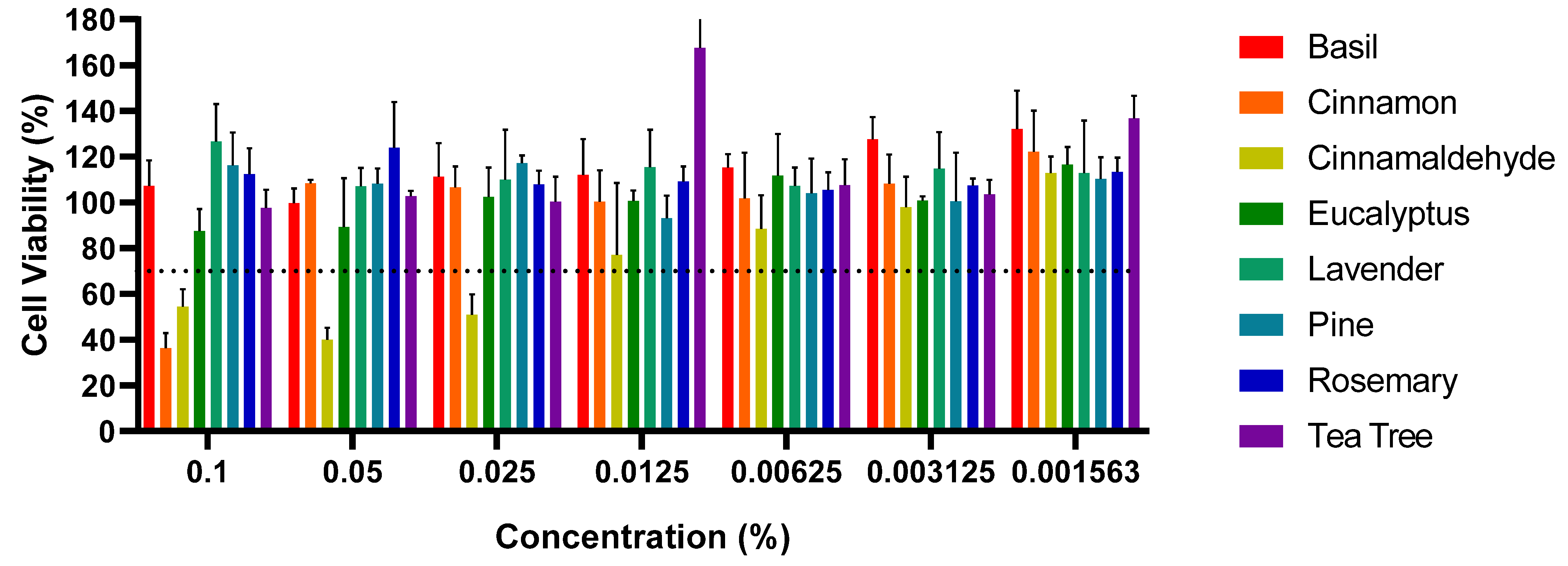
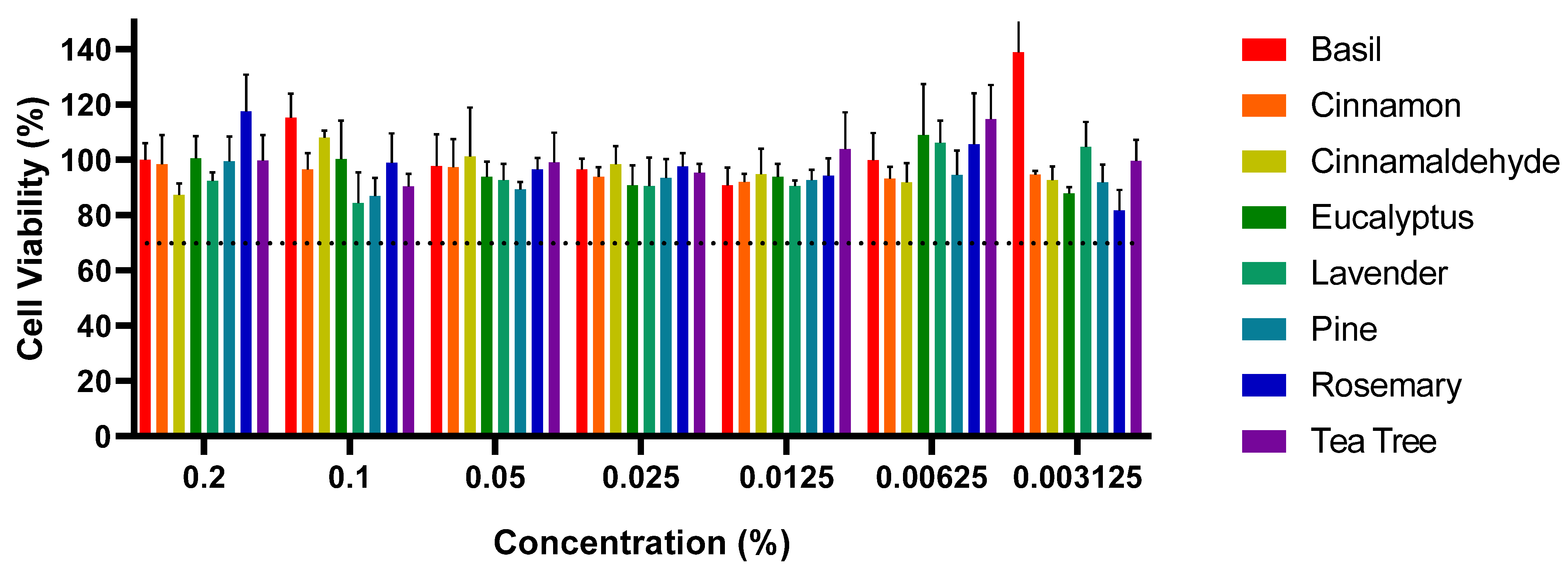
The results from this study highlight the cytotoxic effects of certain EOs and EOEs on Vero cells, specifically cinnamon EO and cinnamaldehyde; meanwhile, other EOs, such as basil and eucalyptus, did not exhibit significant cytotoxicity at the selected concentrations, with cell viability remaining above 70% [45]. The observed cytotoxicity of cinnamon EO and cinnamaldehyde is consistent with previous reports in the literature, where various EOs and their bioactive compounds demonstrated cytotoxic effects at higher concentrations, typically above 0.02%. For example, studies have shown that EOs like clove, cinnamon, and eucalyptus exhibit varying degrees of toxicity on different cell lines at high concentrations [50]. In this study, cinnamaldehyde demonstrated cytotoxicity at concentrations ranging from 0.0125% to 0.1%, consistent with previous research suggesting its toxicity at higher levels. Likewise, cinnamon EO displayed cytotoxicity at a concentration of 0.1%, aligning with prior studies that demonstrated its antibacterial efficacy and potential cytotoxicity at elevated concentrations [56,57,58,62,68]. During the healing phase of an infected wound, the continuous recruitment of eukaryotic cells to the wound site minimizes the impact on non-target cells. Therefore, although high concentrations of these EOs can cause cytotoxic effects, the wound environment will likely ensure minimal damage to the healthy eukaryotic cells involved in tissue repair [84,85,86]. Previous studies have shown that EOs at appropriate concentrations can be safe and effective for wound healing by promoting antibacterial activity and stimulating tissue repair without significant cytotoxicity [87,88,89]. Further research is needed to determine the optimal concentrations for various applications in order to fully understand their potential for clinical use, particularly in the management of nosocomial pathogens [88].

5. Conclusions

This study investigated the antibacterial and cytotoxic properties of various EOs and their emulsified formulations against clinically relevant nosocomial pathogens. The findings highlight the considerable antibacterial and antibiofilm potential of EOs, particularly those containing cinnamaldehyde, in combating antibiotic-resistant and biofilm-forming bacteria.
Cinnamaldehyde exhibited the most potent antibacterial activity among the tested compounds, with MICs ranging from 1.31 to 2.62 mg/mL. It was effective against the tested Gram-positive and Gram-negative pathogens, including S. aureus, MRSA, S. epidermidis, B. subtilis, S. Enteritidis, and P. aeruginosa. Other EOs, such as cinnamon, eucalyptus, and pine, also demonstrated antibacterial activity, although their efficacy against P. aeruginosa was comparatively limited.
Notably, cinnamaldehyde showed significant biofilm-disrupting capabilities, completely eradicating biofilms formed by S. aureus, MRSA, and P. aeruginosa within one hour. This underscores its potential as a valuable therapeutic agent for treating persistent biofilm-associated infections, often resistant to conventional therapies.
This study also highlights the value of aqueous EO emulsions stabilized using OSA-starch as an effective strategy to enhance the use of plant-derived oils in various biomedical and industrial fields. Continued research into their long-term stability and broader biological effects will be crucial to harness their potential across different applications.
Cytotoxicity analyses revealed that cinnamaldehyde and cinnamon EO were cytotoxic at concentrations above 0.1%. In contrast, other EOs, including basil, eucalyptus, and lavender, did not exhibit significant cytotoxicity at the tested concentrations, indicating their potential safety for therapeutic use when applied at appropriate doses.

Supplementary Materials

The following supporting information can be downloaded at https://www.mdpi.com/article/10.3390/pr13051531/s1.

Author Contributions

Conceptualization, J.S. and R.R.; methodology, H.S., J.B., T.H. and R.L.; validation, J.S. and R.R.; investigation, H.S., J.B., T.H. and R.L.; resources, J.S. and R.R.; data curation, J.S.; writing—original draft preparation, H.S., R.L. and J.B.; writing—review and editing J.S., R.L. and R.R.; visualization, H.S. and T.H; supervision, J.S.; project administration, J.S. and R.R.; funding acquisition, J.S. All authors have read and agreed to the published version of the manuscript.

Funding

This research received no external funding.

Data Availability Statement

The original contributions presented in the study are included in the article, further inquiries can be directed to the corresponding authors.

Acknowledgments

This study is based upon work from COST Action EURESTOP, CA21145, supported by COST (European Cooperation in Science and Technology).

Conflicts of Interest

The authors declare no conflicts of interest.

Abbreviations

The following abbreviations are used in this manuscript:
ATCCAmerican tissue culture collection
DMEMDulbecco’s Modified Eagle’s Medium
EOEssential oil
EOEEssential oil emulsion
FBSFetal bovine serum
GC-MSGas Chromatography-Mass Spectrometry
MBCMinimal inhibitory concentration
MHBMueller–Hinton Broth
MICMinimal bactericidal concentration
MRSAMethicillin-resistant Staphylococcus aureus
OSA-starchStarch sodium octenylsuccinate
PDIPolydispersity index
SDStandard deviation

References

  1. Otto, M. Staphylococcus epidermidis—The “accidental” Pathogen. Nat. Rev. Microbiol. 2009, 7, 555–567. [Google Scholar] [CrossRef] [PubMed]
  2. Haidar, A.; Muazzam, A.; Nadeem, A.; Atique, R.; Saeed, H.A.; Naveed, A.; Sharif, J.; Perveen, A.; Fatima, H.R.; Samad, A. Biofilm Formation and Antibiotic Resistance in Pseudomonas aeruginosa. Microbe 2024, 3, 100078. [Google Scholar] [CrossRef]
  3. Ben-Amram, H.; Azrad, M.; Cohen-Assodi, J.; Sharabi-Nov, A.; Edelstein, S.; Agay-Shay, K.; Peretz, A. Biofilm Formation by Hospital-Acquired Resistant Bacteria Isolated from Respiratory Samples. J. Epidemiol. Glob. Health 2024, 14, 291–297. [Google Scholar] [CrossRef] [PubMed]
  4. Yong, Y.Y.; Dykes, G.A.; Choo, W.S. Biofilm Formation by Staphylococci in Health-Related Environments and Recent Reports on Their Control Using Natural Compounds. Crit. Rev. Microbiol. 2019, 45, 201–222. [Google Scholar] [CrossRef]
  5. Baker, S.J.; Payne, D.J.; Rappuoli, R.; De Gregorio, E. Technologies to Address Antimicrobial Resistance. Proc. Natl. Acad. Sci. USA 2018, 115, 12887–12895. [Google Scholar] [CrossRef]
  6. Larsson, D.G.J.; Flach, C.-F. Antibiotic Resistance in the Environment. Nat. Rev. Microbiol. 2022, 20, 257–269. [Google Scholar] [CrossRef]
  7. Stepanović, S.; Vuković, D.; Dakić, I.; Savić, B.; Švabić-Vlahović, M. A Modified Microtiter-Plate Test for Quantification of Staphylococcal Biofilm Formation. J. Microbiol. Methods 2000, 40, 175–179. [Google Scholar] [CrossRef]
  8. Omwenga, E.O.; Awuor, S.O. The Bacterial Biofilms: Formation, Impacts, and Possible Management Targets in the Healthcare System. Can. J. Infect. Dis. Med. Microbiol. 2024, 2024, 1542576. [Google Scholar] [CrossRef] [PubMed]
  9. Stewart, P.S.; William Costerton, J. Antibiotic Resistance of Bacteria in Biofilms. Lancet 2001, 358, 135–138. [Google Scholar] [CrossRef]
  10. Dostert, M.; Trimble, M.J.; Hancock, R.E.W. Antibiofilm Peptides: Overcoming Biofilm-Related Treatment Failure. RSC Adv. 2021, 11, 2718–2728. [Google Scholar] [CrossRef]
  11. Høiby, N.; Ciofu, O.; Johansen, H.K.; Song, Z.; Moser, C.; Jensen, P.Ø.; Molin, S.; Givskov, M.; Tolker-Nielsen, T.; Bjarnsholt, T. The Clinical Impact of Bacterial Biofilms. Int. J. Oral. Sci. 2011, 3, 55–65. [Google Scholar] [CrossRef]
  12. Tan, L.; Huang, Y.; Shang, W.; Yang, Y.; Peng, H.; Hu, Z.; Wang, Y.; Rao, Y.; Hu, Q.; Rao, X.; et al. Accessory Gene Regulator (Agr) Allelic Variants in Cognate Staphylococcus aureus Strain Display Similar Phenotypes. Front. Microbiol. 2022, 13, 700894. [Google Scholar] [CrossRef] [PubMed]
  13. Christensen, G.D.; Simpson, W.A.; Bisno, A.L.; Beachey, E.H. Adherence of Slime-Producing Strains of Staphylococcus Epidermidis to Smooth Surfaces. Infect. Immun. 1982, 37, 318–326. [Google Scholar] [CrossRef] [PubMed]
  14. Christensen, G.D.; Simpson, W.A.; Younger, J.J.; Baddour, L.M.; Barrett, F.F.; Melton, D.M.; Beachey, E.H. Adherence of Coagulase-Negative Staphylococci to Plastic Tissue Culture Plates: A Quantitative Model for the Adherence of Staphylococci to Medical Devices. J. Clin. Microbiol. 1985, 22, 996–1006. [Google Scholar] [CrossRef]
  15. Borges, A.; Lopez-Romero, J.C.; Oliveira, D.; Giaouris, E.; Simões, M. Prevention, Removal and Inactivation of Escherichia coli and Staphylococcus aureus Biofilms Using Selected Monoterpenes of Essential Oils. J. Appl. Microbiol. 2017, 123, 104–115. [Google Scholar] [CrossRef] [PubMed]
  16. Condò, C.; Anacarso, I.; Sabia, C.; Iseppi, R.; Anfelli, I.; Forti, L.; De Niederhäusern, S.; Bondi, M.; Messi, P. Antimicrobial Activity of Spices Essential Oils and Its Effectiveness on Mature Biofilms of Human Pathogens. Nat. Prod. Res. 2020, 34, 567–574. [Google Scholar] [CrossRef]
  17. Ayaz, M.; Ullah, F.; Sadiq, A.; Ullah, F.; Ovais, M.; Ahmed, J.; Devkota, H.P. Synergistic Interactions of Phytochemicals with Antimicrobial Agents: Potential Strategy to Counteract Drug Resistance. Chem.-Biol. Interact. 2019, 308, 294–303. [Google Scholar] [CrossRef]
  18. Cahyana, Y.; Putri, Y.S.E.; Solihah, D.S.; Lutfi, F.S.; Alqurashi, R.M.; Marta, H. Pickering Emulsions as Vehicles for Bioactive Compounds from Essential Oils. Molecules 2022, 27, 7872. [Google Scholar] [CrossRef]
  19. Cheesman, M.; Ilanko, A.; Blonk, B.; Cock, I. Developing New Antimicrobial Therapies: Are Synergistic Combinations of Plant Extracts/Compounds with Conventional Antibiotics the Solution? Phcog. Rev. 2017, 11, 57. [Google Scholar] [CrossRef]
  20. Chouhan, S.; Sharma, K.; Guleria, S. Antimicrobial Activity of Some Essential Oils-Present Status and Future Perspectives. Medicines 2017, 4, 58. [Google Scholar] [CrossRef]
  21. Elgayyar, M.; Draughon, F.A.; Golden, D.A.; Mount, J.R. Antimicrobial Activity of Essential Oils from Plants Against Selected Pathogenic and Saprophytic Microorganisms. J. Food Prot. 2001, 64, 1019–1024. [Google Scholar] [CrossRef] [PubMed]
  22. Plackett, B. Why Big Pharma Has Abandoned Antibiotics. Nature 2020, 586, S50–S52. [Google Scholar] [CrossRef]
  23. Stephens, B.; Plant, J. Evaluation of the Antibacterial Activity of a Sizable Set of Essential Oils. Med. Aromat. Plants 2015, 4. [Google Scholar] [CrossRef]
  24. El-Abbasy, U.K.; Abdel-Hameed, M.A.; Hatterman-Valenti, H.M.; El-Shereif, A.R.; Abd El-Khalek, A.F. Effectiveness of Oregano and Thyme Essential Oils as Alternatives for Sulfur Dioxide in Controlling Decay and Gray Mold and Maintaining Quality of ‘Flame Seedless’ Table Grape (Vitis vinifera L.) during Cold Storage. Agronomy 2023, 13, 3075. [Google Scholar] [CrossRef]
  25. Man, A.; Santacroce, L.; Iacob, R.; Mare, A.; Man, L. Antimicrobial Activity of Six Essential Oils Against a Group of Human Pathogens: A Comparative Study. Pathogens 2019, 8, 15. [Google Scholar] [CrossRef]
  26. Mo, T.; Os, A. Plant Essential Oil: An Alternative to Emerging Multidrug Resistant Pathogens. J. Microbiol. Exp. 2017, 5, 00163. [Google Scholar] [CrossRef]
  27. Nazzaro, F.; Fratianni, F.; De Martino, L.; Coppola, R.; De Feo, V. Effect of Essential Oils on Pathogenic Bacteria. Pharmaceuticals 2013, 6, 1451–1474. [Google Scholar] [CrossRef]
  28. Semeniuc, C.A.; Pop, C.R.; Rotar, A.M. Antibacterial Activity and Interactions of Plant Essential Oil Combinations against Gram-Positive and Gram-Negative Bacteria. J. Food Drug Anal. 2017, 25, 403–408. [Google Scholar] [CrossRef]
  29. Budzyńska, A.; Wieckowska-Szakiel, M.; Sadowska, B.; Kalemba, D.; Rózalska, B. Antibiofilm Activity of Selected Plant Essential Oils and Their Major Components. Pol. J. Microbiol. 2011, 60, 35–41. [Google Scholar] [CrossRef]
  30. Alanazi, A.D.; Almohammed, H.I. Therapeutic Potential and Safety of the Cinnamomum zeylanicum Methanolic Extract Against Chronic Toxoplasma gondii Infection in Mice. Front. Cell. Infect. Microbiol. 2022, 12, 900046. [Google Scholar] [CrossRef] [PubMed]
  31. Reichling, J.; Schnitzler, P.; Wiesenhofer, K. Comparative Study on the Cytotoxicity of Different Myrtaceae Essential Oils on Cultured Vero and RC-37 Cells. Pharmazie 2008, 63, 830–835. [Google Scholar] [CrossRef]
  32. Reichling, J.; Schnitzler, P.; Suschke, U.; Saller, R. Essential Oils of Aromatic Plants with Antibacterial, Antifungal, Antiviral, and Cytotoxic Properties—An Overview. Complement. Med. Res. 2009, 16, 79–90. [Google Scholar] [CrossRef] [PubMed]
  33. Valdivieso-Ugarte, M.; Gomez-Llorente, C.; Plaza-Díaz, J.; Gil, Á. Antimicrobial, Antioxidant, and Immunomodulatory Properties of Essential Oils: A Systematic Review. Nutrients 2019, 11, 2786. [Google Scholar] [CrossRef] [PubMed]
  34. Yammine, J.; Chihib, N.-E.; Gharsallaoui, A.; Dumas, E.; Ismail, A.; Karam, L. Essential Oils and Their Active Components Applied as: Free, Encapsulated and in Hurdle Technology to Fight Microbial Contaminations. A Review. Heliyon 2022, 8, e12472. [Google Scholar] [CrossRef]
  35. Linke, A.; Weiss, J.; Kohlus, R. Factors Determining the Surface Oil Concentration of Encapsulated Lipid Particles: Impact of the Emulsion Oil Droplet Size. Eur. Food Res. Technol. 2020, 246, 1933–1943. [Google Scholar] [CrossRef]
  36. Yammine, J.; Chihib, N.-E.; Gharsallaoui, A.; Ismail, A.; Karam, L. Advances in Essential Oils Encapsulation: Development, Characterization and Release Mechanisms. Polym. Bull. 2024, 81, 3837–3882. [Google Scholar] [CrossRef]
  37. Navikaite-Snipaitiene, V.; Spirikavice, K.; Siugzdaite, J.; Grigonyte, E.B.; Rutkaite, R. Preparation and Biological Activity Studies of Octenyl Succinic Anhydride Starch-Based Emulsions Containing Natural Essential Oils and Their Components. Appl. Sci. 2024, 14, 4050. [Google Scholar] [CrossRef]
  38. Akbari, A.; Bahmani, K.; Kafkas, N.E.; Bilgin, O.F.; Hamijo, T.; Izadi Darbandi, A.; Farhadpour, M. Evaluation of Seed Yield, Essential Oil Compositions, and Fatty Acid Profiles in Advanced Fennel (Foeniculum Vulgare Mill) Breeding Populations. Biocatal. Agric. Biotechnol. 2024, 57, 103118. [Google Scholar] [CrossRef]
  39. CLSI M100-S24; Performance Standards for Antimicrobial Susceptibility Testing; 24th Informational Supplement. Clinical and Laboratory Standards Institute: Wayne, PA, USA, 2014.
  40. Balouiri, M.; Sadiki, M.; Ibnsouda, S.K. Methods for in Vitro Evaluating Antimicrobial Activity: A Review. J. Pharm. Anal. 2016, 6, 71–79. [Google Scholar] [CrossRef]
  41. Mosmann, T. Rapid Colorimetric Assay for Cellular Growth and Survival: Application to Proliferation and Cytotoxicity Assays. J. Immunol. Methods 1983, 65, 55–63. [Google Scholar] [CrossRef]
  42. Sienkiewicz, M.; Łysakowska, M.; Pastuszka, M.; Bienias, W.; Kowalczyk, E. The Potential of Use Basil and Rosemary Essential Oils as Effective Antibacterial Agents. Molecules 2013, 18, 9334–9351. [Google Scholar] [CrossRef] [PubMed]
  43. Singh, I.R.; Pulikkal, A.K. Preparation, Stability and Biological Activity of Essential Oil-Based Nano Emulsions: A Comprehensive Review. OpenNano 2022, 8, 100066. [Google Scholar] [CrossRef]
  44. Ishak, A.; Mazonakis, N.; Spernovasilis, N.; Akinosoglou, K.; Tsioutis, C. Bactericidal versus Bacteriostatic Antibacterials: Clinical Significance, Differences and Synergistic Potential in Clinical Practice. J. Antimicrob. Chemother. 2025, 80, 1–17. [Google Scholar] [CrossRef]
  45. ISO 10993-5:2009; Biological Evaluation of Medical Devices—Part 5: Tests for In Vitro Cytotoxicity. ISO: Geneva, Switzerland, 2009.
  46. Burt, S. Essential Oils: Their Antibacterial Properties and Potential Applications in Foods—A Review. Int. J. Food Microbiol. 2004, 94, 223–253. [Google Scholar] [CrossRef] [PubMed]
  47. Ultee, A.; Bennik, M.H.J.; Moezelaar, R. The Phenolic Hydroxyl Group of Carvacrol Is Essential for Action against the Food-Borne Pathogen Bacillus cereus. Appl. Environ. Microbiol. 2002, 68, 1561–1568. [Google Scholar] [CrossRef]
  48. Khan, M.S.A.; Zahin, M.; Hasan, S.; Husain, F.M.; Ahmad, I. Inhibition of Quorum Sensing Regulated Bacterial Functions by Plant Essential Oils with Special Reference to Clove Oil. Lett. Appl. Microbiol. 2009, 49, 354–360. [Google Scholar] [CrossRef] [PubMed]
  49. Donsì, F.; Ferrari, G. Essential Oil Nanoemulsions as Antimicrobial Agents in Food. J. Biotechnol. 2016, 233, 106–120. [Google Scholar] [CrossRef]
  50. Russo, R.; Corasaniti, M.T.; Bagetta, G.; Morrone, L.A. Exploitation of Cytotoxicity of Some Essential Oils for Translation in Cancer Therapy. Evid.-Based Complement. Altern. Med. 2015, 2015, 397821. [Google Scholar] [CrossRef]
  51. Nhouchi, Z.; Watuzola, R.; Pense-Lheritier, A.M. A Review on Octenyl Succinic Anhydride Modified Starch-based Pickering-emulsion: Instabilities and Ingredients Interactions. J. Texture Stud. 2022, 53, 581–600. [Google Scholar] [CrossRef]
  52. Buranasuksombat, U.; Kwon, Y.J.; Turner, M.; Bhandari, B. Influence of Emulsion Droplet Size on Antimicrobial Properties. Food Sci. Biotechnol. 2011, 20, 793–800. [Google Scholar] [CrossRef]
  53. Sharif, H.R.; Abbas, S.; Majeed, H.; Safdar, W.; Shamoon, M.; Khan, M.A.; Shoaib, M.; Raza, H.; Haider, J. Formulation, Characterization and Antimicrobial Properties of Black Cumin Essential Oil Nanoemulsions Stabilized by OSA Starch. J. Food Sci. Technol. 2017, 54, 3358–3365. [Google Scholar] [CrossRef]
  54. Kim, Y.-G.; Lee, J.-H.; Kim, S.-I.; Baek, K.-H.; Lee, J. Cinnamon Bark Oil and Its Components Inhibit Biofilm Formation and Toxin Production. Int. J. Food Microbiol. 2015, 195, 30–39. [Google Scholar] [CrossRef] [PubMed]
  55. Xu, J.; Lin, Q.; Sheng, M.; Ding, T.; Li, B.; Gao, Y.; Tan, Y. Antibiofilm Effect of Cinnamaldehyde-Chitosan Nanoparticles against the Biofilm of Staphylococcus aureus. Antibiotics 2022, 11, 1403. [Google Scholar] [CrossRef] [PubMed]
  56. Piovezan, M.; Sayuri Uchida, N.; Fiori Da Silva, A.; Grespan, R.; Regina Santos, P.; Leite Silva, E.; Kenji Nakamura Cuman, R.; Machinski Junior, M.; Martha Graton Mikcha, J. Effect of Cinnamon Essential Oil and Cinnamaldehyde on Salmonella Saintpaul Biofilm on a Stainless Steel Surface. J. Gen. Appl. Microbiol. 2014, 60, 119–121. [Google Scholar] [CrossRef] [PubMed]
  57. Lee, J.-E.; Jung, M.; Lee, S.-C.; Huh, M.-J.; Seo, S.-M.; Park, I.-K. Antibacterial Mode of Action of trans-Cinnamaldehyde Derived from Cinnamon Bark (Cinnamomum verum) Essential Oil against Agrobacterium tumefaciens. Pestic. Biochem. Physiol. 2020, 165, 104546. [Google Scholar] [CrossRef]
  58. Doyle, A.A.; Stephens, J.C. A Review of Cinnamaldehyde and Its Derivatives as Antibacterial Agents. Fitoterapia 2019, 139, 104405. [Google Scholar] [CrossRef]
  59. Pandey, V.K.; Tripathi, A.; Srivastava, S.; Dar, A.H.; Singh, R.; Farooqui, A.; Pandey, S. Exploiting the Bioactive Properties of Essential Oils and Their Potential Applications in Food Industry. Food Sci. Biotechnol. 2023, 32, 885–902. [Google Scholar] [CrossRef]
  60. Sakkas, H.; Gousia, P.; Economou, V.; Sakkas, V.; Petsios, S.; Papadopoulou, C. In Vitro Antimicrobial Activity of Five Essential Oils on Multi-Drug Resistant Gram-Negative Clinical Isolates. J. Intercult. Ethnopharmacol. 2016, 5, 212. [Google Scholar] [CrossRef]
  61. Nostro, A.; Roccaro, A.S.; Bisignano, G.; Marino, A.; Cannatelli, M.A.; Pizzimenti, F.C.; Cioni, P.L.; Procopio, F.; Blanco, A.R. Effects of Oregano, Carvacrol and Thymol on Staphylococcus aureus and Staphylococcus epidermidis Biofilms. J. Med. Microbiol. 2007, 56, 519–523. [Google Scholar] [CrossRef]
  62. Shu, C.; Ge, L.; Li, Z.; Chen, B.; Liao, S.; Lu, L.; Wu, Q.; Jiang, X.; An, Y.; Wang, Z.; et al. Antibacterial Activity of Cinnamon Essential Oil and Its Main Component of Cinnamaldehyde and the Underlying Mechanism. Front. Pharmacol. 2024, 15, 1378434. [Google Scholar] [CrossRef]
  63. Zuzarte, M.; Sousa, C.; Alves-Silva, J.; Salgueiro, L. Plant Monoterpenes and Essential Oils as Potential Anti-Ageing Agents: Insights from Preclinical Data. Biomedicines 2024, 12, 365. [Google Scholar] [CrossRef] [PubMed]
  64. Shin, J.; Prabhakaran, V.-S.; Kim, K. The Multi-Faceted Potential of Plant-Derived Metabolites as Antimicrobial Agents against Multidrug-Resistant Pathogens. Microb. Pathog. 2018, 116, 209–214. [Google Scholar] [CrossRef] [PubMed]
  65. Zhang, Y.; Liu, X.; Wang, Y.; Jiang, P.; Quek, S. Antibacterial Activity and Mechanism of Cinnamon Essential Oil against Escherichia coli and Staphylococcus aureus. Food Control 2016, 59, 282–289. [Google Scholar] [CrossRef]
  66. El Atki, Y.; Aouam, I.; El Kamari, F.; Taroq, A.; Nayme, K.; Timinouni, M.; Lyoussi, B.; Abdellaoui, A. Antibacterial Activity of Cinnamon Essential Oils and Their Synergistic Potential with Antibiotics. J. Adv. Pharm. Technol. Res. 2019, 10, 63. [Google Scholar] [CrossRef]
  67. Abdelatti, M.A.I.; Abd El-Aziz, N.K.; El-Naenaeey, E.Y.M.; Ammar, A.M.; Alharbi, N.K.; Alharthi, A.; Zakai, S.A.; Abdelkhalek, A. Antibacterial and Anti-Efflux Activities of Cinnamon Essential Oil against Pan and Extensive Drug-Resistant Pseudomonas aeruginosa Isolated from Human and Animal Sources. Antibiotics 2023, 12, 1514. [Google Scholar] [CrossRef]
  68. Firmino, D.F.; Cavalcante, T.T.A.; Gomes, G.A.; Firmino, N.C.S.; Rosa, L.D.; De Carvalho, M.G.; Catunda, F.E.A., Jr. Antibacterial and Antibiofilm Activities of Cinnamomum Sp. Essential Oil and Cinnamaldehyde: Antimicrobial Activities. Sci. World J. 2018, 2018, 7405736. [Google Scholar] [CrossRef]
  69. Li, X.-Z.; Zhang, L.; Poole, K. Role of the Multidrug Efflux Systems of Pseudomonas aeruginosa in Organic Solvent Tolerance. J. Bacteriol. 1998, 180, 2987–2991. [Google Scholar] [CrossRef]
  70. Langeveld, W.T.; Veldhuizen, E.J.A.; Burt, S.A. Synergy between Essential Oil Components and Antibiotics: A Review. Crit. Rev. Microbiol. 2014, 40, 76–94. [Google Scholar] [CrossRef]
  71. Fratini, F.; Pecorini, C.; Resci, I.; Copelotti, E.; Nocera, F.P.; Najar, B.; Mancini, S. Evaluation of the Synergistic Antimicrobial Activity of Essential Oils and Cecropin A Natural Peptide on Gram-Negative Bacteria. Animals 2025, 15, 282. [Google Scholar] [CrossRef]
  72. Lahmar, A.; Bedoui, A.; Mokdad-Bzeouich, I.; Dhaouifi, Z.; Kalboussi, Z.; Cheraif, I.; Ghedira, K.; Chekir-Ghedira, L. Reversal of Resistance in Bacteria Underlies Synergistic Effect of Essential Oils with Conventional Antibiotics. Microb. Pathog. 2017, 106, 50–59. [Google Scholar] [CrossRef]
  73. Roy, R.; Tiwari, M.; Donelli, G.; Tiwari, V. Strategies for Combating Bacterial Biofilms: A Focus on Anti-Biofilm Agents and Their Mechanisms of Action. Virulence 2018, 9, 522–554. [Google Scholar] [CrossRef] [PubMed]
  74. Sharma, D.; Misba, L.; Khan, A.U. Antibiotics versus Biofilm: An Emerging Battleground in Microbial Communities. Antimicrob. Resist. Infect. Control 2019, 8, 76. [Google Scholar] [CrossRef]
  75. Kalia, V.C.; Patel, S.K.S.; Lee, J.-K. Bacterial Biofilm Inhibitors: An Overview. Ecotoxicol. Environ. Saf. 2023, 264, 115389. [Google Scholar] [CrossRef] [PubMed]
  76. Yin, L.; Guo, Y.; Xv, X.; Dai, Y.; Li, L.; Sun, F.; Lv, X.; Shu, G.; Liang, X.; He, C.; et al. Cinnamaldehyde Nanoemulsion Decorated with Rhamnolipid for Inhibition of Methicillin-Resistant Staphylococcus aureus Biofilm Formation: In Vitro and In Vivo Assessment. Front. Microbiol. 2024, 15, 1514659. [Google Scholar] [CrossRef] [PubMed]
  77. Kashi, M.; Noei, M.; Chegini, Z.; Shariati, A. Natural Compounds in the Fight against Staphylococcus aureus Biofilms: A Review of Antibiofilm Strategies. Front. Pharmacol. 2024, 15, 1491363. [Google Scholar] [CrossRef]
  78. Didehdar, M.; Chegini, Z.; Tabaeian, S.P.; Razavi, S.; Shariati, A. Cinnamomum: The New Therapeutic Agents for Inhibition of Bacterial and Fungal Biofilm-Associated Infection. Front. Cell. Infect. Microbiol. 2022, 12, 930624. [Google Scholar] [CrossRef]
  79. Topa, S.H.; Subramoni, S.; Palombo, E.A.; Kingshott, P.; Rice, S.A.; Blackall, L.L. Cinnamaldehyde Disrupts Biofilm Formation and Swarming Motility of Pseudomonas aeruginosa. Microbiology 2018, 164, 1087–1097. [Google Scholar] [CrossRef]
  80. Rossi, M.W.; Heuertz, R.M. Cinnamaldehyde Inhibits MRSA Biofilm Formation and Reduces Cell Viability. Am. Soc. Clin. Lab. Sci. 2017, 30, 214–218. [Google Scholar] [CrossRef]
  81. Kot, B.; Sytykiewicz, H.; Sprawka, I.; Witeska, M. Effect of Trans-Cinnamaldehyde on Methicillin-Resistant Staphylococcus aureus Biofilm Formation: Metabolic Activity Assessment and Analysis of the Biofilm-Associated Genes Expression. Int. J. Mol. Sci. 2019, 21, 102. [Google Scholar] [CrossRef]
  82. Papa, R.; Garzoli, S.; Vrenna, G.; Sabatino, M.; Sapienza, F.; Relucenti, M.; Donfrancesco, O.; Fiscarelli, E.; Artini, M.; Selan, L.; et al. Essential Oils Biofilm Modulation Activity, Chemical and Machine Learning Analysis—Application on Staphylococcus aureus Isolates from Cystic Fibrosis Patients. Int. J. Mol. Sci. 2020, 21, 9258. [Google Scholar] [CrossRef]
  83. Millezi, A.F.; Costa, K.A.D.; Oliveira, J.M.; Lopes, S.P.; Pereira, M.O.; Piccoli, R.H. Antibacterial and Anti-Biofilm Activity of Cinnamon Essential Oil and Eugenol. Cienc. Rural 2019, 49, e20180314. [Google Scholar] [CrossRef]
  84. Goswami, A.G.; Basu, S.; Banerjee, T.; Shukla, V.K. Biofilm and Wound Healing: From Bench to Bedside. Eur. J. Med. Res. 2023, 28, 157. [Google Scholar] [CrossRef] [PubMed]
  85. Liang, Y.; Liang, Y.; Zhang, H.; Guo, B. Antibacterial Biomaterials for Skin Wound Dressing. Asian J. Pharm. Sci. 2022, 17, 353–384. [Google Scholar] [CrossRef]
  86. Zielińska, M.; Pawłowska, A.; Orzeł, A.; Sulej, L.; Muzyka-Placzyńska, K.; Baran, A.; Filipecka-Tyczka, D.; Pawłowska, P.; Nowińska, A.; Bogusławska, J.; et al. Wound Microbiota and Its Impact on Wound Healing. Int. J. Mol. Sci. 2023, 24, 17318. [Google Scholar] [CrossRef] [PubMed]
  87. Mazutti Da Silva, S.M.; Rezende Costa, C.R.; Martins Gelfuso, G.; Silva Guerra, E.N.; De Medeiros Nóbrega, Y.K.; Gomes, S.M.; Pic-Taylor, A.; Fonseca-Bazzo, Y.M.; Silveira, D.; Magalhães, P.D.O. Wound Healing Effect of Essential Oil Extracted from Eugenia Dysenterica DC (Myrtaceae) Leaves. Molecules 2018, 24, 2. [Google Scholar] [CrossRef]
  88. Salas-Oropeza, J.; Jimenez-Estrada, M.; Perez-Torres, A.; Castell-Rodriguez, A.E.; Becerril-Millan, R.; Rodriguez-Monroy, M.A.; Canales-Martinez, M.M. Wound Healing Activity of the Essential Oil of Bursera Morelensis, in Mice. Molecules 2020, 25, 1795. [Google Scholar] [CrossRef]
  89. Shaaban, H.A. Essential Oil as Antimicrobial Agents: Efficacy, Stability, and Safety Issues for Food Application. In Essential Oils—Bioactive Compounds, New Perspectives and Applications; Santana De Oliveira, M., Almeida Da Costa, W., Gomes Silva, S., Eds.; IntechOpen: London, UK, 2020; ISBN 978-1-83962-697-5. [Google Scholar]
Figure 1. Biofilm formation evaluation by the tube method using cinnamaldehyde. MRSA: Methicillin-resistant S. aureus.
Processes 13 01531 g001
Figure 2. Cell viability after treatment with essential oils for 72 h on Vero cells. Control sample (untreated cells) = 100% viability. According to the guidelines in ISO 10993-5 (2009) [45] a material concentration is considered non-cytotoxic when cell viability remains above 70% (dotted line).
Processes 13 01531 g002
Figure 3. Cell viability after treatment with essential oils emulsions for 72 h on Vero cells. The control sample (untreated cells) = 100% viability. According to the guidelines in ISO 10993-5 (2009) [45], a material concentration is considered non-cytotoxic when cell viability remains above 70% (dotted line).
Processes 13 01531 g003
Figure 4. Microscopic observation of Vero cells after treatment with the EOs and their emulsions.
Processes 13 01531 g004
Table 1. Characteristics of prepared emulsions containing different EOs.
Essential Oil EmulsionDroplet Diameter, Mean ± SD (nm)PDI, Mean ± SD
Basil EOE661.6 ± 12.20.16 ± 0.05
Cinnamon EOE475.1 ± 14.30.25 ± 0.01
Cinnamaldehyde E258.8 ± 58.30.26 ± 0.09
Eucalyptus EOE819.4 ± 18.20.31 ± 0.01
Lavender EOE426.5 ± 4.50.19 ± 0.03
Pine EOE649.1 ± 7.80.16 ± 0.06
Rosemary EOE516.3 ± 36.60.22 ± 0.02
Tea tree EOE433.5 ± 10.80.19 ± 0.04
Table 2. Inhibition zone diameter of essential oils for bacterial strains determined by disc agar diffusion assay.
Bioactive CompoundsInhibition Zone Diameter (mm)
S. aureusMRSAS. epidermidisB. subtilisS. EnteritidisP. aeruginosa
Basil EO10.04 ± 0.05 g9.96 ± 0.41 f9.83 ± 0.42 f9.24 ± 0.26 g<6.0 ± 0.00 f<6.0 ± 0.00 c
Cinnamon EO14.17 ± 0.06 e14.05 ± 0.49 e18.46 ± 0.54 c30.00 ± 0.44 b19.90 ± 0.40 b<6.0 ± 0.00 c
Cinnamaldehyde36.45 ± 0.05 a47.08 ± 0.89 a50.22 ± 0.45 a43.88 ± 0.56 a31.83 ± 0.17 a24.37 ± 0.25 a
Eucalyptus EO28.04 ± 0.06 b28.58 ± 0.49 b30.00 ± 0.44 b21.00 ± 0.55 c14.61 ± 0.31 d<6.0 ± 0.00 c
Lavender EO21.37 ± 0.31 c11.06 ± 0.95 f13.81 ± 0.34 d19.75 ± 0.59 c,d8.65 ± 0.39 f<6.0 ± 0.00 c
Pine EO18.25 ± 0.70 d20.91 ± 0.89 d13.49 ± 0.43 d16.23 ± 0.71 e18.80 ± 0.26 c<6.0 ± 0.00 c
Rosemary EO9.11 ± 0.25 h7.00 ± 0.05 g8.10 ± 0.26 h10.89 ± 0.53 f8.00 ± 0.22 e<6.0 ± 0.00 c
Tea tree EO20.20 ± 0.71 c24.44 ± 0.42 c17.91 ± 0.25 c18.30 ± 0.68 d14.81 ± 0.29 d7.50 ± 0.22 b
Ampicillin11.53 ± 0.14 f8.00 ± 0.15 f12.62 ± 0.15 e21.71 ± 0.45 c12.14 ± 0.16 e-
Notes: Three results (mean ± SD, n = 3) read from the same column and marked with the same letter do not differ significantly at a threshold α = 5% (Two-way ANOVA; Tukey’s test). In contrast, results marked with different letters show a significant difference. NA: Not Applicable. MRSA: Methicillin-resistant S. aureus.
Table 3. Inhibition zone diameter of essential oils emulsions for bacterial strains determined by disc agar diffusion assay.
Emulsion of Bioactive CompoundInhibition Zone Diameter (mm)
S. aureusMRSAS. epidermidisB. subtilisS. EnteritidisP. aeruginosa
Cinnamon EOE11.18 ± 0.17 b,c11.48 ± 0.27 b11.99 ± 0.18 c9.62 ± 0.24 c<6.0 ± 0.00 c9.65 ± 0.55 a
Cinnamaldehyde E12.07 ± 0.28 a,b21.82 ± 0.34 a23.00 ± 0.25 a16.67 ± 0.44 b15.24 ± 0.29 a10.33 ± 0.58 a
Eucalyptus EOE10.83 ± 0.30 c,d12.22 ± 0.30 b16.88 ± 0.51 b9.65 ± 0.20 c<6.0 ± 0.00 c<6.0 ± 0.00 c
Lavender EOE12.48 ± 0.14 a10.23 ± 0.25 c10.78 ± 0.25 d<6.0 ± 0.00 d<6.0 ± 0.00 c<6.0 ± 0.00 c
Pine EOE11.60 ± 0.17 b<6.0 ± 0.00 e11.78 ± 0.22 c<6.0 ± 0.00 d<6.0 ± 0.00 c<6.0 ± 0.00 c
Tea tree EOE9.40 ± 0.28 e8.85 ± 0.31 d9.72 ± 0.45 e<6.0 ± 0.00 d<6.0 ± 0.00 c7.00 ± 0.50 b
Ampicillin11.53 ± 0.14 b8.00 ± 0.1512.62 ± 0.1521.71 ± 0.45 a12.14 ± 0.16 b<6.0 ± 0.00 c
Notes: Three results (mean ± SD, n = 3) read from the same column and marked with the same letter do not differ significantly at a threshold α = 5% (Two-way ANOVA; Tukey’s test). In contrast, results marked with different letters show a significant difference. NA: Not Applicable. MRSA: Methicillin-resistant S. aureus.
Table 4. MIC, MBC, and MBC/MIC ratios of the essential oils were tested against different bacterial strains.
Bioactive SubstanceConcentration (mg/mL)Bacterial Strains
S. aureusMRSAS. epidermidisB. subtilisS. EnteritidisP. aeruginosa
Cinnamon EOMIC2.565.125.122.562.565.12
MBC5.125.125.122.565.12>5.12
MBC/MIC21112>1
CinnamaldehydeMIC1.311.311.311.311.312.62
MBC2.622.621.311.312.622.62
MBC/MIC221121
Eucalyptus EOMIC2.282.282.282.284.564.56
MBC2.284.564.564.564.564.56
MBC/MIC122211
Lavender EOMIC>4.43>4.43>4.43>4.43>4.43>4.43
MBC>4.43>4.43>4.43>4.43>4.43>4.43
MBC/MICNDNDNDNDNDND
Pine EOMIC>4.31>4.31>4.31>4.31>4.31>4.31
MBC>4.31>4.31>4.31>4.31>4.31>4.31
MBC/MICNDNDNDNDNDND
Tea tree EOMIC2.234.472.232.234.474.47
MBC4.474.47>4.442.23>4.474.47
MBC/MIC21>21>11
AmpicillinMIC0.170.350.170.020.352.81
MBC0.170.700.350.045.6211.25
MRSA: Methicillin-resistant S. aureus, MIC: minimal inhibitory concentration, MBC: minimal bactericidal concentration, ND: no data.
Table 5. MIC, MBC, and MBC/MIC ratios of the essential oil emulsions were tested against different bacterial strains.
Bioactive SubstanceConcentrations (mg/mL)Bacterial Strains
S. aureusMRSAS. epidermidisB. subtilisS. Enteritidis
Cinnamon EOEMIC13.506.7527.006.7513.50
MBC27.0027.0027.00>54.0013.50
MBC/MIC241>81
Cinnamaldehyde EMIC13.756.876.876.876.87
MBC13.7513.756.87>55.0013.75
MBC/MIC111>82
Eucalyptus EOEMIC26.5026.5013.2513.2526.50
MBC26.5026.5013.25>53.0053.00
MBC/MIC111>42
Tea tree EOEMIC46.0046.0046.0046.0046.00
MBC46.0046.0046.00>46.00>46.00
MBC/MIC111>1>1
AmpicillinMIC0.170.350.170.020.35
MBC0.170.700.350.045.62
MRSA: Methicillin-resistant S. aureus, MIC: minimal inhibitory concentration, MBC: minimal bactericidal concentration.
Table 6. Results of cinnamaldehyde treatment on biofilm formed by S. aureus, MRSA, and P. aeruginosa.
BacteriaThe Evaluation of the Amount of Biofilm Formed by Bacteria
Before TreatmentAfter Treatment
S. aureusGram-positiveweak positivenegative
MRSAmoderate positivenegative
P. aeruginosaGram-negativemoderate positivenegative
The amount of biofilm formed was scored as negative, weak positive, moderate positive, and strong. MRSA: Methicillin-resistant S. aureus.
Disclaimer/Publisher’s Note: The statements, opinions and data contained in all publications are solely those of the individual author(s) and contributor(s) and not of MDPI and/or the editor(s). MDPI and/or the editor(s) disclaim responsibility for any injury to people or property resulting from any ideas, methods, instructions or products referred to in the content.

