Article

pH-Responsive Gold Nanoparticle/PVP Nanoconjugate for Targeted Delivery and Enhanced Anticancer Activity of Withaferin A

by
Velmurugan Sekar
1,
Amutha Santhanam
1,* and
Paulraj Arunkumar
2,*
1
Nanobiotherapeutics Laboratory, National Centre for Nanoscience and Nanotechnology, University of Madras, Guindy Campus, Chennai 600025, Tamil Nadu, India
2
School of Chemical Engineering, Chonnam National University, Gwangju 61186, Republic of Korea
*
Authors to whom correspondence should be addressed.
Processes 2025, 13(5), 1290; https://doi.org/10.3390/pr13051290
Submission received: 10 March 2025 / Revised: 15 April 2025 / Accepted: 21 April 2025 / Published: 23 April 2025
(This article belongs to the Special Issue Composite Materials Processing, Modeling and Simulation)

Abstract

:
The development of advanced high-capacity nanoparticle-based drug loading, precise targeting, low toxicity, and excellent biocompatibility is critical for improving cancer therapeutics. Withaferin A, a natural steroidal lactone derived from Physalis minima, exhibits potential biological activity and holds promise as a therapeutic agent. In this study, a novel nanoconjugate (NC) was developed using gold nanoparticles (AuNPs) functionalized with polyvinylpyrrolidone (PVP), Withaferin A drug, and folic acid for targeted drug delivery in cancer treatment. The AuNPs–PVP–Withaferin A–FA nanoconjugate was synthesized through a layer-by-layer assembly process and was confirmed using UV–visible and FTIR spectroscopy. The hydrodynamic radius, surface charge, and morphology of the NC were characterized using dynamic light scattering (DLS), zeta potential analysis, and electron microscopy, respectively. The nanoformulation demonstrated a pH-responsive drug release, with 92% of Withaferin A released at pH 5, mimicking the tumor microenvironment. In vitro cytotoxicity studies conducted on MCF-7 cells using MTT assays, dual dye staining, and protein expression analysis revealed that the nanoconjugate effectively induced apoptosis in cancer cells. These outcomes emphasize the prospect AuNPs–PVP–Withaferin A–FA nanoconjugate as a targeted and efficient Withaferin A delivery system for cancer therapy, leveraging the inherent anticancer properties of Withaferin A.

1. Introduction

Breast cancer persists as a significant worldwide medical issue, particularly for women, and is a prominent reason of morbidity and mortality worldwide [1]. Despite advancements in cancer therapy, many patients with advanced breast cancer develop resistance to conventional treatments, underscoring the requirement for more potent therapeutic approaches [2]. Apoptosis induction is a promising approach for early-stage cancer treatment; however, current methods for cancer detection and therapy are often limited by poor selectivity, cytotoxicity, instability, and biocompatibility issues, as well as undesirable side effects [3]. These limitations highlight the urgent need for innovative chemotherapeutic agents that can effectively inhibit or slow cancer progression.
The main treatment method has been chemotherapy; however, it often comes with serious side effects and limited patient compliance. To address these challenges, researchers have turned to herbal medicine, exploring natural compounds with anticancer properties [4]. A member of the Solanaceae family, Physalis minima is a medicinal plant that has long been used in India for its diuretic, tonic, and purgative qualities. It has also been shown to have analgesic, anti-inflammatory, and anthelmintic effects. The Munda tribe of Chota Nagpur in India has used its leaf extract for earache relief [5,6,7]. One of the key bioactive compounds derived from medicinal plants is Withaferin A, a withanolide known for its potent anticancer properties [8,9]. One potential approach in the realm of biomedical research is the combination of nanotechnology and plant bioactive chemicals. Bioactive substances generated from plants have many different medicinal uses. In the synthesis of nanoparticles, these phytochemicals can act as natural reducing and stabilizing agents, improving biocompatibility and medicinal efficacy when paired with nanotechnology [10]. The synthesis of withanolides involves the oxidation of C-26, C-23, and C-22 through the triterpenoid pathway, forming lactone rings essential for their anticancer activity [11,12]. Withaferin A is primarily sourced from Withania somnifera, but recent studies indicate that Physalis minima also contains substantial amounts of this compound, making it a promising alternative for sustainable Withaferin A production [13,14]. However, its clinical application is hindered by challenges such as poor bioavailability, nonspecific biodistribution, and lack of targeted delivery [15]. To address these limitations, nanotechnology-based drug delivery systems have emerged as a promising solution, offering enhanced drug stability, targeted delivery, and improved therapeutic outcomes [16].
Gold nanoparticle (AuNPs)-based nanocarriers have garnered major interest in cancer therapy because of their distinct physicochemical properties, biocompatibility, and ability to functionalize with targeting ligands [10]. Polymer-functionalized AuNPs using polyvinylpyrrolidone (PVP) provide a stable platform for drug delivery, enabling the conjugation of bioactive molecules such as Withaferin A. The implementation of targeting ligands, such folic acid (FA), which identify particular receptors that are overexpressed on particular cancer cell types, can further functionalize nanocarriers intended for active drug targeting [17,18]. FA specifically binds to biomarkers while also promoting cell proliferation. FA-functionalized nanostructures can enable sustained drug release to cellular targets, thereby enhancing the therapeutic efficacy of anticancer agents such as Withaferin A. Folate-conjugated nanomaterial research has accelerated significantly in the field of cancer biology, especially in the creation of innovative methods for cancer screening and treatment [18]. FA conjugation onto PVP-functionalized AuNPs has been known to enhance targeted drug delivery to cancer cells with high efficiency [18,19]. The layer-by-layer (LBL) assembly technique has garnered substantial interest for delivering nanoconjugates on the AuNP surface that involves stabilizing and adsorbing anticancer drugs using an oppositely charged PVP polymer [20]. The LBL assembly technique, in combination with carbodiimide coupling chemistry using 1-ethyl-3-[3-dimethylaminopropyl] carbodiimide (EDC) and N-hydroxysuccinimide (NHS), facilitates efficient conjugation of FA and drug molecules onto AuNPs, ensuring targeted and sustained drug release [21].
In this study, we developed a pH-responsive AuNPs–PVP nanoconjugate for the targeted delivery of Withaferin A drug, derived from Physalis minima, a medicinal plant rich in withanolides. This nanoconjugate intends to overcome the limits of traditional drug delivery systems by increasing the bioavailability, stability, and targeted delivery of Withaferin A to breast cancer cells. The therapeutic efficacy of this system was evaluated, with a focus on its ability to induce apoptosis and inhibit cancer cell proliferation. Our findings demonstrate the exciting potential of this nanoconjugate as a unique and effective technique for breast cancer treatment by combining the synergistic effects of natural products and nanotechnology.

2. Materials and Methods

2.1. Materials

Gold (III) chloride, polyvinylpyrrolidone (PVP), folic acid (FA), N-(3-dimethylamino propyl)-N-ethylcarbodiimide hydrochloride (EDC HCl), N-hydroxy succinimide (NHS), Dulbecco’s Modified Eagle’s Medium, L-glutamine, phosphate-buffered saline (PBS), 3-(4,5 dimethylthiozol-2-yl)-2,5-diphenyltetrazoliumbromide, 2′7′diacetyl dichloro fluorescein, sodium dodecyl sulfate, trypan blue, trypsin-EDTA, ethylene diamine tetra acetic acid, acridine orange, ethidium bromide, rhodamine-123, Withaferin A (CAS Number: 5119-48-2), and Triton X-100 were obtained from Sigma-Aldrich, St. Louis, MO, USA. The solvents acetone, dimethyl sulfoxide (DMSO), and methanol used in the study were purchased from Spectrochem, Mumbai, India (HPLC grade).

2.2. Characterization Studies

The preliminary formation of the AuNPs–PVP–Withaferin A–FA nanoconjugate was analyzed using a PerkinElmer UV Lambda 650 UV–VIS spectrophotometer. The functional groups were analyzed using an FTIR spectrometer (Thermo iS50 SMART ITX, Waltham, MA, USA). HORIBA SZ-100 (Tokyo, Japan) for Windows [Z Type] Ver 2.20 was used to measure the hydrodynamic particle size and surface stability. The size and structure of the nanoconjugate were investigated by TEM (FEI Tecnai G220 S-TWIN, Hillsboro, OR, USA). The Withaferin A drug was confirmed by the HPLC chromatography technique. The Kinet DS3 program was utilized to assess the drug release kinetic profile of the AuNPs–PVP–FA–Withaferin A nanoconjugate, and the Origin 2022 version was used to plot each graph. The TEM particle size distribution graph was plotted using Image J 1.5 a software.

2.3. Physalis minima-Mediated AuNPs and Withaferin A

Physalis minima plant-mediated biogenic AuNP synthesis was carried out according to our earlier report [22]. An aqueous plant extract of Physalis minima was prepared and filtered through Whatman filter paper No. 1. Then, the synthesis of Physalis minima-mediated AuNPs was carried out by taking 0.1 mM gold (III) chloride solution and mixed with Physalis minima extract of varying concentrations for 1 h while keeping it away from light for bioreduction, and the resultant sample was centrifuged, dried, and purified to obtain Physalis minima-mediated AuNPs. Withaferin A was extracted using a priorly reported method [23] with few modifications. The plant was effectively cleansed three times with distilled water before drying at room temperature in the shade for 7 days before being powdered. Powder samples were kept at 4 °C for future use. Physalis minima powder (1.0 g) was extracted with 50 mL of methanol in a Soxhlet apparatus for 24 h under refluxing conditions. The extract was filtered through Whatman filter paper (Grade 1), and the solvent was extracted with a vacuum oven at 60 °C. The viscous extract was then placed on glass Petri dishes and allowed to evaporate completely. After that, the extracted powder was elucidated by silica gel containing column chromatography, which was then collected, vacuum-dried, and dissolved in methanol for further purification. A Teflon-coated membrane with a 0.22 μm hole size was used to filter the plant extracted powder for purification and subjected to HPLC analysis for analyzing sample purity. Commercial Withaferin A was used as the control for the HPLC analysis.

2.4. Functionalization of Physalis minima-Mediated AuNPs

2.4.1. Preparation of PVP-Capped Gold Nanoparticles

The PVP polymer and Physalis minima-mediated AuNPs (100 mg/mL) [22] were combined in a 1:1 (w/v) ratio and stirred at 650 RPM for 2 h. The resulting sample formed a fine coating of PVP on the surface of AuNPs, which can be coupled further with other nanoparticles or activated ligands for the anticancer treatment [24].

2.4.2. Folic Acid (FA)-Conjugated AuNPs

The conjugation of folic acid with the PVP-Capped AuNPs required the conversion of folic acid to active folate. The activation of FA was accomplished using the previously mentioned procedure with a few changes [25]. An amount of 2 mg of FA was liquefied in 20 mL of DMSO to produce a transparent solution. The COOH groups in FA were activated by mixing FA in DMSO solution with 0.4 M of EDC/0.1 M of NHS in a ratio of FA/EDC/NHS = 2:1:1. The resultant solution was then stirred at 650 RPM constantly for 1 h.

2.4.3. Preparation of AuNPs–PVP–Withaferin A NC

Withaferin A (10 mg/mL) was liquefied in acetone and added to the AuNPs–PVP (100 mg/mL) solution in a dropwise manner with stirring at 650 RPM and heating at 50 °C continuously for 3 h. The pellet was then washed several times to remove unbounded Withaferin A to obtain Withaferin A–AuNPs–PVP. The FA-conjugated AuNPs were then added to 5 mL of Withaferin A–AuNPs–PVP solution. The conjugated AuNPs–PVP–FA containing Withaferin A was rinsed several times with deionized water before being suspended in PBS. The nanoconjugate was then stored at 4 °C for further use. Figure 1 exemplifies the synthesis of AuNPs–PVP–Withaferin A–FA nanoconjugate using the LBL method.

2.5. HPLC Analysis of Withaferin A Plant Extract

HPLC was used to estimate Withaferin A in the plant extract both quantitatively and qualitatively. Withaferin A was identified by the 227 nm peak in the HPLC analysis using a reverse-phase (RP) C-18 column. The quantification of Withaferin A was accomplished by comparing the resultant graph with a calibration of standard compounds.

2.6. Evaluation of Withaferin A Loading Efficiency

Withaferin A’s drug loading efficiency (DLE) was determined using the dialysis method [25]. The AuNPs–PVP–Withaferin A–FA nanoconjugate was placed in a porous dialysis tube and dialyzed at room temperature against PBS (pH 5.0, 6.0, and 7.0) for 24 h. The drug loading was measured by recording the optical density of UV–visible spectra.
Drug   loading   % = Amount   of   drug   added Free   drug Total   drug   added × 100

2.7. In Vitro Release of Withaferin A

A Dialysis Method (12 kDa) used for the in vitro release of Withaferin A was carried out by taking 5.0 mL of AuNPs–PVP–Withaferin A–FA in a beaker with 150 mL of PBS buffer (pH 5, 6, and 7) and continuously stirring at 300 rotations per minute. A volume of release buffer solution equivalent to 2000 µL of material was collected and substituted at prearranged intervals. Using UV–visible spectroscopy, the drug’s released concentration was examined as a function of time. The cumulative drug release % was calculated by dividing the total amount of drug released by the total drug added using the formula below:
Cumulative   drug   release   % = Amount   of   drug   released Total   drug   added × 100

2.8. Cell Culture Maintenance

NIH-3T3 cells and MCF-7 cell lines were obtained from the cell repository of the National Centre for Cell Science (NCCS), Pune, India. The cell line was preserved through the addition of 10% FBS in DMEM Media, a nutrient medium. To avoid contamination, the medium was preserved with 100 mg/mL streptomycin and 100 μ/mL penicillin. The medium was kept humid at 37 °C by adding 5% CO2 to the media containing cell lines.

2.9. MTT Assay (Cell Viability Assay Method)

The material’s cytotoxicity was determined using MCF-7 and NIH-3T3 cells [26]. The MTT assay was used to assess the cell viability of Withaferin A-loaded AuNPs–PVP–FA on breast cancer and normal fibroblast cells. MCF-7 and NIH-3T3 cells were collected, counted using a hemocytometer, and planted in 96-well plates at a density of 1 × 10⁴. The cells were then cultured for 24 h to enhance adherence. Samples were introduced to wells at concentrations ranging from 10 to 100 μg/mL. A humidified 95% air and 5% CO2 incubator was used to incubate all of the treated cells for 24 h at 37 °C. A multi-well plate reader was used to detect the absorbance of the purple precipitated formazan, which was then dissolved in 100 µL of concentrated DMSO to determine the cell viability. Using a control, the percentage of stable cells was used to express the results. The values of half-maximum inhibitory concentration (IC50) were computed.
Inhibitory   of   cell   proliferation   % = Mean   absorbance   of   the   control Mean   absorbance   of   the   sample Mean   absorbance   of   the   control × 100
The IC50 values were calculated from the sample dosage response curve that had a 50% reduction in cytotoxicity compared with the control cells. All the experiments were replicated three times. Cell viability was determined using the Sigma Plot 10.0 program.

2.10. Acridine Orange/Ethidium Bromide (AO/EtBr) Dual Staining Technique for the Evaluation of Apoptotic Induction

In a 96-well plate, MCF-7 cells were seeded at a density of 5 × 10⁴ cells per well, and the plate was incubated for 24 h. The cells were exposed to 10 and 100 μg/mL samples for 24 h before being isolated, cleaned in cold PBS, and stained for 5 min at room temperature using a solution of AO (100 μg/mL) and EtBr (100 μg/mL). Fluorescence microscopy was used to analyze the labelled cells at a magnification of 20×. Following treatment, the cells were extracted and given three PBS washes. Under a fluorescence microscope, plates were stained for 5 min with 100 μg/mL of (1:1 ratio) AO/EtBr.

2.11. Implementation of DAPI Labelling for Analyzing Apoptotic Induction

MCF-7 and NIH-3T3 cells (1 × 10⁵ cells/coverslip) were cultivated and treated at their IC50 concentration with the AuNPs–PVP–Withaferin A–FA nanoconjugate. The cells were fixed with methanol: acetic acid (3:1, v/v) and then cleaned with PBS. After a 20 min dark exposure, the cells were stained with 1 mg/mL DAPI (4,6-diamidino-2-phenylindole, dihydrochloride). A fluorescent microscope fitted with the proper filter was used to capture stained images.

2.12. Western Blotting

Western blot investigation was used to examine caspase-3 expression using a standard protocol [27]. MCF-7 cells were seeded at 1 × 10⁵ cells/mL and incubated for 24 h. The AuNPs–PVP–Withaferin A–FA nanoconjugate was administered with concentrations of 20, 40, 60, and 80 µg/mL to the cells. The cells were lysed with 200 μL of lysis buffer after being rinsed with ice-cold PBS. Cell homogenates were centrifuged at 12,000× g at 4 °C for 15 min. Protein concentrations were measured using a Bio-Rad Protein Assay Kit (Hercules, CA, USA), with β-actin protein serving as a loading control. Protein in equal quantities (50 μg) was placed on a 10% Bio-Rad precast gel and subsequently moved to a PVDF membrane. After that, the membranes were incubated at 4 °C overnight with caspase-3 primary antibodies (EMD Millipore, Burlington, NJ, USA). Following incubation, each membrane was treated with a secondary antibody (Santa Cruz Biotechnology, Dallas, TX, USA) for an hour at room temperature. This process was repeated four times with 0.05% Tween 20 Tris-buffered saline (TBST) for 15 min each time. Using a thermal assay kit (SuperSignal West Femto Luminol/Enhancer Solution, Thermo Fisher Scientific, Waltham, MA, USA) and Quantity One software, version 4.6 (Bio-Rad Laboratories Inc., Hercules, CA, USA), immunoreactivity was found on the membranes following four rounds of TBST washing.

2.13. Data Analysis

Each measurement was made three times, and the results were reported as mean ± standard error of the mean. Student’s t-test and, when appropriate, ANOVA was used to evaluate the data. A p-value of less than 0.05 was used to indicate the statistical significance.

3. Results

3.1. HPLC Analysis of Withaferin A from Plant Extract

HPLC was used to analyze the plant extract’s Withaferin A content [23]. The HPLC of standard Withaferin A exhibits a retention time of 8.76 min, confirming the presence of Withaferin A (Figure 2A). The Withaferin A from the plant extract shows peaks at 8.77 min and 14.19 min of retention times, which confirm the presence of steroidal lactones and Withanoloid-IV in trace amounts, respectively (Figure 2B). These findings are consistent with earlier assertions that the herbal plant contains Withaferin A [23,24,28].

3.2. XRD and FTIR Analysis

The XRD results of synthesized AuNPs–PVP–Withaferin A–FA match with the standard Au with JCPDS Card 044-0784, as shown in Figure 3a, and suggest no structural changes even after functionalization and drug loading. The FTIR results (Figure 3b) show bands at 3403, 3421, 3436, 3443, and 3416 cm−1 for the stretching bands of OH and NH functional groups of AuNPs and the nanoconjugates. The stretching bands at 2935, 2026, 2073, and 2928 cm−1 on the AuNPs, AuNPs–PVP, and activated folic acid spectrum denote the functional group of C–H stretching and C=C stretching vibration. The vibrational modes at 1425, 1356, 1331, and 1271 cm−1 in the activated folic acid, Withaferin A, AuNPs–PVP–FA NCs, and AuNPs are due to O–H stretching vibration. A minor stretching vibration at 662 cm−1 is responsible for the C-I functional group in AuNPs–PVP (Supplementary Materials). The presence of Withaferin A drug in the AuNPs–PVP–Withaferin A–FA nanoconjugate was confirmed from the bands at 1210, 1358, 1631, and 1745 cm−1, as reported earlier [29,30].

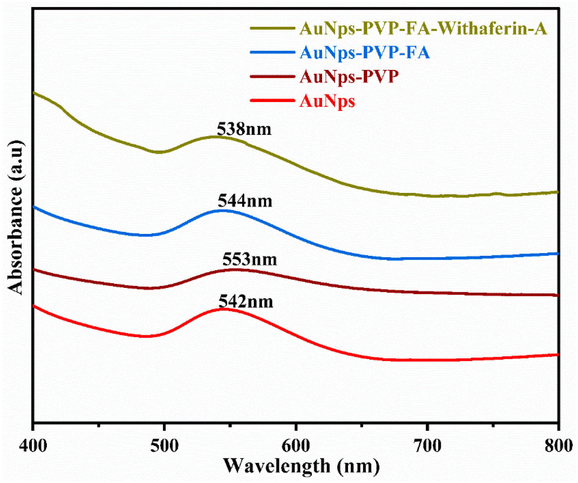
3.3. UV–Visible Spectroscopy Analysis

The prepared AuNPs–PVP–FA with and without Withaferin A drug was analyzed using a UV–visible spectrophotometer. Figure 4 shows a typical surface plasmon resonance (SPR) absorption peak at 546 nm for the AuNPs (100 mg/mL) alone. There was a shift in absorption peak to 553 nm for AuNPs–PVP, indicating that PVP had coated on the surface of phyto-fabricated AuNPs and increased in the particle size of AuNPs with PVP coating following conjugation (Figure 4). The modification of folic acid on AuNPs–PVP shifted the SPR peak toward 544 nm in AuNPs–PVP–FA, which corresponds to the π-π* transition of the C–C bond of FA. Withaferin A with the AuNPs–PVP–FA nanoconjugate showed the SPR band at 538 nm due to the active surface functionalized with FA and Withaferin A loaded onto AuNPs–PVP–FA, which is in accordance with previous reports [31,32].

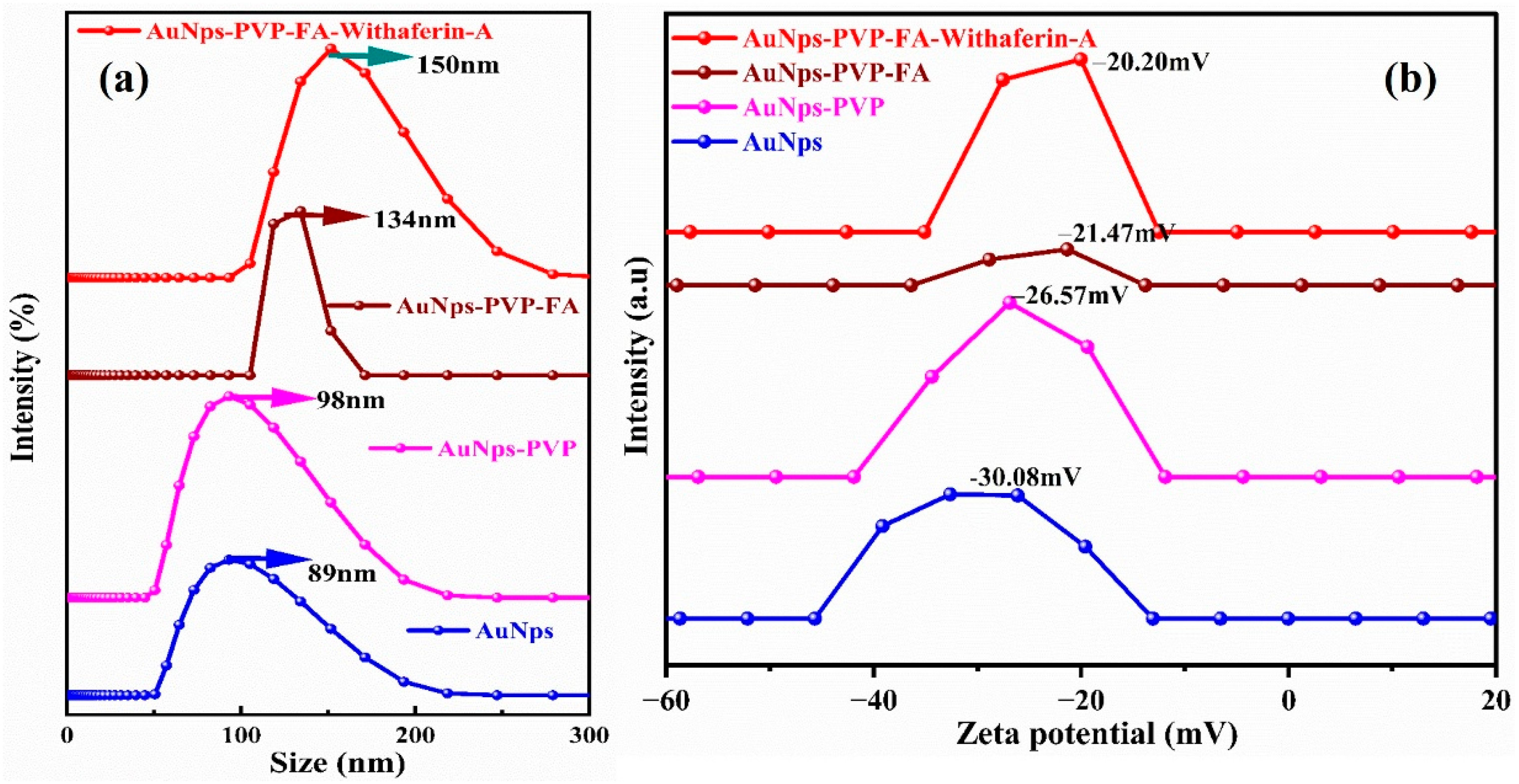
3.4. Hydrodynamic Particle Size and Surface Charge Analysis

The dynamic light scattering (DLS) results disclose that the typical hydrodynamic particle sizes of AuNPs and AuNPs–PVP are 89 nm and 98 nm, with PDI values of 0.3 and 0.5, respectively, indicating a high monodispersed index. The partial cross-linking between the AuNPs and PVP particles with FA during conjugation is responsible for an increase in the hydrodynamic size of Au–PVP–FA with 134 nm (PDI 0.12) compared with AuNPs and AuNPs–PVP, as shown in Figure 5. On the other hand, the hydrodynamic diameter of Au–PVP–FA grafted with Withaferin A is 150 nm (PDI 0.15) due to an increase in shell volume with the integration of AuNPs, PVP capping, and FA, responsible for the notable rise in the particle sizes of the Au–PVP–FA–Withaferin A nanoconjugate. The zeta potential of AuNPs is −30.08 mV, which shifted significantly to −26.57 mV after PVP coating in AuNPs–PVP. AuNPs–PVP–FA–Withaferin A is effectively loaded with hydrophobic oxygen-rich Withaferin A and FA owing to the hydrophilic interaction between PVP and hydrophilic polymer, which stabilizes AuNPs–PVP. The addition of partially negatively charged NHS–folate to AuNPs–PVP reduced the negative potential value of AuNPs–PVP–FA. The reduced negative potential value of the AuNPs–PVP–FA-loaded Withaferin A nanoconjugate is suitable for long-term storage without particle aggregation. Previous studies have shown that particles with a positive charge facilitate opsonization system clearance. Consequently, for medication delivery at locations other than the reticuloendothelial system, a negative zeta potential of approximately −10 to −30 mV is appropriate [33,34]. The greater negative charge of the nanoconjugate on the nanoparticle’s surface results in the high attachment rate of cells and other components.

3.5. Morphological Analysis of AuNPs–PVP–FA with Withaferin A Drug

Figure 6a shows HR-TEM images of AuNPs–PVP–FA with Withaferin A that are spherically shaped similar to AuNPs [22] even with capping and conjugation of drugs, where the structure of AuNPs remains the same. The elemental distribution revealed the presence of Au, Na, carbon, and oxygen, as shown in Figure 6b. The SAED pattern (Figure 6c) shows nanoparticle formation, and the particle size distribution graph (Figure 6d) of AuNPs–PVP–FA–Withaferin A shows an average particle size of 38 nm. Earlier reports of biogenic AuNPs synthesized from the fruit extract [35] and seed oil [36] were also spherically shaped.

3.6. Drug Loading and Drug Release Studies

The drug loading efficiency of the AuNPs–PVP–FA–Withaferin A nanoconjugate at different pH (5, 6, 7), concentrations and the amount of drug loaded are shown in Table 1. Figure 7 shows that the amount of drug loading (Figure 7a,b) relies on the drug and drug carrier. The drug loading efficiency is increased due to the equal amount of PVP and AuNPs (1:1) at pH 5. The drug loading results show that the concentration of AuNPs and the amount of drug play a major role in drug loading.

3.7. Drug Release Study (In Vitro)

The Withaferin A drug released from the AuNPs–PVP–FA–Withaferin A nanoconjugate has been investigated at different pH’s. Three distinct pH values were used for the experiments: pH 5—the estimated pH of lysosome/endosomes; pH 6—the pH of the surrounding tissue around the tumor; and pH 7—the pH of biological blood. The drug molecules’ release was dependent on the pH medium and duration of drug release. The release of the drug was increased gradually and then maintained at pH 7 and pH 6, with release ratios of 53 ± 70% (pH 7) and 48 ± 83% (pH 6) in 24 h. However, at a lower pH of pH 5, the release rate of Withaferin A was 58 ± 92% and was significantly faster. The lower pH levels led the bioactive compound to protonate, delivering chemisorbed drug molecules into the medium. Moreover, at lower pH, the surface charge of AuNPs–PVP–FA–Withaferin A turned positive, which pointed to the drug’s electrostatic interaction with AuNPs–PVP–FA and aided in the drug’s release. This finding shows that Withaferin A exhibits characteristics of pH-stimuli drug release.
The majority of the drug may stay in the carrier for an extended amount of time (pH 7.4) at usual biological circumstances, suggesting the possibility of a longer exposure of Withaferin A in the body fluid and a significant decrease in the adverse effects on normal tissues. Conversely, a lower pH may cause a faster release, such as around the tumor site or within the tumor cells’ endosome and lysosome. The absorption of drug-loaded AuNPs–PVP–FA–Withaferin A by tumor cells by the process of endocytosis would result in a significant improvement in the efficacy of cancer treatment, making drug release suitable for the anticancer drug delivery system. The drug release was confirmed from the kinetics models of cumulative drug release analyzed using KinetDS3 software. The results of release models’ respective to R2 values are followed for the pH 5.0 Kors Myer–peppers R2 = 0.9906, zero-order reaction R2 = 0.9561, first-order R2 = 0.9657, and Hickson cross well R2 = 0.9812; for the pH 6.0 Kors Myer –peppers R2 = 0.9897, zero-order reaction R2 = 0.9849, first-order R2 = 0.8997, and Hickson cross well R2 = 0.9371; and for the pH 7.0 Kors Myer–peppers R2 = 0.9234, zero-order reaction R2 = 0.9449, first-order R2 = 0.9211, and Hickson cross well R2 = 0.8852. These findings correlated with the previous reports (Table 2) [37,38].

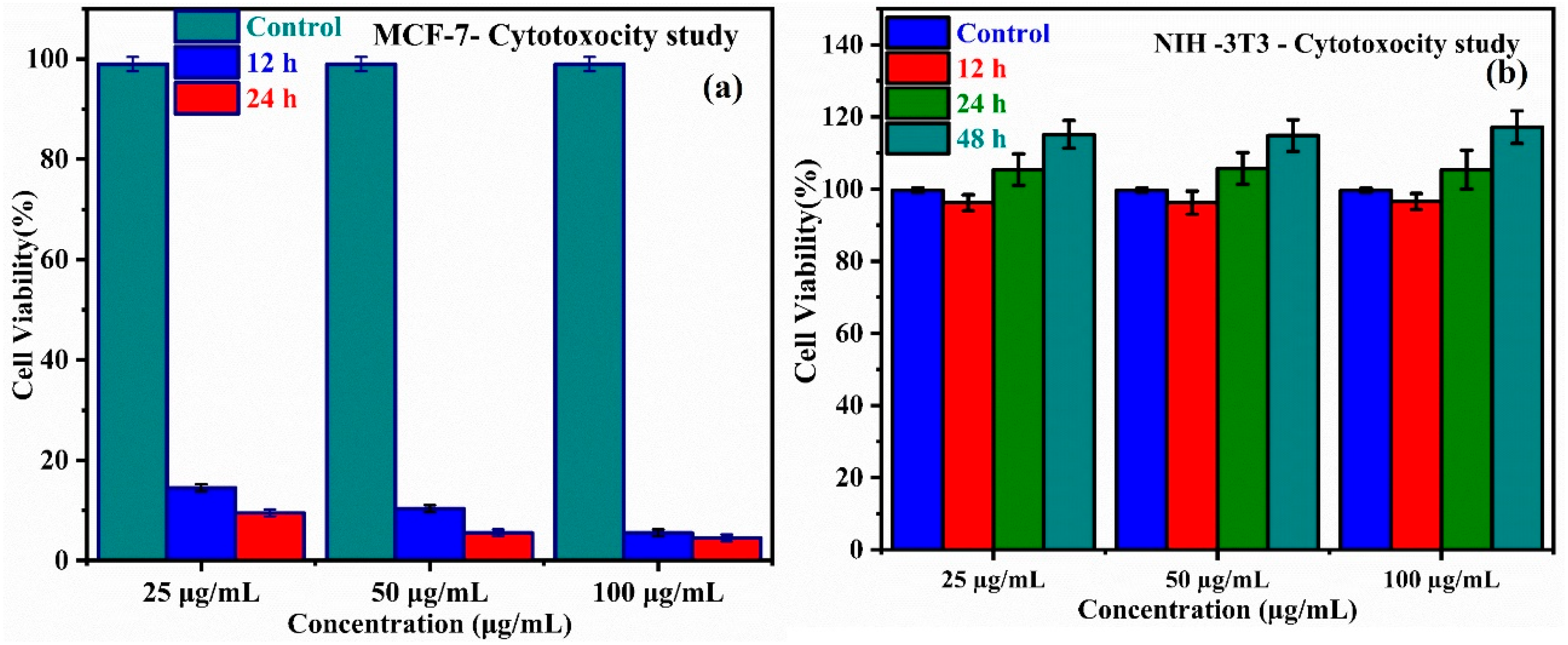
3.8. In Vitro Cytotoxicity and Cell Viability

Among the most crucial factors for a toxicological investigation of the response of cells to harmful substances is the cell viability assay, which may provide knowledge regarding cell survival, cell death, and inhibited metabolic functions. MCF-7 (cancer cell) and NIH-3T3 fibroblast cells (normal cell) were treated with AuNPs–PVP–FA–Withaferin A ranging from 25 to 100 µg/mL, and cytotoxicity was assessed to investigate the efficacy of Withaferin A drug delivery using the AuNPs–PVP–FA–Withaferin A nanoconjugate as the carrier. The results indicate that the drug concentrations between 70 and 100 µg/mL had a considerably greater effect on apoptosis than lower concentrations (20 and 40 µg/mL). This suggests that MCF-7 breast cancer cells are subjected to a dose-dependent cytotoxic impact. The cytotoxicity analysis suggests that AuNPs–PVP–FA–Withaferin A may facilitate the induction of apoptosis in MCF-7 cells. Additionally, the results of NIH-3T3 fibroblast cell line treatment do not show any cytotoxic effect at 48 h. For MCF-7 cells, the IC50 value is 20.86 ± 22.11 µg/mL, and for NIH-3T3 fibroblast cells, an IC50 value of 99.65 ± 101.56 µg/mL, which concurs with other reports (Figure 8) [39,40].

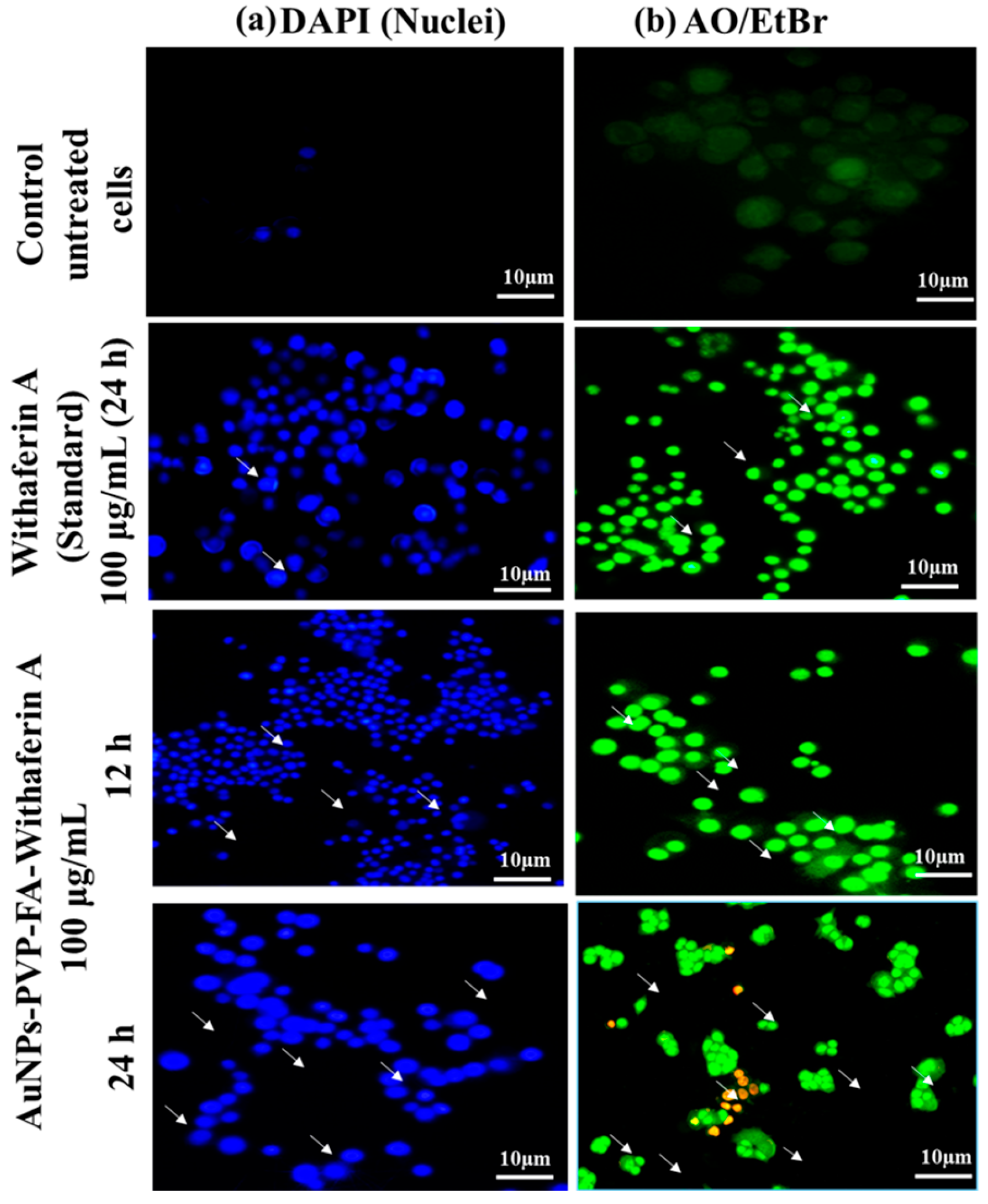
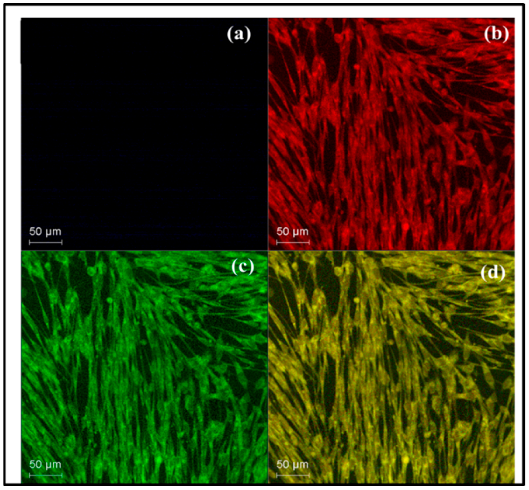
3.9. Morphological Observations of Cell Viability

The morphological changes of MCF -7 cells after treatment with the Withaferin A drug are shown in Figure 9. After treating MCF-7 cells with AuNPs–PVP–FA–Withaferin A, the induction of apoptosis was examined and the cells were stained with AO/EtBr. The most obvious morphological alterations seen in the treated cells consisted of cytoplasmic condensation, cell shrinkage, creation of cell surface protuberances at the plasma membrane, and aggregation of nuclear chromatin into dense masses beneath the nuclear membrane. Nuclear shrinkage and blabbing resulted in the formation of apoptotic cells and apoptotic bodies, which were identified as orange-colored entities. Conversely, necrotic cells lost their membrane integrity, which is why they fluoresced red when examined under a fluorescent microscope. On the other hand, the NIH-3T3 fibroblast cells treated with AuNPs–PVP–FA–Withaferin A did not exhibit any considerable morphological changes, as shown in Figure 10.
The nuclear counterstain DAPI was introduced to observe the nuclear fragmentation in MCF 7 cells with induced the AuNPs–PVP–FA–Withaferin A nanoconjugate. It was observed that common apoptotic changes of nucleus fragmentation, chromatin condensation, and the formation of apoptotic bodies were found in the nuclear morphology analysis of MCF-7 cells. It is relevant to mention that similar research has demonstrated the same results of nuclear fragmentation [41]. Shah et al. 2020 [42] validated that the nanocarrier-based delivery of the Withaferin A drug is appropriate for cancer therapy. The NIH-3T3 cells do not show any changes in the nuclear fragmentation after 48 h.
The nuclear and AO/EtBr staining results showed alterations in cytoskeletal structures and interactions with other cells as a potential cause of changes in the MCF-7 cellular shape. The morphological changes in MCF-7 breast cancer cells were visible because actin strands provide a fundamental component for preserving cell structure and support processes like adhesion and motility early apoptosis (Figure 9a,b). The NIH-3T3 cell results reveal the development of fibroblast cells after 48 h. The nanoconjugate of Withaferin A does not show cytotoxicity to the fibroblast cells due to its dynamic composition and acts as support and as a nutrition source, as shown in Figure 10. Prior research involving AuNPs and noisomes [43,44] substantiates the cytotoxic effects of the nanoconjugate, although biogenic AuNPs derived from plants [45,46] showed significant anticancer characteristics and comparable apoptotic effects.

3.10. Western Blotting Analysis

Western blotting is an effective method for investigating the effects of AuNPs–PVP–FA–Withaferin A on pro- and anti-apoptotic protein expression levels. Figure 11 represents the presence of pro- and anti-apoptotic proteins of Bax, p53, caspase-3, and β-actin at different concentrations of nanoconjugate. To figure it out, the apoptosis (cell death) was induced via intrinsic or extrinsic pathways due to mitochondrial toxicity generated by AuNPs–PVP–FA with Withaferin A or inside the mitochondria via the activation of Bax, p53, caspase-3, and β-actin proteins. The expression of these proteins increases statistically with the increasing concentration of AuNPs–PVP–FA–Withaferin A.
The Bax protein family of pro-apoptotic Bcl-2 proteins regulates apoptosis in both normal and cancerous cells, making it a crucial regulator of cell death and an essential entry point for mitochondrial malfunction [47,48]. Similarly, the G-action pool, cell migration, and cell proliferation are all directly regulated by β-actin. Cysteine-aspartic acid proteases are also referred to as caspases, and they are well known for being essential to both the start and completion of apoptosis. Breaking down cellular DNA requires an activation of caspase-3. The MCF-7 cells treated with the AuNPs–PVP–FA–Withaferin A drug show that caspase-3, similarly p53, was overexpressed (Figure 11). Caspases are essential participants in the process of programmed cell death, or apoptosis. Among these, caspase-3 effectively cleaves several vital cellular proteins once it is activated. Increased caspase-3 activity is commonly interpreted as an apoptotic signal and an indication of apoptosis. The tumor suppressor gene p53 is found in the cell nucleus and is essential for regulating both cell division and death. In about 50% of all cases of human cancer and nearly all forms of disease, the most often changed gene in malignancies is the p53 gene, which affects humans. The expression of these genes is the reason for the cell death of MCF-7 breast cancer cells and was supported by previous results [49]. The nanoconjugate appears to induce apoptosis through an intrinsic (mitochondrial) pathway. Preliminary mechanistic investigations suggest that treatment with the nanoconjugate results in mitochondrial membrane depolarization, leading to the release of cytochrome c into the cytosol. This event subsequently activates caspase and the downstream effector caspase-3, indicating the involvement of the intrinsic apoptotic pathway, resulting in programmed cell death (apoptosis). Caspase-3 cleaves key cellular proteins, leading to cell death. Additionally, the upregulation of pro-apoptotic proteins such as Bax further supports mitochondria-mediated apoptosis. Furthermore, it increased the expression of a gene (p53) point to a concurrent involvement of the apoptosis. The activation of these pathways suggests that the nanoconjugate exerts a robust pro-apoptotic effect, possibly through enhanced cellular uptake, improved intracellular trafficking, and sustained release of the therapeutic payload towards the apoptosis [50].

4. Conclusions

This study highlights the development of a AuNPs–PVP–FA–Withaferin A nanoconjugate as an efficient drug delivery system for cancer therapy. Compared with free Withaferin A, the nanoconjugate exhibited improved targeting capability, sustained drug release, and enhanced cytotoxic effects on MCF-7 breast cancer cells. The AuNP-based nanocarrier displayed a spherical morphology, a zeta potential of −20.20 mV, a polydispersity index (PDI) of 0.4, and a hydrodynamic size of 150 nm, as characterized by dynamic light scattering (DLS). The pH-responsive drug release profile showed sustained and controlled Withaferin A release over 24 h, with the highest release observed at pH 5, mimicking the tumor microenvironment. Cellular assays, including MTT and Western blot analysis, confirmed that the folate-modified nanoconjugate effectively inhibited cancer cell viability and migration while demonstrating biocompatibility in NIH-3T3 fibroblast cells over 48 h. These findings suggest that the AuNPs–PVP–FA–Withaferin A nanoconjugate enhances the therapeutic efficacy of Withaferin A through folate receptor-mediated targeting and controlled drug release. Therefore, this nanocarrier system holds promise for advancing Withaferin A-based cancer therapy with minimal toxicity to normal cells.

Supplementary Materials

The following supporting information can be downloaded at: https://www.mdpi.com/article/10.3390/pr13051290/s1, Table S1: Wavenumbers and Surface functional groups.

Author Contributions

V.S.—conceptualization, methodology, software, validation, analysis, investigation, resources, data curation, writing—original draft, and writing—review and editing; A.S.—conceptualization, methodology, validation, formal analysis, investigation, data curation, writing—original draft, writing—review and editing, visualization, and supervision; P.A.—formal analysis, data curation, writing—original draft, writing—review and editing, and supervision. All authors have read and agreed to the published version of the manuscript.

Funding

This research received no external funding.

Data Availability Statement

The raw data supporting the conclusions of this article will be made available by the authors on request.

Acknowledgments

The authors acknowledge the MHRD-RUSA 2.0 project (C3/RI&QI/RUSA2.0/Theme/Group-2/Award/2021/034, dated 3 February 2021 and NCNSNT-DST-FIST for the XRD analysis.

Conflicts of Interest

The authors declare no conflicts of interest.

Abbreviations

The following abbreviations are used in this manuscript:
AuNPsgold nanoparticles
PVPpolyvinylpyrrolidone
FAfolic acid
DLEfrug loading efficiency
DLSdynamic light scattering
ZETAZero Energy Thermonuclear Analysis
HPLChigh-performance liquid chromatography
FTIRFourier-transform infrared spectroscopy
NCnanoconjugate
TEMtransmission electron microscopy
SEMscanning electron microscopy
UVultraviolet
SPRsurface plasmon resonance
DAPI-406-diamidino-2-phenylindole
TMB3,3′,5,5′-tetramethylbenzidine
Ao/EtBracridine orange/ethidium bromide
MTT3-(4,5-dimethylthiazol-2-yl)-2,5-diphenyl-2H-tetrazolium bromide
IC50half-maximum inhibitory concentration
CCK8Cell Counting Kit 8
FA–Withaferin A AuNPs–PVP NCsfolic acid–Withaferin A–gold nanoparticles–polyvinylpyrrolidone nanoconjugates

References

  1. Ortiz-Casas, B.; Galdámez-Martínez, A.; Gutiérrez-Flores, J.; Ibañez, A.B.; Panda, P.K.; Santana, G.; de la Vega, H.A.; Suar, M.; Rodelo, C.G.; Kaushik, A.; et al. Bio-acceptable 0D and 1D ZnO nanostructures for cancer diagnostics and treatment. Mater. Today 2021, 50, 533–569. [Google Scholar] [CrossRef]
  2. Siegel, R.L.; Kratzer, T.B.; Giaquinto, A.N.; Sung, H.; Jemal, A. Cancer statistics, 2025. CA Cancer J. Clin. 2025, 75, 10–45. [Google Scholar] [CrossRef] [PubMed]
  3. Rawla, P. Epidemiology of Prostate Cancer. World J. Oncol. 2019, 10, 63–89. [Google Scholar] [CrossRef] [PubMed]
  4. Halder, T.; Ghosh, B. Cytological, genetical and phytochemically stable meta-Topolin (mT)-induced mass propagation of underutilized Physalis minima L. for production of Withaferin A. Biocatal. Agric. Biotechnol. 2021, 33, 102012. [Google Scholar] [CrossRef]
  5. Kaur, G.; Chauhan, K.; Anand, N.; Kaur, S. Evaluation of In Vitro and In Vivo Protective Efficacy of Bauhinia variegata Against Leishmania donovani in Murine Model. Acta Parasitol. 2021, 66, 812–826. [Google Scholar] [CrossRef]
  6. Behl, T.; Sharma, A.; Sharma, L.; Sehgal, A.; Zengin, G.; Brata, R.; Fratila, O.; Bungau, S. Exploring the multifaceted therapeutic potential of Withaferin A and its derivatives. Biomedicines 2020, 8, 571. [Google Scholar] [CrossRef]
  7. Misra, L.N.; Lal, P.; Kumar, D. Isolation and Characterization of Steroids of Nutraceutical Value in Physalis minima. Prev. Nutr. Food Sci. 2006, 11, 133–139. [Google Scholar] [CrossRef]
  8. Sivasankarapillai, V.S.; Nair, R.M.K.; Rahdar, A.; Bungau, S.; Zaha, D.C.; Aleya, L.; Tit, D.M. Overview of the anticancer activity of Withaferin A, an active constituent of the Indian ginseng Withania somnifera. Environ. Sci. Pollut. Res. 2020, 27, 26025–26035. [Google Scholar] [CrossRef]
  9. Hahm, E.; Lee, J.; Singh, S.V. Role of mitogen-activated protein kinases and Mcl-1 in apoptosis induction by Withaferin A in human breast cancer cells. Mol. Carcinog. 2013, 53, 907–916. [Google Scholar] [CrossRef]
  10. Huang, M.; He, J.-X.; Hu, H.-X.; Zhang, K.; Wang, X.-N.; Zhao, B.-B.; Lou, H.-X.; Ren, D.-M.; Shen, T. Withanolides from the genus Physalis: A review on their phytochemical and pharmacological aspects. J. Pharm. Pharmacol. 2019, 72, 649–669. [Google Scholar] [CrossRef]
  11. Meng, Q.; Fan, J.; Liu, Z.; Li, X.; Zhang, F.; Zhang, Y.; Sun, Y.; Li, L.; Liu, X.; Hua, E. Cytotoxic Withanolides from the Whole Herb of Physalis angulata L. Molecules 2019, 24, 1608. [Google Scholar] [CrossRef] [PubMed]
  12. Lavie, D.; Glotter, E.; Shvo, Y. 1371. Constituents of Withania somnifera Dun. Part IV. The structure of Withaferin A. J. Chem. Soc. 1965, 1371–7517. [Google Scholar] [CrossRef]
  13. Dutta, R.; Khalil, R.; Green, R.; Mohapatra, S.S.; Mohapatra, S. Withania somnifera (Ashwagandha) and Withaferin A: Potential in Integrative Oncology. Int. J. Mol. Sci. 2019, 20, 5310. [Google Scholar] [CrossRef]
  14. Berghe, W.V.; Sabbe, L.; Kaileh, M.; Haegeman, G.; Heyninck, K. Molecular insight in the multifunctional activities of Withaferin A. Biochem. Pharmacol. 2012, 84, 1282–1291. [Google Scholar] [CrossRef]
  15. Hosny, M.; Eltaweil, A.S.; Mostafa, M.; El-Badry, Y.A.; Hussein, E.E.; Omer, A.M.; Fawzy, M. Facile synthesis of gold nanoparticles for anticancer, antioxidant applications, and photocatalytic degradation of toxic organic pollutants. ACS Omega 2022, 7, 3121–3133. [Google Scholar] [CrossRef]
  16. Sharma, J.R.; Sibuyi, N.R.S.; Fadaka, A.O.; Meyer, S.; Madiehe, A.M.; Katti, K.; Meyer, M. Anticancer and Drug-Sensitizing Activities of Gold Nanoparticles Synthesized from Cyclopia genistoides (Honey Bush) Extracts. Appl. Sci. 2023, 13, 3973. [Google Scholar] [CrossRef]
  17. Tagde, P.; Kulkarni, G.T.; Mishra, D.K.; Kesharwani, P. Recent advances in folic acid engineered nanocarriers for treatment of breast cancer. J. Drug Deliv. Sci. Technol. 2020, 56, 101613. [Google Scholar] [CrossRef]
  18. Sargazi, S.; Laraib, U.; Er, S.; Rahdar, A.; Hassanisaadi, M.; Zafar, M.N.; Díez-Pascual, A.M.; Bilal, M. Application of green gold nanoparticles in cancer therapy and diagnosis. Nanomaterials 2022, 12, 1102. [Google Scholar] [CrossRef]
  19. Lee, Y.J.; Kim, Y.-J.; Park, Y. Folic acid and chitosan-functionalized gold nanorods and triangular silver nanoplates for the delivery of anticancer agents. Int. J. Nanomed. 2022, 17, 1881–1902. [Google Scholar] [CrossRef]
  20. Manju, S.; Sreenivasan, K. Enhanced drug loading on magnetic nanoparticles by layer-by-layer assembly using drug conjugates: Blood compatibility evaluation and targeted drug delivery in cancer cells. Langmuir 2011, 27, 14489–14496. [Google Scholar] [CrossRef]
  21. Gajendiran, M.; Jo, H.; Kim, K.; Balasubramanian, S. Green synthesis of multifunctional PEG-carboxylate π back-bonded gold nanoconjugates for breast cancer treatment. Int. J. Nanomed. 2019, 14, 819–834. [Google Scholar] [CrossRef] [PubMed]
  22. Sekar, V.; Al-Ansari, M.M.; Narenkumar, J.; Al-Humaid, L.; Arunkumar, P.; Santhanam, A. Synthesis of gold nanoparticles (AuNPs) with improved anti-diabetic, antioxidant and anti-microbial activity from Physalis minima. J. King Saud Univ. Sci. 2022, 34, 102197. [Google Scholar] [CrossRef]
  23. Pechyen, C.; Ponsanti, K.; Tangnorawich, B.; Ngernyuang, N. Biogenic synthesis of gold nanoparticles mediated by Spondias dulcis (Anacardiaceae) peel extract and its cytotoxic activity in human breast cancer cell. Toxicol. Rep. 2022, 9, 1092–1098. [Google Scholar] [CrossRef]
  24. Sangwan, R.S.; Chaurasiya, N.D.; Lal, P.; Misra, L.; Tuli, R.; Sangwan, N.S. Withanolide A is inherently de novo biosynthesized in roots of the medicinal plant Ashwagandha (Withania somnifera). Physiol. Plant. 2008, 133, 278–287. [Google Scholar] [CrossRef]
  25. Anadozie, S.O.; Adewale, O.B.; Sibuyi, N.R.S.; Fadaka, A.O.; Isitua, C.C.; Davids, H.; Roux, S. One-pot synthesis, characterization and biological activities of gold nanoparticles prepared using aqueous seed extract of Garcinia kola. Process Biochem. 2023, 128, 49–57. [Google Scholar] [CrossRef]
  26. Mosmann, T. Rapid colorimetric assay for cellular growth and survival: Application to proliferation and cytotoxicity assays. J. Immunol. Methods 1983, 65, 55–63. [Google Scholar] [CrossRef]
  27. Bass, J.J.; Wilkinson, D.J.; Rankin, D.; Phillips, B.E.; Szewczyk, N.J.; Smith, K.; Atherton, P.J. An overview of technical considerations for Western blotting applications to physiological research. Scand. J. Med. Sci. Sports 2016, 27, 4–25. [Google Scholar] [CrossRef]
  28. Nile, S.H.; Nile, A.; Gansukh, E.; Baskar, V.; Kai, G. Subcritical water extraction of withanosides and withanolides from ashwagandha (Withania somnifera L.) and their biological activities. Food Chem. Toxicol. 2019, 132, 110659. [Google Scholar] [CrossRef]
  29. Binupriya, A.R.; Sathishkumar, M.; Vijayaraghavan, K.; Yun, S. Bioreduction of trivalent aurum to nano-crystalline gold particles by active and inactive cells and cell-free extract of Aspergillus oryzae var. viridis. J. Hazard. Mater. 2009, 177, 539–545. [Google Scholar] [CrossRef]
  30. Hira, I.; Kumari, R.; Saini, A.K.; Gullilat, H.; Saini, V.; Kumar, A.; Saini, R. Apoptotic cell death induction through pectin, guar gum and zinc oxide nanocomposite in A549 lung adenocarcinomas. Biointerface Res. Appl. Chem. 2021, 12, 1856–1869. [Google Scholar] [CrossRef]
  31. Mathew, M.S.; Vinod, K.; Jayaram, P.S.; Jayasree, R.S.; Joseph, K. Improved bioavailability of curcumin in gliadin-protected gold quantum cluster for targeted delivery. ACS Omega 2019, 4, 14169–14178. [Google Scholar] [CrossRef] [PubMed]
  32. Dey, S.; Sreenivasan, K. Conjugating curcumin to water soluble polymer stabilized gold nanoparticles via pH responsive succinate linker. J. Mater. Chem. B 2015, 3, 824–833. [Google Scholar] [CrossRef] [PubMed]
  33. Al-Ansari, M.M.; Al-Dahmash, N.D.; Ranjitsingh, A.J.A. Synthesis of silver nanoparticles using gum Arabic: Evaluation of its inhibitory action on Streptococcus mutans causing dental caries and endocarditis. J. Infect. Public Health 2021, 14, 324–330. [Google Scholar] [CrossRef] [PubMed]
  34. Wang, L.; Xu, J.; Yan, Y.; Liu, H.; Karunakaran, T.; Li, F. Green synthesis of gold nanoparticles from Scutellaria barbata and its anticancer activity in pancreatic cancer cell (PANC-1). Artif. Cells Nanomed. Biotechnol. 2019, 47, 1617–1627. [Google Scholar] [CrossRef]
  35. Al-Radadi, N.S. Biogenic proficient synthesis of (Au-NPs) via aqueous extract of Red Dragon Pulp and seed oil: Characterization, antioxidant, cytotoxic properties, anti-diabetic anti-inflammatory, anti-Alzheimer and their anti-proliferative potential against cancer cell lines. Saudi J. Biol. Sci. 2022, 29, 2836–2855. [Google Scholar] [CrossRef]
  36. Hejazi, M.; Arshadi, S.; Amini, M.; Baradaran, B.; Shahbazi-Derakhshi, P.; Sameti, P.; Soleymani, J.; Mokhtarzadeh, A.; Tavangar, S.M. Hyaluronic acid-functionalized gold nanoparticles as a cancer diagnostic probe for targeted bioimaging applications. Microchem. J. 2023, 193, 108953. [Google Scholar] [CrossRef]
  37. Vijayan, R.; Joseph, S.; Mathew, B. Indigofera tinctoria leaf extract mediated green synthesis of silver and gold nanoparticles and assessment of their anticancer, antimicrobial, antioxidant and catalytic properties. Artif. Cells Nanomed. Biotechnol. 2017, 46, 861–871. [Google Scholar] [CrossRef]
  38. Velidandi, A.; Pabbathi, N.P.P.; Dahariya, S.; Baadhe, R.R. Catalytic and eco-toxicity investigations of bio-fabricated monometallic nanoparticles along with their anti-bacterial, anti-inflammatory, anti-diabetic, anti-oxidative and anti-cancer potentials. Colloids Interface Sci. Commun. 2020, 38, 100302. [Google Scholar] [CrossRef]
  39. Nosrati, H.; Seidi, F.; Hosseinmirzaei, A.; Mousazadeh, N.; Mohammadi, A.; Ghaffarlou, M.; Danafar, H.; Conde, J.; Sharafi, A. Prodrug polymeric nanoconjugates encapsulating gold nanoparticles for enhanced X-ray radiation therapy in breast cancer. Adv. Healthc. Mater. 2022, 11, 2270014. [Google Scholar] [CrossRef]
  40. Uzma, M.; Sunayana, N.; Raghavendra, V.B.; Madhu, C.S.; Shanmuganathan, R.; Brindhadevi, K. Biogenic synthesis of gold nanoparticles using Commiphora wightii and their cytotoxic effects on breast cancer cell line (MCF-7). Process Biochem. 2020, 92, 269–276. [Google Scholar] [CrossRef]
  41. Lara-Cruz, C.; Jiménez-Salazar, J.E.; Arteaga, M.; Arredondo, M.; Ramón-Gallegos, E.; Batina, N.; Damián-Matsumura, P. Gold nanoparticle uptake is enhanced by estradiol in MCF-7 breast cancer cells. Int. J. Nanomed. 2019, 14, 2705–2718. [Google Scholar] [CrossRef] [PubMed]
  42. Shah, H.S.; Usman, F.; Ashfaq, M.; Khalil, R.; Ul-Haq, Z.; Mushtaq, A.; Qaiser, R.; Iqbal, J. Preparation and characterization of anticancer niosomal withaferin–A formulation for improved delivery to cancer cells: In vitro, in vivo, and in silico evaluation. J. Drug Deliv. Sci. Technol. 2020, 59, 101863. [Google Scholar] [CrossRef]
  43. Amale, F.R.; Ferdowsian, S.; Hajrasouliha, S.; Kazempoor, R.; Mirzaie, A.; Dakkali, M.S.; Akbarzadeh, I.; Meybodi, S.M.; Mirghafouri, M. Gold nanoparticles loaded into niosomes: A novel approach for enhanced antitumor activity against human ovarian cancer. Adv. Powder Technol. 2021, 32, 4711–4722. [Google Scholar] [CrossRef]
  44. Ayyanaar, S.; Kesavan, M.P. One-pot biogenic synthesis of gold nanoparticles@saponins niosomes: Sustainable nanomedicine for antibacterial, anti-inflammatory and anticancer therapeutics. Colloids Surf. A Physicochem. Eng. Aspects 2023, 676, 132229. [Google Scholar] [CrossRef]
  45. Ke, Y.; Aboody, M.S.A.; Alturaiki, W.; Alsagaby, S.A.; Alfaiz, F.A.; Veeraraghavan, V.P.; Mickymaray, S. Photosynthesized gold nanoparticles from Catharanthus roseus induces caspase-mediated apoptosis in cervical cancer cells (HeLa). Artif. Cells Nanomed. Biotechnol. 2019, 47, 1938–1946. [Google Scholar] [CrossRef]
  46. Naraginti, S.; Li, Y. Preliminary investigation of catalytic, antioxidant, anticancer and bactericidal activity of green synthesized silver and gold nanoparticles using Actinidia deliciosa. J. Photochem. Photobiol. B Biol. 2017, 170, 225–234. [Google Scholar] [CrossRef]
  47. Velmathi, G.; Sekar, V.; Kavitha, N.S.; Albeshr, M.F.; Santhanam, A. Biosynthesis of gold nanoparticles by the extremophile bacterium Deinococcus radiodurans and an evaluation of its application in drug delivery. Process Biochem. 2024, 145, 250–260. [Google Scholar] [CrossRef]
  48. Mobaraki, F.; Momeni, M.; Yazdi, M.E.T.; Meshkat, Z.; Toosi, M.S.; Hosseini, S.M. Plant-derived synthesis and characterization of gold nanoparticles: Investigation of its antioxidant and anticancer activity against human testicular embryonic carcinoma stem cells. Process Biochem. 2021, 111, 167–177. [Google Scholar] [CrossRef]
  49. Selim, M.E.; Hendi, A.A. Gold nanoparticles induce apoptosis in MCF-7 human breast cancer cells. Asian Pac. J. Cancer Prev. 2012, 13, 1617–1620. [Google Scholar] [CrossRef]
  50. Muthukumar, T.; Sudhakumari, N.; Sambandam, B.; Aravinthan, A.; Sastry, T.P.; Kim, J.-H. Green synthesis of gold nanoparticles and their enhanced synergistic antitumor activity using HepG2 and MCF7 cells and its antibacterial effects. Process Biochem. 2016, 51, 384–391. [Google Scholar] [CrossRef]
Figure 1. Synthesis of AuNPs–PVP–Withaferin A–FA nanoconjugate using the layer-by-layer method.
Processes 13 01290 g001
Figure 2. HPLC spectra of (A) standard drug and (B) Withaferin A extracted from Physalis minima plant.
Processes 13 01290 g002
Figure 3. (a) XRD of AuNPs–PVP–Withaferin A–FA and (b) FTIR spectra of AuNPs–PVP, AuNPs–PVP–FA, and AuNPs–PVP–Withaferin A–FA.
Processes 13 01290 g003
Figure 4. UV–visible spectra of AuNPs, AuNPs–PVP, AuNPs–PVP–FA, and AuNPs–PVP–FA with Withaferin A drug.
Processes 13 01290 g004
Figure 5. (a) Dynamic light scattering and (b) zeta potential results of AuNPs, AuNPs–PVP, AuNPs–PVP-FA, and AuNPs–PVP–FA with Withaferin A drug.
Processes 13 01290 g005
Figure 6. (a) HR-TEM, (b) EDS, (c) selected area electron diffraction (SAED) pattern, and (d) particle size distribution of AuNPs–PVP–FA loaded with Withaferin A.
Processes 13 01290 g006
Figure 7. (a,b) Drug loading results at pH 5 and (c) cumulative drug release results of AuNPs–PVP–FA with Withaferin A drug at different pH’s of 5, 6, and 7 (* represents that 1:1 shows high loading efficiency).
Processes 13 01290 g007
Figure 8. Cytotoxicity effect of the AuNPs–PVP–FA–Withaferin A nanoconjugate on the (a) MCF-7 cells and (b) NIH-3T3 fibroblast cells compared with control (untreated cells). The information is presented as mean ± standard deviation (n = 3).
Processes 13 01290 g008
Figure 9. Fluorescence images of (a) DAPI and (b) AO/EtBr. Non-treated (control) and treated with AuNPs–PVP–Withaferin A–FA on the MCF-7 breast cancer cells at different time durations.
Processes 13 01290 g009
Figure 10. Confocal images of AuNPs–PVP–FA–Withaferin A on NIH-3T3 cells with (a) DAPI, (b) RO, and (c,d) AO stains with 100 µg/mL for 48 h.
Processes 13 01290 g010
Figure 11. Expression of proteins in MCF-7 cells after treatment with AuNPs–PVP–FA–Withaferin A, (A) control, (B) 25 µg/mL, and (C) 100 µg/mL.
Processes 13 01290 g011
Table 1. Drug loading efficiency.
Drug Concentration (mg/mL)pH 5 (%)pH 6 (%)pH 7 (%)
1:11:21:41:11:21:41:11:21:4
10073.5264.7669.2362.0265.5168.1171.2569.9672.02
20079.4569.3271.5269.1171.4571.6572.5471.2374.53
30085.1475.1176.5975.0872.8974.2376.8974.1278.98
40090.3981.0384.9679.3279.8378.5978.6878.5881.45
50094.8682.4988.3281.2384.7881.9885.7881.0983.85
Table 2. Drug release models with R2 values.
Kinetics Release Models’ R2 ValuespH 5pH 6pH 7
Zero-order0.95610.98490.9449
First-order0.96570.94970.9211
Hickson cross well0.98120.93710.8852
Kors Myer–peppers0.99060.98970.9234
Disclaimer/Publisher’s Note: The statements, opinions and data contained in all publications are solely those of the individual author(s) and contributor(s) and not of MDPI and/or the editor(s). MDPI and/or the editor(s) disclaim responsibility for any injury to people or property resulting from any ideas, methods, instructions or products referred to in the content.

