Abstract
Olea europaea subsp. laperrinei, endemic to the central Sahara in Algeria, Niger, and Sudan, is recognized for its therapeutic and nutritional benefits, notably its antioxidant and antibacterial properties. This study investigates the phytochemical composition and biological activities of its leaf extracts obtained via aqueous and ethanolic extraction. Total phenols, flavonoids, and tannins were quantified by UV–visible spectrophotometry, while individual phenolic compounds were identified by HPLC-UV. Antioxidant capacity was measured using DPPH radical scavenging, hydrogen peroxide neutralization, and β-carotene bleaching assays. Antibacterial activity was evaluated against nine bacterial strains. The ethanolic extract exhibited higher total phenol (134.72 ± 9.25 mg GAE/g VP) and flavonoid (26.08 ± 1.57 mg QE/g VP) contents, while tannin levels were similar in both extracts. Twenty-seven phenolic compounds, including oleuropein, hydroxytyrosol, rutin, and quercetin, were identified. The ethanolic extract showed superior antioxidant capacity, with an EC50 of 7.48 ± 0.39 µg/mL (DPPH) and IC50 values of 214.04 ± 2.89 µg/mL (H2O2) and 185.63 ± 3.84 µg/mL (β-carotene). Both extracts exhibited antibacterial effects, with inhibition zones up to 23.33 ± 0.76 mm and MIC values as low as 0.78 mg/mL. These findings highlight the potential of Laperrine olive leaves as natural antioxidant and antibacterial agents.
1. Introduction
In traditional medicine, olive leaves are valued for their diuretic properties and efficacy in treating hypertension. These leaves are also used to combat fever and various tropical diseases, including malaria [1]. Extensive research on the Olea genus has revealed a wide range of biological activities. Olive leaves enhance the immune system, delay cellular aging, prevent cardiovascular diseases [2], and exhibit gastroprotective properties [3]. Their antiviral, antimicrobial [4], antileishmanial [5], antibacterial [6,7,8], and insecticidal [9,10,11,12] activities have been well-documented.
Further investigations, such as those conducted by Goldsmith et al. [13], Gutierrez-Venegar et al. [14], and Nediani et al. [15], highlight their antifungal, antidiabetic, hypoglycemic, anti-inflammatory, hypotensive, and anticancer properties. Goldsmith et al. and Ruzzolini et al. [16,17] revealed their selective action against cancer cells, notably without harming non-tumorigenic cells in breast and prostate cancers, and their potential to overcome the resistance of cancer cells to chemotherapy. Olive leaf biomolecules also address allergic and metabolic disorders and combat Alzheimer’s disease [18]. Oleuropein, a key component and a precursor to hydroxytyrosol [19], serves as a scavenger of reactive oxygen species [20], protecting against oxidative damage [21,22]. Additionally, olive leaves are used as forage for goats and sheep, and their inclusion in turkey feed has been shown to improve meat storage quality by reducing lipid oxidation, as observed in the studies by Djenane et al. [23] on turkey and camel meat [24].
Olea europaea subsp. laperrinei, an olive subspecies within the Oleaceae family, is endemic to the arid zones of central Algerian Sahara, including Hoggar, Tefedest, Tassili N’Ajjer, and Mouydir, predominantly in mountainous regions at altitudes up to 2800 m, where annual rainfall does not exceed 100 mm [25]. Recognized as part of the Olea europaea taxonomic complex, this subspecies is closely related to the Mediterranean olive tree and serves as a crucial genetic resource for its drought resistance, making it suitable as a rootstock for various olive tree varieties in arid regions [25].
As a sclerophyllous species, the olive exhibits remarkable adaptation to its environment, which includes dry and arid climates and significant human impact [26]. In response to these challenging conditions, olive populations enhance their defense mechanisms through chemical barriers, including the synthesis of polyphenols [27]. These natural molecules, which are widespread in plants, are recognized for their antioxidant, antibacterial [28], antiradical, and anti-inflammatory properties. Such characteristics are vital in preventing cancer and cardiovascular diseases [29].
The chemical composition analysis conducted by Lahcene et al. [9] on Olea europaea subsp. laperrinei leaf extracts obtained through acid extraction revealed the presence of oleuropein, hydroxytyrosol, tyrosol, caffeic acid, luteolin, apigenin, diosmetin, and quercetin. The study by Djenane et al. [24] revealed the presence of hydroxytyrosol, tyrosol, catechin, caffeic acid, rutin, luteolin-7-glucoside, verbascoside, apigenin-7-glucoside, diosmetin-7-glucoside, oleuropein, and luteolin-4-glucoside. These findings are further confirmed by the work of Bouchoucha et al. [30], who detected hydroxytyrosol, tyrosol, caffeic acid, rutin, luteolin-7-glucoside, verbascoside, quercetin, and oleuropein in Olea europaea subsp. laperrinei leaf extracts.
The in vitro evaluation of the anti-leishmanial effect of Olea europaea subsp. laperrinei leaf extracts against Leishmania infantum highlighted their efficacy in the promastigote form [5]. Moreover, the leaf extracts of Olea europaea subsp. laperrinei exhibit a dual insecticidal effect against the flour moth Ephestia kuehniella. They act both on reproductive parameters and on adult longevity [9].
This preliminary study aims to advance our understanding of the Olea europaea subsp. laperrinei and its potential as a natural source of antioxidants and antibacterial agents. It represents a significant step towards the comprehensive exploration and valorization of the Laperrine olive tree, emphasizing its unique contributions to traditional medicine and modern scientific research.
2. Materials and Methods
2.1. Plant Material
The leaves of Olea laperrinei used in this study were harvested in May 2022 from thirty mature trees at the Ouled Hanghassi station (23°14′50.1″ N, 5°29′13.7″ E) in the Tamanrasset region. The botanical identification was carried out using Ozenda’s flora by Ms. R. Sahki, a taxonomist affiliated with the National Institute of Forestry Research (Algeria). A voucher specimen (LABAB 0001-2022) has been preserved in the laboratory for reference. The leaves were left to air-dry in a shaded, moisture-free environment before being finely ground using an electric grinder (Siyo Lux Electro, Shanghai, China) (particle size: 0.5–1 mm) and kept at room temperature in the dark until further analysis.
2.2. Extraction Procedure
An amount of 20 g of plant powder was mixed with 200 mL of either distilled water or a 70:30 (v/v) distilled water–ethanol (Merck, Darmstadt, Germany) mixture. Each extraction was carried out for 24 h under continuous stirring (100 rpm) at room temperature, protected from light. The extracts were then filtered through Whatman No. 1 filter paper, and the resulting filtrates were freeze-dried using a CHRIST Alpha 1-2 lyophilizer (Martin Christ Gefriertrocknungsanlagen GmbH, Osterode am Harz, Germany). The lyophilized residues were kept at 4 °C.
2.3. Phytochemical Studies
2.3.1. Determination of Total Phenol Content
Each test tube contained 0.25 mL of EA and EE at different concentrations; 1.25 mL of the Folin–Ciocalteu reagent (Sigma-Aldrich, Darmstadt, Germany) diluted to 1/10 and 1 mL of sodium carbonate (75 g/L) chloride (Sigma-Aldrich, Germany) were added. The different solutions were then incubated at 40 °C for 30 min. The absorbance was subsequently measured at 765 nm using a MEDLINE MD 2000 UV-visible spectrophotometer (Medline Industries, Inc., Northfield, IL, USA) [31]. The total polyphenol content was determined by extrapolation on a gallic acid (Biochem Chemopharma, Cosne-Cours-sur-Loire, France) standard curve and expressed in milligrams of gallic acid equivalent per gram of vegetal powder (mg GAE/g VP).
2.3.2. Determination of Total Flavonoid Content
0.5 mL of each extract solution was mixed with 1.5 mL of methanol (Honeywell, Paris, France), 0.1 mL of aluminum chloride (10%) (Sigma-Aldrich, Germany), 0.1 mL of potassium acetate (1 M) (Sigma-Aldrich, Germany), and 2.8 mL of distilled water. The mixture was incubated for 30 min at room temperature. The reading was performed at 415 nm [32]. The standard curve was established with quercetin (Sigma-Aldrich, Germany). The linear regression equation was used to calculate the total flavonoid content, which was expressed in milligrams of quercetin equivalent per gram of vegetal powder (mg QE/g VP).
2.3.3. Determination of Total Tannin Content
Aliquots of 500 μL of each sample were incubated with 1 mL of bovine serum albumin (BSA) (Sigma-Aldrich, Germany) at a concentration of 1 mg/mL for 24 h at 4 °C. The precipitate obtained after centrifugation at 750× g at 4 °C for 15 min using a SIGMA centrifuge was dissolved in 2 mL of a surfactant solution containing 1% sodium dodecyl-sulfate (SDS) (Sigma-Aldrich, Germany) and 5% triethanolamine (TEA) (Sigma-Aldrich, Germany) and then supplemented with 500 μL of the FeCl3 reagent (Merck, Germany). After 15 min, the absorbance was measured at 510 nm [33].
The total tannin content, expressed in milligrams of tannic acid equivalent per gram of vegetal powder (mg TAE/g VP), was determined using the linear regression equation obtained from the tannic acid (Sigma-Aldrich, Germany) calibration curve.
2.3.4. HPLC-UV Analysis
The chemical composition of the samples was analyzed by high-performance liquid chromatography (HPLC). Quantification was performed using an HPLC system (Agilent 1100) (Agilent Technologies, Waldbronn, Germany; originally from the USA). equipped with a diode array detector (DAD), a quaternary pump, an automated injector, and an online degasser. Separation was performed using a Hypersil BDS-C18 column (5 µm particle size, 250 mm × 4.6 mm internal diameter) maintained at 30 °C. The eluent system comprised water acidified with 0.2% acetic acid (Merck, Germany) (pH 3.1) and acetonitrile (Merck, Germany), applied under a 30-min linear gradient at a flow rate of 1.5 mL/min, transitioning from 95% aqueous phase to 100% acetonitrile. A 5 µL aliquot of the methanol-dissolved extract (0.3 g/mL) was injected for analysis.
The percentage of each compound in the samples was calculated based on peak area measurements from chromatographic analysis. Five wavelengths (230, 255, 280, 300, and 355 nm) were used to detect polyphenols and identify the maximum number of components in a single injection. Each of these wavelengths is an average for the detection of a specific family of polyphenols. By operating under these conditions, the maximum number of families can be detected, providing a comprehensive analysis of the content of the analyzed samples. The phenolic constituents were identified by comparing their retention times with those of reference standards analyzed under identical conditions, supplemented by UV spectral data.
2.4. In Vitro Antioxidant Activities
2.4.1. Total Antioxidant Capacity
The test consists of mixing 0.1 mL of the plant extract at different concentrations (100 to 700 μg/mL) with 1 mL of the molybdate reagent (0.6 M sulfuric acid, 28 mM NaH2PO4, and 4 mM ammonium molybdate) (Merck, Germany). After incubation for 90 min at 95 °C and cooling the mixture to room temperature, the absorbance was read at 695 nm [34]. Ascorbic acid (Sigma-Aldrich, Gillingham, UK) was employed as the reference standard. TAC is expressed in milligrams of ascorbic acid equivalent per gram of extract (mg AAE/g of extract).
2.4.2. Ferric-Reducing Antioxidant Power Assay
Different concentrations of the extract (40 to 300 μg/mL) were prepared in distilled water and mixed with 2.5 mL of phosphate buffer (200 mM, pH 6.6) and 2.5 mL of potassium ferricyanide (K3Fe(CN)6) (1% w/v) (Sigma-Aldrich, Germany). After incubation for 20 min at 50 °C, 2.5 mL of trichloroacetic acid (10%) (Merck, Germany) was added and the mixture was centrifuged at 750× g for 10 min. 5 mL of the supernatant was mixed with an equal volume of distilled water and 1 mL of FeCl3 (0.1%). Absorbance readings were ultimately recorded at 700 nm, with ascorbic acid serving as the positive control under identical experimental conditions. FRAP is expressed in milligrams of ascorbic acid equivalent per gram of extract (mg AAE/g of extract) [35].
2.4.3. DPPH Radical Scavenging Assay
A volume of 3.75 mL of each extract (5 to 20 μg/mL) was combined with 0.25 mL of DPPH (0.8 mM) (Sigma-Aldrich, Germany). Following a 30-min incubation period at ambient temperature under light-protected conditions, absorbance values were recorded at 517 nm [36].
The free radical scavenging activity (%) was determined using the following equation:
Ac − As/Ab × 100;
Ac = Absorbance of the control;
As = Absorbance of the sample.
The antiradical activity is defined by two parameters:
- -
- Effective concentration (EC50), representing the antioxidant quantity required to reduce the initial DPPH radical concentration by half. A lower EC50 indicates stronger radical scavenging activity.
- -
- The antioxidant activity index (AAI) corresponds to the final concentration of DPPH in the control (μg/mL)/EC50 (μg/mL). The extracts have low antioxidant activity when AAI is <0.5, moderate antioxidant activity when AAI is between 0.5 and 1.0, high antioxidant activity when AAI is between 1.0 and 2.0, and very strong antioxidant activity when AAI is >2.0 [37].
2.4.4. β-Carotene Bleaching Assay
In a flask containing 25 μL of linoleic acid and 200 mg of Tween 40, 2 mg of β-carotene (Merck, Germany) dissolved in 1 mL of chloroform (Merck, Germany) was introduced. After the evaporation of the latter at 40 °C in a vacuum rotavapor (Hanvapor, Hahnshin S&T Co., Ltd., Gimpo, Republic of Korea), 100 mL of oxygen-saturated distilled water was added and the mixture was stirred vigorously. An amount of 2.5 mL of the resulting emulsion was added to 350 μL of different concentrations (100 to 1800 μg/mL) of the tested plant extracts and BHT (Merck, Germany) as a positive control. After incubation at 50 °C for 120 min, the absorbance was measured at 470 nm [38]. The percentage inhibition (I%) was calculated according to the following equation:
(E120 − C120/C0 − C120) × 100
E120 = absorbance of the sample at t120 min;
C120 = absorbance of control at t120 min;
C0 Absorbance of control at t0 min.
2.4.5. Hydrogen Peroxide Radical Scavenging Activity
A hydrogen peroxide solution (40 mM) (Sigma-Aldrich, Germany) was prepared in phosphate buffer (pH 7.4). A 3.4 mL aliquot of the sample at different concentrations (130–510 μg/mL) was added to 0.6 mL of the H2O2 solution. After 10 min of incubation, the absorbance of the reaction mixture was measured at 230 nm [39]. Ascorbic acid served as a positive control. The H2O2 scavenging ability of the extracts and the referent was calculated using the following formula:
Trapping percentage = (1 − Absorbance of Extract/Absorbance of Control) × 100.
2.5. Antibacterial Activity
2.5.1. Bacterial Strains
The antibacterial assays were conducted on nine bacterial strains, including Bacillus cereus ATCC14579, Staphylococcus aureus FRIS6, Staphylococcus aureus LGA251mecC, Escherichia coli ATCC 25922, Pseudomonas aeruginosa ATCC 27853, Klebsiella pneumoniae ATCC 700603, Klebsiella pneumoniae ATCC 4352, Klebsiella pneumoniae 1766 and Klebsiella pneumoniae 825, and Pseudomonas aeruginosa ATCC 27853.
2.5.2. Disk Diffusion Method
Mueller–Hinton agar plates (Condalab, Madrid, Spain) were surface inoculated by swabbing with a standardized bacterial suspension (108 CFU/mL). Then, discs of Whatman paper (6 mm in diameter) were deposited on the plates and 20 µL of the extracts at different concentrations (0.4, 0.6, and 0.8 mg/mL) were placed on the discs. An antibiotic disc (Oxoid, Altrincham, UK) and a disc loaded with 20 µL of sterile distilled water served as positive and negative controls, respectively. The plates were stored for 2 h at 4 °C to enable the diffusion of the extracts while slowing bacterial growth. After an incubation of 24 h at 37 °C, the inhibition zone diameters (IZD) were measured. Depending on their sensitivity to the extracts, the bacteria were categorized as non-susceptible if the IZD was below 8 mm; sensitive if the IZD was between 8 mm and 14 mm; highly sensitive if the IZD was 15 to 19 mm; and when the IZD was over 20 mm, the bacteria were considered extremely sensitive [40].
The Minimum Inhibitory Concentrations (MICs) were evaluated using the same method, with an extract concentration range of 0.09 to 400 mg/mL. MICs are defined as the lowest concentrations resulting in zones of inhibition greater than 8 mm.
2.6. Statistical Analysis
All the results were presented as the mean ± standard deviation of the three tests of each experiment. The Student’s t and ANOVA tests were applied, using Statbox 6 software (In vivo Agrosolutions, Paris, France), to analyze the antioxidant activity results. The antibacterial activity outcomes were analyzed by the Kruskal–Wallis test, using R software i386 3.0.2 (R Foundation for Statistical Computing, Vienna, Austria). Statistical significance levels were set at 5%, 1%, and 0.1%.
3. Results
3.1. Total Phenolic, Flavonoid, and Tannin Content
The yield, expressed as a percentage of the extract’s mass relative to the starting plant material mass, was 17.29 ± 1.42% for the aqueous extract (EA) and 15.98 ± 1.38% for the ethanol–water extract (EE). The t-test revealed no significant difference (p > 0.05) between the two extracts.
The colorimetric methods (Folin–Ciocalteu, aluminum trichloride, and protein–tannin complex formation) performed allowed for the quantitative characterization of the EA and EE extracts. The aim of this study was to compare the effect of the extraction solvent on the total phenolic, flavonoid, and tannin content. The results obtained revealed variability in the calculated contents. Statistical analysis of the data showed that the difference between the means was significant for total phenols (p < 0.05), with 118.85 ± 4.99 mg GAE/g VP recorded for EA and 134.72 ± 9.25 mg GAE/g VP for EE, and for total flavonoids (p < 0.05), with a calculated average of 17.50 ± 2.16 mg QE/g VP for EA and of 26.08 ± 1.57 mg QE/g VP for EE. For total tannins, the t-test shows no significant difference (p > 0.05) between EA and EE.
3.2. HPLC-UV Analysis
The chemical composition of EA (Figure 1A) and EE (Figure 1B) extracts were analyzed by liquid chromatography, revealing the richness of the extracts in phenolic compounds. A total of 27 chemical compounds were identified, as represented in Table 1 and Figure 1. These findings demonstrate the substantial phenolic diversity of the extracts and underscore the efficacy of the extraction methods in isolating these bioactive compounds. The greater number of compounds in the EE extract suggests that the ethanol–water mixture is particularly effective at solubilizing and extracting a wider range of phenolic molecules, highlighting the significance of solvent choice in optimizing the extraction of beneficial phytochemicals.
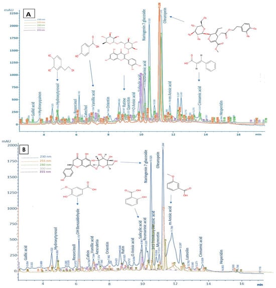
Figure 1.
HPLC chromatograms and chemical structure of the major phytochemicals. (A) Extract EA. (B) Extract EE.

Table 1.
Biomolecules identified in the EA and EE extracts.
3.3. In Vitro Antioxidant Activities
3.3.1. Total Antioxidant Activity
The results obtained from our experiment showed that extracts of the Laperrine olive leaves have a high total antioxidant capacity. The t-test revealed a significant difference between the EA extract and the EE extract (p < 0.01). The TAC was calculated from the linear regression equation obtained from the ascorbic acid calibration curve (Y = 0.0048X + 0.02, R2 = 0.99). The overall antioxidant activity of EA (267.26 ± 2.67 mg AAE/g extract) was found to be lower compared to EE (345.41 ± 3.31 mg AAE/g extract).
3.3.2. Ferric-Reducing Antioxidant Power Assay
The value of the FRAP was determined from the linear regression equation obtained from the ascorbic acid calibration curve (Y = 0.0143X + 0.0062, R2 = 0.99). The EE extract has a greater reducing power than the EA extract (p < 0.01), the calculated values are 258.76 ± 6.69 mg AAE/g extract for EE and 153.72 ± 2.70 mg AAE/g extract for EA. As shown in Figure 2, both extracts exhibited dose-dependent activity, with antioxidant activity increasing proportionally to extract concentration.
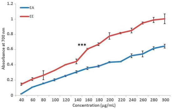
Figure 2.
Ferric reduction antioxidant power of aqueous extract in comparison with ethanolic extract of Olea europaea subsp. laperrinei leaves. Results represent the mean ± SD, n = 3; *** p < 0.001.
The statistical analysis showed a significant difference (p < 0.001) between the EA and EE samples. The latter is indeed more active than the EA extract. We recorded the IC50: 140.52 ± 2.13 μg/mL for the ethanol–water extract and 234.91 ± 7.69 μg/mL for the aqueous extract. We calculated a value of 34.44 ± 0.66 μg/mL in the case of ascorbic acid.
3.3.3. DPPH• Free Radical Scavenging Assay
The antiradical activity of the EA and EE extracts increased dose-dependently (Figure 3). The EE extract demonstrated significantly greater scavenging capacity (p < 0.001) than the EA extract, requiring 7.48 ± 0.39 μg/mL and 10.88 ± 0.22 μg/mL, respectively, to scavenge 50% of the initial DPPH• concentration. Ascorbic acid showed an EC50 of 2.52 ± 0.05 μg/mL.
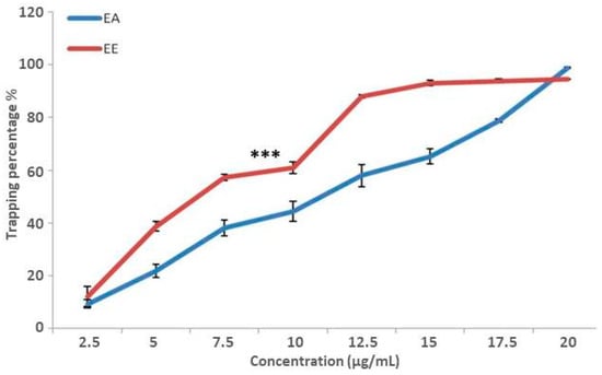
Figure 3.
DPPH• radical trapping capacity of aqueous extract compared with ethanolic extract of Olea europaea subsp. laperrinei leaves; n = 3; *** p < 0.001.
This result was confirmed by the calculation of the antioxidant activity index which places the EE extract in the same class as ascorbic acid (AAI > 2), indicating very strong antioxidant activity, whereas the EA extract demonstrated strong antioxidant activity (AAI < 2). In addition, the maximum percentage inhibition of the DPPH• radical scavenging was 98.84 ± 0.25% with 20 μg/mL of the EA extract and 93.85 ± 1.16% with 17.5 μg/mL of the EE extract.
3.3.4. β-Carotene Bleaching Assay
Figure 4 shows that both extracts effectively inhibited linoleic acid oxidation, with the inhibition percentage increasing proportionally to their concentrations. The EE extract exhibited a significantly higher inhibitory effect on linoleic acid oxidation than the aqueous EA extract, as evidenced by its higher maximum inhibition percentage (87.41 ± 5.76% at 900 μg/mL for EE compared to 83.91% at 1800 μg/mL for EA and 83.39 ± 6.82% at 100 μg/mL for BHT).
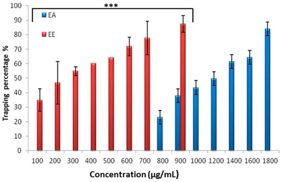
Figure 4.
The percentage of inhibition of β-carotene bleaching by the aqueous extract compared with the ethanolic extract of leaves of Olea europaea subsp. laperrinei; n = 3; *** p < 0.001.
The IC50 value, representing the extract concentration needed for 50% inhibition, was determined through a graphical analysis of the inhibition percentage as a function of extract concentration. This analysis revealed the EE extract as significantly more effective (p < 0.001), with an IC50 of 185.63 ± 3.84 μg/mL, compared to the EA extract, which had an IC50 of 1187.99 ± 93.23 μg/mL.
3.3.5. Hydrogen Peroxide Radical Scavenging Activity
While hydrogen peroxide (H2O2) itself exhibits relatively low reactivity, it becomes hazardous upon penetrating biological membranes, where it can transform into the hydroxyl radical (OH•). This radical is notorious for initiating lipid peroxidation and DNA mutations, illustrating the critical need for effective H2O2 neutralization within biological systems. The capacity of plant extracts to mitigate H2O2’s potential harm is largely due to their phenolic compounds, which donate electrons to H2O2, transforming it into harmless water (H2O). This process effectively obstructs the genesis of damaging hydroxyl radicals. The proficiency of the Laperrine olive leaf extracts in scavenging hydrogen peroxide is depicted in Figure 5, demonstrating their ability to sequester H2O2 in a concentration-dependent manner. The EA extract showcased an impressive H2O2 neutralization efficiency of 85.96 ± 3.91% at 470 μg/mL, while the EE extract exhibited a commendable efficacy of 83.70 ± 6.26% at 270 μg/mL. For comparison, ascorbic acid, a recognized antioxidant, achieved an 84.78 ± 2.37% neutralization rate at only 80 μg/mL. These findings underline the significant antioxidant potential of Laperrine olive leaf extracts, particularly in their dose-dependent capacity to counteract hydrogen peroxide-induced oxidative stress. Such properties highlight the extracts’ utility in preventive health strategies and their potential integration into therapeutic formulations to safeguard cellular integrity against oxidative damage.
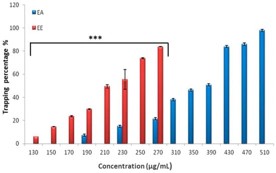
Figure 5.
Trapping capacity of hydrogen peroxide by the aqueous extract and the ethanolic extract of Olea europaea subsp. laperrinei leaves. Results are the mean ± SD, n = 3; *** p < 0.001.
A comparison of the averages obtained after calculating the concentration required to reduce H2O2 by 50% showed a significant difference (p < 0.001) between the extracts tested, with the EE extract (IC50 = 214.04 ± 2.89 µg/mL) having a greater hydrogen peroxide scavenging capacity than the EA extract (IC50 = 351.30 ± 5.30 µg/mL).
3.4. Antibacterial Activity
The evaluation of the antibacterial activity showed notable variations in the sensitivity of the bacterial strains. S. aureus FRIS6, S. aureus LGA251, and P. aeruginosa were highly sensitive to the aqueous extract, with inhibition diameters of 18.66 ± 1.15 mm, 17.33 ± 1.52 mm, and 15.33 ± 0.57 mm, respectively. B. cereus, E. coli, K. pneumoniae 700603, K. pneumoniae 4352, K. pneumoniae 1766, and K. pneumoniae 825 were all sensitive to the aqueous extract, with IZDs varying from 10.00 mm to 12.83 ± 1.04 mm. Thus, aqueous extract showed equivalent activity against Gram-positive and Gram-negative bacteria. While the ethanolic extract demonstrated higher effectiveness on Gram-positive strains. S. aureus FRIS6 and S. aureus LGA251 were extremely sensitive, with IZDs of 23.33 ± 0.76 mm and 20.66 ± 0.76 mm, respectively, which is significantly (p < 0.001) different from B. cereus (14.15 ± 0.86 mm). For Gram-negative bacteria, P. aeruginosa was highly sensitive (IZD = 19 mm) to the ethanolic extract, with a significant (p < 0.001) difference in sensitivity compared to E. coli and K. pneumoniae strains, which were categorized as sensitive to this extract (Figure 6).
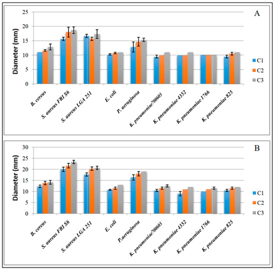
Figure 6.
Inhibition zone diameters induced by different extract concentrations on the bacterial strains tested. (A) Aqueous extract; (B) ethanolic extract; C1: 0.4 g/mL; C2: 0.6 g/mL; C3: 0.8 g/mL.
As a result, S. aureus LGA251, S. aureus FRI S6, and P. aeruginosa were selected for MIC determination. The analysis of inhibition diameters in relation to concentrations revealed an exponential trend. Figure 7 illustrates the kinetics of IZDs for the specified strains based on the type of extract tested. For S. aureus FRI S6, a progressive increase in inhibition diameters was observed starting from 6.25 mg/mL, indicating that higher concentrations promote larger inhibition zones, with the maximum inhibition diameter recorded at 400 mg/mL (Figure 7A).
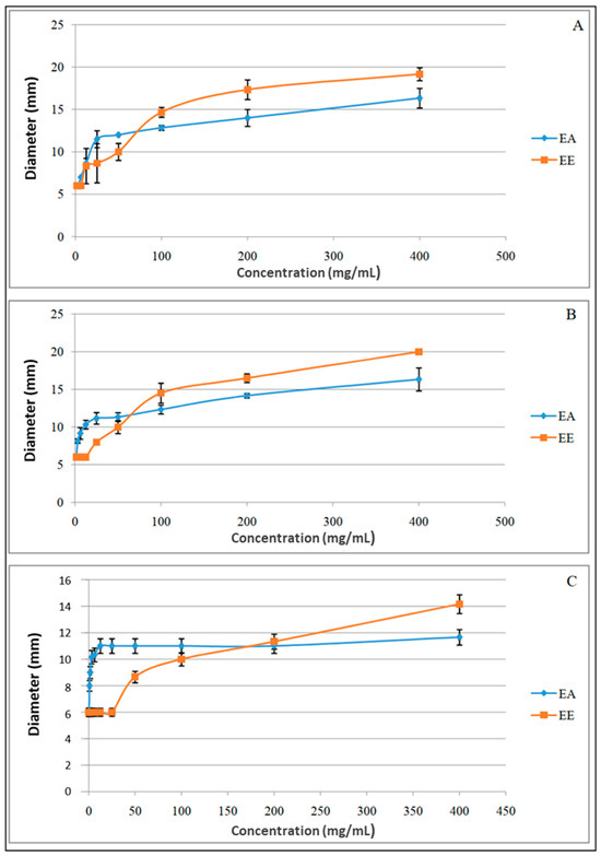
Figure 7.
Variation of IZDs with extract concentration for: (A) S. aureus FRI S6; (B) S. aureus LGA 251; (C) P. aeruginosa; EA: aqueous extract; EE: ethanolic extract.
For the ethanolic extract, a progressive increase in IZDs was noted, starting from 12.5 mg/mL, reaching a maximum of 19.10 ± 0.73 mm at 400 mg/mL. For S. aureus LGA251 (Figure 7B), inhibition zones began increasing from 3.12 mg/mL, peaking at 16.30 ± 1.52 mm at 400 mg/mL. The ethanolic extract exhibited IZDs increasing progressively from 8.00 mm at 25 mg/mL to 20.00 mm at 400 mg/mL.
For P. aeruginosa (Figure 7C), at 0.78 mg/mL of the aqueous extract, an inhibition zone of 8.00 mm was observed. Between 12.5 mg/mL and 200 mg/mL, inhibition diameters remained at 11.00 mm, and then slightly increased to 11.60 ± 0.57 mm at 400 mg/mL. The ethanolic extract showed an increase in inhibition diameters starting from 50 mg/mL, ranging from 8.60 ± 1.52 mm to 14.10 ± 1.4 mm.
Table 2 reports the MIC values for the three tested strains. The MIC of the aqueous extract was 6.25 mg/mL for S. aureus FRI S6, 1.56 mg/mL for S. aureus LGA 251, and 0.78 mg/mL for P. aeruginosa, while for the ethanolic extract, the MIC obtained was 12.5 mg/mL for S. aureus FRI S6, 25 mg/mL for S. aureus LGA 251, and 50 mg/mL for P. aeruginosa.
Table 2.
MIC values for aqueous (EA) and ethanolic (EE) extracts.
4. Discussion
According to Luis et al. [41], the yield varies depending on the solvent used. However, this observation does not appear to apply in the case of the Laperrine olive tree leaves, as both yields surpassed the 11% documented by Ahmed et al. [42] from an ethanolic extraction of Olea europaea leaves, suggesting potentially higher extraction efficiency for Laperrine olive tree leaves. The results obtained may be attributed to the physicochemical characteristics of the extracts. Addad et al. [43] showed that the extraction yield varies according to the extraction time, pH, temperature, and chemical profile of the extract.
A comparison of the total phenolic content of EA and EE extracts with 70 medicinal plants classified according to the total phenolic content of aqueous extracts (ranging between 0.6 mg CAE/g dry extract and 145.87 mg CAE/g dry extract) [44] suggests that the leaves of Olea laperrinei exhibit a remarkably high concentration of polyphenols, surpassing those in Olea europaea leaves (Table 3). This high polyphenol content is likely attributable to the arid environmental conditions in which the Laperrine olive tree thrives, as polyphenols are known to be synthesized under hostile environmental conditions in response to stress. These findings suggest that the extreme conditions of the Laperrine olive tree’s habitat may enhance its phytochemical content, particularly polyphenols, contributing to its potential as a reservoir of natural antioxidants and other therapeutic phytochemicals.
Table 3.
Comparison of the total phenol content of Olea europaea subsp. laperrinei leaves with that of Olea europaea L. leaves.
Research on phenolic compounds underscores their sensitivity to environmental, physiological, and genetic factors, influencing their bioavailability within species. Their concentration can vary with seasonal changes, micro-environmental effects, and the maturation stage of the plant. Notably, stress from intense solar radiation significantly stimulates polyphenol synthesis.
The total flavonoid content in Laperrine olive leaves surpasses the levels reported by Abaza et al. [46] in the Olea europaea variety Chetoui, with aqueous and ethanolic leaf extracts yielding 6.23 ± 0.62 mg CE/g and 15.83 ± 1.26 mg CE/g dry weight, respectively. Studies indicate that the total tannin content is higher in Laperrine olive leaves than in Olea europaea, with Brahmi et al. [46] reporting 79.70 mg CE/100 g and 73.05 mg CE/100 g dry weight for the Chemlal and Neb Jmel varieties, respectively. However, the tannin content in Olea europaea subsp. laperrinei was approximately 22.79 g TAE/kg dry matter, closely aligning with values reported by Mebirouk-Boudechiche et al. [48] for Olea europaea leaves.
The phytochemical analysis performed on the EA and EE extracts revealed the presence of two major fractions among the phenolic compounds, namely flavonoids and tannins, whose contents were particularly high. This richness appears to be a response of the species to its environmental conditions and clearly shows the close relationship between the biosynthesis of secondary metabolites and the extreme environmental conditions to which the Laperrine olive tree is exposed. It should be noted, however, that compared to the EA extract, the total phenol and flavonoid contents were significantly higher in the EE extract. This result confirms that the ethanol–water mixture is a better extraction solvent compared to water alone.
This study found that the total tannin content exceeds the total flavonoid content in Laperrine olive leaves, attributed to the adaptation of the species to the dry central Sahara environment, characterized by limited, xerophytic conditions, superficial soil, and significant pastoral pressure. Tannin synthesis often increases under environmental stress, affecting woody plant leaves in relatively high concentrations.
Selecting an appropriate extraction solvent is crucial for optimizing polyphenol levels [49]. Polar solvents like water can extract a high concentration of polyphenols but may also co-extract impurities such as organic acids, sugars, and soluble proteins. Mailoa et al. [50] and subsequent research suggested that a mixture of an organic solvent and water, particularly ethanol, enhances polyphenol solubility [51], making ethanol–water blends efficient for dissolving a broad range of phenolic compounds [52]. Our findings corroborate this, demonstrating more efficient phenolic compound extraction using ethanol–water mixtures compared to water alone, aligning with Mohammedi and Atik [53].
Chromatographic analysis of Laperrine olive leaf extracts revealed a diverse array of phenolic compounds, underscoring the chemical richness inherent to Olea europaea. Notably, phenolic alcohols such as hydroxytyrosol and oleuropein, characteristic of the olive species, were identified. The extracts also contained simple phenols (such as resorcinol and catechol), a diphenol (hydroxyquinone), phenolic acids deriving from hydroxybenzoic acid (gallic acid, salicylic acid, vanillic acid, and 3,4,5-trimethoxybenzoic acid) and phenylpropanoids (caffeic acid, rosmarinic acid, and cinnamic acid). Additionally, two isomers of anisic acid 2-methoxybenzoic acid (o-anisic acid) and 3-methoxybenzoic acid (m-anisic acid), along with multiple flavonoid families, including flavones (luteolin 7-glucoside, orientin, vitexin 2-O rhamnoside), flavonols (quercetin, rutin, catechin, myricetin), and flavanones (hesperidin, naringenin 7-glucoside), were detected. Other identified compounds included a phenolic aldehyde isomer of hydroxybenzaldehyde (p-hydroxybenzaldehyde), an aromatic compound from the coumarin family (aesculetin), and an alkaloid (caffeine). These components align with those found in Olea europaea subsp. sylvestris and Olea europaea L., including quercetin, rutin, vanillic acid, salicylic acid, luteolin, aesculetin, cinnamic acid, caffeine, caffeic acid, and apigenin [10,54]. For the Olea europaea variety Bouriche, common compounds shared with the Laperrine olive leaves extract include oleuropein, rutin, and luteolin 7-glucoside [55]. Moreover, our findings align with those of Lahcene et al. [9] following acid hydrolysis and Djenane et al. [24] and Bouchoucha et al. [30] from leaf extracts of Laperrine olive leaves, confirming the significant phytochemical diversity and potential bioactivity of these extracts.
The total antioxidant capacity of EA and EE extracts significantly surpassed the results obtained by Khlif et al. [56] from a methanol–water extract of Olea europaea variety Chetoui (73.94 ± 1.98 mg EE/g extract). The efficacy of the antioxidant activity of the plant extracts is attributed to the presence of polyphenols, serving as reducing agents. This aligns with the positive linear correlation established between the TAC and the total phenolic content of the extracts, with correlation coefficients of 0.99 for the EA extract and 0.89 for the EE extract, underscoring a significant relationship. Consequently, these findings suggest that the polyphenols in the Laperrine olive tree extracts are key contributors to their antioxidant potency. This is consistent with the insights from Jan et al. [57], highlighting that a plant’s high total phenol content is directly related to its antioxidant capabilities, reinforcing the value of Laperrine olive leaves as a potent natural source of antioxidants.
Comparative analysis reveals that the Laperrine olive tree extracts exhibit a higher reducing potential than those obtained from Olea europaea leaves, suggesting their pronounced ability to act as electron or H+ proton donors. Reducing agents, which exert antioxidant action through electron or hydrogen atom donation and free radical chain disruption, are crucial for this activity. This is supported by data from Hayes et al. [58], who reported a methanolic extract content of 30.1 ± 0.1 mg TRE/100 g of dry weight, and Goldsmith et al. [13], who reported values of 22.55 ± 19.17 mg TRE/g dry residue and 232.12 ± 4.89 mg TRE/g dry residue for aqueous and ethanolic extracts, respectively.
The reducing power of Olea europaea subsp. laperrinei leaf extracts is higher than that of Olea europaea leaf extracts. Indeed, Ferreira et al. [1] noted an IC50 of 1.17103 µg/mL obtained from a methanolic extract, and Xie et al. [44] obtained a value of 1.55103 µg/mL for an ethanolic extract. These values confirm that our extracts have a high reducing power, as the lower the value of the IC50, the greater the reduction power of the plant extract. Ebrahimazadeh et al. [59] noted that levels of phenolic compounds and flavonoids lead to a higher reducing power of plant extracts. This agrees with our results, as we have established a positive correlation between the reduction power of the extracts tested and the content of polyphenols and total flavonoids (R2 > 0.9).
Furthermore, the results of the highly correlated preliminary assays (TAC and FRAP; R2 = 0.99) demonstrated the remarkable antioxidant capacity of Olea europaea subsp. laperrinei leaf extracts.
The antioxidant activity of Olea laperrinei was found to be strong, exhibiting significant scavenging activity against DPPH. Indeed, extracts of Laperrine olive leaves showed superior free radical scavenging activity compared to the aqueous (66.04 µg/mL) and methanolic (54.04 µg/mL) extracts reported by [30], which themselves displayed better activity than those of Olea europaea. Arab et al. [60] reported an EC50 of 0.29103 µg/mL for a methanolic extract, while Khlif et al. [56] and Mkaouar et al. [61] reported significantly higher EC50 values for their extracts. This suggests that Laperrine extracts are more effective at neutralizing free radicals.
The results obtained in the present study contrast with the literature data for Olea europaea, where the maximum DPPH radical trapping rate is notably lower. DPPH•, a stable nitrogen-centered radical, shifts from violet to yellow upon reduction through hydrogen or electron donation, marking substances capable of this reaction as antioxidants. Thus, Laperrine olive leaf extracts act as hydrogen donors, highlighting their potential therapeutic applications. According to Al Saeghi et al. [62], plant species with high antioxidant activity, such as the Laperrine olive leaf extracts, could be explored for treating various diseases, reinforcing their value in human health.
The results of the β-Carotene bleaching test surpass the inhibition rates reported by Hayes et al. [58] and Saiah et al. [63] for methanolic extracts of Olea europaea leaves, which showed maximum linoleic acid inhibition percentages of 16% at 1 mg/mL and 71.76 ± 3.28% at 4 mg/mL, respectively. The observed difference in inhibitory capacity between the extracts can be attributed to the solubility characteristics of antioxidants within linoleic acid emulsions. Apolar antioxidants, more prevalent in the EE extract, exhibit enhanced antioxidative properties in oil-in-water emulsions due to their localization at the oil–water interface, offering superior lipid oxidation protection. Conversely, polar antioxidants, characteristic of the EA extract, remain in the aqueous phase and consequently exhibit diminished lipid protection efficacy. The rapid discoloration observed in control samples without antioxidants is due to the coupled oxidation of linoleic acid and β-carotene. The incorporation of antioxidants from the extracts effectively delays this oxidation process. As demonstrated by Kubula and Siriamornpun [64], plant-derived antioxidants neutralize free radicals within the β-carotene/linoleic acid system, highlighting the potent antioxidative capabilities of Laperrine olive leaf extracts and their potential applications in enhancing food stability and health benefits.
The extracts from the Laperrine olive leaves display a superior capacity for hydrogen peroxide (H2O2) scavenging compared to an aqueous extract derived from the leaves of Olea europaea, specifically the Mamecik variety, which neutralized 62.27% of H2O2 at a concentration of 500 μg/mL [63]. The scavenging capacity of the EA and EE extracts is attributed to the rich content of total phenols and flavonoids present in Laperrine olive. Supporting this, Orak et al. [65] identified a positive correlation between the scavenging activity against hydrogen peroxide and the total phenols and flavonoids content, aligning with our findings where the correlation coefficient exceeded 0.9 for both extracts. This strong correlation can be explained by the nature of phenolic compounds, whose hydroxyl groups act as effective double electron and H+ proton donors, thus neutralizing reactive oxygen species such as H2O2. This mechanism underscores the critical role of phenolic composition in determining the antioxidant efficacy of plant extracts, particularly in neutralizing potentially damaging reactive oxygen species.
The obtained results show that the phytochemical molecules contained in the EA and EE extracts exert a significant antioxidant effect. Their effectiveness is primarily attributed to their capacity to scavenge free radicals. This is achieved by donating a hydrogen atom or an electron to the free radical, thus converting it into its reduced form. This reduced state prevents it from reaching its biological targets (DNA, proteins, or lipids) and causing alterations that could lead to serious pathologies.
The antioxidant effects revealed in vitro are directly related to the phenolic content of the studied extracts and depend on the various interactions (synergy or antagonism) governing them. Thus, the antioxidant potential favors the EE extract, whose phenolic content is richer than that of the EA extract. Indeed, it demonstrates a strong ability to neutralize free radicals such as DPPH• and H2O2 as well as effectively inhibit β-carotene bleaching. Moreover, the calculation of the antioxidant activity index (AAI > 2) clearly defines it as having a very strong antioxidant activity, with potency similar to that of ascorbic acid.
Numerous studies have highlighted the antibacterial activity of Olea europaea leaf extracts [66,67,68], specifying, however, that this activity is influenced by several factors such as the cultivar, the extraction method used, and the type of solvent employed. These parameters directly affect the total content of phenolic compounds, which are responsible for the antibacterial effects [69]. The EA and EE extracts are mainly composed of oleuropein, hydroxytyrosol, vanillic acid, salicylic acid, and anisic acid, which exhibit strong antimicrobial efficacy when tested individually [70], as well as gallic acid, caffeic acid, and cinnamic acid, whose bactericidal effect is reported by Ecevit et al. [70]. The work done by Al Rimawi et al. [69] showed that oleuropein has remarkable bactericidal activity, particularly against S. aureus, and according to Silvan et al. [71], hydroxytyrosol is highly effective against Campylobacter jejuni, with the MIC determined at 0.25 mg/mL. Ben Hassena et al. [72] further demonstrated the bactericidal potential of hydroxytyrosol-rich extracts against multidrug-resistant ESBL-producing clinical isolates of Escherichia coli and Klebsiella pneumoniae. They reported MIC values as low as 0.3125 mg/mL, with MBC/MIC ratios confirming bactericidal action.
The review by Mumcu [73] further emphasized that the antimicrobial efficacy of olive leaf extracts is enhanced by a synergistic effect between their phenolic compounds, suggesting that their combined action is more potent than individual compounds alone.
The results obtained in our experimentation showing that the ethanolic extract exhibited better antibacterial activity than the aqueous extract are confirmed by the work of Khan et al. [74], who explained that this could be due to the effect of the solvent on the distribution and nature of the phytochemical substances present in the extract. Indeed, the EE extract is characterized not only by higher levels of phenolic compounds but also by the presence of rutin, which is absent from the aqueous extract. This compound has a significant bactericidal effect against methicillin-resistant S. aureus (MRSA), Acinetobacter baumannii, and P. aeruginosa, with an MIC of 0.5 mg/mL [75]. In the same study, Ivanov et al. [75] further reported that rutin significantly interferes with bacterial virulence mechanisms. Specifically, it inhibits biofilm formation, reduces the production of extracellular virulence factors such as elastase, pyocyanine, and rhamnolipids, and downregulates quorum-sensing gene expression in P. aeruginosa and MRSA.
Although the antimicrobial action mechanism is not yet fully elucidated, it has been reported that, due to their hydrophobicity, phenolic compounds can accumulate and intercalate into the bacterial cell wall, leading to its disruption and the alteration of its permeability, resulting in the leakage of cytoplasmic elements such as potassium, glutamate, or inorganic phosphate, thereby inhibiting bacterial growth [73]. Moreover, the particular structure of certain compounds allows them to have specific mechanisms. This is the case for the ortho-diphenolic structure of oleuropein, which makes it capable of disturbing essential pathways for amino acid synthesis in microorganisms [69], and for hydroxytyrosol, whose small size and hydroxyl groups allow it to better penetrate bacteria and interact with intracellular targets [72]. This is further supported by molecular docking results, which revealed strong binding affinities of hydroxytyrosol to key bacterial enzymes, such as DNA gyrase B subunit and peptidoglycan DD-transpeptidase (PBP3), potentially interfering with DNA replication and cell wall synthesis [72].
Similar to several reports, our study also revealed that Gram-positive bacteria are more sensitive than Gram-negative bacteria. Thus, Magyari et al. [76] demonstrated that S. aureus and Streptococcus pyogenes were more sensitive to the EE of olive leaves, with respective inhibition zones of 21 and 24 mm, compared to P. aeruginosa and E. coli, which showed inhibition zones of 10 and 12 mm, respectively. This increased sensitivity of Gram-positive bacteria could be explained by the absence of the outer membrane, which reduces the permeability of antimicrobial agents in Gram-negative bacteria. However, in our present study, and in comparison, to B. cereus, P. aeruginosa showed greater sensitivity to the tested extracts. These results corroborate the work of Sanchez et al. [77], as Yersinia enterocolitica (Gram-negative) is more sensitive than Listeria monocytogenes (Gram-positive) to olive leaf extracts. Furthermore, Pereira et al. [4] indicated that B. cereus was the most sensitive among the tested strains, while B. subtilis was the most resistant to the olive leaf extract. This thus demonstrates the complexity of predicting bacterial sensitivity to extracts, even within strains of the same genus, and the difficulty in understanding the factors it depends on. Moreover, it is important to note that this sensitivity is also related to the method used to evaluate the activity of the extract, which could explain the results obtained for P. aeruginosa in this study. Indeed, this strain was found to be extremely sensitive to the EE using the agar diffusion method (IZD = 19 mm), but it was considered resistant after determining an MIC of 50 mg/mL. Therefore, the choice of method and the proper analysis of the results obtained by each method are crucial for a better assessment of sensitivity [78].
5. Conclusions
In this study, we investigated the phytochemical composition and evaluated the antioxidant and antibacterial activities of ethanolic and aqueous extracts from Laperrine olive leaves. The results clearly demonstrate the richness of these extracts in bioactive compounds, reflecting the Laperrine olive tree’s adaptation to arid environments and highlighting the link between polyphenol biosynthesis and environmental stress. Both extracts exhibited significant antioxidant activity in vitro, attributed to their high phenolic and tannin contents. The ethanolic extract showed superior antioxidant potential, likely due to its higher concentration of key compounds such as oleuropein, hydroxytyrosol, gallic acid, rutin, luteolin-7-glucoside, naringenin-7-glucoside, myricetin, luteolin, and apigenin, which may act synergistically.
Regarding antibacterial activity, both extracts were active against all tested bacterial strains, with notable efficacy against S. aureus and P. aeruginosa. While the ethanolic extract displayed larger inhibition zones, the aqueous extract exhibited lower MIC values, suggesting distinct compound interactions in each extract.
These findings support the potential of Laperrine olive leaves as a source of natural antioxidants and antibacterial agents, with possible applications in therapeutic and agricultural fields. Additionally, Olea europaea subsp. laperrinei represents a valuable genetic resource, particularly for enhancing drought tolerance in cultivated olive varieties.
While the present study provides valuable insights, it is primarily focused on in vitro assessments. In vivo experiments and mechanistic studies may be considered in future research to further elucidate the biological potential of the extracts.
Author Contributions
Conceptualization, S.L., E.B. and K.H.; methodology, S.L., B.S., N.S. and K.H.; software, I.M. and L.T.; validation, K.H., L.T. and R.A.; formal analysis, S.L., I.M., E.B. and L.T.; investigation, S.L., K.B., B.S. and N.S.; resources, L.T. and K.H., data curation, S.L., R.A. and L.T.; writing—original draft preparation, S.L. and K.H.; writing—review and editing, K.H., L.T., M.A., F.M.A., M.T., G.F. and R.A.; visualization, I.M., K.B., B.S. and N.S.; supervision, K.H., L.T. and R.A.; project administration, K.H. and R.A.; funding acquisition, R.A., M.A., F.M.A., M.T., G.F. and N.A. All authors have read and agreed to the published version of the manuscript.
Funding
This research received no external funding.
Data Availability Statement
The original contributions presented in this study are included in the article. Further inquiries can be directed to the corresponding author.
Acknowledgments
The authors would like to express their gratitude to the Algerian Ministry of Higher Education and also thank the Deanship of Scientific Research at Shaqra University for their guidance and support throughout this work.
Conflicts of Interest
The authors declare no conflicts of interest.
References
- Ferreira, I.C.F.; Barros, L.; Soares, M.E.; Bastos, M.L.; Pereira, J.A. Antioxidant activity and phenolic contents of Olea europaea L. leaves sprayed with different copper formulations. Food Chem. 2006, 103, 188–195. [Google Scholar] [CrossRef]
- Lee, O.H.; Lee, B.Y. Antioxidant and antimicrobial activities of individual and combined phenolics in Olea europaea leaf extract. Bioresour. Technol. 2010, 101, 3751–3754. [Google Scholar] [CrossRef]
- Dekanski, D.; Janićijević-Hudomal, S.; Tadić, V.; Marković, G.; Arsić, I.; Mitrović, D.M. Phytochemical analysis and gastroprotective activity of an olive leaf extract. Serbian J. Chem. Soc. 2009, 74, 367–377. [Google Scholar] [CrossRef]
- Pereira, A.P.; Ferreira, I.C.F.R.; Marcelino, F.; Valentão, P.; Andrade, P.B.; Seabra, R.; Estevinho, L.; Bento, A.; Pereira, J.A. Phenolic compounds and antimicrobial activity of olive (Olea europaea L. cv. Cobrançosa) leaves. Molecules 2007, 12, 1153–1162. [Google Scholar] [CrossRef] [PubMed]
- Lahcene, S.; Trabelsi, L.; Salem-Bekhit, M.M.; Rabea, S.; Refat, H.M.; Selim, M.; Alehaidib, S.M.; Sebbane, H.; Msela, A.; Benguerba, Y.; et al. Anti-leishmanial activities of Olea europaea subsp. laperrinei extracts. Cell. Mol. Biol. 2023, 69, 207–213. [Google Scholar] [CrossRef] [PubMed]
- Maazoun, A.M.; Ben Hlel, T.; Haouel-Hamdi, S.; Belhadj, F.; Mediouni-Ben Jemâa, J.; Marzouki, M.N. Screening for insecticidal potential and acetylcholinesterase activity inhibition of Urginea maritima bulbs extract for the control of Sitophilus oryzae (L.). J. Asia Pac. Entomol. 2017, 20, 752–760. [Google Scholar] [CrossRef]
- Stefani, T.; Morales-San Claudio, P.D.C.; Rios, M.Y.; Aguilar-Guadarrama, A.B.; Gonzãlez-Maya, L.; Sãnchez-Carranza, J.N.; Gonzãlez-Ferrara, M.; Camacho-Corona, M.D.R. UPLC-QTOF-MS analysis of cytotoxic and antibacterial extracts of Hechtia glomerata Zucc. Nat. Prod. Res. 2020, 34, 644–648. [Google Scholar] [CrossRef]
- De la Ossa, J.G.; El Kadri, H.; Gutierrez-Merino, J.; Wantock, T.; Harle, T.; Seggiani, M.; Danti, S.; Di Stefano, R.; Velliou, E. Combined antimicrobial effect of bio-waste olive leaf extract and remote cold atmospheric plasma effluent. Molecules 2021, 26, 1890. [Google Scholar] [CrossRef]
- Lahcene, S.; Taibi, F.; Mestar, N.; Ali Ahmed, S.; Boumendjel, M.; Ouafi, S.; Houali, K. Insecticidal effects of the Olea europaea subsp. laperrinei extracts on the flour pyralid Ephestia kuehniella. Cell. Mol. Biol. 2018, 64, 6–12. [Google Scholar]
- Mestar, N.G.; Boudiaf, M.N.; Lahcene, S.; Abbaci, H.; Aiche, G.I.; Metna, B.; Saadoun, N.S.; Taibi, F.; Houali, K. Bio-insecticidal effects of oleaster leaves aqueous extracts against psylla larvae (Euphyllura olivina (Costa)), a primary pest of Olea europaea L. Cell. Mol. Biol. 2018, 64, 35–40. [Google Scholar] [CrossRef]
- Kovalíková, Z.; Kubeš, J.; Skalický, M.; Kuchtickova, N.; Mašková, L.; Tuma, J.; Vachová, P.; Hejnak, V. Changes in content of polyphenols and ascorbic acid in leaves of white cabbage after pestinfestation. Molecules 2019, 24, 2622. [Google Scholar] [CrossRef] [PubMed]
- Karami, S.; Basaki, T.; Khaneghah, A.M. The inhibitory effects of polyphenolic compounds on the damage caused by safflower fly (Acanthiophilus helianthi) in Carthamus spp. Qual. Assur. Saf. Crop Foods 2021, 13, 87–93. [Google Scholar]
- Goldsmith, C.D.; Vuong, Q.V.; Sadeqzadeh, E.; Stathopoulos, C.E.; Roach, P.D.; Scarlett, C.J. Phytochemical properties and anti-proliferative activity of Olea europaea L. leaf extracts against pancreatic cancer cells. Molecules 2015, 20, 12992–13004. [Google Scholar] [CrossRef]
- Gutiérrez-Venegas, G.; Torras-Ceballos, A.; Gómez-Mora, J.; Fernández-Rojas, B. Luteolin, quercetin, genistein, and quercetagetin inhibit the effects of lipopolysaccharide obtained from Porphyromonas gingivalis in H9c2 cardiomyoblasts. Cell. Mol. Biol. Lett. 2017, 22, 19–30. [Google Scholar] [CrossRef] [PubMed]
- Nediani, C.; Ruzzolini, J.; Romani, A.; Calorini, L. Oleuropein, a bioactive compound from Olea europaea L., as a potential preventive and therapeutic agent in non-communicable diseases. Antioxidants 2019, 8, 578. [Google Scholar] [CrossRef]
- Goldsmith, C.D.; Bond, D.R.; Jankowski, H.; Weidenhofer, J.; Stathopoulos, C.E.; Roach, P.D.; Scarlett, C.J. The olive biophenols oleuropein and hydroxytyrosol selectively reduce proliferation, influence the cell cycle, and induce apoptosis in pancreatic cancer cells. Int. J. Mol. Sci. 2018, 19, 1937. [Google Scholar] [CrossRef]
- Ruzzolini, J.; Peppicelli, S.; Andreucci, E.; Bianchini, F.; Scardigli, A.; Romani, A.; La Marca, G.; Nediani, C.; Calorini, L. Oleuropein, the main polyphenol of Olea europaea leaf extract, has an anticancer effect on human BRAF melanoma cells and potentiates the cytotoxicity of current chemotherapies. Nutrients 2018, 10, 1950. [Google Scholar] [CrossRef] [PubMed]
- Batiha, G.E.; Beshbishy, A.M.; Ikram, M.; Mulla, Z.S.; Abd El-Hack, M.E.; Taha, A.E.; Algammal, A.M.; Elewa, Y.H. The pharmacological activity, biochemical properties, and pharmacokinetics of the major natural polyphenolic flavonoid: Quercetin. Foods 2020, 9, 374. [Google Scholar] [CrossRef]
- Peyrol, J.; Riva, C.; Amiot, M. Hydroxytyrosol in the prevention of the metabolic syndrome and related disorders. Nutrients 2017, 9, 306. [Google Scholar] [CrossRef]
- Monteiro, M.; Silva, A.F.R.; Resende, D.; Braga, S.S.; Coimbra, M.A.; Silva, A.M.S.; Cardoso, S.M. Strategies to broaden the applications of olive biophenols oleuropein and hydroxytyrosol in food products. Antioxidants 2021, 10, 444. [Google Scholar] [CrossRef]
- Cheurfa, M.; Abdallah, H.H.; Allem, R.; Noui, A.; Picot-Allain, C.M.N.; Mahomoodally, F. Hypocholesterolaemic and antioxidant properties of Olea europaea L. leaves from Chlef province, Algeria using in vitro, in vivo and in silico approaches. Food Chem. Toxicol. 2019, 123, 98–105. [Google Scholar] [CrossRef] [PubMed]
- Kashyap, P.; Shikha, D.; Thakur, M.; Aneja, A. Functionality of apigenin as a potent antioxidant with emphasis on bioavailability, metabolism, action mechanism and in vitro and in vivo studies: A review. J. Food Biochem. 2021, 46, e13950. [Google Scholar] [CrossRef]
- Djenane, D.; Yangüela, J.; Derriche, F.; Bouarab, L.; Roncales, P. Utilisation des composés de feuilles d’olivier comme agents antimicrobiens; application pour la conservation de la viande fraîche de dinde. Rev. Nat. Technol. 2012, 7, 53–61. [Google Scholar]
- Djenane, D.; Aboudaou, M.; Djenane, F.; García-Gonzalo, D.; Pagán, R. Improvement of the shelf-life status of modified atmosphere packaged camel meat using nisin and Olea europaea subsp. laperrinei leaf extract. Foods 2020, 9, 1336. [Google Scholar] [CrossRef] [PubMed]
- Baali-Cherif, D.; Besnard, G. High genetic diversity and clonal growth in relict populations of Olea europaea subsp. laperrinei (Oleaceae) from Hoggar, Algeria. Ann. Bot. 2005, 96, 823–830. [Google Scholar] [CrossRef] [PubMed]
- Besnard, G.; Baali-Cherif, D. Coexistence of diploids and triploids in a Saharan relict olive: Evidence from nuclear microsatellite and flow cytometry analyses. C. R. Biol. 2009, 332, 1115–1120. [Google Scholar] [CrossRef]
- Bernal, M.; Llorens, L.; Julkunen-Tiitto, R.; Badosa, J.; Verdaguer, D. Altitudinal and seasonal changes of phenolic compounds in Buxus sempervirens leaves and cuticles. Plant Phys. Biochem. 2013, 70, 471–482. [Google Scholar] [CrossRef]
- Manso, T.; Lores, M.; de Miguel, T. Antimicrobial activity of polyphenols and natural polyphenolic extracts on clinical isolates. Antibiotics 2021, 11, 46. [Google Scholar] [CrossRef]
- Havsteen, B.H. The biochemistry and medical significance of the flavonoids. Pharmacol. Ther. 2002, 96, 67–202. [Google Scholar]
- Bouchoucha, S.; Boukhebti, H.; Belhadj, H.; Oulmi, A.; Chaker, A.N. Identification of bioactive compounds and determination of total phenolic and flavonoid contents in leaf extracts originated from Algerian desert Olea europaea subsp. laperinei and Olea europaea subsp. europaea var. sylvestris and evaluation their potential as antioxidants. Prog. Nutr. 2023, 25, 1–15. [Google Scholar]
- Singleton, V.L.; Rossi, J.A. Colorimetry of total phenolics with phosphomolybdic-phosphotungstic acid reagents. Am. J. Enol. Vitic. 1965, 16, 144–158. [Google Scholar] [CrossRef]
- Chang, C.C.; Yang, M.H.; Wen, H.M.; Chern, J.C. Estimation of total flavonoid content in propolis by two complementary colorimetric methods. J. Food Drug Anal. 2002, 10, 178–182. [Google Scholar]
- Hagerman, A.E.; Butler, L.G. Protein precipitation method for the quantitative Determination of tannins. J. Agric. Food Chem. 1978, 26, 809–812. [Google Scholar] [CrossRef]
- Prieto, P.; Pineda, M.; Aguilar, M. Spectrophotometric quantitation of antioxidant capacity through the formation of a phosphomolybdenum complex: Specific application to the Determination of vitamin E. Methods Enzymol. 1999, 269, 337–341. [Google Scholar] [CrossRef]
- Oyaizu, M. Studies on products of browning reaction. Antioxidative activities of products of browning reaction prepared from glucosamine. Jpn. J. Nutr. Diet. 1986, 44, 307–315. [Google Scholar] [CrossRef]
- Sharma, O.P.; Bhat, T.K. DPPH antioxidant assay revisited. Food Chem. 2009, 113, 1202–1205. [Google Scholar] [CrossRef]
- Scherer, R.; Godoy, H.T. Antioxidant activity index (AAI) by the 2,2-diphenyl-1-picrylhydrazyl method. Food Chem. 2009, 112, 654–658. [Google Scholar] [CrossRef]
- Tepe, B.; Sokmen, M.; Akpulat, H.A.; Sokmen, A. Screening of the antioxidant potentials of six Salvia species from Turkey. Food Chem. 2006, 95, 200–204. [Google Scholar] [CrossRef]
- Ruch, R.J.; Cheng, S.J.; Klaunig, J.E. Prevention of cytotoxicity and inhibition of intercellular communication by antioxidant catechins isolated from Chinese green tea. Carcinogenesis 1989, 10, 1003–1008. [Google Scholar] [CrossRef]
- Ponce, A.G.; Fritz, R.; Del Valle, C.; Roura, S.I. Antimicrobial activity of essential oils on the native microflora of organic Swiss chard. LWT-Food Sci. Technol. 2003, 36, 679–684. [Google Scholar] [CrossRef]
- Luis, A.; Gil, N.; Amaral, M.E.; Duarte, A.P. Antioxidant activities of extracts from Acacia melanoxylon, Acacia dealbata, and Olea europaea and alkaloids estimation. Int. J. Pharm. Pharm. Sci. 2012, 4, 225–231. [Google Scholar]
- Ahmed, A.M.; Rabii, N.S.; Garbaj, A.M.; Abolghait, S.K. Antibacterial effect of olive (Olea europaea L.) leaves extract in raw peeled undeveined shrimp (Penaeus semisulcatus). Int. J. Vet. Sci. Med. 2014, 2, 53–56. [Google Scholar] [CrossRef]
- Addab, N.; Fetni, S.; Hamlaoui, F.; Zerguine, A.; MahlouL, K. Comparative evaluation of antioxidant activity of ethanolic extracts from leaves of Olea europaea L. from Eastern Algeria. J. Fac. Med. 2020, 4, 579–586. [Google Scholar]
- Katalinic, V.; Milo, M.; Kulisic, T.; Jukic, M. Screening of 70 medicinal plant extracts for antioxidant capacity and total phenols. Food Chem. 2006, 94, 550–557. [Google Scholar] [CrossRef]
- Xie, P.J.; Huang, L.X.; Zhang, C.H.; Zhang, Y.L. Phenolic compositions and antioxidant performance of olive leaf and fruit (Olea europaea L.) extracts and their structure-activity relationships. J. Funct. Foods 2015, 16, 460–471. [Google Scholar] [CrossRef]
- Brahmi, F.; Mechri, B.; Dhibi, M.; Hammami, M. Variations in phenolic compounds and antiradical scavenging activity of Olea europaea leaves and fruits extracts collected in two different seasons. Ind. Crops Prod. 2013, 49, 256–264. [Google Scholar] [CrossRef]
- Abaza, L.; Ben Youcef, N.; Manai, H.; Mahjoub Haddada, F.; Methenni, K.; Zarrouk, M. Chetoui olive leaf extracts: Influence of the solvent type on phenolics and antioxidant activities. Grsas Y Aceites 2011, 62, 96–104. [Google Scholar] [CrossRef]
- Mebirouk-Boudechiche, L.; Cherif, M.; Boudechiche, L.; Sammar, F. Teneurs en composés primaires et secondaires des feuilles d’arbustes fourragers de la région humide d’Algérie. Rev. Méd. Vét. 2014, 165, 344–352. [Google Scholar]
- Xu, B.J.; Chang, S.K.C. A comparative study on phenolic profiles and antioxidant activities of legumes as affected by extraction solvents. J. Food Sci. 2007, 72, 159–166. [Google Scholar] [CrossRef]
- Mailoa, M.N.; Mahendradatta, M.; Laga, A.; Djide, N. Tannin extract of guava leaves (Psidium guajava L) variation with concentration organic solvents. Int. J. Sci. Technol. Res. 2013, 2, 106–110. [Google Scholar]
- Fernando, C.D.; Soysa, P. Total phenolic, flavonoid contents, in-vitro antioxidant activities, and hepatoprotective effect of aqueous leaf extract of Atalantia ceylanica. BMC Complement. Altern. Med. 2014, 14, 395. [Google Scholar] [CrossRef] [PubMed]
- Alothman, M.; Bhat, R.; Karim, A.A. Antioxidant capacity and phenolic content of selected tropical fruits from Malaysia, extracted with different solvents. Food Chem. 2009, 115, 785–788. [Google Scholar] [CrossRef]
- Mohammedi, Z.; Atik, F. Impact of solvent extraction type on total polyphenols content and biological activity from Tamarix aphylla (L.) karst. Int. J. Pharma Bio Sci. 2011, 2, 609–615. [Google Scholar]
- Ghomari, O.; Sounni, F.; Massaoudi, Y.; Ghanam, J.; Drissi Kaitouni, L.B.; Merzouki, M.; Benlemlih, M. Phenolic profile (HPLC-UV) of olive leaves according to extraction procedure and assessment of antibacterial activity. Biotechnol. Rep. 2019, 23, e00347. [Google Scholar] [CrossRef] [PubMed]
- Rouibah, Z.; Ben Mensour, A.; Rekik, O.; Boumendjel, M.; Taibi, F.; Bouaziz, M.; El Feki, A.; Messarah, M.; Boumendjel, A. Chemical composition, antioxidant activities, in an allergic asthma model, of Olea europaea L. leaf extracts from Collo (Skikda, Algeria). Drug Chem. Toxicol. 2019, 45, 197–208. [Google Scholar] [CrossRef] [PubMed]
- Khlif, I.; Jellali, K.; Michel, T.; Halabalaki, M.; Skaltsounis, A.L.; Allouche, N. Characteristics, phytochemical analysis, and biological activities of extracts from Tunisian Chetoui Olea europaea Variety. J. Chem. 2015, 2418731. [Google Scholar] [CrossRef]
- Jan, S.; Khan, M.R.; Rashid, U.; Bokhari, J. Assessment of antioxidant potential, total phenolics, and flavonoids of different solvent fractions of Monotheca buxifolia fruit. Osong Public Health Res. Perspect. 2013, 4, 246–254. [Google Scholar] [CrossRef]
- Hayes, J.E.; Allen, P.; Brunton, N.; O’Grady, M.N.; Kerry, J.P. Phenolic composition and in vitro antioxidant capacity of four commercial phytochemical products: Olive leaf extract (Olea europaea L.), lutein, sesamol, and ellagic acid. Food Chem. 2011, 126, 948–955. [Google Scholar] [CrossRef]
- Ebrahimzadeh, M.A.; Nabavi, S.M.; Nabavi, S.F.; Eslami, B. Antioxidant activity of the bulb and aerial parts of Ornithogalum sintenisii L. (Liliaceae) at flowering stage. Trop. J. Pharma Res. 2010, 9, 141–148. [Google Scholar] [CrossRef][Green Version]
- Arab, K.; Bouchenak, O.; Yahiaoui, K. Evaluation de l’activité biologique des feuilles de l’olivier sauvage et cultivé. Afr. Sci. 2013, 9, 159–166. [Google Scholar]
- Mkaouar, S.; Krichen, F.; Bahloul, N.; Allaf, K.; Kechaou, N. Enhancement of Bioactive Compounds and Antioxidant Activities of Olive (Olea europaea L.) Leaf Extract by Instant Controlled Pressure Drop. Food Bioprocess Technol. 2018, 11, 1222–1229. [Google Scholar] [CrossRef]
- Al-Saeghi, S.S.; Hossain, M.; Al-Touby, S.S.J. Characterization of antioxidant and antibacterial compounds from aerial parts of Haplophyllum tuberculatum. J. Bioresour. Bioprod. 2022, 7, 52–62. [Google Scholar] [CrossRef]
- Saiah, H.; Allem, R.; El Kebir, F.Z. Antioxidant and antibacterial activities of six Algerian medicinal plants. Int. J. Pharm. Pharm. Sci. 2016, 8, 367–374. [Google Scholar]
- Kubula, J.; Siriamornpun, S. Phenolic contents and antioxidant activities of bitter gourd (Momordica charantia L.) leaf, stem, and fruit fraction extracts in vitro. Food Chem. 2008, 110, 881–890. [Google Scholar] [CrossRef]
- Orak, H.H.; Isbilir, S.S.; Yagar, H. Determination of antioxidant properties of lyophilized olive leaf water extracts obtained from 21 different cultivars. Food Sci. Biotechnol. 2012, 21, 1065–1074. [Google Scholar] [CrossRef]
- Alsulaymani, F.A.; Elmhdwi, M.F.; Gaber, S.; El Aali, N.M.; Mohammed, M.I.; Abduulsalam, A.A. In vitro antioxidant and antibacterial activity of olive leave extract. J. Pharm. Appl. Chem. 2021, 7, 41–47. [Google Scholar]
- Šimat, V.; Skroza, D.; Tabanelli, G.; Čagalj, M.; Pasini, F.; Gómez-Caravaca, A.M.; Fernández-Fernández, C.; Sterniša, M.; Smole Možina, S.; Ozogul, Y.; et al. Antioxidant and antimicrobial activity of hydroethanolic leaf extracts from six Mediterranean olive cultivars. Antioxidants 2022, 11, 1656. [Google Scholar] [CrossRef]
- Martín-García, B.; De Montijo-Prieto, S.; Jiménez-Valera, M.; Carrasco-Pancorbo, A.; Ruiz-Bravo, A.; Verardo, V.; Gómez-Caravaca, A.M. Comparative extraction of phenolic compounds from olive leaves using a sonotrode and an ultrasonic bath and the evaluation of both antioxidant and antimicrobial activity. Antioxidants 2022, 11, 558. [Google Scholar] [CrossRef]
- Al-Rimawi, F.; Sbeih, M.; Amayreh, M.; Rahhal, B.; Mudalal, S. Evaluation of the antibacterial and antifungal properties of oleuropein, Olea europaea leaf extract, and Thymus vulgaris oil. BMC Complement. Med. Ther. 2024, 24, 297. [Google Scholar] [CrossRef] [PubMed]
- Ecevit, K.; Barros, A.A.; Silva, J.M.; Reis, R.L. Preventing microbial infections with natural phenolic compounds. Future Pharmacol. 2022, 2, 460–498. [Google Scholar] [CrossRef]
- Silvan, J.M.; Guerrero-Hurtado, E.; Gutierrez-Docio, A.; Prodanov, M.; Martinez-Rodriguez, A.J. Olive leaf as a source of antibacterial compounds active against antibiotic-resistant strains of Campylobacter jejuni and Campylobacter coli. Antibiotics 2023, 12, 26. [Google Scholar] [CrossRef]
- Ben Hassena, A.; Abidi, J.; Miled, N.; Kulinowski, Ł.; Skalicka-Woźniak, K.; Bouaziz, M. New insights into the antibacterial activity of hydroxytyrosol extracted from olive leaves: Molecular docking simulations of its antibacterial mechanisms. Chem. Biodivers. 2024, 22, e202401714. [Google Scholar] [CrossRef]
- Mumcu, A. Olive leaf: Antimicrobial and antioxidant properties, food applications, and current studies. Eurasian J. Food Sci. Technol. 2023, 7, 23–43. [Google Scholar]
- Khan, H.; Ahmad, W.; Hussain, I.; Imran, M.; Afridi, M.S.; Ullah, S. Phytochemical composition, antioxidant and antimicrobial activities of leaves of Olea europaea wild variety. J. Food Meas. Charact. 2020, 14, 640–648. [Google Scholar] [CrossRef]
- Ivanov, M.; Novović, K.; Malešević, M.; Dinić, M.; Stojković, D.; Jovčić, B.; Soković, M. Polyphenols as inhibitors of antibiotic-resistant bacteria—Mechanisms underlying rutin interference with bacterial virulence. Pharmaceuticals 2022, 15, 385. [Google Scholar] [CrossRef] [PubMed]
- Magyari-Pavel, I.Z.; Moacă, E.-A.; Avram, Ș.; Diaconeasa, Z.; Haidu, D.; Ștefănuț, M.N.; Rostas, A.M.; Muntean, D.; Bora, L.; Badescu, B.; et al. Antioxidant extracts from Greek and Spanish olive leaves: Antimicrobial, anticancer and antiangiogenic effects. Antioxidants 2024, 13, 774. [Google Scholar] [CrossRef] [PubMed]
- Sánchez-Gutiérrez, M.; Bascón-Villegas, I.; Rodríguez, A.; Pérez-Rodríguez, F.; Fernández-Prior, Á.; Rosal, A.; Carrasco, E. Valorisation of Olea europaea L. olive leaves through the evaluation of their extracts: Antioxidant and antimicrobial activity. Foods 2021, 10, 966. [Google Scholar] [CrossRef]
- Hossain, T.J. Methods for screening and evaluation of antimicrobial activity: A review of protocols, advantages, and limitations. Eur. J. Microbiol. Immunol. 2024, 14, 97–115. [Google Scholar] [CrossRef]


Disclaimer/Publisher’s Note: The statements, opinions and data contained in all publications are solely those of the individual author(s) and contributor(s) and not of MDPI and/or the editor(s). MDPI and/or the editor(s) disclaim responsibility for any injury to people or property resulting from any ideas, methods, instructions or products referred to in the content. |
© 2025 by the authors. Licensee MDPI, Basel, Switzerland. This article is an open access article distributed under the terms and conditions of the Creative Commons Attribution (CC BY) license (https://creativecommons.org/licenses/by/4.0/).