1. Introduction
The global health crisis caused by antibiotic-resistant bacteria has intensified the demand for natural antimicrobial agents with novel mechanisms [
1]. Protein hydrolysates, produced through enzymatic or chemical hydrolysis, are increasingly recognized for their antimicrobial potential, as their bioactive peptides exhibit efficacy with minimal risk of resistance development [
2]. Although these hydrolysates are applied in pharmaceuticals [
3], food preservation [
4], and aquaculture [
5,
6], their widespread adoption is hindered by inefficient extraction methods and insufficient stability characterization under practical conditions. Recent studies demonstrate the antimicrobial activity of fish-derived peptides [
7], such as tilapia by-product hydrolysates [
8] and tuna by-product-derived sequences [
9], against multidrug-resistant pathogens. However, scalability and cost-effectiveness remain critical challenges [
10,
11].
Approximately 40–55% of fish processing by-products, including collagen-rich scales, are underutilized, leading to economic losses and environmental burdens [
12,
13]. To address this, enzymatic hydrolysis has emerged as a high-value strategy due to its controllability and efficiency [
7]. While chemical hydrolysis is cost-effective, it suffers from poor food compatibility, amino acid degradation, and potential toxicity [
14]. In contrast, enzymatic hydrolysis enables the precise regulation of substrate concentration, enzyme/substrate ratio, pH, temperature, and duration [
7,
14], facilitating a targeted release of functional peptides. Fish-derived antimicrobial peptides (AMPs) exert broad-spectrum activity by disrupting bacterial membrane integrity (e.g., pore formation) [
9], though their efficacy varies significantly with fish species, hydrolysis protocols, and target bacteria [
7,
14].
Fish scales, as an underexplored collagen source, hold untapped potential for AMP production. Although marine by-products (e.g., mackerel viscera (
Scomber Scombrus) [
15] and yellowfin tuna (
Thunnus albacores) viscera [
16]) have been extensively studied, scale-derived peptides remain poorly characterized despite their unique collagenolytic properties. Existing scale processing strategies have focused on either standalone enzymatic hydrolysis [
17,
18] or combined acid–enzyme approaches [
19,
20], yet these methods often result in suboptimal activity due to compromised peptide functionality. For instance, the dual-enzyme hydrolysis of crucian carp scales achieved inhibition zones of 20.0–25.2 mm against
E. coli and
S. aureus [
17], while the acidic protease digestion of grass carp scales exhibited even weaker efficacy [
18]. These results highlight the need for optimized extraction protocols that preserve bioactive peptide functionality while enhancing antimicrobial efficacy.
This study addresses three critical questions as follows: (1) how can sequential acid-enzyme extraction protocols be optimized for grass carp (Ctenopharyngodon idella) scales to maximize antimicrobial peptide activity? (2) What are the thermal and operational stability profiles of scale-derived hydrolysates compared to conventional single-step methods? (3) What mechanisms drive the antimicrobial efficacy of these peptides against Gram-negative and Gram-positive bacteria? A sequential citric acid–pepsin hydrolysis protocol was developed through a systematic optimization of pH, liquid-to-solid ratio, and hydrolysis duration. The optimized method enhanced collagen solubilization and peptide release, demonstrating superior antimicrobial activity and stability. Membrane permeability assays and morphological analyses further elucidated the mechanistic basis of bacterial inhibition.
This paper explores the antibacterial potential of fish scale protein hydrolysates (FSPHs) from grass carp. The Results Section presents a comprehensive analysis of FSPHs’ antimicrobial properties, beginning with an initial efficacy assessment and advancing to mechanistic investigations. First, the antibacterial activity against target pathogens is evaluated, followed by a quantitative determination of minimum inhibitory and bactericidal concentrations (MICs and MBCs). Subsequent sections examine the hydrolysates’ thermal and freeze–thaw stability and employ membrane permeability assays combined with microscopic observations to explore the underlying antibacterial mechanisms. In the Discussion Section, the findings are critically interpreted and contextualized within the broader scientific literature on bioactive peptide extraction and antimicrobial action. The Conclusion Section synthesizes key outcomes, highlights the scientific significance, and proposes strategic directions for future research in fish-derived antimicrobial development.
2. Materials and Methods
The study employed a sequential characterization of fish scale protein hydrolysates (FSPHs) through four investigative phases, namely (1) optimized preparation using acid–enzyme sequential extraction, (2) a functional characterization of antimicrobial activity, (3) stability profiling under operational stresses, and (4) a mechanistic analysis of bacterial inhibition.
Figure 1 illustrates the overall workflow for the preparation and characterization of FSPH.
2.1. Preparation of FSPH
- (1)
Preparation Process Optimization
Fresh scales of the white grass carp (
Ctenopharyngodon idella) were collected from a local market in Zhongshan City, Guangdong Province. The selection of this cyprinid species was based on preliminary analyses suggesting its higher hydroxyproline content compared to other common freshwater carp species (e.g.,
Crucian carp) [
21], a biochemical indicator of collagen abundance in fish scales [
22]. The optimization of the enzymatic hydrolysis process for fish scales was conducted based on the method established by Gu et al. [
19], with further refinement achieved through orthogonal experimental design (OED; L9(3
4)). Orthogonal experimental design (OED) is a statistical optimization method that systematically evaluates multiple parameters through minimal experimental runs while maintaining mathematical rigor [
23]. The optimization process focused on four critical parameters, namely citric acid concentration (14–18%), solid–liquid ratio (1:17–1:21 g/mL), pepsin concentration (0.6–1.0%), and hydrolysis duration (0.5–2.5 h). By systematically varying these parameters using OED-constructed orthogonal arrays, the optimal conditions were identified based on the antimicrobial efficacy of the resulting fish scale protein hydrolysate (FSPH).
- (2)
Pretreatment
A 5 g portion of the dried scales was minced finely with scissors. The minced scales were then subjected to a liquid-to-solid ratio of 10:1 (mL/g) using a 0.5 mol/L NaOH solution for initial processing. The mixture was stirred magnetically for 3 h, with the solution being replaced every 1.5 h to ensure thorough processing. The samples were rinsed with distilled water until they reached neutrality, a step crucial for the removal of surface lipids and unwanted proteins from the scales. Subsequently, 0.2 mol/L EDTA-2Na [
24] was added at a liquid-to-solid ratio of 15:1 (mL/g) to facilitate decalcification, which was further enhanced through ultrasonic treatment (40 kHz, 50% amplitude, 2 h). The ultrasonic treatment enhances the decalcification process by generating high-frequency sound waves that create cavitation bubbles. These bubbles promote the disruption of the fish scale structure, thereby facilitating a more efficient removal of calcium ions [
25,
26]. The samples were then rinsed with distilled water until a neutral pH was achieved.
- (3)
Optimized Hydrolysis
The optimal enzymatic hydrolysis parameters were obtained through orthogonal optimization. Specifically, the pre-treated grass carp fish scales were mixed with an 18% citric acid solution at a liquid-to-solid ratio of 1:21 (g/mL) and subjected to extraction under a water bath at 50 ± 1 °C for 12 h. The pH of the mixture was adjusted to 1.5, and 0.8% pepsin (1:3000) was added to initiate enzymatic hydrolysis. The enzymatic hydrolysis was carried out in a water bath at 37 ± 1 °C with continuous agitation for 1.5 h until completion.
After enzymatic hydrolysis, the samples were heat-inactivated in a water bath at 80 ± 0.5 °C for 10 min to denature the enzymes. Upon the completion of heat inactivation, the samples were centrifuged at 4 °C and 4000 rpm for 30 min to obtain the crude extract in the supernatant. The crude extract was subsequently concentrated to a final volume of 20 mL through rotary evaporation at 40 ± 1 °C. This process yielded the final product, the FSPH, which was then stored at 4 °C for future analysis and applications.
2.2. Determination of Peptide Content and Antibacterial Activity
The method for determining peptide content in the FSPH is as follows: macromolecular proteins in the protein hydrolysate were precipitated using 10% trichloroacetic acid. After centrifugation and filtration, a dinitrophenylhydrazine reagent was added to the supernatant, and its OD value was measured at 540 nm. Subsequently, the peptide content in the sample was determined by referring to the Gly-Gly-Tyr-Arg tetrapeptide standard curve.
The antibacterial activity of the FSPH against
S. aureus and
E. coli was assessed using the double-layer Oxford cup method [
19,
27]. The specific procedure involved pouring 10–20 mL of sterilized LB solid medium into a sterile culture dish as the bottom layer, allowing it to cool and solidify. Then, a sterilized or flame-sterilized forceps was used to gently place the Oxford cup on top of the LB medium (the Oxford cup should be flamed before placement). Subsequently, 20 mL of semisolid LB medium containing the test bacteria (
E. coli and
S. aureus) was poured into the dish under sterile conditions (bacterial suspension: medium ratio of 1:100, bacterial suspension concentration of 1 × 10
6 CFU/mL). After cooling and solidification, the forceps were used to remove the Oxford cup from the culture dish. Finally, using a pipette, 100 μL of sterile physiological saline, the extracted FSPH, and a hydrochloric acid solution (adjusted to the same pH as the hydrolysate) were added to separate wells. The sterile physiological saline served as a blank control, while the hydrochloric acid solution with the same pH as the antibacterial peptide solution acted as a negative control. After adding all the samples, the culture dishes were placed upright in a 37 °C incubator for 16–24 h, and the diameter of the antibacterial zones was measured. Each experiment with the FSPH was repeated three times.
2.3. Minimal Inhibitory Concentration (Mic) and Minimal Bactericidal Concentration (MBC)
The FSPH was obtained by vacuum freeze-drying (Alpha 1-4 LSC PLUS, Martin Christ Gefriertrocknungsanlagen GmbH, Osterode am Harz, Germany). The hydrolysate was dissolved in sterile water and the pH was adjusted to 1.5, 2.5, 3.5, 4.5, and 5.5, respectively, to investigate the effects of the FSPH at different pH values on the test bacteria. The minimum inhibitory concentration (MIC) of
Escherichia coli and
Staphylococcus aureus was determined using the 96-well plate method [
16], with amoxicillin (CP grade) as the positive control. To prepare the amoxicillin stock solution, sterile water was used to obtain a concentration of 32 μg∙mL
−1, which was then diluted twofold using LB liquid medium to prepare ten concentrations in a 4 mL system. The final concentrations of amoxicillin comprised a series of twofold dilutions, including 16.0, 8.00, 4.00, 2.00, 1.00, 0.500, 0.250, 0.125, 0.0625, and 0.0313 μg∙mL
−1. Similarly, the FSPH at different pH values underwent twofold dilution, resulting in peptide concentrations of 270, 135, 67.4, 33.7, 16.9, 8.43, 4.22, 2.11, 1.05, and 0.530 μg∙mL
−1 per well.
Subsequently, a sterile 96-well cell culture plate was obtained. A bacterial suspension of 100 μL with a viable cell count ranging from 106 to 107 CFU∙mL−1 was added to wells 1 to 11. Then, different concentrations of amoxicillin and FSPH stock solutions (100 μL) were added to wells 1 to 10, respectively. The contents were mixed thoroughly by pipetting. Three replicates were performed for each group, with well 11 containing 100 μL of LB liquid medium as the growth control and well 12 serving as the blank control with 200 μL of LB liquid medium. Subsequently, the 96-well cell culture plate was incubated at a constant temperature of 37 °C for 18–24 h. After incubation, the presence or absence of bacterial sediment at the bottom of each well was observed. The lowest concentration corresponding to the wells without visible bacterial sediment was determined as the minimum inhibitory concentration (MIC) for the FSPH or antibiotic.
2.4. Determination of Heat Stability and Freeze–Thaw Stability
To evaluate the heat stability of the FSPH, a control group was set up using the protein hydrolysate obtained at room temperature. The control group consisted of untreated FSPH that did not undergo any heat treatment. The purpose of this control group was to serve as a baseline for comparison. For the heat stability test, the FSPH was subjected to heat treatment by immersing it in a water bath at various temperatures of 50, 60, 70, 80, 90, and 100 °C. Each temperature was maintained for a duration of 10 min. This test aimed to evaluate the FSPH’s ability to withstand different heat conditions and assess any changes in its properties.
In addition to the heat stability test, the freeze–thaw stability of the FSPH was evaluated. The FSPH underwent freeze–thaw cycles in an environment maintained at −20 °C. Each freeze–thaw cycle lasted 24 h. The freeze–thaw cycles were performed 1, 4, 6, 8, and 10 times, respectively. This test aimed to determine the FSPH’s resistance to repeated freezing and thawing.
Following each treatment, the samples were centrifuged at a speed of 12,000 rpm for 20 min at 4 °C. The supernatant was carefully collected, and its antimicrobial activity was assessed in terms of the inhibition zone diameter against S. aureus and E. coli. Each treatment was performed in triplicate to ensure reliability and accuracy. Untreated samples were used as the control group for comparison.
2.5. Observation of Bacterial Morphological Changes Using Optical Microscopy
The morphological changes in S. aureus and E. coli after treatment with the FSPH were observed using an optical microscope. Bacterial suspensions, activated and adjusted to a concentration of 106 CFU/mL, were prepared. A volume of 200 μL of the bacterial suspension was evenly spread on a culture medium and incubated at a constant temperature of 37 °C until single colonies appeared. Subsequently, 200 μL of the FSPH was added to the treated group, and the cultures were further incubated for 48 h. Afterward, Gram staining and observation of bacterial morphology under an optical microscope were carried out for both the treated and control groups.
2.6. Impact of FSPH on E. coli and S. aureus Membrane Permeability
The effects of the FSPH on the outer membrane of bacterial cells were evaluated using the GENMED N-phenyl-1-naphthylamine (NPN) uptake assay, a fluorescence-based method designed to assess cell membrane integrity. NPN is a hydrophobic fluorescent probe that is weakly fluorescent in aqueous solutions but exhibits intense blue fluorescence when in a phospholipid-rich environment. Upon bacterial outer membrane disruption, NPN penetrates the hydrophobic core of the biological membrane from the external aqueous milieu, resulting in increased fluorescence. This assay was adapted to quantify the extent of bacterial membrane damage, as previously described [
27]. The severity of outer membrane damage was quantified using a multifunctional microplate reader (VICTOR Nivo, PERKINELMER, Waltham, MA, USA).
Furthermore, the impact of the FSPH on the bacterial inner membrane was investigated using the GENMED β-galactosidase release assay, a complementary fluorescence-based technique. β-galactosidase, an endogenous bacterial enzyme, is released into the extracellular environment upon inner membrane damage. When this enzyme encounters methylumbelliferyl-β-D-galactoside, it catalyzes the formation of methylumbelliferone, a compound that emits strong blue fluorescence. This fluorescence serves as an indicator of bacterial membrane integrity [
27]. A multifunctional microplate reader was used to measure the level of damage to the inner membrane.
2.7. Statistical Analysis
The statistical analyses were performed using SPSS Statistics (version 20, IBM Corporation, Armonk, NY, USA). Data were expressed as means ± standard deviation (SD). For comparisons involving three or more groups, a one-way analysis of variance (ANOVA) was performed, followed by Tukey’s post hoc test for pairwise comparisons. For comparisons between two groups, independent t-tests were used. A p-value of <0.05 was considered statistically significant. The normality of data and homogeneity of variance were assessed using the Shapiro–Wilk test and Levene’s test, respectively.
3. Results
The Results Section is structured as follows: (1) antibacterial activity of FSPH against target pathogens (
Section 3.1); (2) quantitative determination of MIC/MBC values under varying pH conditions (
Section 3.2); (3) evaluation of thermal and freeze–thaw stability (
Section 3.3); and (4) membrane permeability and morphological analyses to elucidate antibacterial mechanisms (
Section 3.4 and
Section 3.5). This progression ensures a systematic presentation from efficacy assessment to mechanistic exploration.
3.1. Antibacterial Activity of FSPH
The FSPH, prepared as detailed in
Section 2.1, had a peptide concentration of 3.19 mg∙mL
−1 and a pH of 1.5. The antibacterial activity of the FSPH derived from the scales of grass carp was assessed using
E. coli and
S. aureus as test strains, with the results depicted in
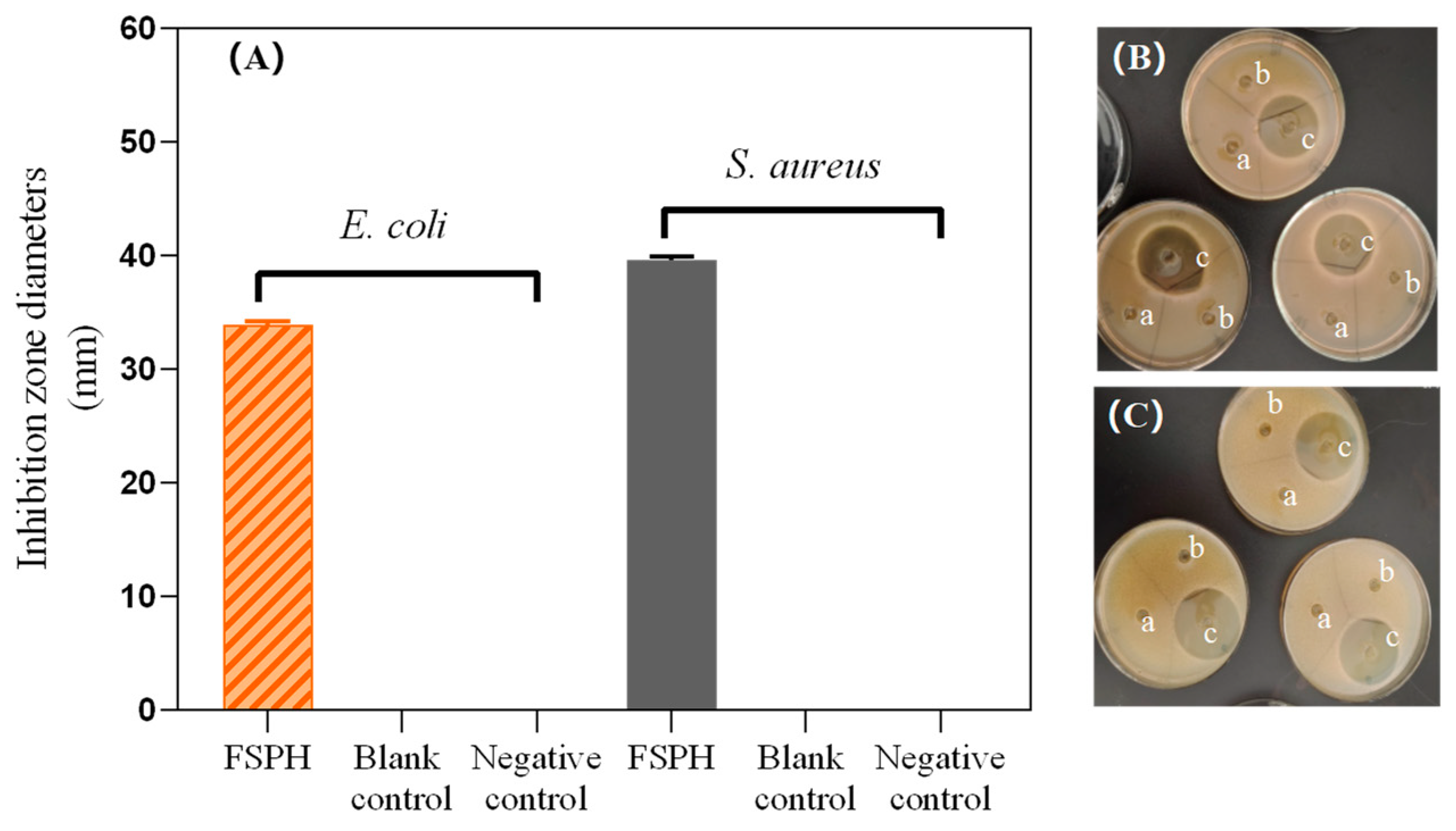
Figure 2. This section is organized as follows: first, the antibacterial zone diameters are presented, followed by a comparison of these findings with previous studies. Specifically, well “a” represents the blank control using physiological saline, well “b” signifies the negative control with saline adjusted to a pH of 1.5, and well “c” is designated for the FSPH treatment group (
Figure 2B,C). The blank control (well “a”) ensures no antibacterial effects from the solvent, while the negative control (well “b”) tests the potential impact of a low pH (1.5). This confirms that the observed antibacterial effects of the FSPH are due to peptides, not the acidic environment. For
E. coli, the antibacterial zone diameter of the FSPH was measured at (34.0 ± 0.3) mm, while for
S. aureus, it was (38.9 ± 0.8) mm.
3.2. Minimum Inhibitory Concentration (MIC) and Minimum Bactericidal Concentration (MBC)
The MIC and MBC values were determined for the FSPH against
E. coli and
S. aureus.
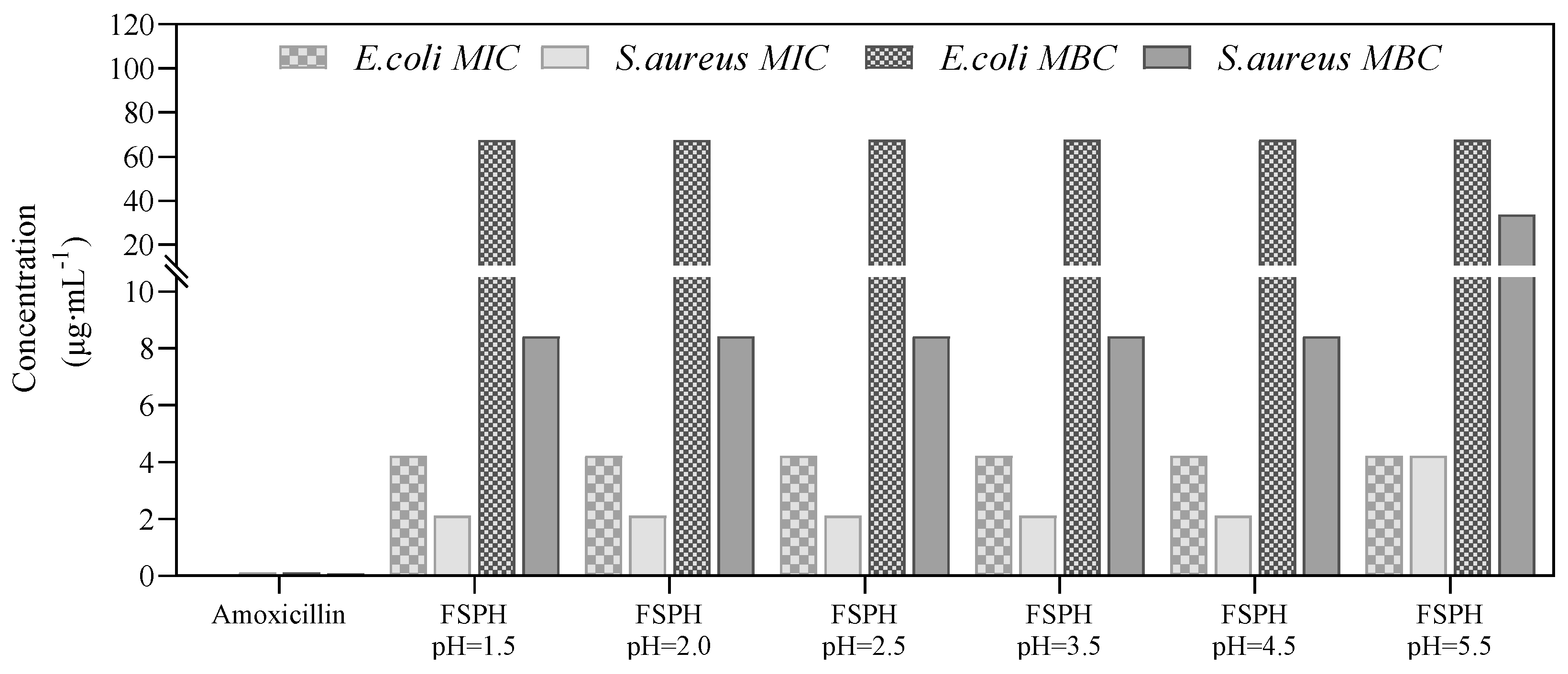
Figure 3 presents the MIC and MBC values for the FSPH under various acidic conditions, along with amoxicillin as a positive control. As depicted in
Figure 3, the FSPH demonstrated effective antibacterial and bactericidal properties against both
E. coli and
S. aureus. However, these effects were significantly weaker than those of amoxicillin, which exhibited MIC and MBC values of 0.0625 μg∙mL
−1 and 0.125 μg∙mL
−1 for
E. coli and 0.125 μg∙mL
−1 and 0.200 μg∙mL
−1 for
S. aureus, respectively.
Under acidic conditions from pH 1.5 to 5.5, the MIC and MBC for the FSPH against E. coli were consistently 4.2 μg∙mL−1 and 67.4 μg∙mL−1, respectively. This suggests that the antibacterial and bactericidal efficacy of the FSPH against E. coli is relatively unaffected by the pH changes associated with protein hydrolysis within this acidic range. The pH of the FSPH was initially very low due to the use of pepsin for enzymatic hydrolysis, which typically operates in acidic conditions. Further assessment of the potential influence of pH on the antibacterial efficacy of the FSPH was conducted by investigating its activity across a range of pH values from 1.5 to 5.5. This range was chosen to evaluate whether the observed antibacterial effects were a result of the acidic environment or if they were primarily due to the bioactive peptides. The results show that the antibacterial and bactericidal effects of the FSPH against E. coli remained consistent within this pH range, confirming that its efficacy is not significantly impacted by the pH conditions used during protein hydrolysis.
For S. aureus, the MIC and MBC of the FSPH remained stable at pH 1.5–4.5, with respective values of 2.11 μg∙mL−1 and 8.43 μg∙mL−1. It should be noted that at a pH of 5.5, there was an increase in both MIC (4.22 μg∙mL−1) and MBC (33.73 μg∙mL−1) values for the protein hydrolysates. This indicates a diminished antibacterial and bactericidal potency of the FSPH against S. aureus as the pH of the hydrolysate approaches neutrality.
3.3. Thermal Stability and Freeze–Thaw Stability of FSPH
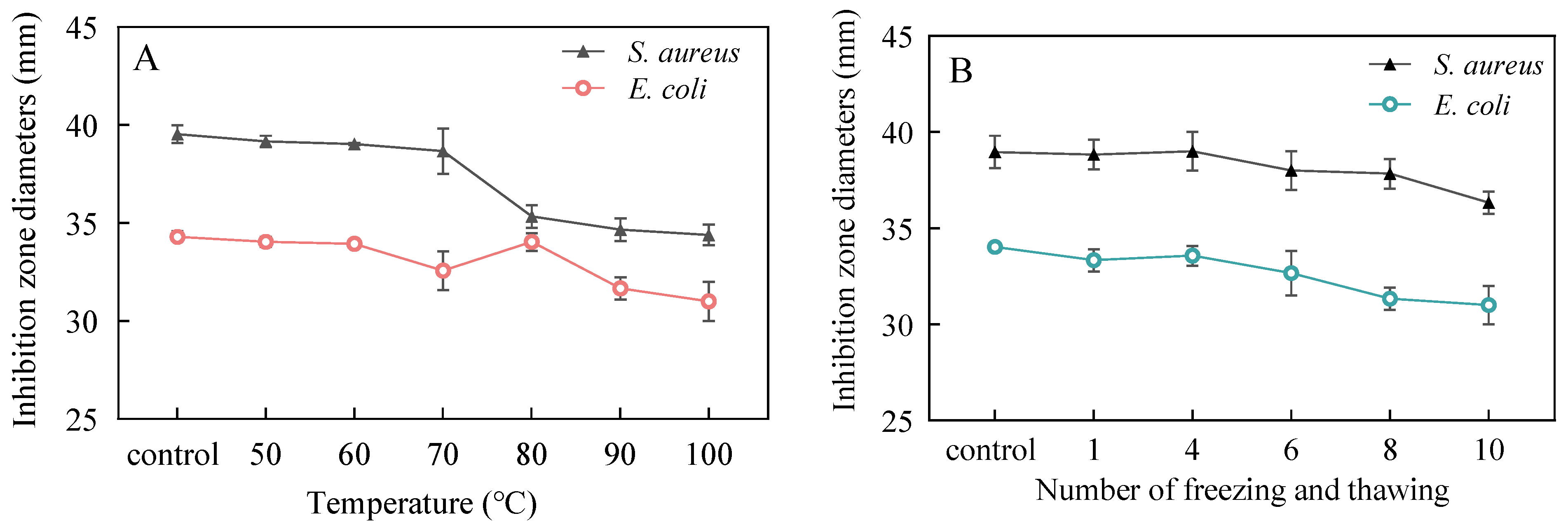
The FSPH was subjected to heating for 10 min at temperatures ranging from 50 to 100 °C, and the antimicrobial activity was measured by the diameter of inhibition zones, as shown in
Figure 4A. The results revealed a slight decrease in the antimicrobial activity against
S. aureus and
E. coli with increasing temperature. Significant differences (
p < 0.05) in antimicrobial activity against S. aureus were observed when the treatment temperature reached 80 °C, 90 °C, and 100 °C compared to the non-heated control group. Similarly, the FSPH treated at 90 °C and 100 °C exhibited significantly different antimicrobial activities against
E. coli (
p < 0.05). The antimicrobial activity was found to be the weakest at a treatment temperature of 100 °C, with inhibition zone diameters exceeding 34 mm and 30 mm in the two groups, respectively. These values correspond to 87.0% and 86.9% of the inhibition zone diameter observed in the control group. These findings indicate that the FSPH demonstrates good thermal stability.
The freezing and thawing cycles were conducted at a temperature of −20 °C with a 24 h interval, and the samples underwent 1, 4, 6, 8, and 10 freeze–thaw cycles. The impact of the number of freeze–thaw cycles on the antimicrobial activity is depicted in
Figure 4B. A decreasing trend in the inhibition zone diameters was observed for both bacterial strains following repeated freeze–thaw cycles, indicating a reduction in the antimicrobial activity of the FSPH. When compared to the control group, the FSPH subjected to 8 and 10 freeze–thaw cycles exhibited a significant difference (
p < 0.05) in antimicrobial activity against
E. coli. Similarly, there were notable differences (
p < 0.05) in the antimicrobial activity against
S. aureus between the treatments subjected to 8 and 10 freeze–thaw cycles. Overall, the experimental groups with the most significant decrease in antimicrobial activity, specifically those subjected to 10 freeze–thaw cycles, demonstrated a mere 9% reduction in the diameter of the inhibition zone. This finding suggests that the FSPH maintains favorable freeze–thaw stability within 10 cycles, highlighting its potential as a stable antimicrobial agent.
3.4. Impact of FSPH on Bacterial Cell Membrane Permeability
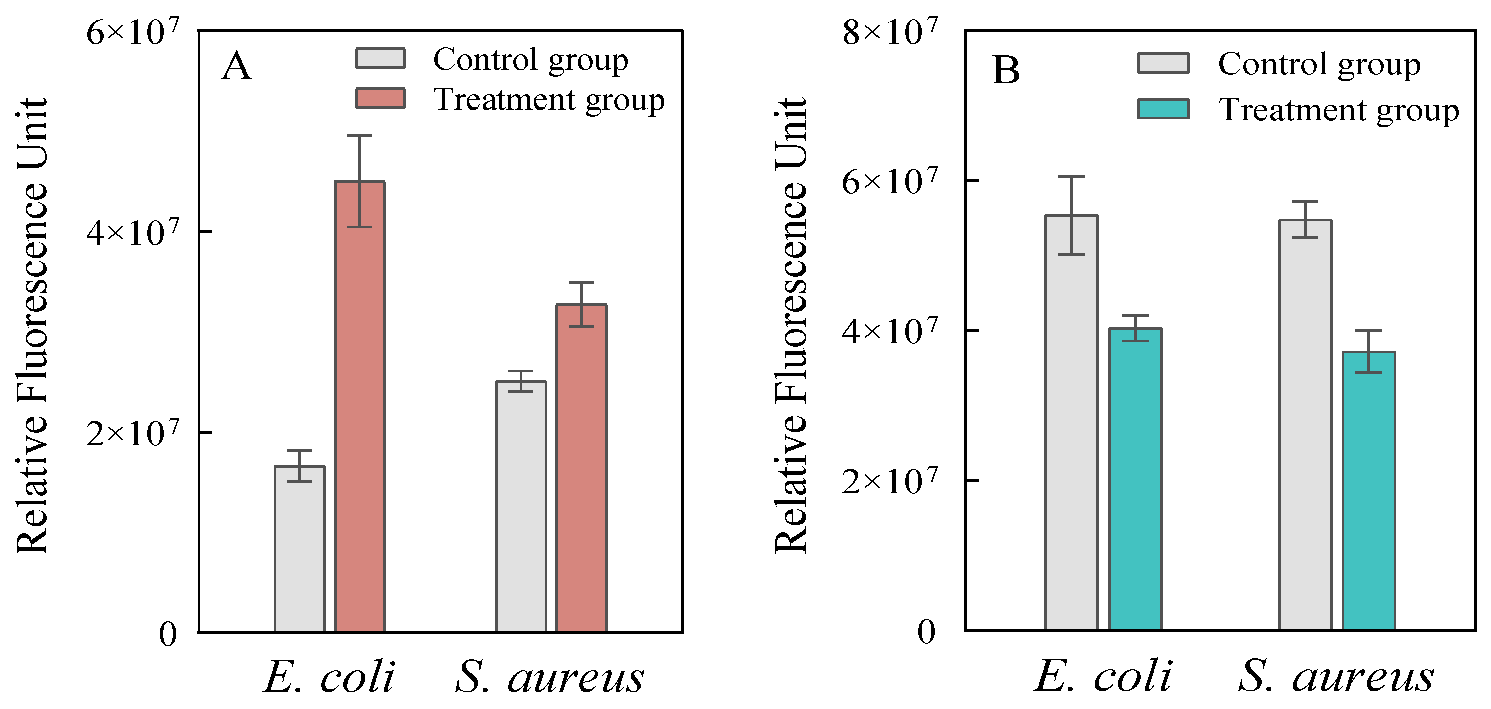
The effect of the FSPH on the permeability of bacterial cell membranes was assessed using the N-phenyl-1-naphthylamine (NPN) fluorescence probe, which is sensitive to changes in membrane integrity [
27]. Relative fluorescence units (RFUs) indicate greater disruption of the outer membrane.
Figure 5A shows a marked increase in NPN fluorescence intensity following treatment with the FSPH for both
E. coli and
S. aureus, suggesting significant disruption of the bacterial outer membranes (
p < 0.001). These results indicate that the antimicrobial action of the FSPH may involve membrane disruption, a mechanism typical of antimicrobial peptides, which primarily target bacterial membranes [
28]. However, further experiments are required to confirm the precise molecular mechanism underlying this effect.
To further investigate membrane disruption, the integrity of the bacterial inner membrane was also evaluated by measuring β-galactosidase activity. The results presented in
Figure 5B show a reduction in the fluorescence intensity of methylumbelliferyl-β-D-galactopyranoside in both
E. coli and
S. aureus after FSPH treatment. This decrease suggests that while the inner membrane may have remained largely intact, some damage occurred or the damage was below the detectable threshold.
3.5. Impact of FSPH on Bacterial Cell Morphology
Following the identification of membrane permeability alterations, their morphological consequences were subsequently examined. In this section, the impact of the FSPH on the morphology of
E. coli and
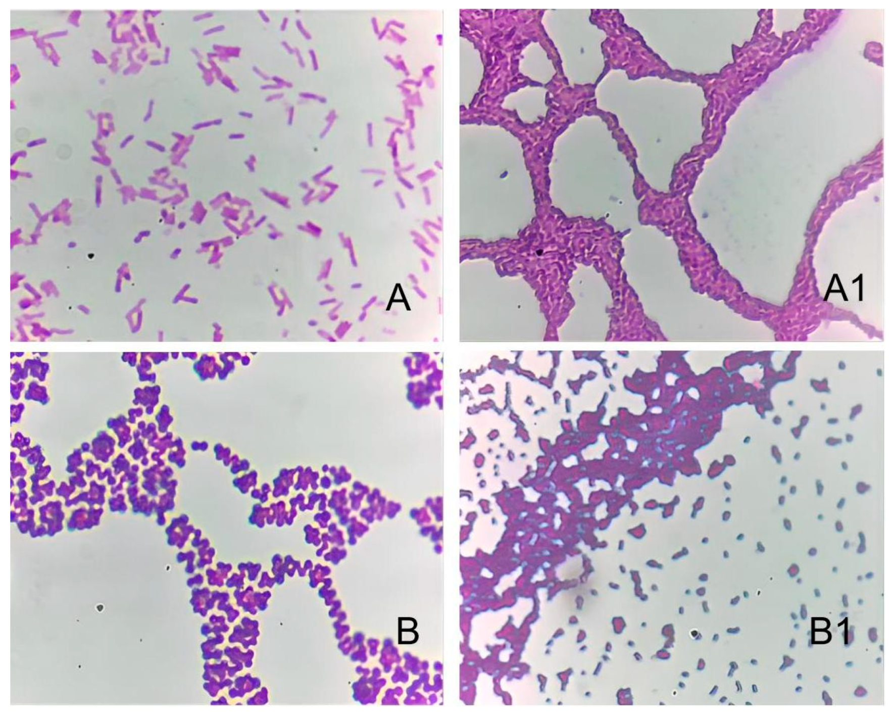
S. aureus after 16 h of treatment was examined. The bacterial cells were subjected to Gram staining, and the resulting morphological changes were observed under a microscope (
Figure 6). As shown in
Figure 6A1, the treatment group exhibited a significant dissolution of the majority of
E. coli cells, resulting in the complete loss of their typical rod-shaped morphology. The cells appear aggregated and dissolved into clumps, as shown throughout the image. In contrast, the control group (
Figure 6A) maintained the usual rod-shaped structure. Similarly, for
S. aureus, the treatment group displayed significant dissolution and deformation of the cells (
Figure 6B1), with most cells appearing disintegrated and clustered together, whereas the control group remained spherical (
Figure 6B). These morphological alterations suggest that prolonged exposure to FSPHs can disrupt the integrity of the bacterial cell walls, leading to the leakage of intracellular contents and ultimately causing cell aggregation or death.
4. Discussion
The fish scale protein hydrolysates (FSPHs) derived from grass carp (
Ctenopharyngodon idella) demonstrated antibacterial activity against both
E. coli and
S. aureus, with inhibition zones measuring 34.0 ± 0.3 mm and 38.9 ± 0.8 mm, respectively. These values indicate that the FSPH possesses potent antibacterial activity, which significantly exceeds the measurements reported by Shi et al. [
17] for protein hydrolysates from crucian carp scales, where values were 17.0 mm for
E. coli and 20.2 mm for
S. aureus.
As summarized in
Table 1, the MIC values of the FSPH (4.2 μg·mL
−1 for
E. coli; 2.1 μg·mL
−1 for
S. aureus) are lower than those of hydrolysates produced through conventional acid–enzyme [
20], double-enzyme [
17], or pepsin-based methods [
29]. The MBC values (67.5 μg·mL
−1 for
E. coli; 8.4 μg·mL
−1 for
S. aureus) also reflect bactericidal activity comparable to synthetic preservatives [
12,
13] and other marine-derived peptides [
29,
30,
31]. These findings align with the existing literature on the antimicrobial potential of fish scale hydrolysates [
17,
19] while providing new data on grass carp as a source.
A noteworthy finding was the thermal and freeze–thaw stability of the FSPH. Despite a 13% reduction in activity after 30 min of exposure to 100 °C, the FSPH retained 87% of its initial antibacterial efficacy. This observation aligns with the findings of Ren X. et al. [
32]. regarding the thermal stability of antimicrobial peptides extracted from fish scales. The stability of the FSPH may be attributed to the extraction process, which involves acid extraction, ultrasonic treatment, and enzymatic hydrolysis. These processes likely induce protein denaturation and conversion into smaller peptides [
33], which have been reported to exhibit enhanced thermal stability under high-temperature conditions [
34,
35]. Such stability characteristics are particularly advantageous for applications in thermally processed foods or topical formulations requiring an extended shelf life.
The impact of FSPHs on bacterial cell morphology was assessed using Gram staining and microscopic observation. Prolonged exposure to the FSPH resulted in significant morphological alterations in both
E. coli and
S. aureus. The bacterial cells exhibited dissolution, deformation, and loss of typical morphology, with
E. coli losing its rod-shaped structure and
S. aureus showing deformation of its spherical shape. These morphological changes indicate damage to the bacterial cell wall, leading to the leakage of intracellular contents and potential cell aggregation or death. Similar morphological disruptions have been reported for other antimicrobial peptides that target bacterial membranes [
36]. The observed cell lysis and disruption strongly suggest that the FSPH exerts its antimicrobial action by targeting the structural integrity of bacterial cells. Membrane permeability assays further supported these findings, with increased fluorescence observed in both
E. coli and
S. aureus following FSPH treatment. This suggests that the antimicrobial action of the FSPH may involve disruption of the bacterial membranes, a mechanism typically associated with antimicrobial peptides [
28,
36].
While the study provides valuable insights into the antibacterial properties and stability of the FSPH, there are notable limitations. The identification of specific peptides responsible for the observed antimicrobial effects remains unresolved. Although peptide content was measured, further analysis using advanced techniques such as LC-MS/MS would be necessary to identify and characterize the active peptides within the hydrolysate. The lack of detailed information on the molecular weight distribution of the peptides also limits the understanding of their structural characteristics and potential applications [
28]. Future studies should aim to identify the specific bioactive peptides in the FSPH and investigate their antimicrobial mechanisms in more detail.
5. Conclusions
This study highlights the potential of FSPHs as sustainable antimicrobial agents derived from grass carp scales. The FSPH, produced through citric acid extraction and enzymatic hydrolysis with pepsin, demonstrated significant antibacterial activity against both Gram-positive (S. aureus) and Gram-negative (E. coli) bacteria, confirming its broad-spectrum efficacy. The MIC and MBC values, along with its stability under acidic conditions (<5.5), suggest that the FSPH can remain effective in diverse environments. Furthermore, the thermal and freeze–thaw stability of the FSPH supports its potential for use in conditions where temperature fluctuations are common.
The antimicrobial action of the FSPH appears to involve disruption of the bacterial cell envelope, as evidenced by increased membrane permeability and subsequent bacterial cell lysis. These findings position the FSPH as a sustainable alternative to synthetic antimicrobials, promoting fishery waste valorization for applications in food, pharmaceuticals, and biotechnology.
However, while permeability assays indicate membrane destabilization, direct evidence of molecular interactions between FSPH peptides and bacterial membranes remains absent. Future studies should focus on elucidating the precise molecular mechanisms involved in FSPH’s antimicrobial activity. Techniques such as molecular docking, fluorescence microscopy, or atomic force microscopy could provide valuable insights into peptide–bacteria interactions and the specific mechanisms underlying the observed antimicrobial effects.
To translate these findings, policymakers and industry leaders should explore integrating fish by-products (e.g., scales) into value-added products like FSPHs, advancing circular economies while reducing reliance on conventional chemicals. Concurrently, further research must identify active peptides within FSPHs, characterize their properties, and optimize production processes for cost efficiency.
This work advances both theoretical and practical knowledge of fish-derived antimicrobials, providing a foundation for their use in food safety and healthcare. Future efforts should bridge the gap between observed efficacy and molecular mechanisms to fully unlock FSPH’s therapeutic and industrial potential.

