Abstract
Due to the characteristics of high salt and high acidity of the electrolyte and the chemical reaction between thiourea (Tu) and metal ions, it is still a problem to quickly and accurately detect Tu concentration in copper electrolyte. An improved spectrophotometric method has been proposed by simplifying the extraction steps of the traditional extraction–spectrophotometry and reducing the dosage of extractant and buffer. The improved method, with a favorable R2 of 0.9991, great precision of 1.67% and excellent spiked recovery of 102.19%, was not affected by the presence of gelatin and copper ions. Moreover, based on the spectrophotometry, a colorimetric method for rapid detection of Tu concentration in copper electrolyte was proposed. Using the standard color card produced enabled the expeditious determination of the concentration range of Tu in copper electrolyte. Since the gray value of the standard color card is linearly related to the Tu concentration, it is feasible to determine the Tu concentration by measuring the gray value of the test strip, and its accuracy was verified by spectrophotometry. The colorimetric method has satisfactory results in industrial practice. This study provides a novel approach for the rapid detection of Tu concentration in the copper electrolysis industry.
1. Introduction
The acceleration of industrialization coupled with the rapid development of the new energy industry has resulted in a significant increase in copper demand and improvement in quality requirements [1]. Electrolytic copper represents a significant source of high-quality copper materials. Industrial practice has proved that the proper addition of gelatin, thiourea (Tu), Avitone, chloride ions and other additives [2,3] in the electrolytic process can effectively avoid various crystallization defects and improve the quality of cathode copper. Among them, Tu is a commonly used additive for copper electrorefining.
Tu, also known as thiocarbamide or isothiourea, is an organic sulfur-containing compound with the chemical formula of CH4N2S. The addition of an appropriate amount of Tu can refine the cathode copper grains and depolarize the cathode [4]. During the electrodeposition process, Tu adsorbed on the active sites of copper substrate hinder the reduction process of copper ions, resulting in slow growth of copper grains and obtaining flat and bright copper with ideal size. When Tu is added simultaneously with gelatin and chloride ions, a synergistic effect can be achieved, resulting in a brighter and denser cathode copper crystal surface compared to using gelatin or chloride ions alone [5,6]. However, when the concentration of Tu in the electrolyte is excessive, it severely inhibits the nucleation rate of sedimentary layers, causing copper grains to become coarse and ultimately leading to uneven copper surfaces. Furthermore, the excessive Tu also increases the sulfur content on the cathode copper and reduces the quality of the product. In addition, the incompletely consumed Tu will remain in the electrolyte, resulting in the decrease in electrolysis efficiency when its concentration accumulates to a certain level [7]. Therefore, the concentration of Tu in copper electrolyte is a pivotal factor influencing the quality of the copper cathode. This means that monitoring the concentration of Tu in the copper electrolyte and making a timely adjustment during the electrolysis process is crucial for obtaining high-quality cathode copper products.
Due to the pervasive application and certain biological hazards, the monitoring of Tu in daily life has received widespread attention [8,9]. The widely used methods are mainly divided into three categories: electrochemical method, spectrophotometric analysis and instrumental detection. The electrochemical method depends on the difference of potential, current and other electrochemical parameters produced by different concentrations of Tu on the electrode. It exhibits a good linear relationship with Tu concentration within a certain range [10,11,12]. As a typical example, Manea et al. [12] determined the Tu concentration in water with the current of copper oxide–copper electrode in the range of 1–7 mM. The reproducibility experiment yielded a relative standard deviation (RSD) of 3.0%, and the spiked recovery experiment yielded a spiked recovery of 97% with RSD of 3.5%. In addition, several novel electrochemical sensors have been prepared for the detection of Tu. Jodan et al. [13] prepared an electrochemical sensor through modification of glass–carbon electrode with alizarin yellow polymer and Ag nanoparticles. The RSD was 2.2% and the spiked recoveries ranged from 98.4% to 103.5% for the detection of Tu in water. The spectrophotometric method [8,14,15], another commonly used testing method, utilizes the unique properties or the characteristic of Tu reacting with other substances to generate absorbing substances and measures the absorbance of the substance at a specific wavelength to detect the Tu concentration. For instance, Abbasi et al. [14] developed a catalytic kinetic spectrophotometric method using the catalytic effect of Tu on the oxidation of Janus green by potassium iodate. The method can be used for the determination of Tu in fruit juices in the range of 0.01–12.00 mg/L, and the RSD at 8.0 mg/L was 1.68%. Instrumental analysis is also a commonly used method for analyzing Tu with high precision. For example, Huan et al. [16] have detected Tu concentration in water by Raman-assisted synthesized Ag/CeO2 nanomaterials, and the recovery rate in river water ranged from 95% to 104%, with an RSD of less than 3.78%. It is evident that the above methods exhibit greater accuracy and adaptability for the determination of pure Tu and Tu in simple systems. However, the application of these methods to the determination of Tu in electrolyte systems is still a problem.
The key reason for the above problems is the high salt (40 g/L Cu2+) and high acid (190 g/L H2SO4) in the copper electrolyte, as well as the interference of gelatin, Avitone and other additives and impurity ions. Therefore, it is still a challenging task for rapid and accurate detection of Tu concentration in copper electrolyte. Akeneev et al. [11] developed a voltametric method to detect Tu concentration in copper electrolyte in the range of 1.0–30.0 mg/L. However, the method was not only affected by gelatin but also had poor reproducibility and precision of 20% and 30%, respectively. Pedre et al. [17] realized the direct determination of Tu concentration in electrolyte systems by preparing a sensor modified with silver nanoparticles for promoting the oxidation of Tu on the surface of an electrode. Although the method has good accuracy in the presence of certain amounts of gelatin and Avitone, it is still limited by frequent electrode changes and a restricted detection range (1–10 mg/L), which does not correspond to reality. Duan et al. [18] developed a relatively accurate spectrophotometric method for the determination of Tu concentration in copper electrolyte. Unfortunately, it also has shortcomings such as a too narrow linear detection range (0–4 mg/L), large test dose consumption and cumbersome operation. Collet et al. [19] employed ion chromatography to determine the concentration of Tu in their study of the effect of additives on the cathodic plating process. Rethmeier et al. [20] achieved the detection of Tu in copper electrolyte by high-performance liquid chromatography (HPLC) on the condition of using a C18-silica column with double-distilled water as the eluent. The advantage of instrumental detection is that it is not influenced by heavy metals and the results are accurate. However, the expense of the instrument and the high cost of learning make it difficult to apply for rapid measurements in the industrial field. In addition to the above methods, to the best of our present knowledge, relatively few studies have been conducted on the detection methods of Tu concentration in electrolytes.
Another reason for the difficulties in determining the concentration of Tu in copper electrolytes is due to the hydrolysis and accumulation of Tu in an acidic environment and the complex reaction between Tu and copper ions. Firstly, Tu is prone to undergoing hydrolysis reaction as shown in Equation (1) to produce urea (CO(NH2)2), resulting in the increase in analysis results due to differences in molecular structure and element types. Secondly, Tu reacts with Cu2+ in acidic electrolyte to form formamidine disulfide (FDS, C2H6N4S2) due to its reducibility, as shown in Equation (2) [21]. Both the reduced Cu+ and Cu2+ in electrolyte can also be complexed with Tu to form [Cu(Tu)2]2+ or [Cu(Tu)2]+ compounds due to the presence of single electron pairs on sulfur and nitrogen atoms [22]. Tu and its various complexes can adsorb on the surface of copper electrodes and improve the quality of cathode copper. However, it can also desorb from the cathode surface and enter the solution [23,24] due to the dynamic equilibrium between adsorption and desorption. To compensate for the consumption of Tu, intermittent supplementation is necessary during industrial continuous production, resulting in the accumulation of Tu in electrolyte. In addition, Tu can also polymerize to form poly-Tu [24].
CS(NH2)2 + 2H2O → CO(NH2)2 + H2S
2Cu2+ + 4 CS(NH2)2 → S2C2H6N4 + 2[Cu(CS(NH2)2)]+ + 2H+
Due to the above complex reactions, accurate detection of Tu content in industrial electrolytes is extremely difficult, resulting in the fact that determination of the level of Tu concentration in practice still relies on experience. Therefore, it remains highly meaningful to design a method for rapidly determining the concentration range of Tu. The objective of this study is to develop a rapid semi-quantitative analytical method capable of indicating the concentration levels of Tu, thereby facilitating production guidance.
2. Materials and Methods
2.1. Materials
Anhydrous sodium acetate (Analytical Reagent, AR) was purchased from Guangdong Guanghua Technology Co., Ltd. (Shantou, China). Anhydrous ethanol (99%), copper sulfate pentahydrate (AR), glacial acetic acid (AR), ethyl acetate (AR) and potassium iodide (AR) were obtained from Sinopharm Chemical Reagents Co., Ltd. (Shanghai, China). Concentrated sulfuric acid (98%) and iodine (AR) were bought from Macklin biochemical technology Co., Ltd. (Shanghai, China). Tu and gelatin were industrial-grade and received from a domestic copper electrolysis factory and used without purification. The deionized water used in all experiments was produced by the ultrapure water machine in our laboratory.
2.2. Preparation of Standard Solutions
Tu stock solution, iodine standard solution, acetic acid–sodium acetate buffers and simulated electrolyte were prepared before the study. A total of 1.000 g of Tu was accurately weighed and dissolved in deionized water. After Tu dissolved completely, it was diluted with deionized water in a 1000 mL volumetric flask to obtain the stock solution with a concentration of 1000 mg/L. The iodine solution was prepared by dissolving 0.500 g of potassium iodide and 0.250 g of iodine in deionized water and then diluted in a 1000 mL brown volumetric flask. To obtain acetic acid–sodium acetate buffer with pH = 4.2, the mixture containing 33.0 g of anhydrous sodium acetate and 50.0 mL of glacial acetic acid was diluted with deionized water in a 1000 mL volumetric flask. The simulated copper electrolyte containing 40 g/L of Cu2+, 190 g/L of H2SO4 and a certain concentration of Tu was prepared by diluting the solution of 7.8 g of copper sulfate pentahydrate, 5.4 mL of concentrated sulfuric acid and a certain volume of Tu stock solution in a 50 mL volumetric flask.
2.3. Spectrophotometric Assay Procedure
The determination of Tu concentration using traditional spectrophotometry involves two steps: drawing the working curve and measuring the absorbance of the sample. Firstly, the desired volume of Tu stock solution and deionized water were added into a separating funnel by pipettes (100–1000 μL, Eppendorf AG, Hamburg, Germany) to obtain Tu solution with standard concentrations of 0.0, 10.0, 20.0, 30.0, 40.0, 50.0, 60.0 and 70.0 mg/L. After adjusting the pH to 4.2 with 2.0 mL of glacial acetic acid and 20.0 mL of acetic acid sodium acetate buffer solution, 5.0 mL of iodine solution was added to react with Tu. The remaining iodine was extracted with 20.0 mL of ethyl acetate. After the mixture undergoes oscillation and stratification, the aqueous phase was discarded and 5.0 mL of organic phase was transferred to the colorimetric tube. The absorbance of oil phase was measured at a wavelength of 450 nm by a UV-vis spectrophotometer (Shanghai AOE Instruments Co., Ltd., Shanghai, China), using ethyl acetate as reference. Finally, the working curve was obtained by drawing the curve with absorbance as the x-axis and Tu concentration as the y-axis. The absorbance value of samples, including the simulated and industrial electrolyte, was measured using the same procedure as the working curve manufacturing method. The concentration of Tu in specimens can be obtained by substituting the absorbance value into the working curve. In the improved method, the separatory funnel was replaced with a sample vial that simplifies the fussy extraction process to directly extract the oil phase from the upper layer after a few minutes. Moreover, the usage of acetic acid was removed, ethyl acetate was replaced with n-butanol and the amount of buffer solution was reduced by half. These improvements make it more suitable for industrial applications.
To validate the accuracy of the method, this study conducted several fundamental validation experiments, including precision, reproducibility and recovery tests [25,26]. Additionally, linearity and relative error can also be employed to demonstrate the accuracy of the method. In the precision experiment, all samples with known concentrations were repeatedly tested six times and the RSD was calculated according to Equation (3).
where xi is the measured value, is the average of multiple measurements and n is the measuring numbers. In repetitive experiments of simulated electrolyte, samples with known concentrations were also detected six times repeatedly. The RSD and relative error of the result were calculated according to Equation (3) and Equation (4), respectively.
where δ is the relative error, t is the truth-value of the measurement object and xi − t is the absolute error. The spiked recovery experiments were carried out by adding different amounts of Tu to the solution containing a known concentration of Tu. The recovery rate was calculated using Equation (5).
where P is the recovery rate, x1 is the mass of Tu contained in the original sample, x2 is the measured mass of Tu after spiking and x3 is the mass of added Tu. In addition, all presented data are the average of three measurements.
2.4. Colorimetric Method Assay Procedure
When using the colorimetric method to determine the concentration of Tu in the electrolyte, a series of standard solutions containing 0.0, 20.0, 40.0, 60.0 and 80.0 mg/L of Tu were prepared by diluting the stock solution. Afterward, 10 mL of acetic acid–sodium acetate buffer solution and 5.0 mL iodine solution were added into 1.0 mL Tu solution with different concentrations. After 5 min of reaction, starch potassium iodide test strips (Shanghai SSS Reagent Co., Ltd., Shanghai, China) were immersed to display color by the highly sensitive color reaction between starch and residual iodine in solution. Finally, a standard color card was obtained by RGB values after imaging the strips with different color rendering levels [27]. Tu concentration range in the samples was obtained by comparing the colorimetric test strips with the standard color card.
To reflect the color differences of test strips objectively, ImageJ software (v1.54g, National Institutes of Health, Bethesda, MD, USA) was used to obtain the gray values of the chromogenic test strips after taking pictures under the same light source; the pictures were transformed into an 8-bit format with uniform backgrounds, and the gray value of the strips were measured by selecting an area with a fixed position and size. Afterwards, the working curve of color card was established with gray value as the y-axis and Tu concentration as the x-axis. During the detection of Tu concentration in the samples, the gray value obtained with the above method was brought into the working curve, and accurate results could be obtained.
3. Results and Discussion
3.1. Establishment and Verification of Spectrophotometry for Determining Tu Concentration
3.1.1. Establishment of Spectrophotometry
The principle of Tu detection using spectrophotometry is based on the utilization of the quantitative reaction of the reducing Tu with the oxidizing iodine, and the concentration of Tu can be calculated by measuring the remaining iodine. The working curve was drawn as shown in Figure 1a, and the linear regression equation is y = 77.359 − 263.1x, R2 = 0.9991 > 0.99. When the amount of iodine solution added was 5.0 mL, the working curve had a great linear relationship in the range of 0–70.0 mg/L. The absorbance changes over time were obtained by time-scanning the organic phase with a UV-vis spectrophotometer. From Figure 1a, the absorbance decreased only 0.02 after 1800 s; such insignificant changes indicate a stable methodology. Moreover, the precision experiment was carried out by detecting 40 mg/L of pure Tu; from the detection results in Figure 1b, the measured results were around the true value, and the relative error of the spectrophotometric method was less than 4%, with an RSD of 1.67%. Overall, the excellent linear correlation, stable absorbance values, lower relative error and small RSD values of the spectrophotometric method indicate that it was suitable for accurately determining the concentration of Tu.
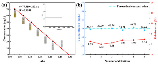
Figure 1.
(a) Working curve and the change in absorbance value over time of spectrophotometric method. (b) Result of precision experiment.
3.1.2. Elimination of Interference Factors in Tu Determination on Spectrophotometry
Due to the presence of excess acidity and copper ions in the complex electrolyte system, the effect of acidity and copper ions on the detection method was investigated as shown in Figure 2a,b. The simulated copper electrolyte containing 70 mg/L of Tu was diluted to different multiples; subsequently, 1 mL was taken and a quantitative buffer was added and the pH value was measured using a pH meter (Mettler, Zurich, Switzerland). Finally, the concentration of Tu was detected by the spectrophotometric method. From Figure 2a, with the increase in the dilution times, the pH value gradually converges to 4.2, the Tu concentration is close to the real value and the relative error also decreases gradually, which indicates that the test results are greatly affected by the acidity. This is due to the presence of 190 g/L of H2SO4 in the electrolyte, and it is difficult to adjust the pH to 4.2 after adding the buffer directly. Therefore, to ensure the accuracy of the detection results, it is necessary to either add a large amount of buffer or add a quantitative amount of buffer after diluting the electrolyte. In order not to increase the usage of buffer while reducing the concentration of copper ions, the copper electrolyte was diluted, and the detection results were accurate when the electrolyte was diluted at least fourfold. In addition, the effect of Cu2+ is illustrated in Figure 2b. The measured concentrations of Tu were close to the true values when the Cu2+ concentration was 0–20 g/L, and the relative error was less than 2.25%. A deviation from the measured results occurred at a further increase in Cu2+ up to 40 g/L, but it was still acceptable. The influence of Cu2+ was negligible in consideration of the need for dilution during the actual measurement.
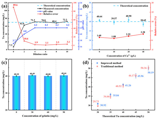
Figure 2.
(a) Effect of acidity on spectrophotometric method. (b) Effect of copper ions on spectrophotometric method. (c) Effect of gelatin on spectrophotometric method. (d) Comparison of traditional and improved spectrophotometric methods.
Gelatin is a commonly used additive in copper electrolysis; therefore, the accuracy of spectrophotometric detection of Tu concentration in the presence of gelatin was also investigated. The results are shown in Figure 2c. With the addition of various concentrations of gelatin, the measured concentration of Tu remained essentially unchanged and the relative errors were all less than 2%, which indicates that the addition of gelatin would not interfere with the detection of Tu.
To prove that the improved spectrophotometric method is workable, different concentrations of Tu were detected by the improved spectrophotometric method and compared with the traditional extraction–spectrophotometry, and the results are shown in Figure 2d. When the Tu concentrations were 30, 35, 40, 45 and 50 mg/L, the relative deviation between the results measured by the improved method and those obtained by the traditional method was 3.92%, 3.35%, 1.81%, 0.46% and 0.34%, respectively. It can be observed that the discrepancies between the two methods are minimal and fall within acceptable limits, indicating that the modified spectrophotometric approach can also be reliably applied for the precise determination of Tu concentrations. However, compared with the traditional method, the improved method simplifies the extraction steps and avoids the use of a large amount of buffer and extractant, which is more convenient and suitable for industrial field conditions.
3.1.3. Accuracy Verification of Tu Concentration Determination by Spectrophotometry
Under the simulated electrolyte, a repeatability experiment and spiked recovery experiment were carried out on the improved spectrophotometric method to validate the feasibility of the method in complex systems. The Tu concentration of the tested sample during the repeatability experiment was 60 mg/L, with the results as shown in Figure 3a. It shows the relative error of Tu concentration in the electrolyte measured by spectrophotometric method is between 0.52% and 3.13%, with an RSD of 2.35%. The spiked recovery experiments were performed in a simulated electrolyte containing a certain amount of Tu, and the results are shown in Table 1. The spiked recovery rate ranges from 99.44% to 104.58%, with an average value of 102.19%. The repeatability experiment and the spiked recovery experiment demonstrated the spectrophotometric method’s high reproducibility and accuracy, thereby satisfying the determination requirements.
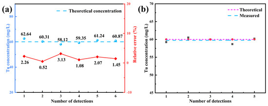
Figure 3.
(a) Results of the repeatability experiment. (b) Determination of Tu concentration in complex simulated electrolyte system by spectrophotometric method.

Table 1.
Results of spiked recovery experiment.
Furthermore, the concentration of Tu in a simulated electrolyte containing gelatin was detected, and the results are as shown in Figure 3b. The spectrophotometric measurement result is close to the theoretical value, which proves that the method is reliable and could be used for the determination of Tu concentration in a complex simulated electrolyte system. Importantly, the presence of gelatin in the electrolyte cannot affect the detection.
3.1.4. Validation of the Method
To verify the field applicability of the spectrophotometric method, Tu in the compound additive prepared by a copper electrolysis plant was detected. The additive solution contained Tu, gelatin and Avitone, with a theoretical concentration of Tu of approximately 9545 mg/L, and sampling occurred at a fixed time daily for six days. It can be seen from Figure 4a that the measured concentration of Tu in the additive is close to the theoretical value; the relative error is within 10%. It is worth noting that the measured concentration is higher than the theoretical concentration. Part of the error may be from systematic errors. Another reason is probably the accumulation of Tu in the electrolytic system due to the prolonged operation of the industrial electrolyte.
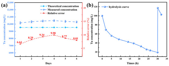
Figure 4.
(a) Validation of spectrophotometric method industrial sites. (b) Hydrolysis curve of Tu in simulated electrolyte at 60 °C (copper electrolyte containing 40 g/L Cu2+, 190 g/L H2SO4 and Tu 100.0 mg/L).
As an attempt to investigate the hydrolysis and accumulation of Tu in the copper electrolyte, the prepared simulated electrolyte was placed in a water bath at 60 °C, consistent with the industrial field. Afterwards, the concentration of Tu at different times was determined by spectrophotometry, and the Tu hydrolysis curve was obtained as shown in Figure 4b. It is obvious that the Tu concentration decreases fastest within 0–2 h, indicating that the hydrolysis was the fastest when Tu was just added. After 2 h, the rate of hydrolysis gradually slowed down with the extension of time. However, even after a 24 h period had elapsed, 25 mg/L of Tu still existed, when 100.00 mg/L of Tu was added at 30 h, the detected result was 117.28 mg/L. The Tu concentration continued to decrease when placed at 60 °C and fell faster when it was just added. It is well known that, in industrial sites, fresh additives were added to the electrolytic cell every 12 or 24 h, and Tu was not completely consumed at that time. Part of the excess Tu in electrolyte comes from the newly added and the other part may come from the desorption of copper complexes [24], while these Tu will slowly accumulate in the electrolyte, gradually increasing the concentration of Tu in the electrolyte. In conclusion, the continuous accumulation of Tu is the foremost reason for the large Tu concentration in the electrolyte.
3.2. Establishment and Verification of Colorimetric Method for Determining Tu Concentration
3.2.1. Establishment of Colorimetric Method
During the color development of a test strip, when the immersion time is too short, the color development is incomplete and, when the immersion time is too long, the generated color-developing substances may dissolve into the solution. Therefore, it is necessary to discuss the effect of immersion time on the color of the test strip. When the test strip was immersed in solution 1, 3, 5 and 10 s, the color rendering degree was observed and photographed. Moreover, the average gray value analysis of the chromogenic test strip was performed by ImageJ software. Figure 5a demonstrates the correspondence between color and depth, while the standardized measurement positions for gray value analysis are illustrated in Figure 5b. As revealed in Figure 5e, when the immersion time is 10 s and the Tu concentration is 10 and 40 mg/L, the color of the test strip is darker and the gray value is smaller. Conversely, when the immersion time is shortened to 1 s, the color of the test strips under different Tu concentrations is light and the gray value is large. At different concentrations, the color development and gray value of the test strip with immersion time of 3 and 5 s are close to each other; hence, the immersion time of the test strip was chosen as 3 s. Afterwards, as shown in Figure 5c, a standard color card was prepared according to the experimental method. The semi-quantitative detection range of the colorimetric method is 0–20, 20–40, 40–60 and 60–80 mg/L. The level of Tu concentration in copper electrolyte can be quickly obtained by comparing with the standard color card. Furthermore, the standard color cards were processed with ImageJ software for gray analysis, and the corresponding relationship between the gray value of the color card and the Tu concentration was obtained as shown in Figure 5d. The linear regression equation of the working curve is y = 1.8203 x − 279, R2 = 0.9821, a more accurate result was acquired by gray analysis.
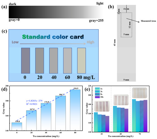
Figure 5.
(a) Correspondence between gray value and color depth. (b) Diagram of gray value reading position. (c) Standard color card for Tu detection. (d) Corresponding relationship between gray value and Tu concentration of standard color card. (e) Effect of immersion time on color display of test strips.
The accuracy of the colorimetric method was verified by repeating the measurement three times with various concentrations of Tu. As illustrated in Figure 6a, the color rendering of the test strips under different Tu concentrations was within the corresponding detection concentration range, indicating that the concentration range of the samples can be determined by colorimetric color cards. Further, gray value analysis of the test strips is as shown in Figure 6b, and the values of the three tests are close, which proves that the display of the color card is relatively stable. Correspondingly, the gray value was converted to Tu concentration by calculation, and the results are shown in Figure 6c. The Tu concentration obtained from the gray analysis was close to the theoretical value. Consequently, the colorimetric method can effectively determine the concentration range of Tu in a sample by naked eye observation, and the gray analysis can provide more precise results, thereby enhancing the accuracy of the assessment.
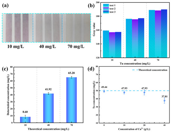
Figure 6.
(a) Visual observation to verify the accuracy of the colorimetric method. (b) Gray value test results of color-revealing test strips. (c) Accuracy experiment results of calculating Tu concentration based on gray value. (d) Effect of copper ions on colorimetric method.
The high concentration of colored ions in the electrolyte may influence the color development of the test strip; hence, the effect of Cu2+ was studied. As illustrated in Figure 6d, at a Cu2+ concentration of 40 g/L, the mean concentration of Tu derived from the gray analysis was 37.8 mg/L, a substantial departure from the actual value of 50 mg/L. Conversely, when the concentration of Cu2+ is reduced to below 20 g/L, the concentration of Tu obtained from the gray analysis approaches the theoretical value, and the colorimetry of the test strip remains basically unaltered. This observation suggests that the colorimetry of the test strip remains unaffected in the presence of Cu2+ concentrations below 20 g/L.
3.2.2. Accuracy Verification and Industrial Application of Colorimetric Method in Electrolyte
During the detection of Tu concentration in the simulated electrolyte, to ensure the complete elimination of the effect of Cu2+, the simulated electrolyte was diluted fourfold so that the Cu2+ concentration was 10 g/L. Then, various concentrations of Tu were added to the diluted simulated electrolytes, ensuring the final concentrations of Tu in the solution were 10, 35 and 60 mg/L, respectively. Afterwards, the Tu concentration was detected by the colorimetric method and the spectrophotometric method to verify the feasibility of the colorimetric method in simulated electrolytes, and the results of the two methods are shown in Table 2. The detection results obtained by the colorimetric method and the spectrophotometric method exhibited a high degree of concordance.
Table 2.
Comparison of results obtained by colorimetric method and spectrophotometric method.
Tu concentration in complex additives was detected in a copper electrolysis plant to validate the applicability of the colorimetric method in industrial sites. Afterwards, the results were compared with those obtained by the spectrophotometric method. As can be seen in Figure 7a, the results of both methods are close to each other, indicating that the colorimetric method can also be used to detect Tu concentration in industrial sites. The measured concentration was slightly higher than the theoretical concentration, which is also related to the accumulation of Tu.
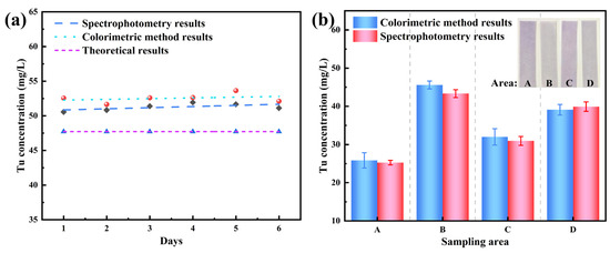
Figure 7.
(a) Accuracy validation of the colorimetric method in industrial sites. (b) Application and validation of the colorimetric method in industrial electrolysis systems.
The industrial applicability of the colorimetric method was systematically validated through a three-tiered analytical protocol. Firstly, visual estimation via the colorimetric method rapidly identified Tu concentration ranges in copper electrolytes within 10 min, enabling preliminary on-site assessment. Subsequently, gray analysis was performed to improve the accuracy of the colorimetric method and obtain more accurate results. Finally, spectrophotometric quantification confirmed the method reliability, and the results are displayed in Figure 7b. The addition amounts of Tu in different areas of the industrial site are consistent with our measured results. The colors of the chromogenic color cards obtained by the colorimetric method detection of Tu concentration in electrolytes from different production areas vary. Therefore, the colorimetric method can be used to rapidly determine the concentration range of Tu in industrial electrolytes, providing theoretical guidance for adjusting additives in industrial settings.
The colorimetric method proposed in this study enables rapid determination of Tu concentration ranges in electrolytes by observing color intensity variations on test strips. As a human-eye-dependent semi-quantitative detection approach, this method demonstrates simplicity and rapidity, making it highly suitable for practical applications [27,28,29]. Based on the chromogenic principle of iodine–starch reactions, starch–potassium iodide test strips were selected as color indicators. When reacting with residual iodine in the solution, the test strips develop a purplish-red coloration, resembling the typical iodine color observed in nonpolar hydrocarbon solvents [30]. In the industry, the colorimetric method has excellent industrial applicability without using any instrument, but there are still some limitations in judging Tu concentration by human eyes, and gray analysis can improve the accuracy but it will affect the convenience of the method, so the accuracy of the colorimetric method is still a problem to be solved.
As demonstrated in Table 3, the two proposed methods in this study demonstrate substantial improvements in detection speed compared with existing techniques for Tu concentration analysis in electrolyte solutions, while exhibiting excellent portability and promising industrial applicability.
Table 3.
Detection methods for Tu concentration in copper electrolytes and acidic electroplating wastewater.
4. Conclusions
This study developed an improved spectrophotometric method by simplifying extraction steps and reducing the dosage of extractant and buffer, which lowered costs and accelerated detection. The optimized method showed excellent linearity (R2 = 0.9991, range: 0–70 mg/L), precision (RSD = 1.67%) and recovery (102.19%), with minimal influence from gelatin/Cu2+. Results aligned with traditional methods, confirming its reliability for Tu concentration detection in copper electrolytes.
Building on the improved spectrophotometric method, a rapid colorimetric method was proposed for industrial applications. This approach completes analysis within 10 min and eliminates copper ion interference by diluting the electrolyte. Additionally, a linear relationship between test strip gray values and Tu concentrations (R2 = 0.9821) was established. Validation tests confirmed its accuracy and consistency with the spectrophotometric results. Field trials confirmed the viability of colorimetric methods for real-time Tu concentration monitoring in copper electrorefining, where timely determination is crucial. This approach combines operational convenience and rapid analysis efficiency, providing a practical solution for industrial applications.
Author Contributions
Writing—original draft, investigation, L.C.; Supervision, Validation, X.Y., Y.L. and Z.Z.; Funding acquisition, Y.X.; Materials and sample provision, W.Z.; Conceptualization, Writing—review and editing Y.W. All authors have read and agreed to the published version of the manuscript.
Funding
This work was supported by Key Research and Development Program of Gansu Province (24YFGA023) and Major Science and Technology Project of Gansu Province (23ZDGA010).
Data Availability Statement
The original contributions presented in this study are included in the article. Further inquiries can be directed to the corresponding author.
Conflicts of Interest
Author Wenqian Zhou was employed by Jinchuan Group Copper and Precious Metals Co., Ltd. The remaining authors declare that the research was conducted in the absence of any commercial or financial relationships that could be construed as a potential conflict of interest.
References
- Stefanie, K.; Stefan, P. Sector-Level Estimates for Global Future Copper Demand and the Potential for Resource Efficiency. Resour. Conserv. Recycl. 2023, 193, 106941. [Google Scholar]
- Cheng, J.; Ding, L.; Zhao, J.; Wang, T.; Wang, R.; Chen, C.; Niu, Y. Roles of some additives in continuous electrolytic refining of cathode copper. Electroplat. Finish. 2020, 39, 383–389. [Google Scholar]
- Fang, Y.; Pan, M.; Huang, H.; Shao, Y.; He, Y.; Chen, B.; Guo, Z. Current situation and prospect of additives in copper electrolysis deposition process. Min. Metall. 2021, 30, 61–69. [Google Scholar]
- Quinet, M.; Lallemand, F.; Ricq, L.; Hihn, J.-Y.; Delobelle, P.; Arnould, C.; Mekhalif, Z. Influence of Organic Additives on the Initial Stages of Copper Electrodeposition on Polycrystalline Platinum. Electrochim. Acta 2009, 54, 1529–1536. [Google Scholar] [CrossRef]
- Collet, T.; Wouters, B.; Hallemans, N.; Ramharter, K.; Lataire, J.; Hubin, A. The Time-Varying Effect of Thiourea on the Copper Electroplating Process with Industrial Copper Concentrations. Electrochim. Acta 2023, 437, 141412. [Google Scholar] [CrossRef]
- Zhang, H.; Xiong, J.; Zhong, S.; Zhang, P.; Shen, K. Mechanism of Action and Detection Method of Copper Electrowinning Additive. Nonferrous Met. Eng. Res. 2016, 37, 14–16. [Google Scholar]
- Kang, M.S.; Kim, S.K.; Kim, K.; Kim, J.J. The Influence of Thiourea on Copper Electrodeposition: Adsorbate Identification and Effect on Electrochemical Nucleation. Thin Solid Film. 2008, 516, 3761–3766. [Google Scholar]
- Duca, G.; Lis, A.; Gladchi, V.; Travin, S. Indirect Photolysis of Cysteine and Thiourea in the Aquatic Environment. Inorg. Chim. Acta 2023, 557, 121682. [Google Scholar] [CrossRef]
- Wang, G.; Dong, Y.; Zhu, X.; Zhang, W.; Wang, C.; Jiao, H. Ultrasensitive and Selective Colorimetric Detection of Thiourea Using Silver Nanoprobes. Analyst 2011, 136, 5256–5260. [Google Scholar] [CrossRef]
- Spataru, N.; Spataru, T.; Fujishima, A. Voltammetric Determination of Thiourea at Conductive Diamond Electrodes. Electroanal 2005, 17, 800–805. [Google Scholar] [CrossRef]
- Akeneev, Y.A.; Zakharova, E.A.; Slepchenko, G.B.; Pikula, N.P. Voltammetric Determination of Thiourea in Copper Refinery Electrolytes. J. Anal. Chem. 2005, 60, 514–517. [Google Scholar] [CrossRef]
- Manea, F.; Radovan, C.; Schoonman, J. Amperometric Determination of Thiourea in Alkaline Media on a Copper Oxide–Copper Electrode. J. Appl. Electrochem. 2006, 36, 1075–1081. [Google Scholar]
- Jodan, I.; Wantala, K.; Amini, N.; Shahmoradi, B.; Ghaslani, M.; Lee, S.M.; Yang, J.; Puttaiah, S.H. Fabrication of a Sensitive Electrochemical Sensor Based on Ag Nanoparticles and Alizarin Yellow Polymer: Application to the Detection of an Environmental Pollutant Thiourea. Korean J. Chem. Eng. 2020, 37, 1609–1615. [Google Scholar] [CrossRef]
- Abbasi, S.; Khani, H.; Hosseinzadeh, L.; Safari, Z. Determination of Thiourea in Fruit Juice by a Kinetic Spectrophotometric Method. J. Hazard. Mater. 2010, 174, 257–262. [Google Scholar] [CrossRef]
- Arab Chamjangali, M.; Bagherian, G.; Goudarzi, N.; Mehrjoo-Irani, S. A New and Sensitive Reaction Rate Method for Spectrophotometric Determination of Trace Amounts of Thiourea in Different Water Samples Based on an Induction Period. J. Anal. Sci. Technol. 2015, 6, 10. [Google Scholar] [CrossRef]
- Jiang, H.; Wang, B.; Tang, R.; Tan, Y.; Qi, M.; Zhang, X. Inhibition to Dual Enzyme-like Activities of Ag/CeO2 Nanozymes for the Detection of Thiourea. Microchem. J. 2023, 185, 108251. [Google Scholar] [CrossRef]
- Pedre, I.; Méndez DeLeo, L.; Sánchez-Loredo, M.G.; Battaglini, F.; González, G.A. Electrochemical Sensor for Thiourea Focused on Metallurgical Applications of Copper. Sens. Actuators B Chem. 2016, 232, 383–389. [Google Scholar]
- Duan, M.; Huang, J.; Yong, F. Extraction—Spectrophotometry determination of thiourea in copper electrolyte. Electroplat. Pollut. Control 1998, 26–27. [Google Scholar]
- Collet, T.; Wouters, B.; Eeltink, S.; Schmidt, P.; Ramharter, K.; Hubin, A. An Ex Situ and Operando Analysis of Thiourea Consumption and Activity during a Simulated Copper Electrorefining Process. Electroanal. Chem. 2022, 920, 116581. [Google Scholar] [CrossRef]
- Rethmeier, J.; Neumann, G.; Stumpf, C.; Rabenstein, A.; Vogt, C. Determination of Low Thiourea Concentrations in Industrial Process Water and Natural Samples Using Reversed-Phase High-Performance Liquid Chromatography. J. Chromatogr. A 2001, 934, 129–134. [Google Scholar] [CrossRef]
- Fabbri, L.; Giurlani, W.; Mencherini, G.; De Luca, A.; Passaponti, M.; Piciollo, E.; Fontanesi, C.; Caneschi, A.; Innocenti, M. Optimisation of Thiourea Concentration in a Decorative Copper Plating Acid Bath Based on Methanesulfonic Electrolyte. Coatings 2022, 12, 376. [Google Scholar] [CrossRef]
- Jin, S.; Ghali, E. Effect of Thiourea on the Copper Cathode Polarization Behavior in Acidic Copper Sulfate at 65 °C. Metall. Mater. Trans. B 2001, 32, 887–893. [Google Scholar]
- Lu, S. Discussion on the function and mechanism of additives in copper electrolysis. Nonferrous Met. Extr. Metall. 1984, 41–46. [Google Scholar]
- Bolzán, A.E.; Wakenge, I.B.; Piatti, R.C.V.; Salvarezza, R.C.; Arvia, A.J. The Behaviour of Copper Anodes in Aqueous Thiourea-Containing Sulphuric Acid Solutions. Open Circuit Potentials and Electrochemical Kinetics. Electroanal. Chem. 2001, 501, 241–252. [Google Scholar] [CrossRef]
- Thakur, D.; Dubey, N.P.; Singh, R. A Review on Spike and Recovery Method in Analytical Method Development and Validation. Crit. Rev. Anal. Chem. 2024, 54, 2053–2071. [Google Scholar] [CrossRef]
- Raposo, F.; Ibelli-Bianco, C. Performance Parameters for Analytical Method Validation: Controversies and Discrepancies among Numerous Guidelines. TrAC Trends Anal. Chem. 2020, 129, 115913. [Google Scholar]
- Fernandes, G.M.; Silva, W.R.; Barreto, D.N.; Lamarca, R.S.; Lima Gomes, P.C.F.; Flávio da S Petruci, J.; Batista, A.D. Novel Approaches for Colorimetric Measurements in Analytical Chemistry—A Review. Anal. Chim. Acta 2020, 1135, 187–203. [Google Scholar]
- Ren, D.; Mei, J.; Bao, J.; Wei, F.; Xu, G.; Yang, J.; Sun, Y.; Hu, Q.; Cen, Y. A Novel Profuse Color Card for Convenient Visual Determination of Iodide in Human Urine Based on Catalytic Oxidation Reaction. J. Pharm. Biomed. 2020, 191, 113580. [Google Scholar] [CrossRef]
- Jia, Y.; Zheng, W.; Zhao, X.; Zhang, J.; Chen, W.; Jiang, X. Mixing-to-Answer Iodide Sensing with Commercial Chemicals. Anal. Chem. 2018, 90, 8276–8282. [Google Scholar]
- Cui, K.; Lv, M.; Wang, R.; Han, L.; Liu, K.; Wang, Y. Experimental study on reaction conditions of iodine and starch. Sci. Technol. Inf. 2007, 29–31. [Google Scholar]


Disclaimer/Publisher’s Note: The statements, opinions and data contained in all publications are solely those of the individual author(s) and contributor(s) and not of MDPI and/or the editor(s). MDPI and/or the editor(s) disclaim responsibility for any injury to people or property resulting from any ideas, methods, instructions or products referred to in the content. |
© 2025 by the authors. Licensee MDPI, Basel, Switzerland. This article is an open access article distributed under the terms and conditions of the Creative Commons Attribution (CC BY) license (https://creativecommons.org/licenses/by/4.0/).