
Fluorometric Sensing of Arsenic in Water: Recent Developments in Metal-Organic Framework-Based Sensors


Abstract

1. Introduction
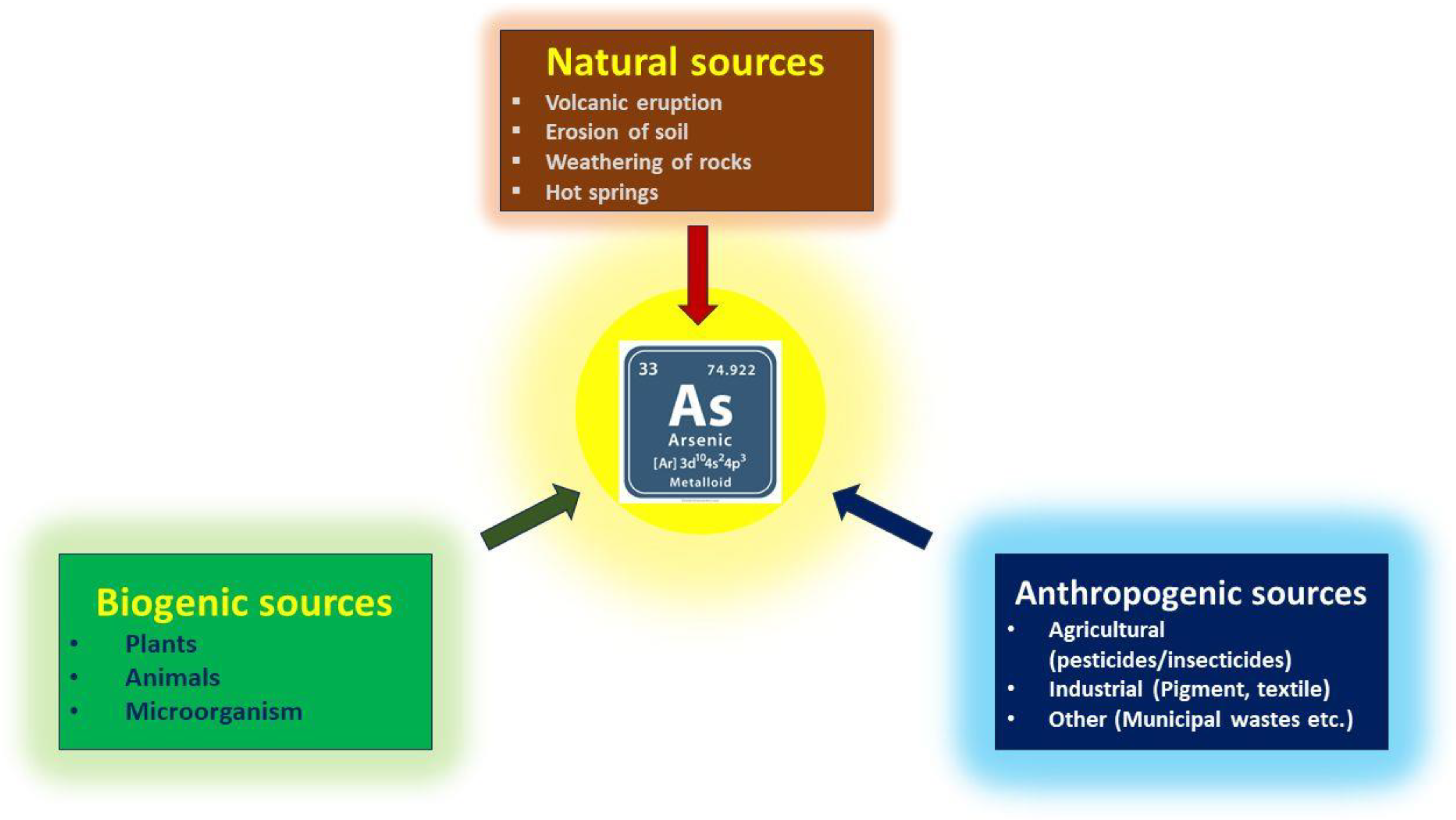
2. The Adverse Impacts of Arsenic
3. Conventional Techniques and Fluorometric Recognition of Arsenic
4. Key Features of MOFs for Fluorometric Detection
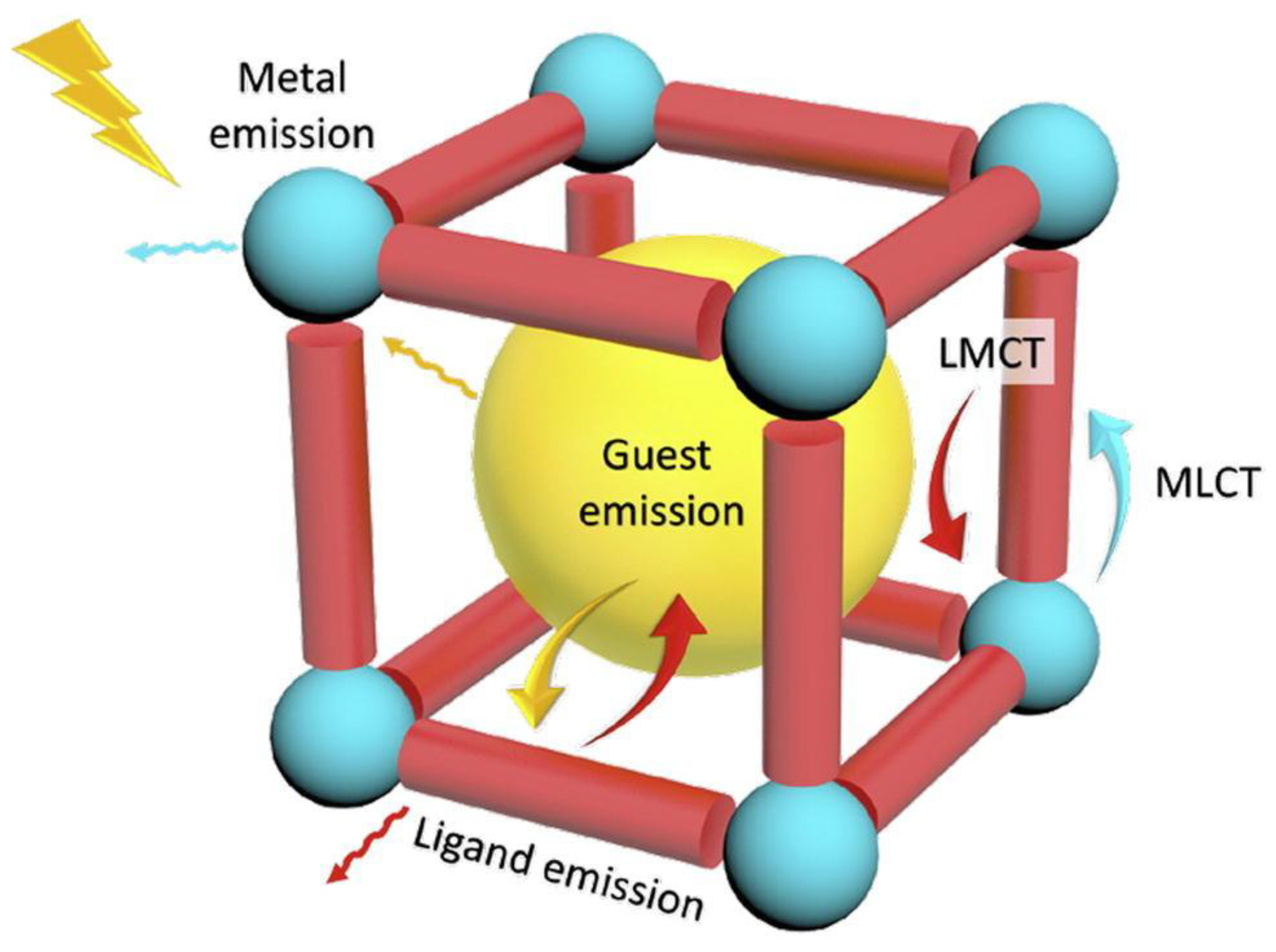
5. Origin of Fluorescence in MOFs
5.1. Ligand Based Fluorescence
5.2. Metal-Centered Fluorescence
5.3. Charge Transfer Fluorescence
5.4. Guest Induced Fluorescence
6. Fluorometric Detection of Arsenic by MOFs
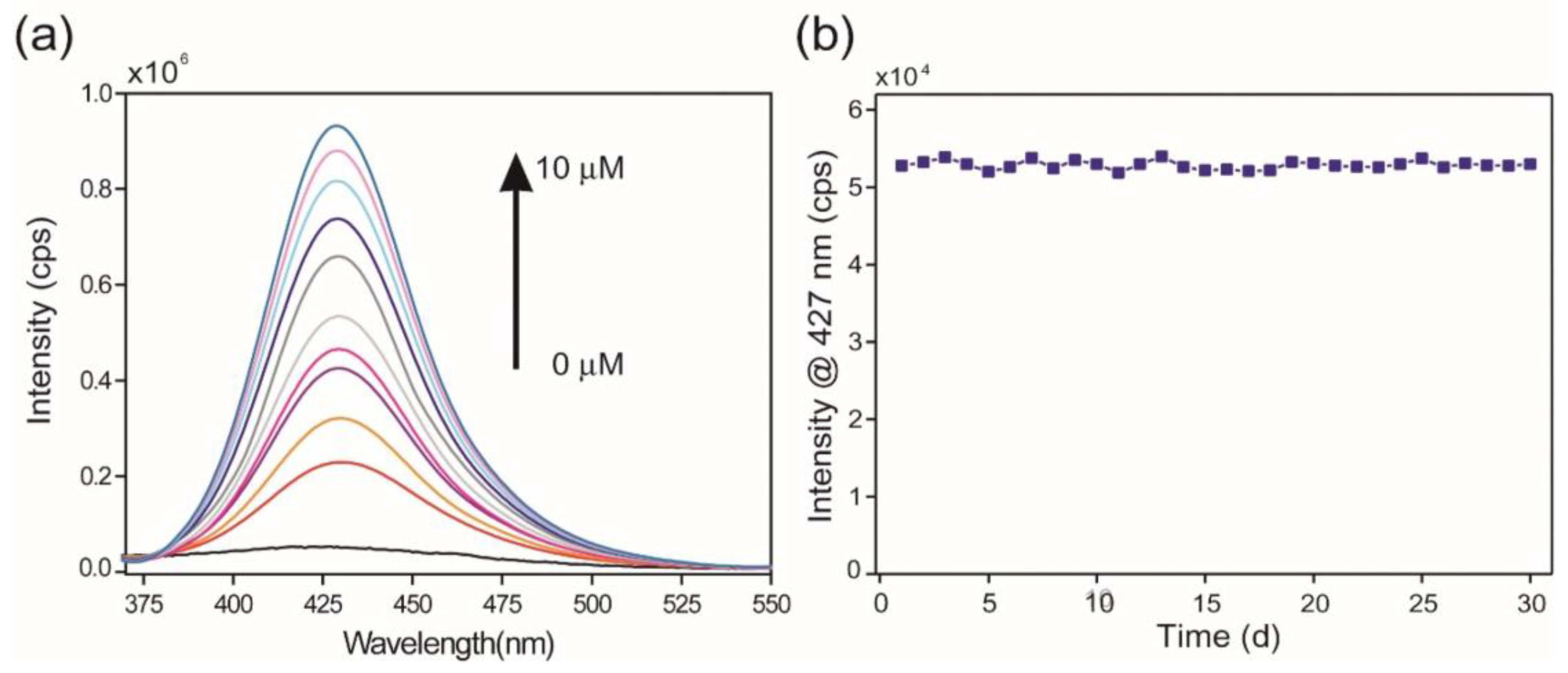
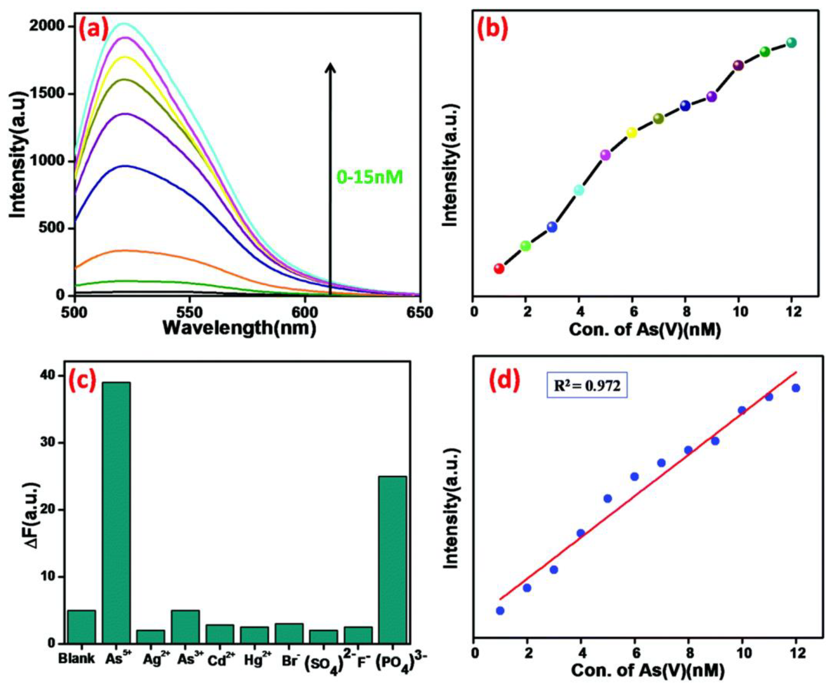
6.1. “Turn On” Sensing of Arsenic
6.2. “Turn Off” Sensing of Arsenic
7. Conclusions and Prospects for the Future
Author Contributions
Funding
Data Availability Statement
Conflicts of Interest
References
- Yoshida, T.; Yamauchi, H.; Sun, G.F. Chronic health effects in people exposed to arsenic via the drinking water: Dose–response relationships in review. Toxicol. Appl. Pharmacol. 2004, 198, 243–252. [Google Scholar] [PubMed]
- Richardson, S.D.; Ternes, T.A. Water analysis: Emerging contaminants and current issues. Anal. Chem. 2005, 77, 3807–3838. [Google Scholar] [CrossRef] [PubMed]
- Ahmad, A.; Bhattacharya, P. Arsenic in Drinking Water: Is 10 µg/L a Safe Limit? Curr. Pollut. Rep. 2019, 5, 1–3. [Google Scholar]
- Olavarria-Fullerton, J.; Wells, S.; Ortiz-Rivera, W.; Sepaniak, M.J.; DeJesus, M.A. Surface-enhanced Raman scattering (SERS) characterization of trace organoarsenic antimicrobials using silver/polydimethylsiloxane nanocomposites. Appl. Spectrosc. 2011, 65, 423–428. [Google Scholar]
- Gamboa-Loira, B.; Cebrián, M.E.; Franco-Marina, F.; López-Carrillo, L. Arsenic metabolism and cancer risk: A meta-analysis. Environ. Res. 2017, 156, 551–558. [Google Scholar]
- Sharma, V.K.; Sohn, M. Aquatic arsenic: Toxicity, speciation, transformations, and remediation. Environ. Int. 2009, 35, 743–759. [Google Scholar]
- Mangalgiri, K.P.; Adak, A.; Blaney, L. Organo arsenicals in Poultry litter: Detection, fate, and toxicity. Environ. Int. 2015, 75, 68–80. [Google Scholar]
- Smedley, P.L.; Kinniburgh, D.G. A review of the source, behaviour and distribution of arsenic in natural waters. Appl. Geochem. 2002, 17, 517–568. [Google Scholar]
- Lee, J.-J.; Kim, Y.-K.; Cho, S.-H.; Park, K.-S.; Chung, I.-J.; Cho, D.; Ryang, D.-W.; Kim, H.-J. Hemolytic anemia as a sequela of arsenic intoxication following long-term ingestion of traditional chinese medicine. J. Korean Med. Sci. 2004, 19, 127–129. [Google Scholar]
- Scott, N.; Hatlelid, K.M.; MacKenzie, N.E.; Carter, D.E. Reactions of arsenic(III) and arsenic(V) species with glutathione. Chem. Res. Toxicol. 1993, 6, 102–106. [Google Scholar]
- Ding, W.-Q.; Labiadh, L.; Xu, L.; Li, X.-Y.; Chen, C.; Fu, M.-L.; Yuan, B. Current advances in the detection and removal of organic arsenic by metal-organic frameworks. Chemosphere 2023, 339, 139687. [Google Scholar] [CrossRef] [PubMed]
- Zhang, L.; Chen, X.-R.; Wen, S.-H.; Liang, R.-P.; Qiu, J.-D. Optical sensors for inorganic arsenic detection. Trends Anal. Chem. 2019, 118, 869–879. [Google Scholar]
- Kolya, H.; Hashitsume, K.; Kang, C.-W. Recent advances in colorimetric detection of arsenic using metal-based nanoparticles. Toxics 2021, 9, 143. [Google Scholar] [CrossRef] [PubMed]
- Qiu, Y.; Yu, S.; Li, L. Research progress in fluorescent probes for arsenic species. Molecules 2022, 27, 8497. [Google Scholar] [CrossRef]
- Thakkar, S.; Dumée, L.F.; Gupta, M.; Singh, B.R.; Yang, W.; Gamboa-Loira, B. Nano–Enabledsensorsfor detectionofarsenicin water. Water Research 2021, 188, 116538. [Google Scholar]
- Devi, P.; Thakur, A.; Lai, R.Y.; Saini, S.; Jain, R.; Kumar, P. Progress in the materials for optical detection of arsenic in water. Trends Anal. Chem. 2019, 110, 97–115. [Google Scholar] [CrossRef]
- Abernathy, C.O.; Thomas, D.J.; Calderon, R.L. Health effects and risk assessment of Arsenic. J. Nutr. 2003, 133, 1536S–1538S. [Google Scholar] [CrossRef]
- Brouwer, O.; Onkenhout, W.; Edelbroek, P.; Kom, J.D.; Wolff, F.D.; Peters, A. Increased neurotoxicity of arsenic in methylenetetrahydrofolate reductase deficiency. Clin. Neurol. Neurosurg. 1992, 94, 307–310. [Google Scholar]
- Civantos, D.P.; Rodríguez, A.L.; Aguado-Borruey, J.M.; Narvaez, J.A.J. Fulminant malignant arrythmia and multiorgan failure in acute arsenic poisoning. Chest 1995, 108, 1774–1775. [Google Scholar]
- Tseng, W.P.; Chu, H.M.; How, S.W.; Fong, J.M.; Yeh, S.; Lin, C.S. Prevalence of skin cancer in an endemic area of chronic arsenicism in Taiwan. J. Natl. Cancer Inst. 1968, 40, 453–463. [Google Scholar]
- Thomas, D.J.; Styblob, M.; Linc, S. The cellular metabolism and systemic toxicity of arsenic. Toxicol. Appl. Pharmacol. 2001, 176, 127–144. [Google Scholar] [PubMed]
- Cullen, W.; McBride, B.; Reglinski, J. The reduction of trimethylarsine oxide to trimethylarsine by thiols: A mechanistic model for the biological reduction of arsenicals. J. Inorg. Biochem. 1984, 21, 45–60. [Google Scholar] [CrossRef]
- Mazumder, D.N.G. Chronic arsenic toxicity & human health. Indian J. Med. Res 2008, 128, 436–447. [Google Scholar]
- Zhang, Q.; Minami, H.; Inoue, S.; Atsuya, I. Differential determination of trace amounts of arsenic (III) and arsenic (V) in seawater by solid sampling atomic absorption spectrometry after preconcentration by coprecipitation with a nickel–pyrrolidine dithiocarbamate complex. Anal. Chim. Acta 2004, 508, 99–105. [Google Scholar] [CrossRef]
- Hung, D.Q.; Nekrassova, O.; Comptons, R.G. Analytical methods for inorganic arsenic in water: A review. Talanta 2004, 64, 269–277. [Google Scholar] [CrossRef]
- Mao, X.; Qi, Y.; Huang, J.; Liu, J.; Chen, G.; Na, X.; Wang, M.; Qian, Y. Ambient-temperature trap/release of arsenic by dielectric barrier discharge and its application to ultratrace arsenic determination in surface water followed by atomic fluorescence spectrometry. Anal. Chem. 2016, 88, 4147–4152. [Google Scholar] [CrossRef]
- Colon, M.; Hidalgo, M.; Iglesias, M. Arsenic determination by ICP-QMS with octopole collision/reaction cell. Overcome of matrix effects under vented and pressurized cell conditions. Talanta 2011, 85, 1941–1947. [Google Scholar] [CrossRef]
- Diesel, E.; Schreiber, M.; Meer, J.R.v.d. Development of bacteria-based bioassays for arsenic detection in natural waters. Anal. Bioanal. Chem. 2009, 394, 687–693. [Google Scholar] [CrossRef]
- Barros, H.; Parra, L.-M.M.; Bennun, L.; Greaves, E.D. Determination of arsenic in water samples by Total Reflection X-Ray Fluorescence using pre-concentration with alumina. Spectrochim. Acta Part B 2010, 65, 489–492. [Google Scholar]
- Jiang, M.; Ma, M.-J.; Yang, M.; Fang, L.; Li, Y.-X.; Zhao, N.-J.; Huang, X.-J. Highly sensitive and stable analysis of trace arsenic(III) and mercury(II) in water by Low-pulse-energy (15 mJ) laser-induced breakdown spectroscopy assisted by active controllable spark discharge and electrochemical enrichment. Sens. Actuators B 2020, 305, 127486. [Google Scholar] [CrossRef]
- Banik, D.; Manna, S.K.; Mahapatra, A.K. Recent development of chromogenic and fluorogenic chemosensors for the detection of arsenic species: Environmental and biological applications. Spectrochim. Acta A 2021, 246, 119047. [Google Scholar]
- Samanta, T.; Shunmugam, R. Colorimetric and fluorometric probes for the optical detection of environmental Hg(II) and As(III) ions. Mater. Adv. 2021, 2, 64–95. [Google Scholar]
- Luong, J.H.T.; Lam, E.; Male, K.B. Recent advances in electrochemical detection of arsenic in drinking and ground waters. Anal. Methods 2014, 6, 6157–6169. [Google Scholar] [CrossRef]
- Jiang, H.-L.; Tatsu, Y.; Lu, Z.-H.; Xu, Q. Non-, Micro-, and Mesoporous Metal–Organic Framework Isomers: Reversible Transformation, Fluorescence Sensing, and Large Molecule Separation. J. Am. Chem. Soc. 2010, 132, 5586–5587. [Google Scholar]
- Lee, M.H.; Yoon, B.; Kim, J.S.; Sessler, J.L. Naphthalimide trifluoroacetylacetonate: A hydrazine-selective chemodosimetric sensor. Chem. Sci. 2013, 4, 4121–4126. [Google Scholar]
- Carter, K.P.; Young, A.M.; Palmer, A.E. Fluorescent sensors for measuring metal ions in living systems. Chem. Rev. 2014, 114, 4564–4601. [Google Scholar]
- Fang, X.; Zong, B.; Mao, S. Metal–organic framework-based sensors for environmental contaminant sensing. Nano-Micro Lett. 2018, 10, 64. [Google Scholar]
- Zhang, Z.; Zhuang, Z.; Song, L.L.; Lin, X.; Zhang, S.; Zheng, G.; Zhan, F. A FRET-based ratiometric fluorescent probe for hydrazine and its application in living cells. J. Photochem. Photobiol. A 2018, 358, 10–16. [Google Scholar] [CrossRef]
- Sun, J.; Zhao, J.; Wang, L.; Li, H.; Yang, F.; Yang, X. Inner filter effect-based sensor for horseradish peroxidase and its application to fluorescence immunoassay. ACS Sens. 2018, 3, 183–190. [Google Scholar]
- Abdelhamid, H.N.; B.-Gómez, A.; M.-Matute, B.; Zou, X. A water-stable lanthanide metal-organic framework for fluorimetric detection of ferric ions and tryptophan. Microchim. Acta 2017, 184, 3363–3371. [Google Scholar]
- Luo, F.; Batten, S.R. Metal–organic framework (MOF): Lanthanide(iii)-doped approach for luminescence modulation and luminescent sensing. Dalton Trans. 2010, 39, 4485–4488. [Google Scholar] [CrossRef] [PubMed]
- Nandi, S.; Mondal, A.; Reinsch, H.; Biswas, S. An ultra-robust luminescent CAU-10 MOF acting as a fluorescent turn-off sensor for Cr2O72− in aqueous medium. Inorg. Chim. Acta 2019, 497, 119078. [Google Scholar] [CrossRef]
- Dalapati, R.; Nandi, S.; Reinsch, H.; Bhunia, B.K.; Mandal, B.B.; Stock, N.; Biswas, S. Fluorogenic naked-eye sensing and live-cell imaging of cyanide by a hydrazine-functionalized CAU-10 metal–organic framework. CrystEngComm 2018, 20, 4194–4201. [Google Scholar] [CrossRef]
- Eddaoudi, M.; Kim, J.; Rosi, N.; Vodak, D.; Wachter, J.; O’Keeffe, M.; Yaghi, O.M. Systematic design of pore size and functionality in isoreticular MOFs and their application in methane storage. Science 2002, 295, 469–472. [Google Scholar] [CrossRef] [PubMed]
- Wang, S.; McGuirk, C.M.; d’Aquino, A.; Mason, J.A.; Mirkin, C.A. Metal–Organic Framework Nanoparticles. Adv. Mater. 2018, 30, 1800202. [Google Scholar] [CrossRef]
- Tibbetts, I.; Kostakis, G.E. Recent Bio-Advances in Metal-Organic Frameworks. Molecules 2020, 25, 1291. [Google Scholar] [CrossRef]
- Gulcay, E.; Erucar, I. Biocompatible MOFs for Storage and Separation of O2: A Molecular Simulation Study. Ind. Eng. Chem. Res. 2019, 58, 3225–3237. [Google Scholar] [CrossRef]
- Huxford, R.C.; Rocca, J.D.; Lin, W. Metal-organic frameworks as potential drug carriers. Curr. Opin. Chem. Biol. 2010, 14, 262–268. [Google Scholar] [CrossRef]
- Leng, X.; Dong, X.; Wang, W.; Sai, N.; Yang, C.; You, L.; Huang, H.; Yin, X.; Ni, J. Biocompatible Fe-based micropore metal-organic frameworks as sustained-release anticancer drug carriers. Molecules 2018, 23, 2490. [Google Scholar] [CrossRef]
- Cui, Y.; Yue, Y.; Qian, G.; Chen, B. Luminescent functional metal–organic frameworks. Chem. Rev. 2012, 112, 1126–1162. [Google Scholar] [CrossRef]
- Zhao, B.; Chen, X.-Y.; Cheng, P.; Liao, D.-Z.; Yan, S.-P.; Jiang, Z.-H. Coordination Polymers Containing 1D Channels as Selective Luminescent Probes. J. Am. Chem. Soc. 2004, 126, 15394–15395. [Google Scholar] [PubMed]
- Xu, H.; Liu, F.; Cui, Y.; Chen, B.; Qian, G. A luminescent nanoscale metal–organic framework for sensing of nitroaromatic explosives. Chem. Commun. 2011, 47, 3153–3155. [Google Scholar]
- Rieter, W.J.; Taylor, K.M.L.; An, H.; Lin, W.; Lin, W. Nanoscale metal–organic frameworks as potential multimodal contrast enhancing agents. J. Am. Chem. Soc. 2006, 128, 9024–9025. [Google Scholar] [CrossRef] [PubMed]
- Li, X.; Wang, X.-W.; Zhang, Y.-H. Blue photoluminescent 3D Zn(II) metal-organic framework constructing from pyridine-2,4,6-tricarboxylate. Inorg. Chem. Commun. 2008, 11, 832–834. [Google Scholar]
- Eliseeva, S.V.; B€unzli, J.-C.G. Lanthanide luminescence for functional materials and bio-sciences. Chem. Soc. Rev. 2010, 39, 189–227. [Google Scholar]
- Moore, E.G.; Samuel, A.P.S.; Raymond, K.N. From Antenna to assay: Lessons learned in lanthanide luminescence. Acc. Chem. Res. 2009, 42, 542–552. [Google Scholar] [CrossRef]
- Wei, Y.; Yu, Y.; Wu, K. Highly Stable Five-Coordinated Mn(II) Polymer [Mn(Hbidc)]n (Hbidc=1H-Benzimidazole-5,6-dicarboxylate): Crystal Structure, Antiferromegnetic Property, and Strong Long-Lived Luminescence. Cryst. Growth Des. 2008, 8, 2087–2089. [Google Scholar]
- Wang, X.W.; Chen, J.-Z.; Liu, J.-H. Photoluminescent Zn(II) Metal–Organic Frameworks Built from Tetrazole Ligand: 2D Four-Connected Regular Honeycomb (4363)-net. Cryst. Growth Des. 2007, 7, 1227–1229. [Google Scholar]
- An, J.; Shade, C.M.; Chengelis-Czegan, D.A.; Petoud, S.; Rosi, N.L. Zinc-Adeninate Metal–Organic Framework for Aqueous Encapsulation and Sensitization of Near-infrared and Visible Emitting Lanthanide Cations. J. Am. Chem. Soc. 2011, 133, 1220–1223. [Google Scholar]
- Karmakar, A.; Joarder, B.; Mallick, A.; Samanta, P.; Desai, A.V.; Basu, S.; Ghosh, S.K. Aqueous phase sensing of cyanide ions using a hydrolytically stable metal–organic framework. Chem. Commun. 2017, 53, 1253–1256. [Google Scholar] [CrossRef]
- Zhang, Y.; Yuan, S.; Day, G.; Wang, X.; Yang, X.; Zhou, H.-C. Luminescent sensors based on metal-organic frameworks. Coord. Chem. Rev. 2018, 354, 28–45. [Google Scholar]
- Lustig, W.P.; Mukherjee, S.; Rudd, N.D.; Desai, A.V.; Li, J.; Ghosh, S.K. Metal–organic frameworks: Functional luminescent and photonic materials for sensing applications. Chem. Soc. Rev. 2017, 46, 3242–3285. [Google Scholar] [CrossRef] [PubMed]
- Müller-Buschbaum, K.; Beuerle, F.; Feldmann, C. MOF based luminescence tuning and chemical/physical sensing. Microporous Mesoporous Mater. 2015, 216, 171–199. [Google Scholar]
- Taylor, J.M.; Dawson, K.W.; Shimizu, G.K.H. A water-stable metal–organic framework with highly acidic pores for proton-conducting applications. J. Am. Chem. Soc. 2013, 135, 1193–1196. [Google Scholar]
- Cao, X.; Lin, W.; Zheng, K.; He, L. A near-infrared fluorescent turn-on probe for fluorescence imaging of hydrogen sulfide in living cells based on thiolysis of dinitrophenyl ether. Chem. Commun. 2012, 48, 10529–10531. [Google Scholar]
- Chen, W.; Liu, W.; Liu, X.-J.; Kuang, Y.-Q.; Yu, R.-Q.; Jiang, J.-H. A novel fluorescent probe for sensitive detection and imaging of hydrazine in living cells. Talanta 2017, 162, 225–231. [Google Scholar]
- Alezi, D.; Belmabkhout, Y.; Suyetin, M.; Bhatt, P.M.; Weselinski, L.J.; Solovyeva, V.; Adil, K.; Spanopoulos, I.; Trikalitis, P.N.; Emwas, A.H.; et al. MOF Crystal Chemistry Paving the Way to Gas Storage Needs: Aluminum-Based soc-MOF for CH4, O2, and CO2 Storage. J. Am. Chem. Soc. 2015, 137, 13308–13318. [Google Scholar]
- Li, B.; Wen, H.-M.; Zhou, W.; Chen, B. Porous metal–organic frameworks for gas storage and separation: What, how, and why? J. Phys. Chem. Lett. 2014, 5, 3468–3479. [Google Scholar] [CrossRef]
- Dalapati, R.; Sakthivel, B.; Dhakshinamoorthy, A.; Buragohain, A.; Bhunia, A.; Jainak, C.; Biswas, S. A highly stable dimethyl-functionalized Ce(IV)-based UiO-66 metal-organic framework material for gas sorption and redox catalysis. CrystEngComm 2016, 18, 7855–7864. [Google Scholar]
- Li, R.; Ren, X.; Ma, H.; Feng, X.; Lin, Z.; Li, X.; Hu, C.; Wang, B. Nickel-substituted zeolitic imidazolate frameworks for time-resolved alcohol sensing and photocatalysis under visible light. J. Mater. Chem. A 2014, 2, 5724–5729. [Google Scholar] [CrossRef]
- Liu, D.; Lu, K.; Poon, C.; Lin, W. Metal–organic frameworks as sensory materials and imaging agents. Inorg. Chem. 2014, 53, 1916–1924. [Google Scholar] [PubMed]
- Kumar, N.; Bhalla, V.; Kumar, M. Recent developments of fluorescent probes for the detection of gasotransmitters (NO, CO and H2S). Coord. Chem. Rev. 2013, 257, 2335–2347. [Google Scholar] [CrossRef]
- Xie, D.; Ma, Y.; Gu, Y.; Zhou, H.; Zhang, H.; Wang, G.; Zhang, Y.; Zhao, H. Bifunctional NH2-MIL-88(Fe) metal–organic framework nanooctahedra for highly sensitive detection and efficient removal of arsenate in aqueous media. Mater. Chem. A 2017, 5, 23794–23804. [Google Scholar]
- Yang, J.; Dai, Y.; Zhu, X.; Wang, Z.; Li, Y.; Zhuang, Q.; Shi, J.; Gu, J. Metal–organic frameworks with inherent recognition sites for selective phosphate sensing through their coordination-induced fluorescence enhancement effect. J. Mater. Chem. A 2015, 3, 7445–7452. [Google Scholar]
- Liu, S.; Liu, M.; Guo, M.; Wang, Z.; Wang, X.; Cui, W.; Tian, Z. Development of Eu-based metal-organic frameworks (MOFs) for luminescence sensing and entrapping of arsenate ion. J. Lumin. 2021, 236, 118102. [Google Scholar]
- Maity, D.; Mandal, S.K.; Guha, B.; Roy, P. A salicylaldehyde based dual chemosensor for zinc and arsenate ion detection: Biological application. Inorg. Chim. Acta. 2021, 519, 120258. [Google Scholar] [CrossRef]
- Lohar, S.; Sahana, A.; Banerjee, A.; Banik, A.; Mukhopadhyay, S.K.; Matalobos, J.S.; Das, D. Antipyrine based arsenate selective fluorescent probe for living cell imaging. Anal. Chem. 2013, 85, 1778–1783. [Google Scholar]
- Dutta, S.; Let, S.; Shirolkar, M.M.; Desai, A.V.; Samanta, P.; Fajal, S.; More, Y.D.; Ghosh, S.K. A luminescent cationic MOF for bimodal recognition of chromium and arsenic based oxo-anions in water. Dalton Trans. 2021, 50, 10133–10141. [Google Scholar]
- Shirolkar, M.M.; Athavale, R.; Ravindran, S.; Rale, V.; Kulkarni, A.; Deokar, R. Antibiotics functionalization intervened morphological, chemical and electronic modifications in chitosan nanoparticles. Nano-Struct. Nano-Obj. 2021, 25, 100657. [Google Scholar]
- Xu, X.; Luo, Z.; Ye, K.; Zou, X.; Niu, X.; Pan, J. One-pot construction of acid phosphatase and hemin loaded multifunctional metal–organic framework nanosheets for ratiometric fluorescent arsenate sensing. J. Hazard. Mater. 2021, 412, 124407. [Google Scholar] [CrossRef]
- Wen, S.-H.; Liang, R.-P.; Zeng, H.-H.; Zhang, L.; Qiu, J.-D. CdSe/ZnS quantum dots coated with carboxy-PEG and modified with the terbium(III) complex of guanosine 5′-monophosphate as a fluorescent nanoprobe for ratiometric determination of arsenate via its inhibition of acid phosphatase activity. Microchim. Acta 2019, 186, 45. [Google Scholar]
- Tong, Y.-J.; Yu, L.-D.; Wu, L.-L.; Cao, S.-P.; Liang, R.-P.; Zhang, L.; Xia, X.-H.; Qiu, J.-D. Aggregation-induced emission of luminol: A novel strategy for fluorescence ratiometric detection of ALP and As(v) with high sensitivity and selectivity. Chem. Commun. 2018, 54, 7487–7490. [Google Scholar]
- Muppidathi, M.; Perumal, P.; Ayyanu, R.; Subramanian, S. Immobilization of ssDNA on a metal–organic framework derived magnetic porous carbon (MPC) composite as afluorescent sensing platform for the detection of arsenate ions. Analyst 2019, 144, 3111–3118. [Google Scholar] [PubMed]
- Tan, H.; Tang, G.; Wang, Z.; Li, Q.; Gao, J.; Wu, S. Magnetic porous carbon nanocomposites derived from metal-organic frameworks as a sensing platform for DNA fluorescent detection. Anal. Chim. Acta 2016, 940, 136–142. [Google Scholar]
- Kim, J.S.; Quang, D.T. Calixarene-Derived Fluorescent Probes. Chem. Rev. 2007, 107, 3780–3799. [Google Scholar]
- Zheng, D.; Zou, R.; Lou, X. Label-Free Fluorescent Detection of Ions, Proteins, and Small Molecules Using Structure-Switching Aptamers, SYBR Gold, and Exonuclease I. Anal. Chem. 2021, 8, 3554–3560. [Google Scholar] [CrossRef]
- Zhu, K.; Fan, R.; Wu, J.; Wang, B.; Lu, H.; Zheng, X.; Sun, T.; Gai, S.; Zhou, X.; Yang, Y. MOF-on-MOF membrane with cascading functionality for capturing dichromate ions and p-Arsanilic Acid turn-on sensing. ACS Appl. Mater. Interfaces 2020, 12, 58239–58251. [Google Scholar]
- Huang, Q.S.; Wang, C.; Wei, W.; Ni, B.J. Magnetic poly(anilineco-5-sulfo-2-anisidine) as multifunctional adsorbent for highly effective co-removal of aqueous Cr(VI) and 2,4-Dichlophenol. Chem. Eng. J. 2020, 387, 124152. [Google Scholar]
- Gu, H.; Rapole, S.B.; Sharma, J.; Huang, Y.; Cao, D.; Colorado, H.A.; Luo, Z.; Haldolaarachchige, N.; Young, D.P.; Walters, B.B.; et al. Magnetic polyaniline nanocomposites toward toxichexavalent chromium removal. RSC Adv. 2012, 2, 11007–11018. [Google Scholar]
- Yang, R.; Wang, Y.; Li, M.; Hong, Y. A new carbon/ferrous sulfide/iron composite prepared by an in situ carbonization reduction method from hemp (Cannabis sativa L.) stems and its Cr(VI) removal abilitIty. ACS Sustain. Chem. Eng. 2014, 2, 1270–1279. [Google Scholar] [CrossRef]
- Wang, T.; Zhang, L.; Li, C.; Yang, W.; Song, T.; Tang, C.; Meng, Y.; Dai, S.; Wang, H.; Chai, L.; et al. Synthesis of core–shell magnetic Fe3O4@poly(m-Phenylenediamine) particles for chromium reduction and adsorption. Environ. Sci. Technol 2015, 49, 5654–5662. [Google Scholar] [CrossRef] [PubMed]
- Kelemen, S.R.; Afeworki, M.; Gorbaty, M.L.; Kwiatek, P.J.; Solum, M.S.; Hu, J.Z.; Pugmire, R.J. XPS and15N NMR study of nitrogen forms in carbonaceous solids. Energy Fuels 2002, 16, 1507–1515. [Google Scholar] [CrossRef]
- Raizada, M.; Sama, F.; Ashafaq, M.; Shahid, M.; Khalid, M.; Ahmad, M.; Siddiqi, Z.A. Synthesis, structure and magnetic studies of lanthanide metal–organic frameworks (Ln–MOFs): Aqueous phase highly selective sensors for picric acid as well as the arsenic ion. Polyhedron 2018, 139, 131–141. [Google Scholar] [CrossRef]
- Caracelli, I.; Schpector, J.Z.; Haiduc, I.; Tiekink, E.R.T. Main group metal lone-pair⋯π(arene) interactions: A new bonding mode for supramolecular associations. CrystEngComm 2016, 18, 6960–6978. [Google Scholar] [CrossRef]
- Zukerman-Schpector, J.; Otero-de-la-Roza, A.; Luaña, V.; Tiekink, E.R.T. Supramolecular architectures based on As(lone pair)⋯π(aryl) interactions. Chem. Commun 2011, 47, 7608–7610. [Google Scholar] [CrossRef]
- Lv, J.; Wang, B.; Xie, Y.; Wang, P.; Shu, L.; Su, X.; Li, J.-R. Selective detection of two representative organic arsenic compounds in aqueous medium with metal–organic frameworks. Environ. Sci. Nano 2019, 6, 2759–2766. [Google Scholar] [CrossRef]
- Hu, Z.; Deibert, B.J.; Li, J. Luminescent metal-organic frameworks for chemical sensing and explosive detection. Chem. Soc. Rev. 2014, 43, 5815–5840. [Google Scholar] [CrossRef]
- Alshammari, K.F.; Subaihi, A.; Alharbi, A.; Khalil, M.A.; Shahat, A. Efficient dual sensor based on modified NH2-UiO-66(Zr) MOF for sensitive and rapid monitoring of ultra-trace arsenic (III) in aqueous media. J. Mol. Liq. 2023, 389, 122787. [Google Scholar] [CrossRef]
- Liu, G.; Li, B.; Shao, J. Ratiometric fluorescence anion sensor based on inhibition of excited-state intramolecular proton transfer (ESIPT). J. Incl. Phenom. Macrocycl. Chem. 2014, 78, 97–102. [Google Scholar] [CrossRef]
- Roy, S.B.; Maity, A.; Rajak, K.K. A turn-off fluorescence sensor for cyanide detection which in turn inhibit 2-way ESIPT investigated by experimental and theoretical study. Inorg. Chem. Commun. 2017, 76, 81–86. [Google Scholar] [CrossRef]
- Hassani, F.; Larki, A.; Ghomi, M.; Pourreza, N. Gold nanoparticles embedded Fe-BTC (AuNPs@Fe-BTC) metal-organic framework as a fluorescence sensor for the selective detection of As(III) in contaminated waters. Spectrochim. Acta Part A 2023, 302, 123104. [Google Scholar] [CrossRef] [PubMed]
- Guo, Y.; Han, Z.; Min, H.; Chen, Z.; Sun, T.; Wang, L.; Shi, W.; Cheng, P. Bilanthanide Metal-Organic Frameworks for Instant Detection of 17β-Estradiol, a Vital hysiological Index. Small Struct. 2022, 3, 2100113. [Google Scholar]
- Zhao, Y.; Zeng, H.; Zhu, X.-W.; Lu, W.; Li, D. Metal–organic frameworks as photoluminescent biosensing platforms: Mechanisms and applications. Chem. Soc. Rev 2021, 50, 4484–4513. [Google Scholar] [PubMed]
- Dam, G.K.; Fajal, S.; Dutta, S.; Let, S.; Desai, A.V.; Ghosh, S.K. Hydrolytically stable luminescent cationic MOF for selective detection of toxic organic arsenic in water. ACS Appl. Opt. Mater. 2023, 1, 1217–1226. [Google Scholar]
- Salinas, Y.; Martinez-Manez, R.; Marcos, M.D.; Sancenon, F.; Costero, A.M.; Parra, M.; Gil, S. Optical chemo sensors and reagents to detect explosives. Chem. Soc. Rev. 2021, 41, 1261–1296. [Google Scholar]
- Zhao, D.; Swager, T.M. Sensory responses in solution vs solid state: A fluorescence quenching study of poly (iptycenebutadiynylene)s. Macromolecules 2005, 38, 9377–9384. [Google Scholar]
- Chen, S.; Yu, Y.L.; Wang, J.H. Inner filter effect-based fluorescent sensing systems: A review. Anal. Chim. Acta 2018, 999, 13–26. [Google Scholar]
- Shukla, S.; Singh, S.; Mitra, M.D. Photosensitizer Modulated Turn–off Fluorescence System and Molecula rLogic Functions for Selective Detection of Arsenic(III). Chemistry Select 2020, 5, 13609–13618. [Google Scholar]
- Wang, J.-X.; Yin, J.; Shekhah, O.; Bakr, O.M.; Eddaoudi, M.; Mohammed, O.F. Energy Transfer in Metal–Organic Frameworks for Fluorescence Sensing. ACS Appl. Mater. Interfaces 2022, 14, 9970–9986. [Google Scholar]
- Haldar, R.; Bhattacharyya, S.; Maji, T.K. Luminescent metal–organic frameworks and their potential applications. J. Chem. Sci. 2020, 132, 99. [Google Scholar] [CrossRef]
- El-Bindary, M.A.; Shahat, A.; El-Deen, I.M.; El-Afify, M.A.M.; Hassan, N. Synthesis of novel fluorescent sensor based on a modified amino Al-MOF for rapid, sensitive, and selective detection of arsenic in aqueous solution. Appl. Organomet Chem. 2023, 37, e7102. [Google Scholar] [CrossRef]
- Bag, R.; Sikdar, Y.; Sahu, S.; Islam, M.M.; Mandal, S.; Goswami, S. Experimental and Theoretical Exploration of ESIPT in a Systematically Constructed Series of Benzimidazole Based Schiff Base Probes: Application as Chemosensors. Chem. Eur. J. 2023, 29, e2022. [Google Scholar] [CrossRef] [PubMed]
- Nandi, S.; Sahana, A.; Sarkar, B.; Mukhopadhyay, S.K.; Das, D. Pyridine Based Fluorescence Probe: Simultaneous Detection and Removal of Arsenate from Real Samples with Living Cell Imaging Properties. J. Fluoresc. 2015, 25, 1191–1201. [Google Scholar] [PubMed]


| Sl. No. | MOFs | Analytical Performance Characteristic | Species Detected | Mode | Ref. |
|---|---|---|---|---|---|
| 1 | NH2-MIL-88(Fe) | LOD = 56 nM (around 4.2 ppb) | As (V) | “Turn On” | [73] |
| 2 | Functionalized Ln-MOFs | LOD = 17.8 nM | AsO4 3− | “Turn On” | [75] |
| 3 | iMOF-4C | LOD = 8.3 µM and 9.9 µM | HAsO42−, and HAsO32− | “Turn On” | [78] |
| 4 | ACP/hemin@Zn-MOF | LOD = 1.05 μg/L | As (V) | “Turn On” | [80] |
| 5 | MPC/FAM-ssDNA | LOD = 630 pM | As(V) | “Turn On” | [82] |
| 6 | Cu(I)-tpt and Cu(II)-tpt | LOD = 0.0556 μg/L | p-Arsanilic acid | “Turn On” | [87] |
| 7 | [Ln3(PDC)3Cl3(H2O)]n | LOD = 1.46 ppb | As3+ | “Turn Off” | [93] |
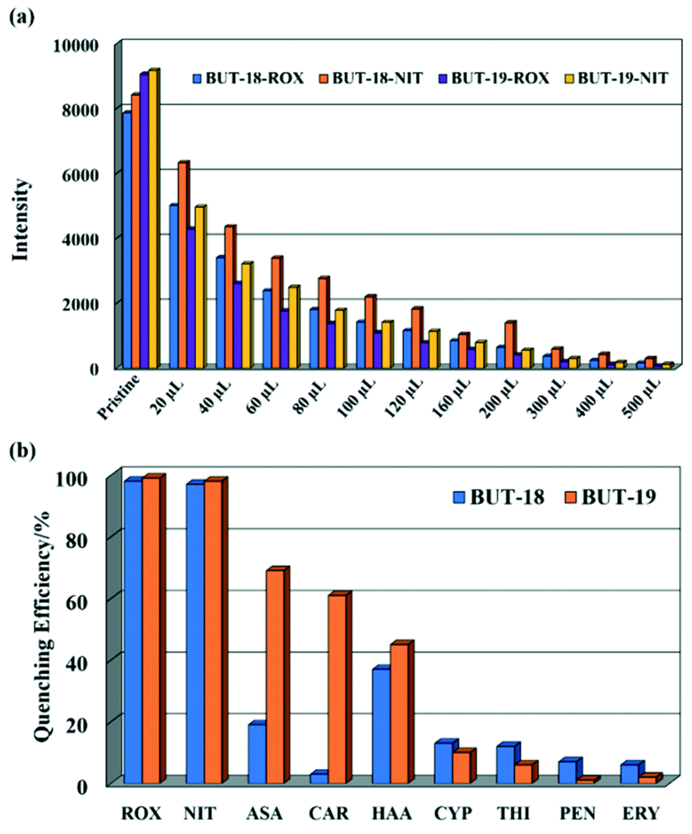
| 8 | Al(CTTA) (BUT-18) and Al(CETA) (BUT-19) | LOD = 15.7 and 32.2 ppb (BUT-18), 13.5 and 13.3 ppb (BUT-19) | Roxarsone (ROX) and Nitarsone (NIT) | “Turn Off” | [96] |
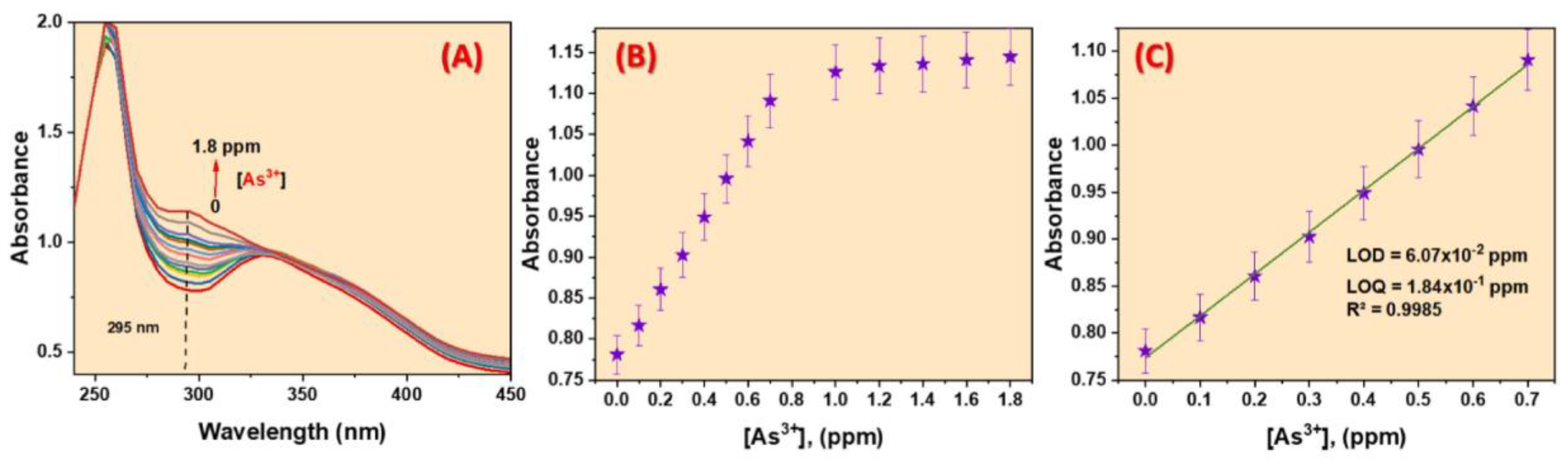
| 9 | Functionalized NH2-UiO-66(Zr) | LOD = 88.3 ppb | As3+ | “Turn Off” | [98] |
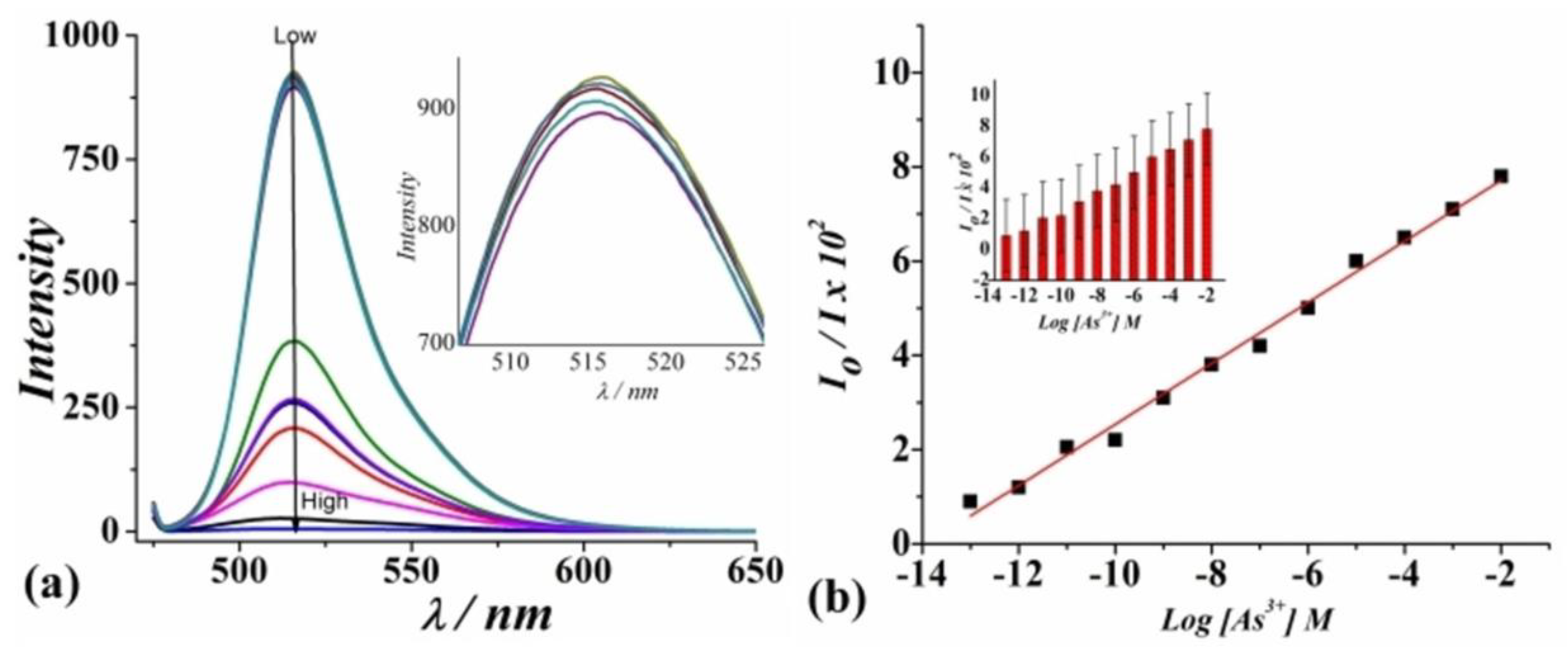
| 10 | AuNPs@Fe-BTC MOF | LOD = 0.2 ng mL−1 | As (III) | “Turn Off” | [101] |
| 11 | iMOF-12C | LOD = 3.95 ppb for ROX and 1.35 ppb for NIT | Roxarsone(ROX) and Nitarsone(NIT) | “Turn Off” | [104] |
| 12 | Fluorescein/AgNPs/PB-MOF | LOD = 0.150 ppm | As(III) | “Turn Off” | [108] |
| 13 | 2HA=N-MIL-53(Al) | LOD = 0.15 μg/L | As(III) | “Turn Off” | [111] |
Disclaimer/Publisher’s Note: The statements, opinions and data contained in all publications are solely those of the individual author(s) and contributor(s) and not of MDPI and/or the editor(s). MDPI and/or the editor(s) disclaim responsibility for any injury to people or property resulting from any ideas, methods, instructions or products referred to in the content. |
© 2025 by the authors. Licensee MDPI, Basel, Switzerland. This article is an open access article distributed under the terms and conditions of the Creative Commons Attribution (CC BY) license (https://creativecommons.org/licenses/by/4.0/).
Share and Cite
Nandi, S.; Dalapati, R. Fluorometric Sensing of Arsenic in Water: Recent Developments in Metal-Organic Framework-Based Sensors. Processes 2025, 13, 923. https://doi.org/10.3390/pr13030923
Nandi S, Dalapati R. Fluorometric Sensing of Arsenic in Water: Recent Developments in Metal-Organic Framework-Based Sensors. Processes. 2025; 13(3):923. https://doi.org/10.3390/pr13030923
Chicago/Turabian StyleNandi, Soutick, and Rana Dalapati. 2025. "Fluorometric Sensing of Arsenic in Water: Recent Developments in Metal-Organic Framework-Based Sensors" Processes 13, no. 3: 923. https://doi.org/10.3390/pr13030923
APA StyleNandi, S., & Dalapati, R. (2025). Fluorometric Sensing of Arsenic in Water: Recent Developments in Metal-Organic Framework-Based Sensors. Processes, 13(3), 923. https://doi.org/10.3390/pr13030923