Article

Olive Tree Twigs as an Attractive Green Source of Antioxidant and Antibiofilm Biomolecules

1
Analytical Biochemistry and Biotechnology Laboratory, Mouloud Mammeri University of Tizi Ouzou, Tizi-Ouzou 15000, Algeria
2
Biochemistry and Microbiology Department, Mouloud Mammeri University of Tizi Ouzou, Tizi-Ouzou 15000, Algeria
3
Natural Ressources Laboratory, Mouloud Mammeri University of Tizi Ouzou, Tizi-Ouzou 15000, Algeria
4
Department of Medical Laboratory, College of Applied Medical Sciences-Shaqra, Shaqra University, Shaqra 11961, Saudi Arabia
5
Medical Laboratories Department, College of Applied Medical Sciences in Al-Quwayiyah, Shaqra University, Shaqra 11961, Saudi Arabia
6
Central Military Laboratory and Blood Bank Department of Microbiology Division, Prince Sultan Military Medical City, Riyadh 12233, Saudi Arabia
7
Laboratoire de Maitrise Des Energies Renouvelables, Faculté des Sciences de la Nature et de la Vie, Université de Bejaia, Bejaia 06000, Algeria
8
Marine Biodiversity Laboratory, National Institute of Marine Sciences and Technology INSTM, University of Carthage, Tunis 1054, Tunisia
*
Authors to whom correspondence should be addressed.
Processes 2025, 13(2), 559; https://doi.org/10.3390/pr13020559
Submission received: 22 December 2024 / Revised: 8 February 2025 / Accepted: 10 February 2025 / Published: 17 February 2025

Abstract

:
Biofilms represent complex three-dimensional microbial communities that can harbor strains highly resistant to antimicrobial agents. These structures, which form on both biotic and abiotic surfaces, are associated with food spoilage and increased complications in hospitalized patients. Consequently, there is significant interest in developing novel biofilm and infection control strategies, particularly those focusing on natural molecules with dual antimicrobial and antibiofilm properties. In this study, olive tree twigs from three varieties of Olea europea chemlal (CH), Azeradj (AZ), and wild-type Olea europaea sylvestris (W) were collected from the Kabylia region in Algeria. The samples underwent systematic extraction and were evaluated for their antioxidant activity using 2,2-diphenyl-1-picrylhydrazyl (DPPH) radical scavenging assay, antimicrobial properties via disk diffusion assay, minimum inhibitory concentration (MIC), and antibiofilm capabilities. Results demonstrated that olive tree twig extracts exhibited substantial antioxidant activity and significant antibacterial and antibiofilm potential. The antioxidant activity, measured through DPPH radical scavenging, showed IC50 values ranging from 38.12 ± 1.52 µg/mL to 148.7 ± 1.23 µg/mL. When tested against six pathogenic bacterial strains, including both ATCC reference strains and milk isolates, the MIC values ranged from 1.18 mg/mL to 4.71 mg/mL. Notably, sub-inhibitory concentrations significantly reduced biofilm formation across most tested strains, with inhibition rates varying from 21% to 90.43%. The effectiveness of biofilm inhibition was dependent on the bacterial strain, olive tree variety, and extract concentration used. Statistical analysis confirmed the significance of these results (p < 0.05). Given the demonstrated antioxidant, antibacterial, and antibiofilm properties of these olive tree twig extracts, they show promise for further development as surface disinfectants and potential applications in food safety and infection control. Additional research is warranted to fully characterize their mechanisms of action and optimize their practical applications.

1. Introduction

Biofilms represent complex matrix-enclosed microbial aggregates capable of forming on both biotic and abiotic surfaces, including medical devices [1,2] and equipment in dairy and food processing industries [1,3,4]. These formations present significant challenges to hygiene standards, public health, and economic stability.
As a fundamental bacterial survival mechanism, biofilm formation plays a critical role in the development and persistence of multi-drug resistance [5]. Unlike planktonic cells, biofilms exhibit exceptional resistance to adverse conditions through their protective extracellular polymeric substances [6]. This resistance has been exacerbated by the indiscriminate use of antibiotics in both human and veterinary medicine, leading to the emergence of novel resistance mechanisms in bacterial pathogens [7]. Notably, many foodborne pathogens and food processing contaminants demonstrate significant biofilm-forming capabilities. In dairy processing environments, biofilm-forming bacteria, particularly Bacillus species, frequently colonize processing lines, potentially causing equipment biocorrosion and biodeterioration [8].
The control and elimination of biofilms present formidable challenges. While conventional physical and mechanical approaches, including disinfectants and antimicrobials [9], have been developed, these methods often prove inadequate against biofilm-related contaminations due to their enhanced resistance mechanisms. Moreover, synthetic antimicrobial agents and food additives may pose risks to human health through consumption or exposure [10]. These challenges necessitate a reevaluation of current practices and emphasize the importance of appropriate antimicrobial stewardship and preventive measures for microbial control. These concerns have catalyzed new research directions, highlighting the urgent need for novel, environmentally sustainable approaches to combat biofilm-related infections [1].
Recent research has increasingly focused on natural antimicrobials derived from biological resources. Various medicinal plants and natural by-products contain bioactive compounds that demonstrate antioxidant properties, inhibit pathogen growth, and extend food shelf life. These bioproducts show considerable potential for applications in food preservation and packaging [11,12,13,14].
Plant bioactive compounds, particularly phenolic compounds found in olive trees and their by-products, serve essential defensive functions. While some compounds enter the food chain, others accumulate as antimicrobial agents [15]. The scientific literature documents extensive investigations into dietary polyphenols’ antimicrobial activity, which varies according to polyphenol structure, bacterial strain, and concentration [16,17].
The Mediterranean region harbors diverse plant species, notably Olea europaea L., a globally significant tree crop cultivated for its edible oil and fruits. These long-lived, evergreen trees generate substantial by-products during cultivation. Pruning and harvesting operations produce significant quantities of residual materials, including olive leaves, which industries utilize for their bioactive compounds in food additives, dietary supplements, cosmetics, and nutraceuticals [18]. These leaves serve as reservoirs of phenolic compounds with documented biological activities relevant to human health [7].
Olive cultivation generates approximately 22 kg of leaves and branches annually per tree [19]. The conventional practice of burning these residues contributes significantly to greenhouse gas emissions in the Mediterranean region [20]. However, these by-products represent valuable sources of polyphenols with applications across food, pharmaceutical, and cosmetic industries. Growing consumer interest in natural products has stimulated research into naturally derived medicines and food processing alternatives.
The current literature reveals a significant gap regarding the in vitro application of polyphenols from Algerian olive tree twigs as antioxidant and antibiofilm agents. Existing research primarily focuses on phenolic extracts from olive leaves or olive mill wastewater [21,22,23,24]. Therefore, this study investigates the antioxidant activity of olive tree-derived polyphenols and evaluates their potential in biofilm prevention.

2. Materials and Methods

2.1. Sample Collection

This study examined olive tree twigs from three varieties collected from two locations in the Kabylia region of Algeria: Mekla (Location 1) and Beni Douala (Location 2). The varieties included *Chemlal* (CH1 and CH2), *Azeradj* (AZ1 and AZ2), and wild-type *Olea europaea sylvestris* (W1 and W2). Location 1 (36°40′24.661″ N, 4°15′50.771″ E) stands at 325 m above sea level, while Location 2 (36°37′08″ N, 4°04′57″ E) is situated at 804 m above sea level. Samples were collected from unpolluted natural habitats, free from phytosanitary products. The collected twigs underwent multiple water rinses to remove dust and impurities, followed by air-drying at ambient temperature for 15 days in darkness.

2.2. Polyphenol Recovery and Quantification

The dried twigs were mechanically ground and sieved to obtain particles smaller than 1 mm, removing fiber residues. For each sample, 5 g of processed material underwent extraction using 50 mL of ethyl acetate (w/v). The extraction process was conducted in flasks under agitation for two hours at room temperature in darkness. The extracts were subsequently filtered and centrifuged at 4000× g. The filtered extracts were collected separately and concentrated to dryness using a rotary vacuum evaporator (HAHN SHIN) at 40 °C. The dried extracts were then reconstituted in a minimal volume of 10% dimethyl-sulfoxide solution (DMSO) in distilled water. Insoluble fractions were removed via micro-centrifugation (SANYO MSE, Microcentaur, UK).
Total phenolic content was determined following the Folin–Ciocalteu method with modifications to reach a final reactional volume suitable for 1 mL spectrophotometer cuvettes [25]. Briefly, 0.5 mL of Folin–Ciocalteu reagent was mixed with 0.1 mL of extract. The mixtures were allowed to stand for 8 min in the absence of light. After that, 0.4 mL of 7.5% Na2CO3 were added; then, the mixtures were incubated in the dark at 40 °C for 15 min. The absorbance at 765 nm was recorded using UV–visible spectrophotometer (Biotech Engineering Management Co., Ltd. (United Kingdom). According to the calibration curve with gallic acid as the standard, the whole phenolic content was estimated; then, the results were defined in terms of mg of gallic acid equivalents (GAEs).

2.3. DPPH Radical Scavenging Activity

The antioxidant capacity of olive tree twig extracts was evaluated using the DPPH (2,2-diphenyl-1-picrylhydrazyl) free radical assay [26]. Serial dilutions of each raw extract were prepared to determine the IC50 value. For each diluted solution of extract (20 µL), 1 mL of ethanolic DPPH solution (0.1 mM) was added. The reduction in absorbance was counted at 517 nm after 30 min using the UV–visible spectrophotometer. The blank sample comprised 1 mL of the DPPH solution and 20 µL of ethanol. Ascorbic acid was used to generate a standard curve using different concentrations varying from 20 to 160 µg/mL. The percentage of inhibition (PI) (%) was calculated as the percentage of reduced DPPH radical based on the subsequent formula:
PI (%) = [(control OD − sample OD)/Control OD] × 100

2.4. Antimicrobial Activity Tests

2.4.1. Microorganisms Strains Used for Testing

The bacterial strains utilized in this research are listed in Table 1. A matrix-assisted laser desorption/ionization time-of-flight mass spectrometry (MALDI-TOF MS) analysis of whole-cell proteomes was employed to identify several strains of food origin, which were isolated from goat milk samples. Notably, Bacillus licheniformis was isolated from cow milk. The remaining strains are ATCC reference strains.

2.4.2. Antimicrobial Susceptibility Using the Disk Diffusion Method

The antimicrobial susceptibility screening of the extracts, diluted serially in 10% DMSO, was conducted using the disk diffusion method on Mueller Hinton Agar (MHA) to assess their antimicrobial activities. Fresh bacterial cultures, incubated for 18 h at 37 °C, were used to prepare microbial suspensions with a turbidity close to 106–107 CFU/mL. These suspensions were subsequently plated on Mueller Hinton Agar. Disks (6 mm in diameter) were immersed in a solution containing 20 µL of each extract or the control (DMSO alone), dried separately in a sterile Petri dish, and then placed onto the inoculated MHA plates. The plates were incubated for 24 h at 37 °C before measuring the inhibition zone diameters in millimeters (including the diameter of the disk). The results are presented as mean ± standard deviation.

2.4.3. Minimal Inhibitory Concentrations

Minimal inhibitory concentrations (MICs) were determined using a 96-well microplate broth serial dilution method, following the manual of the Clinical and Laboratory Standards Institute (CLSI) [27] as described by Sánchez et al. [28], with modifications to the volumes utilized. These values indicate the lowest concentration of extract at which bacterial growth is not visibly detectable. Each microplate well was filled with 300 µL of broth containing approximately 5 × 10⁵ CFU/mL of the target bacteria along with varying concentrations. The selected extracts were chosen based on inhibition zone testing effectiveness against six bacterial strains.

2.4.4. Antimicrobial Activity Using the Viability Testing Method

The experimental trials were conducted based on the viability testing method described by Medina et al. [29] for lactic acid bacteria, with modifications to the volumes used, incubation time, and temperature. A volume of 50 µL of each diluted extract was inoculated with 50 µL of a standardized target bacterial strain in saline solution (10⁷–10⁸ CFU/mL). The concentration for each extract (mg/mL) was 4.0, 9.4, 4.7, 8.5, 11.3, and 4.7, respectively, for AZ1, CH1, W1, AZ2, CH2, and W2. The homogenized mixture was incubated for 4 h, then streaked onto nutrient agar for visual inspection of surviving bacteria after 24 h of incubation, compared to controls without extract (saline water and DMSO).

2.4.5. Antibiofilm Testing

Biofilm Formation Assay

The ability of extracts to inhibit biofilm formation was evaluated in biofilm-producing strains. Biofilm formation was assessed using the crystal violet assay in a 96-well microplate. The procedure involved removing planktonic cells after incubation by washing the plates three times with Phosphate-Buffered Saline (PBS). After drying, the wells were stained with crystal violet solution for 15 min, followed by five washes with PBS to remove excess crystal violet from the biofilm.

Antibiofilm Activity Testing Using a Crystal Violet Assay

The ability of extracts to inhibit biofilm formation was evaluated in biofilm-producing strains. Biofilm formation was assessed using a crystal violet assay in a 96-well microplate. The procedure involved removing planktonic cells after incubation by washing the plates three times with Phosphate-Buffered Saline (PBS). After drying, the wells were stained with crystal violet solution for 15 min, followed by washing steps with PBS to remove excess crystal violet from the biofilm.
The effect of polyphenol extracts from olive tree twigs on biofilm formation was assessed both qualitatively and quantitatively.
The capability of the extracts to affect biofilm formation was evaluated following the 96-well microtiter plate method, using various concentrations of each extract in trypticase yeast soya-glucose (2%) broth as described by Sánchez et al. [28] with modifications. Exponentially growing cells from overnight cultures were adjusted to approximately 0.5 McFarland turbidity, diluted in trypticase yeast soya-glucose broth. Subsequently, 50 µL of this suspension was dispensed as inoculum into each well. The final extract concentrations ranged from 1/2 MIC to 1/8 MIC, with 200 µL per well, and the microplates were incubated at 37 °C for 24 h. A medium without extracts and/or bacterial cells served as the untreated and cultivated control. The controls received 10% DMSO and culture broth instead of the extracts and inoculum, respectively.
After incubation, unattached cells were washed out three times with PBS buffer. Each well was then treated with 1% crystal violet solution for approximately 15 min, followed by three washes with PBS. Subsequently, 200 µL of ethanol was added to each well, and the optical density (OD) was recorded using a microplate reader (Bio-Tek EL 311, Vermont, USA) at an absorbance measurement of 590 nm. The inhibitory effect and biofilm growth were determined using Equations (1) and (2), respectively.
Biofilm inhibition (%) = (OD control − OD test/OD control) × 100
Biofilm growth (%) = (OD test/OD control) × 100
To assess the antibiofilm activity of the extracts, six bacterial strains were selected based on their susceptibility observed in the previous antibacterial activity tests. In this phase of the trials, four reference strains were used: S. aureus ATCC 25923, P. aeruginosa ATCC 27853, B. cereus ATCC 14579, and E. faecalis WDCM 0009. Additionally, two foodborne isolates, K. oxytoca strain (b) and B. licheniformis, were included. The selected extracts, CH1, W2, and AZ2, for antibiofilm testing were only chosen for their wide effectiveness demonstrated in the inhibition zone testing.

2.5. Data Analysis

The experiments were conducted in triplicate, and the results were expressed as mean ± standard deviation. Data analysis was performed using GraphPad Prism (version 5.00) for Windows. The biological activity results were statistically analyzed with the ANOVA variance test, employing a significance level of p < 0.05.

3. Results

3.1. Polyphenol Yield and Content

The highest yield was recorded for the AZ2 extract, measuring 13.65 ± 0.37 mg GAE/g dry weight (DW), followed closely by the CH2 extract at 13.59 ± 0.34 mg GAE/g DW. The CH1 extract yielded 11.31 ± 0.93 mg GAE/g DW, while the W2 extract produced 7.54 ± 0.42 mg GAE/g DW (Figure 1). The lowest yield was observed for the AZ1 extract at 4.94 ± 0.03 mg GAE/g DW, followed by the W1 extract at 5.33 ± 0.50 mg GAE/g DW. Given that different varieties were collected from two locations, the variation in phenolic compound yields may be attributed to agronomic conditions, the age of the olive trees, the olive cultivar, and the physico-chemical properties of the soil. As previously noted, the yields of phenolic compounds in plants are influenced by both biotic and abiotic stress, as these secondary metabolites serve as defense mechanisms against such threats.
As illustrated in Figure 2, the used polyphenol concentration (mg/mL) in the various extracts for antibacterial testing was 8.04 ± 0.05, 18.85 ± 1.54, 9.34 ± 0.83, 17.07 ± 0.46, 22.65 ± 0.06, and 9.43 ± 0.52, respectively, for the AZ1, CH1, W1, AZ2, CH2, and W2 extracts. It is important to note that these extracts were obtained from a 10% DMSO supernatant after centrifugation. In addition to the factors previously mentioned affecting yields, the variability in polyphenol concentration may also be influenced by the concentration and recovery procedures and/or the potential loss of biomolecules in the precipitated insoluble fractions. Concentration could be affected by the concentration and recovery procedures and/or the loss of variable biomolecules in the precipitated insoluble fractions.

3.2. Antioxidant Activity

The results displayed in (Figure 3 and Figure 4) reveal that all raw extracts exhibit significant free radical scavenging activity.
The raw extracts and their 10−1 dilution exhibited high, dose-independent antioxidant activity, ranging from 74.74 ± 4.13% to 87.73 ± 1.33%. This finding confirms that all tested extracts are rich in potent antioxidant biomolecules. However, from the 10−1 to 10−4 dilutions, the extracts demonstrated dose-dependent activity (Figure 3).
Regarding the IC50 values (Figure 4), which are crucial for comparing and characterizing the extracts, they varied among the different varieties, ranging from 38.12 ± 1.52 µg/mL to 148.7 ± 1.23 µg/mL. The CH1, CH2, and W1 extracts were significantly (p < 0.005) the most effective inhibitors of the DPPH radical, with IC50 values of approximately 56.35 ± 3.09 µg/mL, 40.16 ± 1.23 µg/mL, and 38.12 ± 1.52 µg/mL, respectively. In comparison, ascorbic acid exhibited an IC50 value of 80 ± 1.7 µg/mL.

3.3. Antibacterial Susceptibility Testing

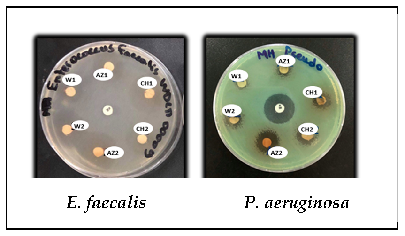
3.3.1. Disk Diffusion Method

Regarding antimicrobial potential, the analyzed extracts revealed widespread antibacterial activity. According to the results displayed in Table 2, olive tree twig raw extracts exhibit antibacterial activity that varies depending on the strain or extract. The obtained inhibition zones varied between 6.5 ± 0.07 mm to 25.9 ± 0.14 mm. The best activities, as indicated by the largest inhibition zones, were recorded particularly against E. faecalis (Figure 5), with inhibition ranging from 12.5 ± 0.71 mm to 25.9 ± 0.14 mm. Moreover, the results revealed that the extracts AZ1, CH1, W2, and CH2 showed significant effectiveness against P. aeruginosa ATCC 27853 (Figure 5), having diameters of inhibition zones measuring 9.5 ± 0.71 mm, 10.5 ± 0.71 mm, 10.25 ± 0.35 mm, and 11.5 ± 0.71 mm, respectively, whereas, using the AZ2 extract, it showed a higher diameter of the inhibition zone, at 14.5 ± 0.71 mm. Nevertheless, P. aeruginosa exhibited a lower inhibition zone of 8 ± 1.41 mm diameter for W1 extract. The extracts showed less or no activity against E. coli and K. pneumoniae.

3.3.2. Determination of MICs

The microdilution method was employed to evaluate the antibacterial activity of the extracts. Figure 6 presents the recorded minimum inhibitory concentration (MIC) values of the three extracts against the six tested bacteria, ranging from 1.18 mg/mL to 4.71 mg/mL.

3.3.3. Bactericidal Activity (Viability Testing)

The purpose of this experiment is to assess the direct effect of the extracts on different bacterial strains in the absence of a culture medium, compared to a negative control containing only the strain in physiological water, as well as another control containing the strain in 10% DMSO, which was used to dissolve the extracts.
As anticipated, the tested extracts did not support the viability of the majority of the tested bacterial strains after 4 h of direct exposure (Table 3). Staphylococcus aureus MU50, Klebsiella pneumoniae ATCC 700603, Klebsiella oxytoca, Enterococcus faecalis, and Bacillus licheniformis were inhibited by all extracts. However, Bacillus cereus ATCC 14579 remained resistant to these extracts.
In the absence of nutrients, K. pneumoniae ATCC 700603, which exhibited total resistance in the disk diffusion test, was inhibited by all extracts during direct contact. Additionally, Pseudomonas aeruginosa ATCC 27853 was inhibited by most extracts, with the exception of the W1 extract. This finding corroborates the results obtained from the disk diffusion test, where this strain was resistant only to the same W1 extract.

3.4. Antibiofilm Action

3.4.1. Biofilm Formation Potential

The ability of the tested bacterial species to form biofilms is reported in terms of the resulting optical density (OD) at 590 nm (Figure 7). (2.42 ± 0.15), Pseudomonas aeruginosa ATCC 27853 (1.94 ± 0.00), Bacillus cereus (b) (1.06 ± 0.07), and Staphylococcus aureus ATCC 25923 (0.90 ± 0.23).

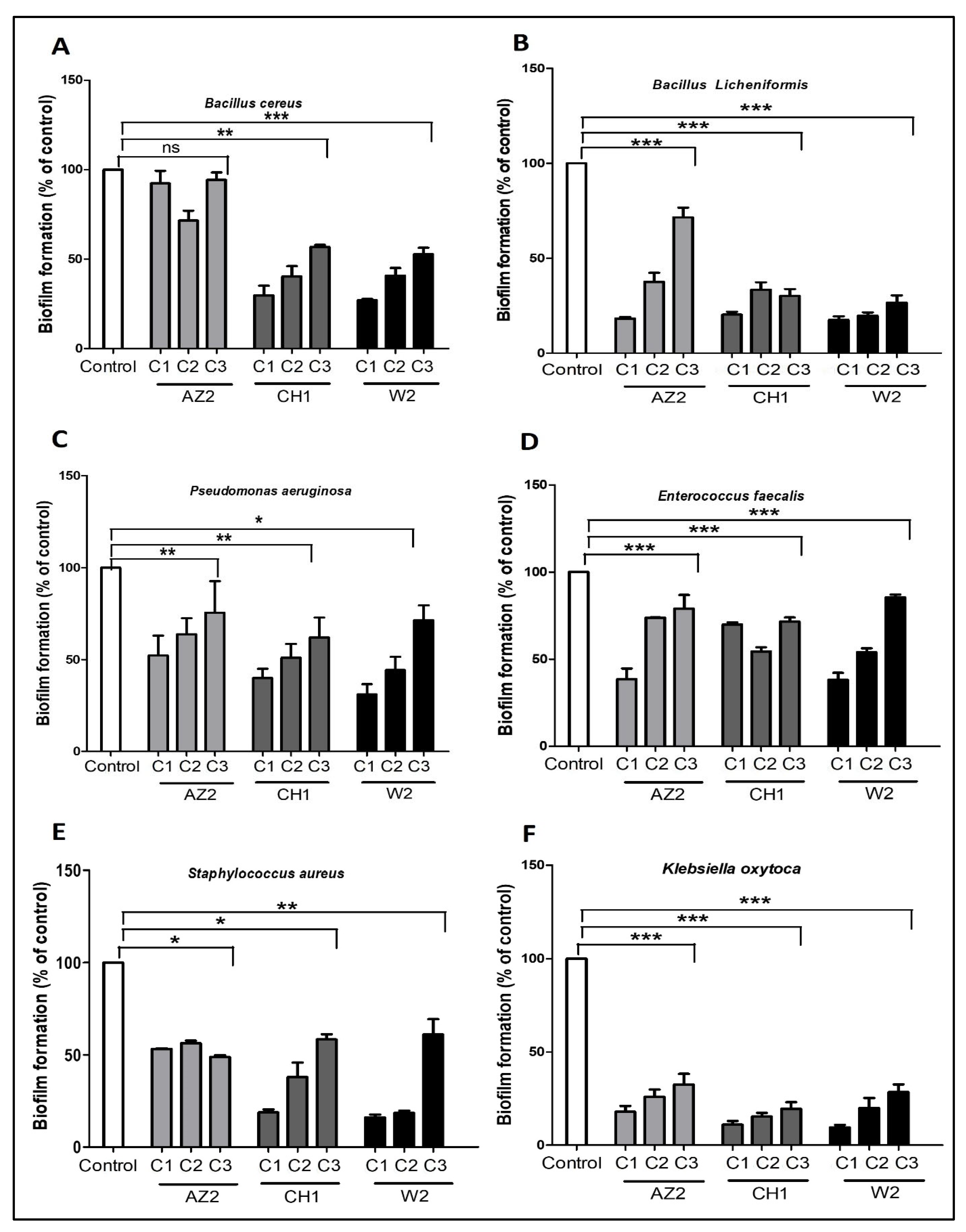
3.4.2. Olive Tree Twig Antibiofilm Potential

Antibiofilm testing trials were conducted using serial dilutions at different concentrations of 1/2 MIC, 1/4 MIC, and 1/8 MIC. The results demonstrated the antibiofilm effectiveness of the extracts by reducing the capability of bacterial adhesion to the polystyrene surface in a 96-well microtiter plate assay, as indicated by a reduction in biofilm growth ratio compared to the control group (100%), which was not exposed to the extracts (Figure 8).
The displayed results (Figure 8A–D) highlight a significant inhibitory effect (p-value < 0.005) of the three olive tree twig extracts against the tested biofilm-forming strains. Quantification of the biofilm formed by Klebsiella oxytoca and Enterococcus faecalis after incubation with polyphenols in the microtiter plate showed a substantial reduction in biofilm amount (p-value < 0.0001). Additionally, all explored extracts exhibited significant antibiofilm activity against Pseudomonas aeruginosa (p-value = 0.004) and Staphylococcus aureus (p-value = 0.003), although their effects were not as pronounced as those observed for K. oxytoca and E. faecalis and Bacillus licheniformis.
The achieved antibiofilm activity indicated that the effect is dose-dependent for most extracts, with some exceptions. Sub-inhibitory concentrations significantly decreased biofilm formation for most strains, with reductions ranging from 21% to 90.43%, depending on the strain, the different olive tree varieties/cultivars, and the concentration used. Antibiofilm results as a function of concentration revealed that extracts CH1, W2, and AZ2, at their minimum used concentration, exhibited the strongest inhibitory effect against K. oxytoca, with reductions exceeding 50% compared to other strains. The most significant inhibition was observed with CH1 (1.18 mg/mL) and W2 (0.59 mg/mL) extracts against K. oxytoca, yielding reductions of 89.02% and 90.43%, respectively, while the weakest effects were noted at lower concentrations.
It was also found that the three extracts, CH1, W2, and AZ2, demonstrated enhanced antibiofilm activity against K. oxytoca at various concentration levels. All higher extract concentrations (1/2 MIC), led to the greatest antibiofilm activity (>50%), except for AZ2 and CH1 extracts, where the values were below 50% when tested against P. aeruginosa ATCC 27853, B. cereus (b), S. aureus ATCC 25923, and E. faecalis, respectively, for the two extracts. Notably, at the same dilution, the W2 extract, corresponding to the wild variety Olea europaea sylvestris, displayed effective antibiofilm activity not only against P. aeruginosa ATCC 27853 (69%) but also against E. faecalis, B. cereus (b), B. licheniformis, S. aureus ATCC 25923, and K. oxytoca, with inhibition rates of 61.77%, 72.87%, 82.45%, 83.9%, and 90.43%, respectively. The Chemlal CH1 extract exerted strong inhibitory effects toward K. oxytoca (89.02%), S. aureus (81.17%), B. licheniformis (79.7%), B. cereus (b) (70.23%), and P. aeruginosa ATCC 27853 (60.02%). No significant antibiofilm effect was recorded for AZ2 against B. cereus (b), although it was effective against K. oxytoca (82.06%) and B. licheniformis (81.77%), followed by E. faecalis (61.5%).

4. Discussion

One of the most notable features of olive leaves is their year-round availability and their rich content of valuable phenolic compounds. Therefore, the current study focused on the valorization of olive tree branches. The results achieved (Figure 1 and Figure 2) demonstrate interesting yields and content of polyphenols, which vary among the different twig extracts. The quantitative variation in total polyphenol content may be influenced by several factors, including olive variety, soil characteristics, and various diseases or stresses affecting the trees. Additionally, the distribution of bioactive constituents might change throughout the olive tree’s life cycle. This variation is contingent upon extreme climatic conditions (such as exposure to sunlight, drought, salinity, and high temperatures), which can stimulate the production of secondary metabolites, particularly polyphenols [30,31].
It is noteworthy that the antioxidant activity, along with the dual antimicrobial and antibiofilm effects observed in this study, may result from synergistic interactions among the various components present in the extract [30,32,33,34]. As outlined in the current work, several studies have reported a positive correlation between high concentrations of phenolic compounds in extracts and their antioxidant activity. The recorded IC50 values varied among the different varieties used, ranging from 38.12 ± 1.52 to 148.7 ± 1.23 µg/mL. As noted by Khelouf et al. [22], olive tree twigs represent a significant source of phenolic compounds with high antioxidant capacities. Despite the promising antioxidant findings, further research is necessary to evaluate their toxicity thresholds in human cell models.
Antioxidant activity typically depends on the location and number of hydroxyl groups in relation to the functional carboxylic groups within the aromatic rings. Oleuropein and hydroxytyrosol, the primary active compounds in olive tree by-products, are well documented for their richness in hydroxyl groups, which confer a high capacity to stabilize free radicals. Consequently, the anti-free radical activity of an extract cannot be predicted solely based on its total phenolic content; it also depends on the structural properties of the biomolecules and the efficacy of the individual compounds present in the extract [31,35,36,37].
Regarding antibacterial trials, the disk diffusion method was initially employed (Table 2), which serves as a qualitative screening tool for antimicrobial activity, particularly when the diameter of inhibition zones is ≥10 mm [38]. As observed, most analyzed extracts exhibited slight inhibition against the tested bacterial strains. However, polyphenolic extracts can exert an inhibitory effect on bacterial growth, even at lower concentrations [21]. The behavior of the bacterial strains in response to the various extracts and concentrations of bioactive compounds may be attributed to their structure, wall composition, and/or metabolic pathways. Nonetheless, although the inhibition zones were not particularly large, this process could be optimized in future studies.
Moreover, it should be noted that the size of inhibition zones for different extracts may be influenced by the physicochemical characteristics of the extracted components. Consequently, a more diffusible but less active extract could yield a larger inhibition diameter than a less diffusible but more potent extract [28,39].
The results revealed that the extracts demonstrate antimicrobial activity against Pseudomonas aeruginosa ATCC 27853, which is particularly noteworthy given its natural virulence and resistance to antibiotics. Furthermore, this bacterium is rarely inhibited by plant extracts or essential oils, making this finding significant [40,41]. Another interesting observation is the efficacy against Enterococcus faecalis WDCM 00009, with zones of inhibition ranging from 12.5 ± 0.71 mm to 25.9 ± 0.14 mm, surpassing that of the positive control (neomycin, 7 ± 0.28 mm). Neomycin, which belongs to the aminoglycoside family, is known for eliciting high-level resistance in Enterococci through aminoglycoside-modifying enzymes [42].
Numerous studies have demonstrated various biological activities of olive tree-derived products [23,43], which contain a complex composition of phenolic compounds [44]. Olive trees are recognized for their resistance to microbial and insect attacks, attributed to their antimicrobial properties. Previous research has shown that olive tree leaves (Olea europaea L., Oleaceae) or their metabolites, such as oleuropein, exhibit effectiveness as antimicrobials both in vitro and in vivo. It has also been reported that olive leaf extracts may exert antimicrobial action against human pathogenic microorganisms [45], including those present in gastric flora [46].
As reviewed by Mandal et al. [47], polyphenols display a range of antimicrobial properties against not only bacteria but also fungi and viruses. They can damage and rupture cell membranes, hinder enzymatic activity, chelate metal ions, generate reactive oxygen species (ROS), inhibit viral entry and replication, and modify the host’s immune response. In their research, Gökmen et al. [48] conducted practical assays to assess the biological antimicrobial effect of olive leaf extracts against ten pathogenic bacteria using the disk diffusion method. They reported inhibition zones of 13.33 ± 2.08 mm and 21.67 ± 1.53 mm against Salmonella typhimurium and Bacillus cereus, respectively.
Additionally, as previously reviewed [49], several reports have demonstrated that olive leaves are rich in bioactive compounds with antioxidant properties, anti-HIV effects, anti-apoptotic and proliferative effects, protective benefits against human leukemia, and the ability to lower lipid levels. Studies on olive leaves have been conducted in various countries to assess their antimicrobial potential, including Algeria [22,24], Morocco [50], Iraq [51,52], Italy [53], Brazil [34], Spain [54,55], and Egypt [56]. All these studies have shown the inhibitory influence of olive leaf extracts on the growth of Staphylococcus aureus. Other research confirms the antimicrobial capacity of olive leaf extracts against Bacillus cereus [30,48,57,58], as well as against Pseudomonas putida, with a reported zone of inhibition of 13 ± 0.90 mm [59].
It is important to underscore the novelty of the present study, as it reveals the antibacterial activities of olive twig extracts—previously underexplored parts of the olive tree compared to the leaves. According to prior research, phenolic compounds not only exhibit significant synergistic effects against B. cereus [57,60] but can also modulate inflammatory and macrophage responses, potentially providing activity against pathogens [61].
In alignment with other studies that have identified Klebsiella pneumoniae and Escherichia coli as the most resistant microorganisms to olive leaf extracts [36,62], the current work also found these two bacteria to be resistant to olive tree twig extracts. However, other studies have indicated that oleuropein, rutin, and hydroxytyrosol significantly impact K. pneumoniae [57,60].
It should be noted that the above and subsequent discussions provide only a brief overview of the biological activities of phenolic compounds and olive leaves, as limited information is available on olive tree twigs. Despite the richness of these tissues in bioactive molecules, notably oleuropein, hydroxytyrosol, and tyrosol, and their potential as raw materials, they remain underexplored [23,37,63,64].
The antibacterial effects of olive tree twigs in vitro were evaluated by measuring their minimum inhibitory concentration (MIC). The recorded values, ranging from 1.18 to 4.71 mg/mL, fall within the range reported for olive leaf extracts, which show MIC values from 0.26 to 64 mg/mL against foodborne pathogens [48,54,59,65]. In the second section of this investigation, we assessed whether the presence of polyphenols could affect bacterial viability in the absence of nutrients or carbon sources. The results (Table 3) highlight the antimicrobial potential of olive tree twig extracts, except for B. cereus, which likely resists due to its spore-forming capacity. This viability is primarily attributed to its ability to produce spores under stressful conditions, ensuring its survival. Indeed, spores are highly resistant to various environmental threats, including antimicrobial agents such as polyphenols [66,67,68].
Regarding Pseudomonas aeruginosa ATCC 27853, it was inhibited by the majority of extracts, except for W1. This finding corroborates the results obtained from the disk diffusion test, where this strain also exhibited resistance to the same extract. In line with this, Markin et al. [45] found that aqueous olive leaf extract at a concentration of 0.6% (w/v) effectively inhibited E. coli, S. aureus, P. aeruginosa, and K. pneumoniae within three hours of contact time, while B. subtilis required a significantly higher concentration (20% w/v) for effective inhibition. Olive tree twigs offer significant advantages, including year-round availability, low cost, and highly efficient biological activities with minimal side effects, making them a potential sustainable antimicrobial agent, particularly for disinfection. Research suggests that the antibacterial characteristics of polyphenols may serve as a promising alternative or supplementary therapy for infectious diseases [47].
The third section of the experiments focused on preventing biofilm formation in six different strains. The results regarding the biofilm formation of the used strains (Figure 8) align with those reported in the literature—P. aeruginosa [69], B. licheniformis [70], Klebsiella sp [71], S. aureus [72,73], B. cereus [67], and K. oxytoca [70]. These strains are recognized as model biofilm-forming pathogenic organisms, underscoring the importance of preventing their adherence to abiotic surfaces, a key objective of this study.
S. aureus is responsible for a wide range of illnesses associated with indwelling medical devices, where it can attach and form biofilms [74]. P. aeruginosa is notorious for causing severe acute and chronic infections in patients with compromised immune systems [69]. K. oxytoca is typically acquired from various environmental sources and is found in milk from cows and buffaloes with mammary gland infections. It produces the cytotoxin tilivalline, which damages the intestinal epithelium [75]. Strains of Bacillus can cause various issues across different industries, particularly in the dairy sector, due to the biofilms formed at the air–liquid interface and in submerged areas, which play a significant role in food contamination. B. cereus produces toxins and is one of the most common causes of food poisoning. It is a spore-bearing rod that is widespread in the environment, with spores capable of persisting under adverse conditions, as recently reviewed by Hongchao [70].
Regarding Bacillus licheniformis, it can adversely affect dairy quality by reducing the shelf life of dairy products through its impact on milk components. In addition to its potential threat to cells and hemolytic activity, it carries toxin genes [76,77]. The current finding (Figure 8) regarding the biofilm formation rate of B. licheniformis (OD = 2.8 ± 0.05) is supported by previous studies, indicating that B. licheniformis isolates can be strong biofilm producers [70,78,79,80,81], posing potential risks to human health at elevated levels [82,83]. Moreover, B. licheniformis is frequently isolated in dairy processing environments, where it can contribute to pipeline biocorrosion and equipment deterioration [8]. Consequently, it affects both the safety and quality of dairy products [84]. Additionally, B. licheniformis can lead to serious health complications in immunocompromised individuals, including bacteremia, peritonitis, esophageal perforation, and sepsis [85,86,87,88]. Recently, Nayeri Fasaei et al. [89] isolated and confirmed B. licheniformis as a causative agent of bovine mastitis in Iran. Although it is widely reported as a probiotic strain [90], its roles in foodborne diseases and the spoilage of dairy products present potential risks in the dairy industry. While the usefulness of B. licheniformis in the dairy sector has been recognized, it is crucial to assess the safety of these bacterial strains on a case-by-case basis [70]. Despite efforts to prevent biofilm formation in dairy environments, effective methods to fully address biofilm-related issues are still needed in the dairy industry [84].
Compared to planktonic cells, biofilm-associated microorganisms can more effectively resist host defenses, antimicrobial agents, and various environmental stressors [73]. Numerous therapeutic options have been proposed to address infections related to medical devices, including the use of effective antibiofilm agents or materials capable of disrupting the biofilm matrix [91]. Therefore, a primary objective of this investigation was to evaluate the effectiveness of olive tree twig extracts as a novel source of antimicrobial agents that could inhibit biofilm development. Additionally, we assessed whether the polyphenol content at various concentrations could influence the strains’ ability to form biofilms. In addition to the destructive effects of plant phenolics on bacteria, the antibiofilm activity experiments for the tested olive tree twig extracts demonstrated a notably gentler antimicrobial effect, leading to reduced biofilm formation (Figure 8). This activity may be attributed to the high organic load and the presence of phenolic compounds, which are inhibitory to many microorganisms. Consistent with the current study, Edziri et al. [30] reported the antibiofilm effects of polyphenolic compounds from olive leaves against S. aureus, B. cereus, and P. aeruginosa. Similarly, Carraro [92] demonstrated the antibiofilm effect of polyphenolic compounds from olive mill wastewater against E. coli, while Matilla Cuenca [74] reported the inhibitory effect of flavonoids on staphylococcal biofilms.
The results presented in (Figure 8) clearly indicate a significant antibiofilm effect (p-value < 0.005) against the tested bacteria, with the exception of the AZ2 extract against B. cereus. When compared to other strains, the AZ2 extract appears less effective against B. cereus, whereas the CH1 and W1 extracts exhibited significant antibiofilm activity (p-value = 0.003) against the same strain. This behavior may be attributed to competition between biomolecules for bacterial target sites or a masking effect among compounds, which can diminish the effectiveness of the more potent biomolecules.
Moreover, it is crucial to emphasize that the antimicrobial activity of these extracts is influenced not only by their chemical composition and the mechanisms of their bioactive compounds but also by interactions such as synergism, antagonism, and chemical reactions between compounds and substances, including nutrients present in the culture medium. These nutrients, potentially derived from the extract, could promote bacterial growth and adherence, thereby masking the antibiofilm action of bioactive compounds.
Conversely, extract dilution may reveal previously masked molecules and increase competition, potentially leading to antagonistic effects or dual actions of both antibiofilm and biofilm-promoting molecules, which could diminish the overall significance of these activities. When utilizing the same leaf extracts, both antibiofilm and biofilm-promoting effects may occur, as noted in previous studies [93]. This hypothesis presents an intriguing avenue for future research aimed at identifying polyphenolic molecular targets to combat infections associated with biofilms.
These anti-adherence results are likely achieved by disrupting bacterial regulatory mechanisms, such as quorum sensing, without adversely affecting bacterial growth [94]. Quorum sensing, a method of chemical communication among bacterial cells, is considered a crucial mechanism in biofilm development, survival, growth, antibiotic resistance, and toxin production in pathogens. Preventing this signaling process may serve as an effective strategy for controlling pathogens, as well as the bacterial toxins that lead to food spoilage and contamination. Some plant phenolic compounds, particularly crude extracts with high phenolic content, exhibit antibiofilm and/or anti-quorum sensing properties [94,95].
In the current study, olive tree twig extracts significantly hindered bacterial cell adherence at sub-MIC levels, indicating that their antibiofilm effect is not linked to the inhibition of planktonic cell growth [96]. The reduction in biofilm formation is largely dose-dependent, with greater effects observed at higher extract concentrations. Although optimal antibiofilm potential was achieved at higher concentrations, a notable reduction in biofilm formation was also recorded at much lower concentrations. This observation is particularly pronounced with the Klebsiella oxytoca strain, where the antibiofilm activity remained significant even after dilution, suggesting that small quantities can effectively target biofilm formation. This may be due to dilution reducing competition among active molecules or allowing for the solubilization of compounds previously masked by lipids or other molecules. This finding underscores the potency and efficacy of these extracts in combating biofilm formation.
Furthermore, the antibiofilm activity is typically attributed to the high concentrations of phenolic compounds present, notably oleuropein, or to a synergistic effect among certain phenols in the extracts. As reviewed by Selim et al. [97], it has been reported that while individual phenolic components of olive leaf extract can exhibit strong activities in vitro, the antioxidant and antimicrobial potential of the entire mixture of phenolic extracts is superior to that of the individual components [32]. As mentioned earlier, the olive tree twig extracts used in this study are rich in phenolic compounds, which are primarily responsible for their bioactivities.
Regarding their mechanisms of antibiofilm action, it has been previously reported that polyphenols can reduce adhesion ligands, neutralize bacterial toxins, and increase membrane permeability, thereby promoting bacterial cell rupture [30]. Moreover, phenolic compounds may prevent biofilm formation through various mechanisms, such as repressing regulatory processes, inhibiting the expression of virulence factors, decreasing motility and adhesion, and modifying bacterial surface charges to prevent cell substratum attachment [94,98,99,100]. Plant extracts rich in phenolics serve as a significant source of inhibitors targeting foodborne pathogenic bacteria, contributing to spoilage [100,101,102].
The growing ineffectiveness of conventional antimicrobials against drug-resistant pathogens has heightened interest in exploring plant phytochemicals. Due to their structural diversity, multi-target action, and ability to interfere with quorum sensing (QS) systems, phytochemicals are less likely to induce resistance compared to traditional drugs. Additionally, they can hinder bacterial functions without significantly impairing bacterial survival [103]. Although olive tree twigs contain a rich array of biologically active compounds, to our knowledge, no previous scientific studies have investigated the antimicrobial and antibiofilm potential of extracts from these three olive tree twig varieties.
Consequently, the results suggest that olive tree twigs from the three local olive varieties—Chemlal, Azeradj, and the wild variety Olea europaea sylvestris—represent an important resource of antioxidants and inhibitory biomolecules against the biofilm-forming bacteria used in this study. The observed differences in bacterial susceptibility to the extracts may stem from the inherent genetic or physiological characteristics of the bacteria. The effectiveness of the extracts can vary depending on the specific methodology employed to assess their antibacterial activity. The antimicrobial properties of polyphenols are related to the extraction method, extract concentration, and exposure time [104]. Despite their promising bioactivities, phenolic extracts from olive twigs necessitate further complementary studies to elucidate the underlying antibacterial mechanisms. Additionally, it is essential to assess variability influenced by factors such as seasonality and cultivation conditions, as well as to evaluate their toxicity and corrosive effects when used for surface disinfection. Furthermore, the assessment of antifungal activity should also be prioritized in future research. Additionally, for food preservation, considerations regarding structural stability, as well as color, taste, and smell, are crucial when incorporated into food packaging.

5. Conclusions

In the current study, olive tree twig extracts were evaluated for their antioxidant and antibacterial performance. The overall findings underscore the dual antioxidant and antibiofilm properties of these extracts. Most of the extracts demonstrated effectiveness in suppressing bacterial viability and biofilm formation in the tested bacteria, although the degree of effectiveness varied. These results highlight the potential of olive tree twig extracts, suggesting promising applications in fields such as infection control and surface disinfection. Their abundance and richness in bioactive compounds indicate that these extracts may serve as a basis for developing biocompatible, eco-friendly antimicrobial and antioxidant agents for applications in the medical and food industries. However, these findings represent only the initial step in the valorization of olive tree twigs. Further, more detailed investigations are needed to explore the inhibitory mechanisms associated with these extracts.

Author Contributions

Conceptualization, S.D., K.M., H.N. and K.H.; methodology, S.D., S.B., T.A.C. and K.H.; software, K.H.; validation, E.-h.N., K.H. and R.A.; formal analysis, T.A.C. and N.S., investigation, H.N. and K.M.; resources, K.H.; data curation, T.A.C., S.B., K.H. and R.A.; writing—original draft preparation, S.D., S.B. and N.S., writing—review and editing, S.D., K.M., H.N., K.H., R.A., M.S.A., A.A.A.I., M.A., F.M.A. (Fawaz M. Almufarriji), L.T. and F.M.A. (Fahad Mohammed Alturaiki); visualization, K.H.; supervision, K.H. and R.A.; project administration, K.H. and R.A.; funding acquisition, K.H., R.A., M.S.A., A.A.A.I., M.A., F.M.A. (Fawaz M. Almufarriji) and F.M.A. (Fahad Mohammed Alturaiki). All authors have read and agreed to the published version of the manuscript.

Funding

This article was prepared utilizing the materials and equipment provided by the Directorate-General for Scientific Research and Technological Development (DGRSDT) in Algeria.

Data Availability Statement

The original contributions presented in this study are included in the article. Further inquiries can be directed to the corresponding authors.

Acknowledgments

The authors would like to express their gratitude to the Algerian Ministry of Higher Education and the Deanship of Scientific Research at Shaqra University for their support of this work.

Conflicts of Interest

The authors declare no conflicts of interest.

References

  1. Carrascosa, C.; Raheem, D.; Ramos, F.; Saraiva, A.; Raposo, A. Microbial Biofilms in the Food Industry—A Comprehensive Review. Int. J. Environ. Res. Public Health 2021, 18, 2014. [Google Scholar] [CrossRef] [PubMed]
  2. Francolini, I.; Donelli, G. Prevention and control of biofilm-based medical-device-related infections. FEMS Immunol. Med. Microbiol. 2010, 59, 227–238. [Google Scholar] [CrossRef] [PubMed]
  3. Elafify, M.; Liao, X.; Feng, J.; Ahn, J.; Ding, T. Biofilm formation in food industries: Challenges and control strategies for food safety. Food Res. Int. 2024, 190, 114650. [Google Scholar] [CrossRef]
  4. LaPointe, G.; Wilson, T.; Tarrah, A.; Gagnon, M.; Roy, D. Biofilm dairy foods review: Microbial Community Tracking from Dairy Farm to Factory: Insights on Biofilm Management for Enhanced Food Safety and Quality. J. Dairy Sci. 2024; in press. [Google Scholar] [CrossRef]
  5. Zafer, M.M.; Mohamed, G.A.; Ibrahim, S.R.M.; Ghosh, S.; Bornman, C.; Elfaky, M.A. Biofilm-mediated infections by multidrug-resistant microbes: A comprehensive exploration and forward perspectives. Arch. Microbiol. 2024, 14, 101. [Google Scholar] [CrossRef]
  6. Bakkiyaraj, D.; Nandhini, J.R.; Malathy, B.; Pandian, S.K. The anti-biofilm potential of pomegranate (Punica granatum L.) extract against human bacterial and fungal pathogens. Biofouling 2013, 29, 929–937. [Google Scholar] [CrossRef]
  7. Slobodníková, L.; Fialová, S.; Rendeková, K.; Kováč, J.; Mučaji, P. Antibiofilm activity of plant polyphenols. Molecules 2016, 21, 1717. [Google Scholar] [CrossRef]
  8. Li, J.; Du, C.; Liu, Z.; Li, X. Electrochemical studies of microbiologically influenced corrosion of X80 steel by nitrate-reducing Bacillus licheniformis under anaerobic conditions. J. Mater. Sci. Technol. 2022, 118, 208–217. [Google Scholar] [CrossRef]
  9. Yin, W.; Xu, S.; Wang, Y.; Zhang, Y.; Chou, S.H.; Galperin, M.Y.; He, J. Ways to control harmful biofilms: Prevention, inhibition, and eradication. Crit. Rev. Microbiol. 2021, 47, 57–78. [Google Scholar] [CrossRef]
  10. Shi, J.; Xu, J.; Liu, X.; Goda, A.; Salem, S.H.; Deabes, M.M.; Ibrahim, M.I.M.; Naguib, K.; Mohamed, S.R. Evaluation of some artificial food preservatives and natural plant extracts as antimicrobial agents for safety. Discov. Food 2024, 4, 89. [Google Scholar] [CrossRef]
  11. Saleh, E.; Morshdy, A.E.; El-Manakhly, E.; Al-Rashed, S.; Hetta, H.F.; Jeandet, P.; Yahia, R.; El-Saber Batiha, G.; Ali, E. Effects of Olive Leaf Extracts as Natural Preservative on Retailed Poultry Meat Quality. Foods 2020, 9, 1017. [Google Scholar] [CrossRef] [PubMed]
  12. Munekata, P.E.S.; Pateiro, M.; Bellucci, E.R.B.; Domínguez, R.; da Silva Barretto, A.C.; Lorenzo, J.M. Strategies to increase the shelf life of meat and meat products with phenolic compounds. Adv. Food Nutr. Res. 2021, 98, 171–205. [Google Scholar] [CrossRef] [PubMed]
  13. Khwaldia, K.; Attour, N.; Matthes, J.; Beck, L.; Schmid, M. Olive byproducts and their bioactive compounds as a valuable source for food packaging applications. Compr. Rev. Food Sci. Food Saf. 2022, 21, 1218–1253. [Google Scholar] [CrossRef] [PubMed]
  14. Madureira, J.; Melgar, B.; Alves, V.D.; Moldão-Martins, M.; Margaça, F.M.A.; Santos-Buelga, C.; Barros, L.; Cabo Verde, S. Effect of Olive Pomace Extract Application and Packaging Material on the Preservation of Fresh-Cut Royal Gala Apples. Foods 2023, 12, 1926. [Google Scholar] [CrossRef]
  15. Chowdhary, V.; Alooparampil, S.V.; Pandya, R.G.; Tank, J. Physiological Function of Phenolic Compounds in Plant Defense System. In Phenolic Compounds—Chemistry, Synthesis, Diversity, Non-Conventional Industrial, Pharmaceutical and Therapeutic Applications; IntechOpen: London, UK, 2022. [Google Scholar] [CrossRef]
  16. Manso, T.; Lores, M.; de Miguel, T. Antimicrobial Activity of Polyphenols and Natural Polyphenolic Extracts on Clinical Isolates. Antibiotics 2021, 11, 46. [Google Scholar] [CrossRef]
  17. Sahin, S.; Bilgin, M. Olive tree (Olea europaea L.) leaf as a waste by-product of table olive and olive oil industry. J. Sci. Food Agric. 2015, 98, 1271–1279. [Google Scholar] [CrossRef]
  18. Satchanska, G. Antibacterial Activity of Plant Polyphenols. In Secondary Metabolites Trends and Reviews; IntechOpen: London, UK, 2022. [Google Scholar] [CrossRef]
  19. Sansoucy, R.; Alibes, X.; Berge, P.; Martilotti, F.; Nefzaoui, A.; Zoïopoulos, P. Olive By-Products for Animal Feed, 43rd ed.; FAO: Washington, DC, USA, 1985; pp. 14–18. [Google Scholar]
  20. Agatonovic-Kustrin, S.; Gegechkori, V.; Morton, D.V.; Tucci, J.; Ehtesham, U.R.; Ku, M.H. The bioprofiling of antibacterials in olive leaf extracts via thin layer chromatography-effect directed analysis (TLC-EDA). J. Pharm. Biomed. Anal. 2022, 219, 114916. [Google Scholar] [CrossRef]
  21. Russo, E.; Spallarossa, A.; Comite, A.; Pagliero, M.; Guida, P.; Belotti, V.; Caviglia, D.; Schito, A.M. Valorization and Potential Antimicrobial Use of Olive Mill Wastewater (OMW) from Italian Olive Oil Production. Antioxidants 2022, 11, 903. [Google Scholar] [CrossRef]
  22. Khelouf, I.; Karoui, I.J.; Lakoud, A.; Hammami, M.; Abderrabba, M. Comparative chemical composition and antioxidant activity of olive leaves Olea europaea L. of Tunisian and Algerian varieties. Heliyon 2023, 9, e22217. [Google Scholar] [CrossRef]
  23. Debs, E.; Abi-Khattar, A.M.; Rajha, H.N.; Abdel-Massih, R.M.; Assaf, J.-C.; Koubaa, M.; Maroun, R.G.; Louka, N. Valorization of Olive Leaves Through Polyphenol Recovery Using Innovative Pretreatments and Extraction Techniques: An Updated Review. Separations 2023, 10, 587. [Google Scholar] [CrossRef]
  24. Djenane, D.; Yangüela, J.; Derriche, F.; Bouarab, L.; Roncales, P. Extrait de feuilles d’olivier; tests in vitro vis-à-vis de Staphylococcus aureus, Salmonella Enteritidis et Pseudomonas aeruginosa; application sur la viande de dinde. Phytothérapie 2012, 10, 10–18. [Google Scholar] [CrossRef]
  25. Singleton, V.L.; Orthofer, R.; Lamuela-Raventós, R.M. Analysis of total phenols and other oxidation substrates and antioxidants by means of Folin-Ciocalteu reagent. Methods Enzymol. 1999, 299, 152–178. [Google Scholar] [CrossRef]
  26. Brand-Williams, W.; Cuvelier, M.E.; Berset, C. Use of a free radical method to evaluate antioxidant activity. Food Sci. Technol. 1995, 28, 25–30. [Google Scholar] [CrossRef]
  27. M07-A10; Methods for Dilution Antimicrobial Susceptibility Tests for Bacteria that Grow Aerobically. 11th ed. Clinical and Laboratory Standards Institute: Wayne, PA, USA, 2018; ISBN 1-56238-836-3.
  28. Sánchez, E.; Rivas, M.C.; Castillo, S.; Leos-Rivas, C.; García-Becerra, L.; Ortiz Martínez, D.M. Antibacterial and Antibiofilm Activity of Methanolic Plant Extracts against Nosocomial Microorganisms. Evid.-Based Complement Altern. Med. 2016, 2016, 1572697. [Google Scholar] [CrossRef]
  29. Medina, E.; Brenes, M.; Romero, C.; García, A.; Castro, A.D. Main antimicrobial compounds in table olives. J. Agric. Chem. 2007, 55, 9817–9823. [Google Scholar] [CrossRef]
  30. Edziri, H.; Jaziri, R.; Chehab, H.; Verschaeve, L.; Flamini, G.; Boujnah, D.; Hammami, M.; Aouni, M.; Mastouri, M. A comparative study on chemical composition, antibiofilm and biological activities of leaves extracts of four Tunisian olive cultivars. Heliyon 2019, 5, e01604. [Google Scholar] [CrossRef]
  31. Addab, N.; Fetni, S.; Hamlaoui, F.; Zerguine, A.; Mahloul, K. Evaluation comparative de l’activité anti-oxydante des extraits éthanoliques des feuilles d’Olea europaea L. de l’Est Algérien. J. Fac. Med. d Oran 2020, 4, 579–586. [Google Scholar] [CrossRef]
  32. Lee, O.-H.; Lee, B.-Y. Antioxidant and antimicrobial activities of individual and combined phenolics in Olea europaea leaf extract. Bioresour. Technol. 2010, 101, 3751–3754. [Google Scholar] [CrossRef]
  33. Muchuweti, M.; Nyamukonda, L.; Chagonda, L.S.; Ndhlala, A.R.; Mupure, C.; Benhura, M. Total phenolic content and antioxidant activity in selected medicinal plants of Zimbabwe. Int. J. Food Sci. Tech. 2006, 41, 28–33. [Google Scholar] [CrossRef]
  34. Antunes, B.F.; Otero, D.M.; Oliveira, F.M.; Jacques, A.C.; Gandra, E.A.; Zambiazi, R.C. Antioxidant and antimicrobial activity of olive trees cultivated in the Campanha Gaúcha region. Braz. J. Dev. 2020, 6, 21791–21805. [Google Scholar] [CrossRef]
  35. Hayes, J.E.; Allen, P.; Brunton, N.; O’Grady, M.N.; Kerry, J.P. Phenolic composition and in vitro antioxidant capacity of four commercial phytochemical products: Olive leaf extract (Olea europaea L.), lutein, sesamol and ellagic acid. Food Chem. 2011, 126, 948–955. [Google Scholar] [CrossRef]
  36. Debib, A.; Boukhatem, M.N. Phenolic Content, Antioxidant and Antimicrobial Activities of “Chemlali” Olive Leaf (Olea europaea L.). Int. J. Pharmacol. Phytochem. Ethnomed. 2017, 6, 38–46. [Google Scholar] [CrossRef]
  37. Rocha-Pimienta, D.M.-V.J.; Ramírez, J.D.-A.R. Pro-/antioxidant and antibacterial activity of olive leaf extracts according to bioavailability of phenolic compounds. Emir. J. Food Agric. 2020, 32, 479–487. [Google Scholar] [CrossRef]
  38. Usman, A.; Abdulrahman, F.I.; Usman, A. Qualitative phytochemical screening and in vitro antimicrobial effects of methanol stem bark extract of Ficus thonningii (Moraceae). Afr. J. Tradit. Complement. Altern. Med. 2009, 6, 289–295. [Google Scholar] [CrossRef]
  39. Savaroglu, F.; Ilhan, S.; Filik-Iscen, C. An evaluation of the antimicrobial activity of some Turkish mosses. J. Med. Plant Res. 2011, 5, 3286–3292. [Google Scholar]
  40. Eftekhar, F.; Yousefzadi, M.; Borhani, K. Antibacterial activity of the essential oil from Ferula gummosa seed. Fitoterapia 2004, 75, 758–759. [Google Scholar] [CrossRef]
  41. Ruberto, G.; Baratta, M.T.; Deans, S.G.; Dorman, H.J.D. Antioxidant and antimicrobial activity of Foeniculum vulgare and Crithmum maritimum essential oils. Planta Med. 2000, 66, 687–693. [Google Scholar] [CrossRef]
  42. Amini, F.; Krimpour, H.A.; Ghaderi, M.; Vaziri, S.; Ferdowsi, S.; Azizi, M.; Amini, S. Prevalence of Aminoglycoside Resistance Genes in Enterococcus Strains in Kermanshah, Iran. Iran. J. Med. Sci. 2018, 43, 487–493. [Google Scholar] [PubMed] [PubMed Central]
  43. Janakat, S.; Al-Nabulsi, A.; Allehdan SA, N.; Olaimat RA, H. Antimicrobial activity of amurca (olive oil lees) extract against selected foodborne pathogens. Food Sci. Technol. 2015, 35, 259–265. [Google Scholar] [CrossRef]
  44. Dermeche, S.; Nadour, M.; Larroche, C.; Moulti-Mati, F.; Michaud, P. Olive mill wastes: Biochemical characterizations and valorization strategies. Process Biochem. 2013, 48, 1532–1552. [Google Scholar] [CrossRef]
  45. Markin, D.; Duek, L.; Berdicevsky, I. In vitro antimicrobial activity of olive leaves. Mycoses 2003, 46, 132–136. [Google Scholar] [CrossRef] [PubMed]
  46. Sudjana, A.N.; D’Orazio, C.; Ryan, V.; Rasool, N.; Ng, J.; Islam, N.; Riley, T.V.; Hammer, K.A. Antimicrobial activity of commercial Olea europaea (olive) leaf extract. Int. J. Antimicrob. Agents 2009, 33, 461–463. [Google Scholar] [CrossRef] [PubMed]
  47. Mandal, M.K.; Domb, A.J. Antimicrobial Activities of Natural Bioactive Polyphenols. Pharmaceutics 2024, 16, 718. [Google Scholar] [CrossRef]
  48. Gökmen, M.; Kara, R.; Akkaya, L.; Torlak, E.; Önen, A. Evaluation of Antimicrobial Activity in Olive (Olea europaea) Leaf Extract. Am. J. Microbiol. 2014, 5, 37–40. [Google Scholar] [CrossRef]
  49. Abaza, L.; Taamalli, A.; Nsir, H.; Zarrouk, M. Olive Tree (Olea europeae L.) Leaves: Importance and Advances in the Analysis of Phenolic Compounds. Antioxidants 2015, 4, 682–698. [Google Scholar] [CrossRef]
  50. Lahdibi-Sahraoui, H.; El Berny, H.; El Karkour, M.; Quasmaoui, H.; Charof, R.; Mennane, Z. Antibacterial Activity of Aqueous and Methanolic Extracts of Olive (Olea europaea L.) Leaves Collected from Different Regions in Morocco. Der Pharma Chem. 2017, 9, 118–124. [Google Scholar]
  51. Al-Khafaji, A.N. Comparison of Antimicrobial Activity of Olive Leaves Extracts and Apple Cider Vinegar Against Bacterial isolates Obtained from Otitis Media. J. Univ. Kerbala 2018, 16, 3. [Google Scholar]
  52. Ibrahim, N.H.; Turkey, A.M.; Abd-al-Rahman, J. Study the effect of olive extract or refined oligonucleus on Staphylococcus aureus isolated from different clinical cases. Ann. Trop. Med. Public Health 2020, 23, SP23923, Corpus ID: 226566157. [Google Scholar] [CrossRef]
  53. Ghalandari, M.; Naghmachi, M.; Oliverio, M.; Nardi, M.; Ghafarian Shirazi, H.R.; Eilami, O. Antimicrobial effect of Hydroxytyrosol, Hydroxytyrosol Acetate and Hydroxytyrosol Oleate on Staphylococcus aureus and Staphylococcus Epidermidis. Electron. J. Gen. Med. 2018, 15, em46. [Google Scholar] [CrossRef]
  54. Sánchez-Gutiérrez, M.; Bascón-Villegas, I.; Rodríguez, A.; Pérez-Rodríguez, F.; Fernández-Prior, A.; Rosal, A.; Carrasco, E. Valorisation of Olea europaea L. Olive Leaves through the Evaluation of Their Extracts: Antioxidant and Antimicrobial Activity. Foods 2021, 10, 966. [Google Scholar] [CrossRef]
  55. Martín-García, B.; De Montijo-Prieto, S.; Jiménez-Valera, M.; Carrasco-Pancorbo, A.; Ruiz-Bravo, A.; Verardo, V.; Gómez-Caravaca, A.M. Comparative Extraction of Phenolic Compounds from Olive Leaves Using a Sonotrode and an Ultrasonic Bath and the Evaluation of Both Antioxidant and Antimicrobial Activity. Antioxidants 2022, 11, 558. [Google Scholar] [CrossRef] [PubMed]
  56. Elnahas, R.A.; Elwakil, B.H.; Elshewemi, S.S.; Olama, Z.A. Egyptian Olea europaea leaves bioactive extract: Antibacterial and wound healing activity in normal and diabetic rats. J. Tradit. Complement. Med. 2021, 11, 427–434. [Google Scholar] [CrossRef]
  57. Pereira, A.P.; Ferreira IC, F.R.; Marcelino, F.; Valentão, P.; Andrade, P.B.; Seabra, R.; Estevinho, L.; Bento, A.; Pereira, J.A. Phenolic compounds and antimicrobial activity of olive (Olea europea L.) leaves. Molecules 2007, 12, 1153–1162. [Google Scholar] [CrossRef]
  58. Ko, K.-W.; Kang, H.J.; Lee, B.Y. Antioxidant, Antimicrobial, and Antiproliferative activities of olive (Olea europaea L) Leaf extracts. Food Sci. Biotechnol. 2009, 18, 818–821. [Google Scholar]
  59. Testa, B.; Lombardi, S.J.; Macciola, E.; Succi, M.; Tremonte, P.; Iorizzo, M. Efficacy of olive leaf extract (Olea europaea L.) in marinated anchovies (Engraulis encrasicolus, L.) process. Heliyon 2019, 5, e01727. [Google Scholar] [CrossRef]
  60. Ahmed, A.M.; Rabii, N.S.; Garbaj, A.M.; Abolghait, S.K. Antibacterial effect of olive (Olea europaea L.) leaves extract in raw peeled undeveined shrimp (Penaeus semisulcatus). Int. J. Veter. Sci. Med. 2014, 2, 53–56. [Google Scholar] [CrossRef]
  61. Lee-Huang, S.; Zhang, L.; Huang, P.; Chang, Y.; Huang, P.L. Anti-HIV activity of olive leaf extract (OLE) and modulation of host cell gene expression by HIV-2003, 1 infection and OLE treatment. Biochem. Biophys. Res. Commun. 2003, 307, 1029. [Google Scholar] [CrossRef]
  62. Coşansu, S.; Kıymetli, O. Antimicrobial Activity of Olive Leaf Extract on Selected Foodborne Pathogens and its Effect on Thermal Resistance of Listeria monocytogenes in Sous Vide Ground Beef. Int. J. Agric. Env. Food Sci. 2021, 5, 236–242. [Google Scholar] [CrossRef]
  63. Aouidi, F.; Perraud-Gaime, I.; Roussos, S.; Hamdi, M. Etude de la répartition quantitative des phénols totaux dans l’olivier en fonction des organes et leur degré de maturité. In For a Renovated, Profitable and Competitive Mediterranean Olive Growing Sector: Proceedings Olivebioteq, Sfax, Tunisia, 15–19 December 2009; Karray, B., Khecharem, J., Sevastianos, R., Eds.; Institut de l’Olivier: Sfax, Tunisia, 2009; pp. 376–379. ISBN 978-9938-9513-0-1. [Google Scholar]
  64. Abaza, L.; Bouslama, L.; Benzekri, R.; Trabelsi, N.; Ameni, T.; Selim, J.; Zarrouk, M. Isolation of an antiviral compound from Tunisian olive twig cultivars. Microb. Path 2019, 128, 245–249. [Google Scholar] [CrossRef]
  65. Masoko, P.; Makgapeetja, D.M. Antibacterial, antifungal and antioxidant activity of Olea africana against pathogenic yeast and nosocomial pathogens. BMC Complement. Altern. Med. 2015, 15, 409. [Google Scholar] [CrossRef]
  66. Ceuppens, S.; Uyttendaele, M.; Drieskens, K.; Rajkovic, A.; Boon, N.; Van De Wiele, T. Survival of Bacillus cereus Vegetative Cells and Spores during In Vitro Simulation of Gastric Passage. J. Food Prot. 2012, 75, 690–694. [Google Scholar] [CrossRef] [PubMed]
  67. Hayrapetyan, H. Bacillus cereus Growth and Biofilm Formation: The Impact of Substratum, Iron Sources, and Transcriptional Regulator Sigma 54. Ph.D. Thesis, Wageningen University, Wageningen, The Netherlands, 2017; 182p. [Google Scholar]
  68. Vidic, J.; Chaix, C.; Manzano, M.; Heyndrickx, M. Food Sensing: Detection of Bacillus cereus Spores in Dairy Products. Biosensors 2020, 10, 15. [Google Scholar] [CrossRef] [PubMed]
  69. Thi, M.T.T.; Wibowo, D.; Rehm, B.H. Pseudomonas aeruginosa Biofilms. Int. J. Mol. Sci. 2020, 21, 8671. [Google Scholar] [CrossRef]
  70. Hongchao, D.; Ma, L.; Xu, Z.; Soteyome, T.; Yuan, L.; Yang, Z.; Jiao, X. Invited review: Role of Bacillus licheniformis in the dairy industry-friends or foes? Dairy Sci. TBC 2024, 107, 7520–7532. [Google Scholar] [CrossRef]
  71. Townsend, M.E.; Moat, J.; Jameson, E. CAUTI’s next top model—Model dependent Klebsiella biofilm inhibition by bacteriophages and antimicrobials. Biofilm 2020, 2, 100038. [Google Scholar] [CrossRef]
  72. Parastan, R.; Kargar, M.; Solhjoo, K.; Kafilzadeh, F. Staphylococcus aureus biofilms: Structures, antibiotic resistance, inhibition, and vaccines. Gene Rep. 2020, 20, 100739. [Google Scholar] [CrossRef]
  73. Shree, P.; Singh, C.K.; Sodhi, K.K.; Surya, J.N.; Singh, D.K. Biofilms: Understanding the structure and contribution towards bacterial resistance in antibiotics. Med. Microecology 2023, 16, 100084. [Google Scholar] [CrossRef]
  74. Matilla-Cuenca, L.; Gil, C.; Cuesta, S.; Rapún-Araiz, B.; Žiemytė, M.; Mira, A.; Lasa, I.; Valle, J. Antibiofilm activity of flavonoids on staphylococcal biofilms through targeting BAP amyloids. Sci. Rep. 2020, 10, 18968. [Google Scholar] [CrossRef]
  75. Validi, M.; Soltan-Dallal, M.M.; Douraghi, M.; Fallah-Mehrabadi, J.; Rahimi-Foroushani, A.; Frohesh-Tehrani, H. Identification of cytotoxin-producing Klebsiella oxytoca strains isolated from clinical samples with cell culture assays. Microb. Pathog. 2017, 113, 1–4. [Google Scholar] [CrossRef]
  76. Sadiq, F.A.; Li, Y.; Liu, T.; Flint, S.; Zhang, G.; Yuan, L.; Pei, Z.; He, G. The heat resistance and spoilage potential of aerobic mesophilic and thermophilic spore forming bacteria isolated from Chinese milk powders. Int. J. Food Microbiol. 2016, 238, 193–201. [Google Scholar] [CrossRef]
  77. Mezian, L.; Chincha, A.I.A.; Vecchione, A.; Ghelardi, E.; Bonatto, J.M.C.; Marsaioli, A.J.; Campelo, P.H.; Benamar, I.; Moussaoui, A.A.; Sant’Ana, A.S.; et al. Aerobic spore-forming bacteria in powdered infant formula: Enumeration, identification by MALDI-TOF mass spectrometry (MS), presence of toxin genes and rpoB gene typing. Int. J. Food Microbiol. 2022, 368, 109613. [Google Scholar] [CrossRef] [PubMed]
  78. Sadiq, F.A.; Flint, S.; Yuan, L.; Li, Y.; Liu, T.; He, G. Propensity for biofilm formation by aerobic mesophilic and thermophilic spore forming bacteria isolated from Chinese milk powders. Int. J. Food Microbiol. 2017, 262, 89–98. [Google Scholar] [CrossRef] [PubMed]
  79. Wang, N.; Gao, J.; Yuan, L.; Jin, Y.; He, G. Metabolomics profiling during biofilm development of Bacillus licheniformis isolated from milk powder. Int. J. Food Microbiol. 2021, 337, 108939. [Google Scholar] [CrossRef] [PubMed]
  80. Yuan, L.; Fan, L.; Liu, S.; Sant’Ana, A.S.; Zhang, Y.; Zhou, W.; Zheng, X.; He, G.; Yang, Z.; Jiao, X. Bacterial community analysis of infant foods obtained from Chinese markets by combining culture dependent and high-throughput sequence methods. Food Res. Int. 2022, 162 Pt B, 112060. [Google Scholar] [CrossRef]
  81. Fan, L.; Liu, S.; Dai, H.; Yuan, L.; Yang, Z.; Jiao, X. Genotype phenotype evaluation of the heterogeneity in biofilm formation by diverse Bacillus licheniformis strains isolated from dairy products. Int. J. Food Microbiol. 2024, 416, 110660. [Google Scholar] [CrossRef]
  82. Yeak, K.Y.C.; Perko, M.; Staring, G.; Fernandez-Ciruelos, B.M.; Wells, J.M.; Abee, T.; Wells-Bennik, M.H.J. Lichenysin Production by Bacillus licheniformis Food Isolates and Toxicity to Human Cells. Front. Microbiol. 2022, 13, 831033. [Google Scholar] [CrossRef]
  83. Zhou, J.; Wu, Y.; Liu, D.; LV, R. The effect of carbon source and temperature on the formation and growth of Bacillus licheniformis and Bacillus cereus biofilms. Food Sci. Technol. 2023, 186, 115239. [Google Scholar] [CrossRef]
  84. Jeyaraman, E.; Eltzov, E. Enhancing food safety: A low-cost biosensor for Bacillus licheniformis detection in food products. Talanta 2024, 276, 126152. [Google Scholar] [CrossRef]
  85. Blue, S.R.; Singh, V.R.; Saubolle, M.A. Bacillus licheniformis Bacteremia: Five Cases Associated with Indwelling Central Venous Catheters. Clin. Infect. Dis. 1995, 20, 629–633. [Google Scholar] [CrossRef]
  86. Ozkocaman, V.; Ozcelik, T.; Ali, R.; Ozkalemkas, F.; Ozkan, A.; Ozakin, C.; Akalin, H.; Ursavas, A.; Coskun, F.; Ener, B.; et al. Bacillus spp. among hospitalized patients with haematological malignancies: Clinical features, epidemics and outcomes. J. Hosp. Infect. 2006, 64, 169–176. [Google Scholar] [CrossRef]
  87. Haydushka, I.A.; Markova, N.; Kirina, V.; Atanassova, M. Recurrent sepsis due to Bacillus licheniformis. J. Glob. Infect. Dis. 2012, 4, 82–83. [Google Scholar] [CrossRef] [PubMed]
  88. La Jeon, Y.; Yang, J.J.; Kim, M.J.; Lim, G.; Cho, S.Y.; Park, T.S.; Suh, J.T.; Park, Y.H.; Lee, M.S.; Kim, S.C.; et al. Combined Bacillus licheniformis and Bacillus subtilis infection in a patient with oesophageal perforation. J. Med. Microbiol. 2012, 61 Pt 12, 1766–1769. [Google Scholar] [CrossRef] [PubMed]
  89. Nayeri Fasaei, B.; Ashrafi Tamai, I.; Joghataei, S.M. First Isolation of Bacillus licheniformis from Bovine Mastitis in Iran. Arch. Razi Inst. 2023, 78, 1690–1697. [Google Scholar] [CrossRef]
  90. Zou, Q.; Cai, M.; Hu, Y.; Ge, C.; Wang, X.; Duan, R. Bacillus licheniformis bloodstream infections associated with oral probiotic administration: Two case reports. Indian J. Med. Microbiol. 2024, 47, 100485. [Google Scholar] [CrossRef]
  91. Karygianni, L.; Ren, Z.; Koo, H.; Thurnheer, T. Biofilm Matrixome: Extracellular Components in Structured Microbial Communities. Trends Microbiol. 2020, 28, 668–681. [Google Scholar] [CrossRef]
  92. Carraro, L.; Fasolato, L.; Montemurro, F.; Martino, M.E.; Balzan, S.; Servili, M.; Novelli, E.; Cardazzo, B. Polyphenols from olive mill waste affect biofilm formation and motility in Escherichia coli K-12. Microb. Biotechnol. 2014, 7, 265–275. [Google Scholar] [CrossRef]
  93. Glasenapp, Y.; Cattò, C.; Villa, F.; Saracchi, M.; Cappitelli, F.; Papenbrock, J. Promoting Beneficial and Inhibiting Undesirable Biofilm Formation with Mangrove Extracts. Int. J. Mol. Sci. 2019, 20, 3549. [Google Scholar] [CrossRef]
  94. Silva, L.N.; Zimmer, K.R.; Macedo, A.J.; Trentin, D.S. Plant Natural Products Targeting Bacterial Virulence Factors. Chem. Rev. 2016, 116, 9162–9236. [Google Scholar] [CrossRef]
  95. Asfour, H.Z. Anti-Quorum Sensing Natural Compounds. J. Microsc. Ultrastruct. 2018, 6, 1–10. [Google Scholar] [CrossRef]
  96. Song, Z.-M.; Zhang, J.-L.; Zhou, K.; Yue, L.-M.; Zhang, Y.; Wang, C.-Y.; Wang, K.-L.; Xu, Y. Anthraquinones as Potential Antibiofilm Agents Against Methicillin-Resistant Staphylococcus aureus. Front. Microbiol. 2021, 12, 709826. [Google Scholar] [CrossRef]
  97. Selim, S.; Albqmi, M.; Al-Sanea, M.M.; Alnusaire, T.S.; Almuhayawi, M.S.; AbdElgawad, H.; Al Jaouni, S.K.; Elkelish, A.; Hussein, S.; Warrad, M.; et al. Valorizing the usage of olive leaves, bioactive compounds, biological activities, and food applications: A comprehensive review. Front. Nutr. 2020, 9, 1008349. [Google Scholar] [CrossRef] [PubMed]
  98. Dubreuil, J.D. Fruit extracts to control pathogenic Escherichia coli: A sweet solution. Heliyon 2020, 6, e03410. [Google Scholar] [CrossRef]
  99. Ebrahimnejad, H.; Ebadi, M.; Mansouri-najand, L. The anti-planktonic and anti-biofilm formation activity of Iranian pomegranate peel hydro-extract against Staphylococcus aureus. Iran. J. Vet. Sci. 2020, 12, 1–9. [Google Scholar] [CrossRef]
  100. Takó, M.; Kerekes, E.B.; Zambrano, C.; Kotogán, A.; Papp, T.; Krisch, J.; Vágvölgyi, C. Plant Phenolics and Phenolic-Enriched Extracts as Antimicrobial Agents against Food-Contaminating Microorganisms. Antioxidants 2020, 18, 165. [Google Scholar] [CrossRef]
  101. Górniak, I.; Bartoszewski, R.; Króliczewski, J. Comprehensive review of antimicrobial activities of plant flavonoids. Phytochem. Rev. 2019, 18, 241–272. [Google Scholar] [CrossRef]
  102. Zambrano, C.; Kerekes, E.B.; Kotogán, A.; Papp, T.; Vágvölgyi, C.; Krisch, J.; Takó, M. Antimicrobial activity of grape, apple and pitahaya residue extracts after carbohydrase treatment against food-related bacteria. LWT Food Sci. Technol. 2019, 100, 416–425. [Google Scholar] [CrossRef]
  103. Ali, I.A.A.; Neelakantan, P. Antibiofilm activity of phytochemicals against Enterococcus faecalis: A literature review. Phytother. Res. 2022, 36, 2824–2838. [Google Scholar] [CrossRef]
  104. Bubonja-Šonje, M.; Knežević, S.; Abram, M. Challenges to antimicrobial susceptibility testing of plant-derived polyphenolic compounds. Arh. Hig. Rada Toksikol. 2020, 71, 300–311. [Google Scholar] [CrossRef]
Figure 1. The polyphenol yields obtained from the olive tree twigs samples used in this study. W: wild; AZ: Azeradj; CH: Chemlal; 1: location 1; 2: location 2.
Processes 13 00559 g001
Figure 2. The polyphenol content in the recovered extracts from the olive tree twig samples.
Processes 13 00559 g002
Figure 3. Antioxidant potential as a function of serial extracts dilutions using the 2,2′-diphenyl-1-picrylhydrazyl radical. W: wild; AZ: Azeradj; CH: Chemlal; 1: location 1; 2: location 2. The presented data indicate that each value represents the mean ± SD from three assays. Statistical analysis was conducted using one-way ANOVA, with significance levels defined as follows: ** p < 0.001, *** p < 0.0001 (n = 3), and “ns” indicating a non-significant difference.
Processes 13 00559 g003
Figure 4. IC50 values are utilized to describe the free radical scavenging action. The mean ± SD is utilized to show that every value describes the average assays and presented data in triplicate.
Processes 13 00559 g004
Figure 5. Photographs illustrating the most effective antimicrobial activities observed against Pseudomonas aeruginosa and Enterococcus faecalis WDCM 00009.
Processes 13 00559 g005
Figure 6. Minimum inhibitory concentration (MIC) values of the three extracts against the six tested bacteria.
Processes 13 00559 g006
Figure 7. The recorded optical density for biofilm forming tested bacteria using the crystal violet method. Each value represents the average of triplicate assays, with data presented as mean ± SD.
Processes 13 00559 g007
Figure 8. Biofilm formation inhibition mediated by olive twig extracts. C1, C2, and C3 represent 1/2, 1/4, and 1/8 MIC extract dilutions, respectively. (AF) represent the result obtained respectively for Bacillus cereus (b), Bacillus licheniformis, Pseudomonas aeruginosa ATCC 27853, Enterococcus faecalis WDCM 00009, Staphylococcus aureus ATCC 25923 and Klebsiella oxytoca. Every value represents the mean of three repeated experiments, with data presented as mean ± SD. The data were analyzed with one-way ANOVA, with * showing p value < 0.01, ** indicating p value < 0.001, *** indicating p value < 0.0001 (n = 3), and “ns” indicating a non-significant difference.
Processes 13 00559 g008
Table 1. List of strains included in the study and their origin.
Strain OriginTarget Strains
ATCC strains-Enterococcus faecalis WDCM 0009
-Staphylococcus aureus ATCC 25923
-Staphylococcus aureus MU 50
-Bacillus cereus ATCC 14579
-Pseudomonas aeruginosa ATCC 27853
-Escherichia coli ATCC 25922
-Klebsiella pneumoniae ATCC 700603
Strain isolates from milk samples-Pseudomonas putida
-Klebsiella oxytoca
-Bacillus licheniformis
-Bacillus cereus (b)
-Bacillus cereus (a)
Table 2. Inhibition zone diameters (mm) exerted by olive tree twig extracts.
Target StrainsOlive Twig ExtractsAntibioticControl (-)
W1AZ1CH1W2AZ2CH2Neomycin
(30 µg/disc)
DMSO
Staphylococcus aureus ATCC 259237.5 ± 0.717.4 ± 0.858.5 ±0.718.25 ± 1.069.5 ± 0.718.25 ±0.3530 ±1.416
Bacillus cereus (a)7.05 ± 0.0716.9 ± 0.147.5 ± 0.718.05 ± 0.359.1 ± 0.288.5 ± 0.7129.75 ± 1.066
Enterococcus faecalis WDCM 0000912.5 ± 0.7119.5 ± 0.7125.5 ± 0.7124.5 ± 0.7125.9 ± 0.1422.5 ± 0.717 ± 0.286
Pseudomonas putida6.9 ± 0.147.1 ± 0.148.75 ± 0.358.88 ± 0.1810.5 ± 0.718.5 ± 0.7118.5 ± 0.716
Bacillus cereus ATCC 145797.75 ± 0.356.9 ± 0.146.75 ± 0.357.75 ± 1.068.5 ± 0.718 ± 0.0030.5 ± 0.716
Escherichia coli ATCC 259226 ± 0.06 ± 0.06 ± 0.06 ± 0.06.9 ± 0.146 ± 0.021.0 ± 1.416
Klebsiella oxytoca8.1 ± 0.717.05 ± 0.718 ± 1.418 ± 0.149.25 ± 2.337.5 ± 0.7115.5 ± 0.716
Bacillus cereus (b)7.05 ± 0.0716.9 ± 0.147.5 ± 0.718.1 ± 0.149 ± 1.417.5 ± 0.7113.5 ± 2.126
Staphylococcus aureus MU506.9 ± 0.146.75 ± 0.357.5 ± 0.718.5 ± 0.718.75 ± 0.358.25 ± 0.3514.5 ± 0.716
Bacillus licheniformis7.95 ± 0.0718.5 ± 0.718.5 ± 0.717.75 ± 0.358.5 ± 0.717.5 ± 0.7132 ± 1.416
Klebsiella pneumoniae ATCC 7006036 ± 0.06 ± 0.06 ± 0.06 ± 0.06.5 ± 0.076 ± 0.016.5 ± 0.716
Pseudomonas aeruginosa ATCC 278538 ± 1.419.5 ± 0.7110.5 ± 0.7110.25 ± 0.3514.5 ± 0.7111.5 ± 0.7120.5 ± 0.716
Table 3. Viability testing of the strains after 4 hours of contact with olive tree twig extracts without nutrients.
Target StrainsOlive Twig ExtractsControlS
W1AZ1CH1W2AZ2CH2SW DMSO
Staphylococcus aureus ATCC 25923-----+++
Bacillus cereus ATCC 14579++++++++
Enterococcus faecalis WDCM 00009------++
Pseudomonas putida------++
Escherichia coli ATCC 25922+----+++
Klebsiella oxytoca------++
Bacillus cereus (b)+--+++++
Staphylococcus aureus MU50------++
Bacillus licheniformis------++
Klebsiella pneumoniae ATCC 700603------++
Pseudomonas aeruginosa ATCC 27853+-----++
W: Wild; AZ: Azeradj; CH: Chemlal; 1: location 1; 2: location 2; SW: Saline water; (+): viable; (-): non-viable.
Disclaimer/Publisher’s Note: The statements, opinions and data contained in all publications are solely those of the individual author(s) and contributor(s) and not of MDPI and/or the editor(s). MDPI and/or the editor(s) disclaim responsibility for any injury to people or property resulting from any ideas, methods, instructions or products referred to in the content.

