Abstract
This work reports the rapid aqueous microwave-assisted synthesis of monoclinic scheelite BiVO4 nanoparticles and their behavior under visible light. X-ray diffraction (XRD) confirms phase-pure BiVO4 with an average crystallite size of ~19 nm, consistent with transmission electron microscopy (TEM) observations, while N2 sorption yields a BET surface area of 7.5 m2/g. UV–Vis diffuse reflectance spectroscopy (DRS) indicates a direct band gap of 2.55 eV. We evaluated the effects of catalyst dosage and initial Acid Orange 7 (AO7) concentration on visible-light degradation efficiency. Up to 77% removal was achieved within 120 min, with kinetics following a pseudo-first-order model (R2 ≈ 0.970–0.996). Under the tested conditions, BiVO4 also exhibited a modest antibacterial effect against Escherichia coli (~0.5 log reduction). These findings demonstrate that microwave-synthesized BiVO4 is a multifunctional material and provides a quantitative baseline for practical wastewater treatment studies under visible light.
1. Introduction
Water represents a key natural resource essential for life support, human health, and ecosystem balance. However, rising living standards are often accompanied by increased industrial production and consumption, which lead to excessive pollution of water, air, and soil [1]. The presence of pollutants such as pesticides, heavy metals, and pharmaceuticals in the environment can cause severe ecological and public health risks [2].
Among various contaminants in aquatic ecosystems, organic dyes pose significant environmental concern because of their extensive industrial application in textiles, leather, paper, food processing, and cosmetics [3,4]. Within this group, azo dyes are the most widely used, accounting for more than 60% of all synthetic dyes produced worldwide [5]. They are characterized by one or more azo bonds, which provide high chemical stability, strong color intensity, and remarkable resistance to light, temperature, and microbial degradation [5,6]. While these properties are desirable in industrial applications, they also make azo dyes persistent in the environment. Moreover, their degradation products, including aromatic amines, are often toxic, mutagenic, or carcinogenic to humans and aquatic life [7]. A representative of this group is AO7, an anionic azo dye widely used in textile dyeing and paper printing. Its complex aromatic structure makes AO7 resistant to conventional wastewater treatment methods, which poses a major environmental challenge [8].
Conventional water purification approaches, including coagulation, activated carbon adsorption, reverse osmosis, ultrafiltration, and biological treatments, are frequently insufficient for the effective elimination of these pollutants [9]. As a result, advanced oxidation processes (AOPs) have attracted considerable attention for the degradation of azo dyes, particularly AO7 [8,10]. These processes generate reactive oxygen species (ROS) capable of mineralizing pollutants into non-toxic end products such as CO2, water, and inorganic ions [11]. Within this context, heterogeneous photocatalytic processes have achieved significant progress, distinguished by their environmental compatibility and demonstrated efficiency in wastewater treatment and air purification [12,13].
Titanium dioxide (TiO2) is the most extensively investigated photocatalyst due to its stability and low toxicity. However, its 3.0–3.2 eV band gap confines activity to the UV range (≈5% of the solar spectrum) [14,15]. Accordingly, recent studies have focused on visible-light-responsive photocatalysts, which are capable of harvesting a larger fraction of solar energy [16]. Among visible-light photocatalysts, BiVO4 stands out owing to its narrower band gap (~2.4 eV) and robust chemical/photostability, while being accessible by straightforward syntheses [17,18,19]. Its electronic structure, involving O 2p and Bi 6s hybrid orbitals, facilitates visible-light absorption more efficiently than TiO2, whose valence band consists only of O 2p orbitals [20]. The monoclinic scheelite phase of BiVO4 is recognized for superior photocatalytic activity compared to tetragonal forms, mainly due to its narrower band gap and favorable charge-transport properties. This has been repeatedly demonstrated in azo-dye degradation under visible-light irradiation [21,22].
The performance of BiVO4 can be improved through material modifications that increase light absorption and limit charge recombination. As reported for Eu3+ doping, the lifetime of photoexcited electrons was extended, enabling ~90% degradation of Methyl Orange in 180 min under visible irradiation [23]. Heterojunction formation has also proven highly effective. BiVO4/TiO2 composite achieved complete degradation of Acid Blue 113 in just 20 min [24], while AgBr-decorated BiVO4 reached ~93% removal of AO7 in 80 min under visible-light irradiation [25].
Beyond its photocatalytic activity, BiVO4 has also been recognized as exhibiting antibacterial effects toward Escherichia coli, primarily through mechanisms involving the generation of ROS and the resulting oxidative stress that compromises cellular membranes and essential biomolecules [17,26]. Microwave-synthesized BiVO4 nanoparticles have been reported to inhibit the growth of Escherichia coli, thereby indicating concurrent photocatalytic and antibacterial activity in aqueous systems [26,27,28]. Furthermore, tetragonal zircon-type BiVO4 nanoparticles (2–8 nm) obtained via an ethylene glycol-assisted method were reported to induce severe bacterial cell wall damage, which was attributed to the synergistic effect of ROS formation and electrostatic interactions between positively charged Bi3+ surface sites and negatively charged bacterial membranes [29,30].
Collectively, these results underscore the potential of BiVO4 as a multifunctional material capable of simultaneously enabling pollutant degradation and efficient microbial inactivation, which is of particular importance for integrated water treatment and disinfection applications.
Considering these favorable properties, this study aims to synthesize, characterize, and evaluate the photocatalytic and antibacterial performance of BiVO4 nanoparticles prepared through a rapid, low-cost, and environmentally friendly microwave-assisted route. This method provides uniform heating, short reaction time, and improved energy efficiency. The obtained BiVO4 samples were characterized by XRD to determine crystal structure and crystallite size, TEM to assess morphology and particle distribution, BET analysis to evaluate specific surface area and porosity, and FTIR to identify vibrational features. To evaluate the photocatalytic performance, AO7 degradation experiments were carried out under visible light at the unadjusted pH, varying both the catalyst dosage and initial dye concentration. Furthermore, the antibacterial activity of BiVO4 nanoparticles was tested against a representative bacterial strain, demonstrating their dual functionality and potential application in integrated wastewater treatment and disinfection systems.
2. Materials and Methods
2.1. Materials
Unless otherwise stated, all reagents were of analytical grade, purchased from Merck (Darmstadt, Germany), and used without further purification. Table 1 summarizes the main chemicals applied in the synthesis and photocatalytic experiments.

Table 1.
The main chemical reagents used in the experiments.
2.2. Synthesis of BiVO4
BiVO4 was obtained through a microwave-assisted aqueous synthesis. Bismuth nitrate pentahydrate (Bi(NO3)3 × 5H2O, 5.0 mL, 0.05 M) and ammonium metavanadate (NH4VO3, 5.5 mL, 0.05 M) were dissolved separately and then combined in a G30 reaction vial containing a magnetic stir bar, giving a total reaction volume of 10.5 mL. The initial pH of the combined precursor solution was approximately 4.5, determined prior to microwave heating and attributable to Bi3+ hydrolysis in the nitrate medium. The mixed solution was stirred for 5 min to ensure uniformity before the vial was closed with a septum and standard cap. The synthesis was conducted in a mono-mode Anton Paar microwave reactor (Anton Paar GmbH, Graz, Austria) under temperature-controlled conditions at 170 °C for 10 min. During the heating phase, the reactor applied up to 300 W of microwave power, reaching the set temperature in about 1 min, and the internal pressure stabilized near 26 bar, within the 30 bar operating limit. After natural cooling to room temperature, the product dispersion was centrifuged at 12,000 rpm for 20 min, and the collected solid was rinsed three times with deionized water and ethanol. No pH adjustment or mineralizing agent was introduced at any step. The washed material was vacuum-dried at 70 °C for 180 min, yielding a pale-yellow, phase-pure monoclinic BiVO4 powder with approximately 98% recovery. Preliminary optimization on the same reactor confirmed that 170 °C for 10 min reproducibly produced pure BiVO4, while longer or hotter programs did not alter the XRD phase composition.
2.3. Characterization
XRD measurements were carried out on a Bruker diffractometer (Cu Kα, 40 kV, 30 mA; Bruker, Billerica, MA, USA). Diffraction data were collected at room temperature over a 2θ range of 10–70°, using a scanning speed of 3° min−1 and a 0.5 mm divergent slit. The average crystallite size was calculated from the most intense diffraction peak according to the Scherrer equation (Equation (1)):
where D is the crystallite size, k is the shape factor, λ is the wavelength, β is the FWHM, and θ is the Bragg angle.
TEM images were obtained using an FEI Tecnai F20 microscope (FEI, Hillsboro, OR, USA) operated at 200 kV after drop-casting the sample dispersion onto lacey carbon grids.
The textural properties of the material were examined by nitrogen adsorption–desorption isotherms recorded at 77 K using a Quantachrome Autosorb iQ3 analyzer (Quantachrome Instruments, Boynton Beach, FL, USA). Prior to measurement, samples were vacuum-degassed by gradual heating until the pressure stabilized below 50 mTorr/min, indicating complete removal of adsorbed gases and moisture. Specific surface area was determined via BET analysis (p/p0 = 0.05–0.30), and pore characteristics were derived from the shape of the isotherm.
FTIR spectroscopy was carried out on a Thermo Scientific Nicolet iS50 (Thermo Fisher Scientific, Waltham, MA, USA) spectrometer equipped with a built-in all-reflective ATR diamond accessory. Spectra were recorded in the range of 400–4000 cm−1 with a spectral resolution of 4 cm−1 at room temperature.
The optical properties were evaluated by UV–Vis DRS using a Shimadzu 1800 spectrophotometer (Shimadzu, Kyoto, Japan) equipped with an integrating sphere in the wavelength range of 300–800 nm. The reflectance data were converted into absorbance using the Kubelka–Munk function (Equation (2)):
where R is the observed reflectance.
The optical band gap (Eg) was then estimated according to the Tauc relation (Equation (3)):
where FKM(R) is the Kubelka–Munk function, R is the observed reflectance in the UV–Vis spectrum, hν is the photon energy, A is a proportionality constant, and n characterizes the electronic transition type. Assuming a direct-allowed transition (n = 1/2), Eg was extracted from the linear portion of (F(R) hν)2 vs. hν extrapolated to (F(R) hν)2 = 0.
2.4. Evaluation of Photocatalytic Activity
The photocatalytic performance of BiVO4 was evaluated by degrading Acid Orange 7 (AO7) in aqueous solution under visible-light irradiation. A 300 W Osram Vitalux lamp was positioned ~30 cm above the reactor, providing broadband emission across the UV–visible–IR range (UVB 280–315 nm: 3.0 W; UVA 315–400 nm: 13.6 W). The incident irradiance at the liquid surface was ~30 mW cm−2, measured with an R-752 universal radiometer equipped with a PH-30 (DIGIRAD) sensor. All tests were performed in a sealed reactor under these standardized conditions. To convey measurement precision, the kinetic plots include standard deviation (SD) error bars reflecting the estimated experimental uncertainty (≈2–3%).
BiVO4 (10, 15, or 20 mg) was dispersed in 50 mL of AO7 solution (15–25 ppm) and magnetically stirred. After a 60 min dark-equilibration period to minimize initial adsorption transients, the suspension was irradiated. A photolysis blank (identical irradiation, no catalyst) was run in parallel.
At predefined times (up to 120 min), 1.5 mL aliquots were withdrawn, clarified by centrifugation (12,000 rpm, 20 min), and analyzed by UV–Vis spectroscopy. AO7 concentration was tracked at its absorption maximum (λmax ≈ 484 nm), and the temporal decay of C/C0 was used to follow decolorization kinetics.
The degradation efficiency (D) was calculated using Equation (4):
where C0 represents the initial concentration of AO7, while Ct corresponds to the concentration after irradiation for time.
The photocatalytic degradation kinetics were analyzed according to the Langmuir–Hinshelwood pseudo-first-order model (Equation (5)):
where Kt is the apparent pseudo-first-order rate constant.
2.5. Antibacterial Properties
Antimicrobial activity of the BiVO4 nanoparticles was examined against the Gram-negative bacterium Escherichia coli ATCC 25922. An overnight culture of E. coli was prepared in tryptic soy broth (Torlak, Belgrade, Serbia) supplemented with 0.6% yeast extract (Torlak, Serbia). The culture was centrifuged, washed twice with sterile physiological saline (0.9% NaCl), and resuspended to a final concentration of approximately 107 CFU/mL. Afterwards, 100 µL of the bacterial suspension was added to 9.9 mL of a BiVO4 nanoparticle suspension (1 mg/mL), which had been previously irradiated with UV light for 30 min to enhance photocatalytic activity. The sample was incubated at 37 °C with shaking at 120 rpm for 4 h, in parallel with a control (E. coli without BiVO4). Following incubation, serial decimal dilutions were prepared and plated on sterile tryptic soy agar (TSA) plates. The plates were incubated for 24 h at 37 °C, after which colony-forming units (CFU) were counted. The percentage of bacterial growth inhibition was calculated as follows (Equation (6)):
where C0 is the number of CFU from the control sample and C is the number of CFU from the treated sample.
The experiment was performed in triplicate and statistical analysis of the results was performed using OriginPro (OriginLab Corporation, Northampton, MA, USA). One-way analysis of variance (ANOVA), followed by Tukey’s test, was used to determine statistical significance. A p-value of p < 0.05 was considered statistically significant.
3. Results and Discussion
3.1. Structural and Morphological Properties of the Microwave-Synthesized BiVO4
Figure 1 shows the structural and morphological characterization of the synthesized BiVO4 powder. The XRD pattern (Figure 1a) matches the JCPDS 01-074-4894 reference, confirming single-phase monoclinic scheelite (clinobisvanite, space group I2/b). The main reflections at 2θ ≈ 18.9°, 28.9°, 30.5°, 35.2°, and 39.7° correspond to the (011), (112), (004), (020), and (211) planes of monoclinic BiVO4, respectively, indicating a highly crystalline, phase-pure material obtained by microwave-assisted synthesis. The average crystallite size, estimated from the most intense peak at 2θ ≈ 28.9° using the Scherrer equation, was ~19 nm, consistent with TEM results.
TEM (Figure 1b) further reveals nanosized particles with a relatively uniform distribution and an average size of ~20 nm, in agreement with the XRD-derived crystallite size (~19 nm). Most nanoparticles are near-spherical to slightly irregular—typical of microwave-assisted rapid nucleation and growth [17,20]. The TEM also indicates that the majority of the particles are discrete, although some degree of agglomeration can be observed, which is commonly reported for BiVO4 nanoparticles owing to their high surface energy [21,26]. The ~19–20 nm crystallite size, together with high phase purity, shortens carrier migration paths and mitigates bulk recombination, which supports the visible-light activity reported later, despite the modest BET surface area.

Figure 1.
(a) XRD pattern of BiVO4 with reference bars (card No: 01-074-4894); (b) TEM image of BiVO4 nanoparticles.
Figure 1.
(a) XRD pattern of BiVO4 with reference bars (card No: 01-074-4894); (b) TEM image of BiVO4 nanoparticles.

3.2. Textural, Vibrational, and Optical Properties of the Microwave-Synthesized BiVO4
Figure 2 summarizes the textural and vibrational characterization. The N2 adsorption–desorption isotherm (Figure 2a) corresponds to IUPAC Type II with negligible hysteresis, consistent with a non-porous to weakly porous compact powder [17,31]. The BET-specific surface area is 7.5 m2/g, a value typical of dense, microwave-synthesized BiVO4 and indicative of limited external porosity [17]. In such systems, photocatalytic performance is governed primarily by visible-light harvesting and charge-carrier dynamics rather than by surface area. The ~20 nm crystallite size shortens e−/h+ migration paths, reduces bulk recombination, and thereby mitigates the influence of the modest BET value [19]. Numerous reports document that materials with relatively low SBET can still deliver satisfactory activity when charge separation and transport are efficient [24]. Related BiVO4 systems with structural or morphological control likewise show moderate visible-light performance at modest surface areas, attributed to nanoscale ordering, morphology tuning, and defect-assisted charge separation [24,25].
The FTIR spectrum (Figure 2b) displays well-resolved bands characteristic of monoclinic scheelite BiVO4. Features at ~805 and ~603 cm−1 are assigned to asymmetric and symmetric stretching modes of VO43− units, while the band near ~510 cm−1 arises from Bi–O vibrations [17,19]. No additional absorptions are observed in the 800–1200 cm−1 and 1400–1700 cm−1 regions, excluding common phosphate/silicate/carbonate impurities; a weak signal at ~1380 cm−1 is consistent with trace nitrates from precursors [19]. Taken together, the sharp internal vibrations and the absence of extraneous bands corroborate phase purity and a well-ordered VO4 framework, in agreement with XRD/TEM. Such structural ordering is conducive to efficient charge-carrier separation with mitigated recombination, supporting the satisfactory visible-light activity observed despite the modest SBET [24]. Finally, the direct band gap of 2.55 eV positions BiVO4 for efficient visible-light harvesting while maintaining sufficient oxidizing power. Combined with nanoscale crystallites, this balance enables interfacial charge transfer and underpins the first-order-like behavior within the L–H window reported in Section 3.3.

Figure 2.
Textural and vibrational characterization of BiVO4: (a) N2 adsorption–desorption isotherm; (b) FTIR spectrum.
Figure 2.
Textural and vibrational characterization of BiVO4: (a) N2 adsorption–desorption isotherm; (b) FTIR spectrum.

Eg was determined from the Tauc plot (Equations (2) and (3)) assuming a direct-allowed transition giving 2.55 eV (Figure 3). This value is consistent with reported monoclinic BiVO4 (≈2.4–2.6 eV) [17]. The consistency of the obtained result with the literature further corroborates the formation of a pure monoclinic scheelite phase, as confirmed by XRD, and underlines the semiconductor’s suitability for visible-light-driven photocatalytic applications. The band gap position ensures efficient utilization of the solar spectrum while maintaining sufficient oxidative potential, thus providing a favorable balance between light absorption and redox activity.
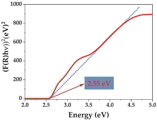
Figure 3.
Tauc’s plot for BiVO4 from Kubelka–Munk-transformed UV–Vis DRS; the linear fit of (F(R) hν)2 vs. hν extrapolated to (F(R) hν)2 = 0 yields and Eg = 2.55 eV (direct, n = 1/2).
3.3. Photocatalytic Performance of the Microwave-Synthesized BiVO4
AO7 in aqueous solution served as the model pollutant to evaluate the visible-light photocatalytic activity of BiVO4. Prior to irradiation, dye–catalyst suspensions were magnetically stirred in the dark for 60 min to reach adsorption–desorption equilibrium; the concentration after this step was taken as C0. A blank conducted under identical visible-light irradiation but without BiVO4 showed only ~10% AO7 loss over 120 min, indicating minimal direct photolysis. Accordingly, the decreases observed under irradiation in the presence of BiVO4 are attributed to photocatalytic conversion rather than adsorption or photolysis. The influences of catalyst dosage and initial dye concentration are examined below; pH was not systematically adjusted and remained near its native value (~4.5).
The direct band gap (2.55 eV) enables visible-light absorption, while ~19–20 nm crystallites shorten carrier migration paths and facilitate interfacial charge transfer. Under the native pH, dye–surface electrostatics together with light-utilization constraints govern the observed trends in the apparent rate constant k with dosage and initial AO7 concentration. These factors rationalize the increase in k with catalyst mass up to 15–20 mg and its higher value at lower AO7 loads.
3.3.1. The Effect of Catalyst Dosage on the Degradation of Acid Orange 7
Figure 4 shows the time-resolved UV–Vis spectra of AO7 under visible-light irradiation in the presence of 15 mg BiVO4, with a slight hypsochromic shift (λmax ≈ 484 nm). The shoulder within 440–520 nm progressively collapses, while the near-UV features around 250–320 nm attenuate more slowly, consistent with the stepwise loss of conjugation in the dye chromophore [32].
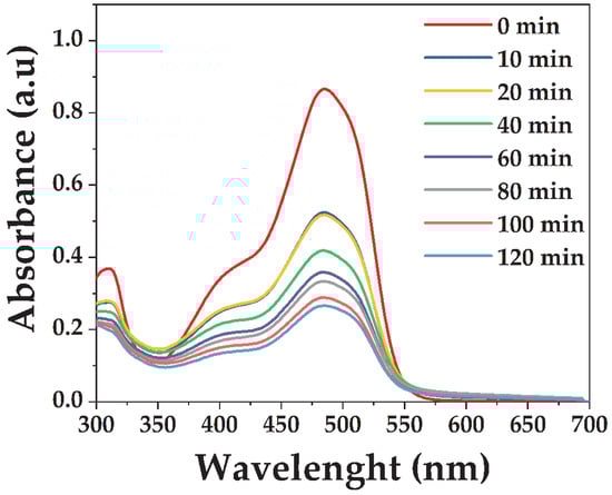
Figure 4.
UV–Vis absorption spectra of the AO7 solution (20 ppm) for 15 mg of BiVO4.
Figure 5 shows the effect of BiVO4 dosage on AO7 degradation and the temporal decay of C/C0 at 484 nm. Figure 5a presents the temporal decay of the normalized concentration (C/C0) at the absorption maximum of AO7 (λmax ≈ 484 nm). In the absence of BiVO4, only about 10% of AO7 was removed after 120 min, confirming that the degradation is primarily photocatalyst-driven. Increasing the catalyst dosage from 10 to 15 mg markedly enhanced degradation efficiency, with ~68% removal compared to ~55% for the lower loading. A further increase to 20 mg resulted in only a slight additional improvement, reaching ~72% after 120 min, indicating that the system operates close to its optimum catalyst loading.
The corresponding pseudo-first-order kinetic plots (Figure 5b) exhibit good linearity, consistent with the Langmuir–Hinshelwood model. The calculated rate constants increased with catalyst dosage, being lowest for 10 mg and highest for 20 mg, although the difference between 15 and 20 mg remained relatively small. This trend reflects a balance between the number of active surface sites and the efficiency of light utilization. At higher dosages, more reactive sites are available; however, excessive amounts can lead to light scattering, shielding effects, and partial particle aggregation, which ultimately limit further improvements in photocatalytic activity [33,34].
Based on these results, a dosage of 15 mg BiVO4 was used to study the effect of initial AO7 concentration, balancing efficiency and avoiding high-loading drawbacks.

Figure 5.
Effect of BiVO4 catalyst dosage on the photocatalytic degradation of AO7 (20 ppm) under visible-light irradiation, including direct photolysis are as follows: (a) temporal decay of normalized concentration (C/C0) with time; (b) pseudo-first-order kinetic plots with SD error bars.
Figure 5.
Effect of BiVO4 catalyst dosage on the photocatalytic degradation of AO7 (20 ppm) under visible-light irradiation, including direct photolysis are as follows: (a) temporal decay of normalized concentration (C/C0) with time; (b) pseudo-first-order kinetic plots with SD error bars.

3.3.2. Influence of the Initial Acid Orange 7 Concentration on Visible-Light Photodegradation Performance
After establishing 15 mg BiVO4 as the optimal catalyst dosage, the effect of the initial AO7 concentration was investigated. Figure 6a shows that the degradation efficiency decreases with increasing dye concentration. At 15 ppm, about 77% of AO7 was removed within 120 min, while the efficiency declined to ~69% at 20 ppm and ~70% at 25 ppm. This reduction is associated with stronger coloration of the solution at higher concentrations, which limits light penetration, as well as the saturation of active sites by excess dye molecules, reducing the availability of reactive oxygen species for photocatalytic reactions [35,36,37].
Kinetic plots (Figure 6b) follow pseudo-first-order kinetics, with linear fits yielding apparent rate constants of 0.00983 min−1 for 15 ppm, 0.00963 min−1 for 20 ppm, and 0.00811 min−1 for 25 ppm. The higher value obtained at 15 ppm indicates that the reaction proceeds faster at lower pollutant loads, while the decrease in k with increasing concentration reflects competition among dye molecules for both photons and active sites. Correlation coefficients (R2 > 0.95) further support the applicability of the Langmuir–Hinshelwood kinetic model (Table 2) [38,39].
Table 2.
Apparent rate constants (k) and correlation coefficients (R2) for AO7 degradation over BiVO4 under visible light.

Figure 6.
Effect of initial AO7 concentration (15 ppm, 20 ppm, and 25 ppm) on the photocatalytic degradation over BiVO4 (15 mg) under visible-light irradiation are as follows: (a) temporal decay of normalized concentration (C/C0) with time; (b) pseudo-first-order kinetic plots with SD error bars.
Figure 6.
Effect of initial AO7 concentration (15 ppm, 20 ppm, and 25 ppm) on the photocatalytic degradation over BiVO4 (15 mg) under visible-light irradiation are as follows: (a) temporal decay of normalized concentration (C/C0) with time; (b) pseudo-first-order kinetic plots with SD error bars.

The kinetic parameters extracted from the pseudo-first-order fits are compiled in Table 2, allowing side-by-side comparison across both series (mass and concentration).
In comparison with conventional TiO2 P25, the superior visible-light response of BiVO4 becomes evident. Because of its wide band gap (~3.2 eV), TiO2 P25 primarily absorbs UV light and typically exhibits very low apparent rate constants (k < 1 × 10⁻³ min−1) under visible-only irradiation [11,14]. In contrast, reported values for BiVO4 during azo-dye degradation commonly fall within (8 × 10⁻³)–(1.5 × 10⁻²) min−1, depending on morphology, synthesis route, and interfacial design [17,19]. Heterojunctions (e.g., BiVO4/TiO2) and plasmonic or metal modifications (e.g., Ag–BiVO4) further improve charge separation and kinetics. The present microwave-synthesized BiVO4 is consistent with these observations, as summarized in Table 3.
Table 3.
Comparison of pseudo-first-order rate constants (k) and experimental conditions for dye degradation under visible light over TiO2 P25 and BiVO4-based photocatalysts.
3.4. Possible Photocatalytic Pathway for the Degradation of Acid Orange 7
Figure 7 outlines a plausible route for AO7 degradation on visible-light-active BiVO4. Upon irradiation, photoexcited electron–hole pairs generate reactive oxygen species (●O2, ●OH) in parallel with direct hole oxidation (h+). Azo-bond cleavage initiates the sequence, which proceeds along two principal trajectories. On the benzene-derived branch, sulfonated aromatic amines evolve through stepwise oxidation to sulfonated hydroxybenzenes and, after desulfonation, to benzoic acid-type derivatives. On the naphthalene-derived branch, hydroxylated intermediates form and convert into additional hydroxylated naphthalenes, quinone species, and ultimately benzoic acid-type compounds. Prolonged oxidation on both branches plausibly promotes aromatic ring opening to low-molecular-weight aliphatic carboxylic acids and, in late stages, mineralization [10,32]. The sequence in Figure 7 accords with our spectral trends—most notably the attenuation of the 484 nm chromophore band—and with mechanisms reported for AO7 and structurally related azo dyes [42].
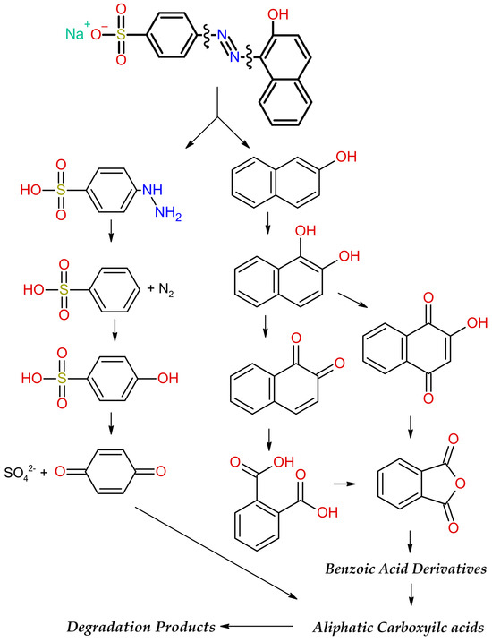
Figure 7.
Plausible visible-light-driven AO7 degradation mechanism over BiVO4, from azo-bond cleavage to oxidation intermediates and lower-molecular-weight products (Modified from [42]).
Although the proposed pathway is consistent with well-established degradation routes, it should be regarded as a literature-informed mechanistic hypothesis. The intermediates are representative rather than experimentally verified in this study, and no claim of complete mineralization is made. Verification would require complementary analyses such as LC–MS for organic intermediates, ion chromatography for SO42− and small acids, TOC monitoring for mineralization degree, and radical-quenching/EPR tests to elucidate the roles of ●OH, ●O2–, and h+.
3.5. Antibacterial Properties
The results demonstrated that BiVO4 nanoparticles exhibited a measurable antibacterial effect against Escherichia coli ATCC 25922, used as a model Gram-negative pathogen. After 4 h of incubation with UV-preactivated BiVO4 nanoparticles, the number of viable bacterial cells was reduced compared to the untreated control (Figure 8). Based on colony counting from serial decimal dilutions, the bacterial concentration in the control group was 2.4 × 10⁶ CFU/mL, while the BiVO4-treated sample averaged 7.3 × 10⁵ CFU/mL. This corresponds to approximately 67% inhibition, or a reduction of ~0.5 log units.
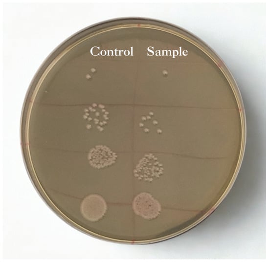
Figure 8.
Photography of the TSA plate with serial dilutions of untreated E. coli (left) and the sample treated with UV-activated BiVO4 nanoparticles (right).
One-way ANOVA with Tukey’s test showed a statistically significant reduction in viable E. coli counts (p < 0.05). Although the decrease did not reach the often-cited 1-log threshold for strong antibacterial activity, UV-preactivated BiVO4 produced a moderate, reproducible effect under ambient (non-UV) conditions, which is relevant where continuous UV exposure is impractical. The nanoscale morphology (~19 nm) and the visible-light-active direct band gap (2.55 eV) of the synthesized monoclinic BiVO4 promote efficient charge separation and ROS generation—features that likely underlie the observed antibacterial response [26]. This effect is plausibly mediated by nanoparticle adhesion to the bacterial envelope with ensuing membrane perturbation, aided by the high surface-area-to-volume ratio, together with oxidative stress from ROS associated with photoactive surface states. Electrostatic interactions between positively charged surface sites and the negatively charged cell envelope can further increase permeability and facilitate ROS-mediated damage to lipids, proteins, and nucleic acids, thereby reducing culturability [43]. For context, TiO2 (P25) showed a higher E. coli reduction (~1.7 log after 180 min under visible light) compared to the present BiVO4 system [44]. This difference primarily arises from the much stronger irradiation and more favorable reactor configuration used in that study, rather than from any intrinsic antibacterial superiority of TiO2 under visible light. By contrast, Ag-modified BiVO4 commonly achieves near-complete inactivation under visible light. For example, Ag/BiVO4 nanostructures have fully photoinactivated E. coli within 60 min under LED visible irradiation [45]. This superior performance is ascribed to localized surface-plasmon-assisted excitation and the role of Ag nanoparticles as electron sinks, which suppress electron–hole recombination and enhance interfacial ROS generation.
4. Conclusions
Microwave-assisted synthesis yielded phase-pure monoclinic BiVO4 with nanosized (~19 nm) crystallites, a specific surface area of 7.5 m2/g, and a direct band gap of 2.55 eV, enabling efficient utilization of visible light. The structural and electronic features enabled stable photocatalytic performance toward AO7 degradation, with the reaction following pseudo-first-order kinetics and achieving a degradation efficiency of approximately 77% after 120 min under visible irradiation. A moderate but statistically significant antibacterial effect was also observed under ambient, non-UV conditions, confirming the ability of UV-preactivated BiVO4 to retain residual ROS activity and surface reactivity. The results demonstrate that efficient charge separation and interfacial processes can occur even at modest surface areas when crystallinity and band-gap alignment are optimized. Future work may explore Eu3+ doping or other modifications to improve visible-light activity and charge-carrier lifetime. In addition, mechanistic analyses such as radical scavenging, EPR spectroscopy, and LC-MS/TOC measurements are recommended to verify the dominant reactive species and clarify the photocatalytic degradation mechanism.
Author Contributions
Conceptualization, D.M.; investigation, N.T. and B.V.; characterization, A.P. and I.M.; antibacterial experiments, S.D.; data curation, J.P.F.; writing—original draft preparation, N.T. and B.V.; writing—review and editing, D.M.; supervision, D.M. All authors have read and agreed to the published version of the manuscript.
Funding
This research was funded by the Ministry of Science, Technological Development, and Innovation of the Republic of Serbia, grant number 451-03-136/2025-03/200017.
Data Availability Statement
The original contributions presented in this study are included in the article. Further inquiries can be directed to the corresponding authors.
Acknowledgments
The authors are thankful to Yehezkel Yogev, Tanja Barudžija, and Dušan Mijin for providing TEM, XRD, and FTIR measurements.
Conflicts of Interest
The authors declare no conflicts of interest.
References
- Moghimi Dehkordi, M.; Pournuroz Nodeh, Z.; Soleimani Dehkordi, K.; Salmanvandi, H.; Rasouli Khorjestan, R.; Ghaffarzadeh, M. Soil, Air, and Water Pollution from Mining and Industrial Activities: Sources of Pollution, Environmental Impacts, and Prevention and Control Methods. Results Eng. 2024, 23, 102729. [Google Scholar] [CrossRef]
- Shetty, S.S.; D, D.; S, H.; Sonkusare, S.; Naik, P.B.; Kumari N, S.; Madhyastha, H. Environmental Pollutants and Their Effects on Human Health. Heliyon 2023, 9, e19496. [Google Scholar] [CrossRef] [PubMed]
- Tkaczyk, A.; Mitrowska, K.; Posyniak, A. Synthetic Organic Dyes as Contaminants of the Aquatic Environment and Their Implications for Ecosystems: A Review. Sci. Total Environ. 2020, 717, 137222. [Google Scholar] [CrossRef]
- Slama, H.B.; Chenari Bouket, A.; Pourhassan, Z.; Alenezi, F.N.; Silini, A.; Cherif-Silini, H.; Oszako, T.; Luptakova, L.; Golińska, P.; Belbahri, L. Diversity of Synthetic Dyes from Textile Industries, Discharge Impacts and Treatment Methods. Appl. Sci. 2021, 11, 6255. [Google Scholar] [CrossRef]
- Benkhaya, S.; M’rabet, S.; El Harfi, A. Classifications, Properties, Recent Synthesis and Applications of Azo Dyes. Heliyon 2020, 6, e03271. [Google Scholar] [CrossRef]
- Kusumlata; Ambade, B.; Kumar, A.; Gautam, S. Sustainable Solutions: Reviewing the Future of Textile Dye Contaminant Removal with Emerging Biological Treatments. Limnol. Rev. 2024, 24, 126–149. [Google Scholar] [CrossRef]
- Dutta, S.; Adhikary, S.; Bhattacharya, S.; Roy, D.; Chatterjee, S.; Chakraborty, A.; Banerjee, D.; Ganguly, A.; Nanda, S.; Rajak, P. Contamination of Textile Dyes in Aquatic Environment: Adverse Impacts on Aquatic Ecosystem and Human Health, and Its Management Using Bioremediation. J. Environ. Manag. 2024, 353, 120103. [Google Scholar] [CrossRef]
- Daneshvar, N.; Aber, S.; Hosseinzadeh, F. Study of C.I. Acid Orange 7 Removal in Contaminated Water by Photo Oxidation Processes. Glob. NEST J. 2008, 10, 16–23. [Google Scholar] [CrossRef]
- Kathi, S.; El Din Mahmoud, A. Trends in Effective Removal of Emerging Contaminants from Wastewater: A Comprehensive Review. Desalination Water Treat. 2024, 317, 100258. [Google Scholar] [CrossRef]
- Iervolino, G.; Vaiano, V.; Pepe, G.; Campiglia, P.; Palma, V. Degradation of Acid Orange 7 Azo Dye in Aqueous Solution by a Catalytic-Assisted, Non-Thermal Plasma Process. Catalysts 2020, 10, 888. [Google Scholar] [CrossRef]
- Tanos, F.; Razzouk, A.; Lesage, G.; Cretin, M.; Bechelany, M. A Comprehensive Review on Modification of Titanium Dioxide-Based Catalysts in Advanced Oxidation Processes for Water Treatment. ChemSusChem 2024, 17, e202301139. [Google Scholar] [CrossRef]
- Pham, H.-C.; Kim, K.-S. Effect of TiO2 Thin Film Thickness on NO and SO2 Removals by Dielectric Barrier Discharge-Photocatalyst Hybrid Process. Ind. Eng. Chem. Res. 2013, 52, 5296–5301. [Google Scholar] [CrossRef]
- Guo, Q.; Ma, Z.; Zhou, C.; Ren, Z.; Yang, X. Single Molecule Photocatalysis on TiO2 Surfaces: Focus Review. Chem. Rev. 2019, 119, 11020–11041. [Google Scholar] [CrossRef]
- Etacheri, V.; Di Valentin, C.; Schneider, J.; Bahnemann, D.; Pillai, S.C. Visible-Light Activation of TiO2 Photocatalysts: Advances in Theory and Experiments. J. Photochem. Photobiol. C Photochem. Rev. 2015, 25, 1–29. [Google Scholar] [CrossRef]
- Kaiba, A.; Alansi, A.M.; Oubelkacem, A.; Chabri, I.; Hameed, S.T.; Afzal, N.; Rafique, M.; Qahtan, T.F. Sunlight-Driven Synthesis of TiO2/(MA)2SnCl4 Nanocomposite Films for Enhanced Photocatalytic Degradation of Organic Pollutants. Catalysts 2025, 15, 214. [Google Scholar] [CrossRef]
- Ishigaki, T.; Nakada, Y.; Tarutani, N.; Uchikoshi, T.; Tsujimoto, Y.; Isobe, M.; Ogata, H.; Zhang, C.; Hao, D. Enhanced Visible-Light Photocatalytic Activity of Anatase-Rutile Mixed-Phase Nano-Size Powder given by High-Temperature Heat Treatment. R. Soc. Open Sci. 2020, 7, 191539. [Google Scholar] [CrossRef]
- Kamble, G.S.; Natarajan, T.S.; Patil, S.S.; Thomas, M.; Chougale, R.K.; Sanadi, P.D.; Siddharth, U.S.; Ling, Y.-C. BiVO4 As a Sustainable and Emerging Photocatalyst: Synthesis Methodologies, Engineering Properties, and Its Volatile Organic Compounds Degradation Efficiency. Nanomaterials 2023, 13, 1528. [Google Scholar] [CrossRef]
- Dolić, S.D.; Jovanović, D.J.; Štrbac, D.; Far, L.Đ.; Dramićanin, M.D. Improved Coloristic Properties and High NIR Reflectance of Environment-Friendly Yellow Pigments Based on Bismuth Vanadate. Ceram. Int. 2018, 44, 22731–22737. [Google Scholar] [CrossRef]
- Dolić, S.D.; Jovanović, D.J.; Smits, K.; Babić, B.; Marinović-Cincović, M.; Porobić, S.; Dramićanin, M.D. A Comparative Study of Photocatalytically Active Nanocrystalline Tetragonal Zyrcon-Type and Monoclinic Scheelite-Type Bismuth Vanadate. Ceram. Int. 2018, 44, 17953–17961. [Google Scholar] [CrossRef]
- Jeong, S.Y.; Choi, K.S.; Shin, H.-M.; Kim, T.L.; Song, J.; Yoon, S.; Jang, H.W.; Yoon, M.-H.; Jeon, C.; Lee, J.; et al. Enhanced Photocatalytic Performance Depending on Morphology of Bismuth Vanadate Thin Film Synthesized by Pulsed Laser Deposition. ACS Appl. Mater. Interfaces 2017, 9, 505–512. [Google Scholar] [CrossRef]
- Jelić, S.T.; Ćirković, J.; Jovanović, J.; Novaković, T.; Podlogar, M.; Mitrić, J.; Branković, G.; Branković, Z. High Efficiency Solar Light Photocatalytic Degradation of Mordant Blue 9 by Monoclinic BiVO4 Nanopowder. Mater. Chem. Phys. 2025, 333, 130341. [Google Scholar] [CrossRef]
- Regmi, C.; Dhakal, D.; Lee, S.W. Visible-Light-Induced Ag/BiVO4 Semiconductor with Enhanced Photocatalytic and Antibacterial Performance. Nanotechnology 2018, 29, 064001. [Google Scholar] [CrossRef] [PubMed]
- Zhang, A.; Zhang, J. Effects of Europium Doping on the Photocatalytic Behavior of BiVO4. J. Hazard. Mater. 2010, 173, 265–272. [Google Scholar] [CrossRef]
- Drisya, K.T.; Solís-López, M.; Ríos-Ramírez, J.J.; Durán-Álvarez, J.C.; Rousseau, A.; Velumani, S.; Asomoza, R.; Kassiba, A.; Jantrania, A.; Castaneda, H. Electronic and Optical Competence of TiO2/BiVO4 Nanocomposites in the Photocatalytic Processes. Sci. Rep. 2020, 10, 13507. [Google Scholar] [CrossRef]
- Ran, J.-H.; Fei, X.; Ni, L.; Telegin, F. Enhanced Photocatalytic Degradation of Acid Orange 7 by AgBr/BiVO4 under Visible Light. J. Fiber Bioeng. Inform. 2018, 11, 151–161. [Google Scholar] [CrossRef]
- Bulut, D.T. Exploring the Dual Role of BiVO4 Nanoparticles: Unveiling Enhanced Antimicrobial Efficacy and Photocatalytic Performance. J. Sol-Gel Sci. Technol. 2025, 114, 198–222. [Google Scholar] [CrossRef]
- Pramila, S.; Nagaraju, G.; Mallikarjunaswamy, C.; Latha, K.C.; Chandan, S.; Ramu, R.; Rashmi, V.; Lakshmi Ranganatha, V. Green Synthesis of BiVO4 Nanoparticles by Microwave Method Using Aegle marmelos Juice as a Fuel: Photocatalytic and Antimicrobial Study. Anal. Chem. Lett. 2020, 10, 298–306. [Google Scholar] [CrossRef]
- Marinković, D.; Righini, G.C.; Ferrari, M. Advances in Synthesis and Applications of Bismuth Vanadate-Based Structures. Inorganics 2025, 13, 268. [Google Scholar] [CrossRef]
- Marinković, D.; Righini, G.C.; Ferrari, M. Synthesis, Optical, and Photocatalytic Properties of the BiVO4 Semiconductor Nanoparticles with Tetragonal Zircon-Type Structure. Photonics 2025, 12, 438. [Google Scholar] [CrossRef]
- Li, B.; Gao, X.; Qu, J.; Xiong, F.; Xuan, H.; Jin, Y.; Yuan, H. Visible-Light-Driven Antimicrobial Activity and Mechanism of Polydopamine-Reduced Graphene Oxide/BiVO4 Composite. Int. J. Mol. Sci. 2022, 23, 7712. [Google Scholar] [CrossRef]
- Thommes, M.; Kaneko, K.; Neimark, A.V.; Olivier, J.P.; Rodriguez-Reinoso, F.; Rouquerol, J.; Sing, K.S.W. Physisorption of Gases, with Special Reference to the Evaluation of Surface Area and Pore Size Distribution (IUPAC Technical Report). Pure Appl. Chem. 2015, 87, 1051–1069. [Google Scholar] [CrossRef]
- Zhang, S.-J.; Yu, H.-Q.; Li, Q.-R. Radiolytic Degradation of Acid Orange 7: A Mechanistic Study. Chemosphere 2005, 61, 1003–1011. [Google Scholar] [CrossRef]
- Iqbal, N.; Huang, X.; Mohamedali Hamid, K.; Yuan, H.; Batool, I.; Yang, Y. Efficient Visible-Light-Driven Photocatalysis of BiVO4@Diatomite for Degradation of Methoxychlor. Catalysts 2025, 15, 672. [Google Scholar] [CrossRef]
- Maran, M.A.; Zheng, A.L.T.; Tan, H.Y.; Sarbini, S.R.; Tan, K.B.; Boonyuen, S.; Wong, K.K.S.; Chung, E.L.T.; Lease, J.; Andou, Y. Assessing the Photocatalytic Performance of Hydrothermally Synthesized Fe-Doped BiVO4 Under Low-Intensity UV Irradiation. Arab. J. Sci. Eng. 2025, in press. [Google Scholar] [CrossRef]
- Nunes, M.J.; Lopes, A.; Pacheco, M.J.; Ciríaco, L. Visible-Light-Driven AO7 Photocatalytic Degradation and Toxicity Removal at Bi-Doped SrTiO3. Materials 2022, 15, 2465. [Google Scholar] [CrossRef] [PubMed]
- Reza, K.M.; Kurny, A.; Gulshan, F. Parameters Affecting the Photocatalytic Degradation of Dyes Using TiO2: A Review. Appl. Water Sci. 2017, 7, 1569–1578. [Google Scholar] [CrossRef]
- Khan, S.; Noor, T.; Iqbal, N.; Yaqoob, L. Photocatalytic Dye Degradation from Textile Wastewater: A Review. ACS Omega 2024, 9, 21751–21767. [Google Scholar] [CrossRef]
- Asenjo, N.G.; Santamaría, R.; Blanco, C.; Granda, M.; Álvarez, P.; Menéndez, R. Correct Use of the Langmuir–Hinshelwood Equation for Proving the Absence of a Synergy Effect in the Photocatalytic Degradation of Phenol on a Suspended Mixture of Titania and Activated Carbon. Carbon 2013, 55, 62–69. [Google Scholar] [CrossRef]
- Phanichphant, S.; Nakaruk, A.; Chansaenpak, K.; Channei, D. Evaluating the Photocatalytic Efficiency of the BiVO4/rGO Photocatalyst. Sci. Rep. 2019, 9, 16091. [Google Scholar] [CrossRef] [PubMed]
- Mancuso, A.; Sacco, O.; Vaiano, V.; Bonelli, B.; Esposito, S.; Freyria, F.S.; Blangetti, N.; Sannino, D. Visible Light-Driven Photocatalytic Activity and Kinetics of Fe-Doped TiO2 Prepared by a Three-Block Copolymer Templating Approach. Materials 2021, 14, 3105. [Google Scholar] [CrossRef]
- Abdullahi, N.; Saion, E.; Shaari, A.H.; Al-Hada, N.M.; Keiteb, A. Optimisation of the Photonic Efficiency of TiO2 Decorated on MWCNTs for Methylene Blue Photodegradation. PLoS ONE 2015, 10, e0125511. [Google Scholar] [CrossRef]
- Viswanathan, B. Photocatalytic Degradation of Dyes: An Overview. Curr. Catal. 2018, 7, 99–121. [Google Scholar] [CrossRef]
- Franco, D.; Calabrese, G.; Guglielmino, S.P.P.; Conoci, S. Metal-Based Nanoparticles: Antibacterial Mechanisms and Biomedical Application. Microorganisms 2022, 10, 1778. [Google Scholar] [CrossRef] [PubMed]
- Haque, F.; Blanchard, A.; Laipply, B.; Dong, X. Visible-Light-Activated TiO2-Based Photocatalysts for the Inactivation of Pathogenic Bacteria. Catalysts 2024, 14, 855. [Google Scholar] [CrossRef]
- Narváez, B.; Mendoza-Mendoza, E.; Peralta-Rodríguez, R.; Bach, H.; Barriga-Castro, E.; Segovia-Sandoval, S.; Martinez-Gutierrez, F. Visible-Leds-Induced Enhanced Photocatalytic and Antibacterial Activity of BiVO4 Based Green Photocatalysts Decorated with Silver and Graphene. J. Photochem. Photobiol. A Chem. 2023, 447, 115191. [Google Scholar] [CrossRef]


Disclaimer/Publisher’s Note: The statements, opinions and data contained in all publications are solely those of the individual author(s) and contributor(s) and not of MDPI and/or the editor(s). MDPI and/or the editor(s) disclaim responsibility for any injury to people or property resulting from any ideas, methods, instructions or products referred to in the content. |
© 2025 by the authors. Licensee MDPI, Basel, Switzerland. This article is an open access article distributed under the terms and conditions of the Creative Commons Attribution (CC BY) license (https://creativecommons.org/licenses/by/4.0/).