Article

Disruption of Early Streptococcus mutans Biofilm Development on Orthodontic Aligner Materials

by
Matea Badnjević
1,2,*,†,
Mirna Petković Didović
2,†,
Ivana Jelovica Badovinac
3,
Sanja Lučić Blagojević
4,
Marko Perčić
5,
Stjepan Špalj
1,6 and
Ivana Gobin
2,7
1
Faculty of Dental Medicine, University of Rijeka, Krešimirova 40/42, 51000 Rijeka, Croatia
2
Faculty of Medicine, University of Rijeka, Braće Branchetta 20, 51000 Rijeka, Croatia
3
Centre for Micro- and Nanosciences and Technologies, Faculty of Physics, University of Rijeka, Radmile Matejčić 2, 51000 Rijeka, Croatia
4
Faculty of Chemical Engineering and Technology, University of Zagreb, Trg Marka Marulića 19, 10000 Zagreb, Croatia
5
Centre for Micro- and Nanosciences and Technologies, Faculty of Engineering, University of Rijeka, Radmile Matejčić 2, 51000 Rijeka, Croatia
6
Faculty of Dental Medicine and Health, Josip Juraj Strossmayer University of Osijek, 31000 Osijek, Croatia
7
Teaching Institute of Public Health of Primorje-Gorski Kotar County, Krešimirova 52a, 51000 Rijeka, Croatia
*
Author to whom correspondence should be addressed.
The authors contributed equally to this work.
Processes 2025, 13(10), 3069; https://doi.org/10.3390/pr13103069
Submission received: 22 August 2025 / Revised: 19 September 2025 / Accepted: 24 September 2025 / Published: 25 September 2025
(This article belongs to the Special Issue Microbial Biofilms: Latest Advances and Prospects)

Abstract

(1) Background: This study aimed to determine the optimum parameters for the treatment of Streptococcus mutans biofilm on clear dental aligners. (2) Methods: A 24-h-old S. mutans biofilm was grown on polyurethane (PU) and poly(ethylene terephthalate glycol) (PETG) aligners. These samples were treated with three chlorhexidine digluconate (CHX)-based antiseptic solutions, manual brushing, and a combination of both, with varying exposure times. The number of adhered bacteria was determined in both untreated and treated samples after sonication. Materials were analyzed with atomic force and scanning electron microscopy, and surface free energy (SFE) values were determined using three different models. (3) Results: Our findings indicated that control strategies do not depend on the type of material. PU and PETG surfaces exhibited similar SFE values (41–45 mJ/m2). Differences in surface roughness were insufficient to cause significant changes in S. mutans behavior. The highest efficacy of all three tested antiseptics was established for the exposure time of 1 min, with efficacy deteriorating just after 3 min. (4) Conclusions: The efficacy of CHX against S. mutans early biofilm is material-independent and time-dependent. The optimal exposure time of 1 min should be combined with brushing, with a general recommendation of the antiseptic-first approach.

Graphical Abstract

1. Introduction

Increasing patient demand for more discreet, comfortable, and easy-to-clean braces has led to numerous studies on orthodontic clear aligners over the last decade [1,2,3,4]. Despite many advantages, the issues that occur with the continuous use of clear aligners, such as odor development and clear aesthetic appearance loss, cannot be neglected. Clear aligners reduce the natural ability of dental and periodontal tissues to buffer and rinse away saliva. They also provide additional surfaces for bacteria to adhere, promoting the formation of bacterial biofilms that lead to caries and enamel demineralization [5,6,7]. Even though the bacteria adhere to oral surfaces indirectly through the pellicle, it is well established that their number and behavior mostly depend on the outermost atomic state of the original solid surface beneath the pellicle [8,9,10,11]. Surface free energy (SFE) and surface roughness of the solid substrate material are recognized as crucial parameters in plaque formation and development [12]. Materials commonly used for clear aligners are based on polyesters like poly(ethylene terephthalate glycol) (PETG) and polyurethanes (PU) [13]. PETG is a derivative of a well-known polyester poly(ethylene-terephthalate), PET. It is obtained by copolymerization of the two monomers that constitute PET, i.e., ethylene glycol and terephthalic acid, with cyclohexanedimethanol (CHDM) (Figure 1a). The CHDM is incorporated into the polymer backbone in place of the ethylene glycol, effectively intercalating a bulky cyclohexane ring between two ethylene carbons. This results in several important property modifications, including easier processing, improved 3D printability, higher glass transition temperature and a wider working range compared to PET [14,15]. Nonetheless, the crucial improvement for orthodontic applications arises from the fact that bulky cyclohexane ring prevents crystallization: while PET is a semicrystalline polymer, PETG is amorphous, which makes it highly transparent and translucent, while retaining good mechanical properties, resistance to solvents and low levels of gloss [2,16]. Moreover, the transparency is maintained throughout its working temperature range, from −70 °C to 70 °C [17]. On the other hand, PUs are characterized by the urethane (or carbamate, –NH–CO–O–, Figure 1b) link between their monomers. These monomers are di- or tri-isocyanates and various polyols, enabling customized tailoring to meet the needs of a particular product. For the fabrication of clear aligners, a thermoplastic PU is used owing to its good balance of flexibility, elasticity, durability, toughness, along with its biocompatibility, transparency, and solvent resistance.
The best-recognized method of maintaining oral hygiene is manual tooth brushing; however, inadequate oral hygiene habits make it difficult to control plaque [18]. Studies have shown that brushing for 2 min removes only half of the plaque from the teeth [19]. The use of antiseptic mouthwash in combination with brushing is thus a frequent part of oral hygiene routine. Among various substances, CHX is considered the “gold standard” of oral antiseptics, known for decades not just for its antibacterial activities against salivary bacteria, but also for its remarkable plaque-inhibiting activity [20,21]. CHX, typically used in the form of a digluconate salt, belongs to the bisbiguanidine class of compounds (Figure 1c). Its antibacterial activity is owed to a perfect hydrophilic/lipophilic balance within its structure. It is a strongly basic molecule, with two protons uniformly distributed over all its ten nitrogen atoms [22]. This cationic nature enables it to interact with bacteria that carry negative surface charge at physiological pH. Its mode of action involves several steps: first, it adsorbs to a bacterial surface and damages the membrane, which increases the permeability to cytoplasm; then, it enters the cytoplasm and causes the precipitation of cytoplasmic substances, disabling the bacteria from repairing cell membranes and rendering them non-viable [22,23,24].
It is known that improper care of clear aligners deteriorates their optical and mechanical properties [25,26,27,28]. For instance, it has been found that the PETG mechanical properties deteriorate after a certain amount of exposure even to pure distilled water [29], emphasizing that the contact of materials and solutions should be kept to a minimum. Prevention and impediment of material deterioration are of crucial importance since those are the essential factors that determine the treatment outcome [2]. Nevertheless, a recent review has shown that there are still no clear guidelines for cleaning and disinfecting of aligners [4].
The aim of this study was therefore to identify optimal parameters for the control of biofilm formation. These parameters include (a) the duration of exposure of the aligners to 0.12% chlorhexidine digluconate (CHX)-containing antiseptics within a realistic time frame (1–5 min); (b) the choice of the most effective commercially available antiseptic liquid and (c) the optimal routine sequence, which includes mechanical brushing in addition to the duration of exposure, as these two are the most popular and accessible cleaning methods for the general population. These parameters were analyzed on the two most commonly used clear aligner materials, PETG and PU [2,3]. The optimal parameters were considered to be those that minimize bacterial adhesion, for which S. mutans was selected as the common caries-forming oral bacterium [30,31,32]. The working hypothesis was that the efficacy of all antiseptics improves with longer exposure time and reaches a plateau after a certain number of minutes, which may vary depending on the aligner material, and that mechanical brushing prior to antiseptic exposure improves the efficacy of the routine.

2. Materials and Methods

2.1. Aligner Materials

  • PU plates (10 mm × 10 mm; Zendura, Bay Materials, Fremont, CA, USA)
  • PETG plates (10 mm × 10 mm; Erkodur, Erkodent Erich Kopp GmbH, Pfalzgrafenweiler, Germany)

2.2. Antiseptic Solutions

  • Curasept ADS 212 (Curasept, Saronno, Italy) with 0.12% CHX
  • Perioplus (Curasept, Flawil, Switzerland) with 0.12% CHX
  • Solution of pure 0.12% CHX prepared in distilled water. The antiseptics used in this study have been described in a previous publication [33].

2.3. Biofilm Treatment

The aligner materials were immersed overnight in 50% artificial saliva consisted of porcine gastric mucin (Sigma-Aldrich, Burlington, MA, USA) (0.25% w/v), sodium chloride (Chem-Lab NV, Zedelgem, Belgium) (0.35% w/v), potassium chloride p.a. (Kemika, Zagreb, Croatia) (0.02% w/v), calcium chloride dihydrate p.a. (Kemika, Zagreb, Croatia) (0.02% w/v), yeast extract (Biolife, Milan, Italy) (0.2% w/v), Lab-Lemco powder (Oxoid, Basingstoke, UK) (0.1% w/v), proteose peptone (Biolife, Milan, Italy) (0.5% w/v), all dissolved in double-distilled water (ddH2O; Sigma-Aldrich, Burlington, MA, USA), and urea p.a. (Kemika, Zagreb, Croatia) at 0.05% (v/v) [34]. After 24 h, the materials were incubated in a microtiter plate with 1 mL of a S. mutans (strain ATCC 25175) suspension with a concentration of 107 colony-forming units/milliliter (CFU/mL) and 1.00 mL Brain Heart Infusion (BHI) for an additional 24 h under capnophilic conditions in a stirred incubator at 37 °C to develop an early biofilm. After 24 h, the materials were rinsed three times with phosphate-buffered saline (PBS) and treated separately: (1) manual brushing (without toothpaste in circular motions for 5 s); (2) exposure to CHX (0.12%) for 1 min; (3) manual brushing followed by exposure to CHX (0.12%) for 1 min; (4) exposure to CHX (0.12%) for 1 min followed by manual brushing; and (5) exposure to CHX (0.12%) for 5 min. After treatment, each material was rinsed with PBS and sonicated for 1 min in an ultrasonic bath (Bactosonic, Bandelin®, Berlin, Germany) at an intensity of 40 kHz. Tenfold dilutions were prepared, and the bacteria were spread on plates that were incubated under capnophilic conditions. After 48 h, the viable count of the bacteria (CFU/mL) was calculated by counting the colonies that had grown. All tests were performed in triplicate and repeated three times. Untreated bacteria in the 24-h-old early biofilm served as a control.

2.4. Atomic Force Microscopy (AFM)

A Bruker Dimension Icon in tapping mode was used to obtain high-resolution scans of surface detail using a Bruker SNL-10 type D (low stiffness) silicon nitride cantilever with a 2 nm silicon tip radius. The scans were performed with scan sizes of 30 µm2 with 512 scan lines, each with 512 data points acquired per line. The data obtained was processed to determine the values of surface roughness parameters after tilt and bow corrections using the proprietary Bruker Nanoscope Analysis software v1.5 (Bruker, Billerica, MA, USA).

2.5. SFE

A Data Physics OCA 20 goniometer (Data Physics Instruments GmbH, Filderstadt, Germany) was used for this experiment. Contact angles were determined using the sessile drop method. Water, formamide, and diiodomethane (99+%, AcrosOrganics, Geel, Belgium) were the test fluids. The surface of the aligner was cleaned with 95% ethanol (Kemika, Zagreb, Croatia) and dried. Then, 0.3 µL of the test liquid was applied. Ten measurements were conducted on each sample with the same test liquid, and average contact angle values for individual liquids were used for calculations. The values of the dispersive and polar components of the test liquids, used to calculate the SFE of the tested sample according to the Owens–Wendt, Wu, and Van Oss–Chaudhury–Good (OCG or acid–base) models, are detailed in our previous work [31].

2.6. Scanning Electron Microscopy (SEM)

The SEM images were acquired using a Jeol JSM-7800F SEM device (Jeol Ltd., Tokyo, Japan). The surface morphology was analyzed with a secondary electron detector at an electron beam acceleration voltage of 10 kV and a working distance of 10 mm. Prior to SEM analysis, a 10 nm thick Au/Pd coating was deposited on the sample surfaces using Gatan’s argon ion beam system (PECS II) to prevent charging. The bacterial biofilm was prepared by fixation (60% methanol, 30% chloroform, and 10% glacial acetic acid in PBS), dehydration (in an ethanol gradient), and drying [35].

3. Results

3.1. Time–Kill Curves

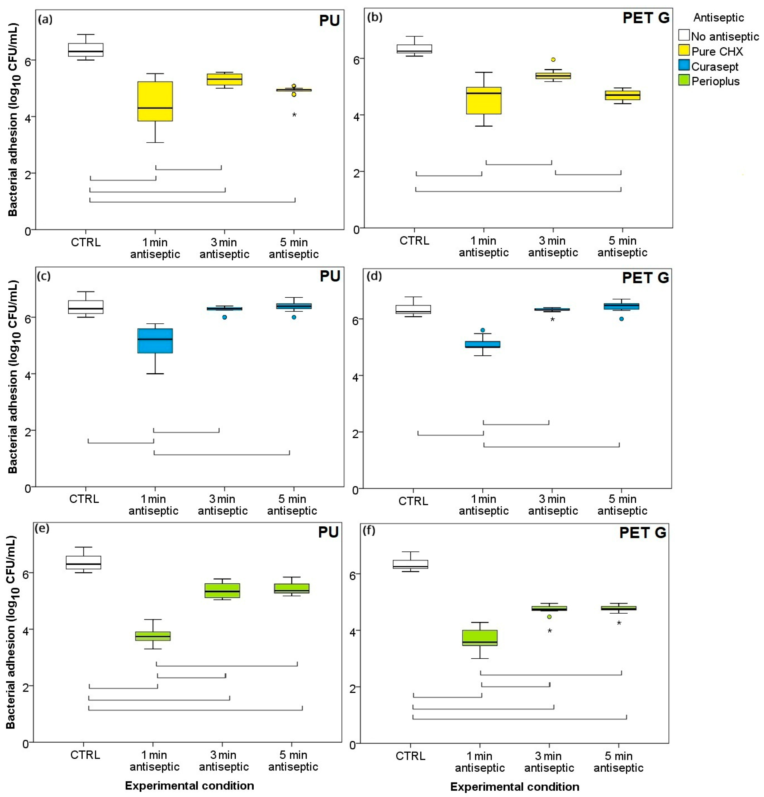
In the initial phase of this research, we investigated the time–kill curves of three types of antiseptics for S. mutans on PU and PETG surfaces to establish how the efficacy of a particular antiseptic correlates with the duration of the exposure. For the pure CHX (Figure 2a,b), the results showed a statistically significant (p < 0.05) decrease in the number of adhered bacteria compared to those of the control on both surfaces for all examined exposure durations, except the 3-min exposure on PETG. However, for both surfaces, the 3-min exposure seemed to be less efficient than the 1-min one, whereas the 5-min exposure showed no significant difference either to the 1-min or to 3-min exposures (except for 3- vs. 5-min on PETG).
The time–kill curves for the Curasept antiseptic (Figure 2c,d) exhibited a similar counterintuitive shape, with an even more pronounced decrease in efficacy for the 3- and 5-min exposures compared to the 1-min one. In comparison to pure CHX, the efficacy of Curasept was further diminished by longer exposures, with the number of adhered bacteria reaching the level of the control. The results were remarkably similar on PU (Figure 2c) and PETG (Figure 2d).
With Perioplus antiseptic, the number of adhered bacteria was consistently the lowest after 1-min exposure (Figure 2e,f). Longer exposures were significantly less efficient than the 1-min exposure, but—unlike Curasept—did exhibit a statistically significant improvement compared to the control. Similar results were obtained for both examined surfaces, with a slight difference; longer exposures were more efficient on PETG (Figure 2f) than on PU (Figure 2e).

3.2. One Minute Exposure and Brushing: Individual Treatments vs. Combination

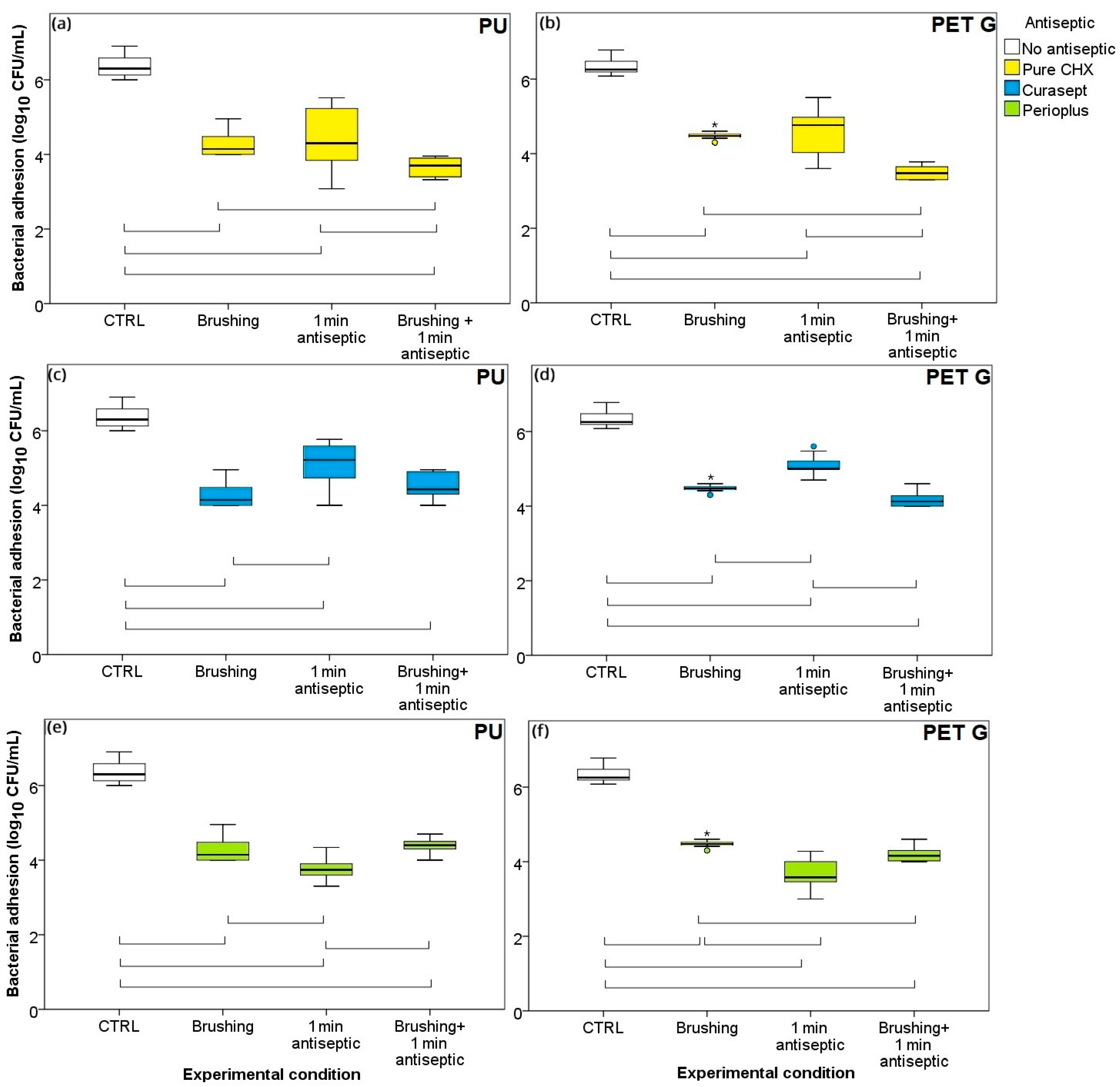
The results of the time–kill curves (Figure 2) unequivocally demonstrated that 1 min was the optimum time for exposure with all three antiseptics. The remainder of the research, therefore, focused on 1-min exposure, and its combination with mechanical brushing was further examined. The results for pure CHX (Figure 3a,b) expectedly showed a statistically significant reduction in the number of adhered bacteria when any treatment or treatment combination was employed compared to that in the control. The effect of mechanical brushing was equally efficient as that of the 1-min exposure. However, the combination of brushing and exposure was more efficient than the individual treatments, showing a statistically significant difference. Comparing the data on PU (Figure 3a) and that on PETG (Figure 3b) aligners, the results showed that the treatments exhibited similar effects.
The 1-min exposure with Curasept antiseptic was less effective than brushing (Figure 3c,d). For the PU aligner, combining brushing and exposure to antiseptic did not yield a significant improvement over the individual treatments. However, for PETG, the best results were obtained with the brushing/antiseptic combination. The results showed a significant improvement with the combination compared to the exposure-only treatment, but not significantly compared to the brushing alone, again demonstrating the weak antiseptic properties of Curasept. The results for Perioplus antiseptic (Figure 3e,f) showed that the number of adhered bacteria was the lowest after 1-min exposure, with a significant difference compared to that with brushing on both materials and even compared to that with the combined treatment (significant difference on PU).
For an easier comparison of efficacies of the antiseptics, the number of adhered bacteria after mechanical brushing, 1-min exposure, or the combination of both is pooled and shown in Figure 4. The results on both materials showed that a significant improvement over brushing was obtained only in the case of Perioplus. The 1-min exposure with Curasept removed fewer bacteria than brushing alone on both materials. Also, on both materials, combining brushing and exposure yielded the best results when pure CHX was used. Surprisingly, the brushing/exposure combination with Perioplus and Curasept did not yield a significant improvement compared to brushing alone.

3.3. Ordering of Treatments: Brushing-First or Antiseptic-First?

In the final series of measurements, we aimed to examine whether the order of treatments in the exposure/brushing combination was a relevant factor. For this purpose, we used PU aligners and all three antiseptics. The results (Figure 5) expectedly showed a statistically significant decrease in the number of adhered bacteria when any combination of treatments was applied for all three antiseptics, compared to that in the control. Regarding the order within the treatment, a certain improvement (decrease) in the number of adhered bacteria was observed when the antiseptic was used first and followed by brushing, compared to the reverse order, for all three tested antiseptics. However, a statistically significant difference was obtained only in the case of Curasept.
To elucidate the differences in the results between PU and PETG aligners, SFE values were determined from static contact angles of the three probe liquids (Table 1). Static water contact angles (WCA) are also used as a rough measure of surface hydrophobicity, with the 90° angle being the cutoff value [36,37,38,39]. The WCA of the PETG (approximately 77.3°) was somewhat lower than that in the literature for a PETG retainer from another manufacturer (approximately 82°), indicating somewhat lower hydrophobicity [40]. Similarly, the WCA values for the PU aligner (approximately 80.4°) were also lower than those for typical PU materials (82–93°). Generally, these WCA values categorize the examined materials as weakly hydrophobic (90° > WCA > 56–65°; [36]), with PU being more hydrophobic than PETG.
WCA was used, together with formamide and diiodomethane contact angles, to obtain the SFE of PETG and PU aligners using three typical mathematical models (Table 2). Within each model, the dispersive (nonpolar) component is presented first (γds), followed by the polar component (γps), then the total SFE as the sum of the two (γs). For the acid–base model, the nonpolar component is known as Lifshitz–van der Waals (γLWs), whereas the polar component is subdivided into acid and base interactions (γ+s and γ-s, respectively). The signs + and − in the superscript designate electron-accepting (acidic) or electron-donating (basic) abilities of the surface [41].
The results obtained from all three models showed very similar total SFE values for PETG and PU aligners. For both materials, the dispersive components were an order of magnitude higher than polar, indicating the dominance of non-polar interactions. Polar interactions, although weak for both materials, were stronger for PETG. Additionally, the acid-base model revealed that—within polar interactions—the basic character prevails for both materials. This model also revealed the differences between the two materials, namely, the higher value of the basic γs component for PETG compared to PU (7.9 mJ/m2 vs. 4.9 mJ/m2). This indicates that the PETG surface had a more basic character, i.e., more electron-donating abilities than PU. Note that the acidic component of PETG is almost zero. Hence, it can be inferred that the higher value of the polar component of PETG compared to PU can be attributed to basic, not acidic, interactions. Regarding the absolute values of total SFE, results were similar for both materials, with values of around 41–45 mJ/m2, depending on the model. Generally, surfaces with SFE of ~20–25 mJ/m2 are considered low energy surfaces, those with ~30–35 mJ/m2 are considered medium energy surfaces and those with >50 mJ/m2 are considered high energy surfaces [9]. Note that SFE value for S. mutans, determined spectrophotometrically in our previous work, was 31 mJ/m2 [31].

3.4. Analysis of the SEM Micrographs

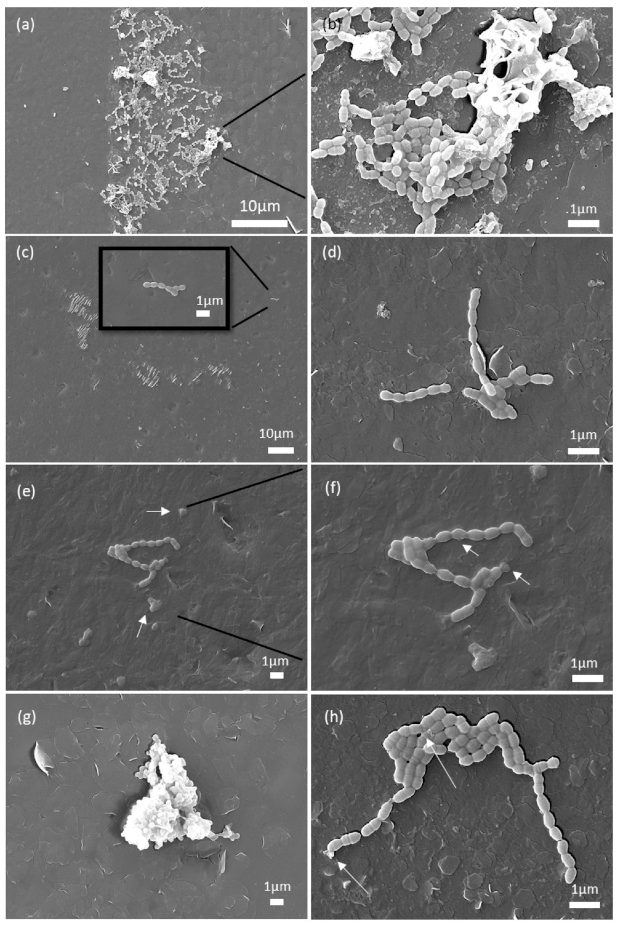
The results thus far showed that control strategies with CHX for S. mutans on clear dental aligners do not depend on the type of material used for their production. Therefore, the SEM analysis was performed only on the PU samples. After 5-min CHX treatment, damaged cells with a changed morphology and disintegrated membranes were observed on the surface of the PU (Figure 6). Accumulations of bacteria embedded in a thicker layer of EPS (Short arrows on Figure 6f) were also noticeable compared to untreated bacteria and bacteria treated for 1 min. Membrane damage was visible on some bacteria within the biofilm (Long arrows on Figure 6e,f). Brushing treatment led to sloughing of the biofilm (Figure 6g), which was evidenced by the presence of biofilm accumulations, where biofilm disintegration, partial detachment, and formation of the aggregates occurred. The number of individual cells and smaller clusters of bacteria was reduced on the PU. The combination of brushing and 1-min CHX treatment also led to a reduction in the number of individual cells; if they were present, they were visibly damaged. In larger clusters of cells, damage to the cell membrane and leakage of intracellular contents were visible (Figure 6h).

3.5. Surface Roughness Analysis

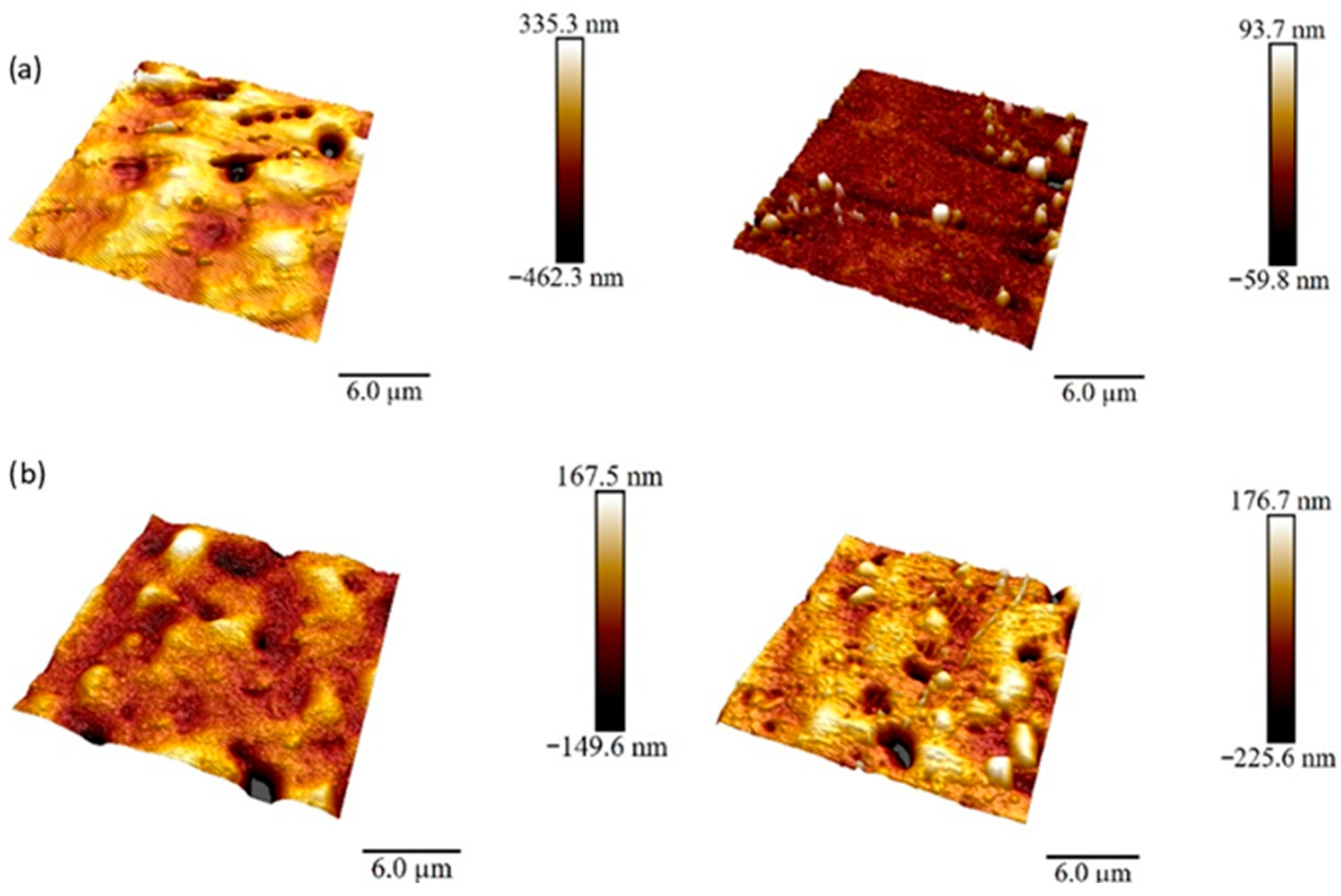
To examine the potential differences in surface roughness between PETG and PU, AFM micrographs were recorded, from which we obtained the values for root mean square roughness (Rq), arithmetical mean height (Ra), and maximum roughness depth (Rmax) (Figure 7, Table 3). The results from two representative micrographs for each material showed that all parameters were higher for PETG than those for PU, which was in accordance with the visual evaluation of the SEM micrographs. Alongside higher surface roughness, the results showed that the PETG surface was more heterogeneous, with Ra values spanning almost one order of magnitude (9.45–71.0 nm), a characteristic not observed for PU.

4. Discussion

4.1. Aligner Materials

The results of this study revealed counterintuitive time–kill curves for CHX-based antiseptics on S. mutans. The optimal efficacy on both examined aligner materials was established after 1-min exposure, with a decrease in efficacy already after the 3-min treatment. With Curasept antiseptic, the number of adhered bacteria even returned to the level of the control after 3- and 5-min exposure. Compared to mechanical brushing, the 1-min treatment with antiseptic showed significant improvement only in the case of Perioplus. The combination of brushing and antiseptics yielded better results than those of separate treatments for pure CHX solution, which was not the case for the two commercial antiseptics. Regarding the order of treatments within the combination, somewhat better results were obtained when the antiseptic was applied first, but with a statistically significant difference only in the case of Curasept.
Regarding the aligner materials’ surface, the results indicated moderate differences in SFE, surface roughness and heterogeneity. Slightly higher surface roughness and heterogeneity was measured for PETG, which can most likely be attributed to differences in polymer microstructure. Danielle et al. [25] reported X-ray diffraction (XRD) diffractograms and differential scanning calorimetry (DSC) thermograms of PETG and PU aligners equal to those analyzed here. The XRD results were interpreted as indicative of higher crystallinity of PU than that of PETG; however, the DSC results did not exhibit any melting peaks for either material. If PETG indeed contained some crystalline phase, it would cause microstructural differences that would be reflected in higher surface roughness [31]. For PTFE membranes used in dental practice, it was found that a higher degree of crystallinity resulted in lower nanoscale roughness, which promoted the adhesion of oral bacteria [31]. It is plausible that the nanoscale heterogeneity of PETG surfaces could exert a more pronounced influence in multispecies biofilms, where microbial colonization is shaped by interspecies interactions and spatial organization. Surface irregularities may promote micro-niche formation, differential adhesion, and localized gradients of nutrients or antimicrobials, thereby affecting community structure and resilience. In contrast to single-species systems, multispecies biofilms may exploit such topographical features to establish cooperative or competitive microenvironments.
The WCA and SFE results showed that PETG is more hydrophilic than PU and that its polar SFE component can be attributed to its basic moieties. At first, this appears counterintuitive as PUs—alongside carbonyl groups, which can also act as Lewis bases—contain more amino groups, whereas PETG contains only carbonyl groups. However, the basicity of the amino group is reduced owing to resonance within the urethane linkage. It is also known that contributions to the polar SFE component can come from double and triple covalent bonds [36]. Therefore, our results indicate that the aromatic moieties in the PETG chain are a primary source of the higher value of the basic SFE component.
Despite these variances, the time–kill curves, effect of treatments, and antiseptic efficacies were remarkably similar on both materials, with no significant difference. Previous studies have shown that surface roughness and SFE have a major influence on the adhesion and retention of microorganisms [12]. Supragingivally, an increase in these two factors leads to faster growth and maturation of the plaque, which is why low surface tension and smooth surfaces were found to be preferable. Our results indicate that the differences in surface roughness and SFE between PETG and PU aligners were too low to cause any significant differences in S. mutans adhesion, biofilm formation, and its interaction with CHX. Chemical differences of the surface beneath the pellicle did not affect the control strategies for the early S. mutans biofilm.

4.2. Decreased Effectiveness of CHX After Longer Exposures

The time–kill curves revealed counterintuitive biofilm behavior: prolonged exposure (3- and 5-min) to any of the tested antiseptics on both types of aligners was significantly less efficient than that with 1-min exposure. The SEM micrographs showed that CHX did not induce surface damage on either PETG or PU aligners during those exposure times; hence, a potential increase in surface roughness because of a chemical reaction with CHX could be dismissed as a plausible explanation. On another note, biofilms hinder the permeation of antiseptics to bacterial cells, disabling the killing of bacteria that are further away from the biofilm surface and rendering bacteria more tolerant to antimicrobials [24,42,43,44,45]. CHX can be deactivated as an effective antimicrobial by reacting with proteins and ions in the saliva and within the biofilm [22,45]. These reasons alone, however, would cause a plateau in the time–kill curves and do not provide an explanation for the return of the bacterial count to levels similar to the control after (3–5)-min exposures. A plateau, described as ‘an initial rapid kill usually followed by a tailing off,’ was indeed recorded when CHX was applied to numerous other microbial species [[45] and ref. within].
In our study, the time–kill curves showed a recovery of viable bacteria counts after the initial 1-min exposure drop. Fitzgerald et al. [46] showed that CHX can indeed be taken up by bacteria (E. coli and P. aeruginosa) extremely fast. Specifically, the maximum uptake was observed within a contact time of only 20 s. For S. mutans, it has been shown that some CHX is absorbed within the first 6–7 s [22]. This peculiar shape of the time–kill curves is in agreement with several other studies that reported the rapid recovery of urinary pathogens in the biofilm from the initial bactericidal effects of CHX [47,48]. Forbes et al. [49] found that Serratia marcescens and Klebsiella pneumoniae biofilm-forming abilities increased after CHX exposure. This effect was attributed to the upregulation of efflux pumps, and it was concluded that repeated exposures to certain microbicides can result in temporary phenotypic adaptations or the selection of stable genetic mutations. The increase in biofilm-forming ability was considered an adaptation to antimicrobial stress. Similar observations of biofilm adaptation to antimicrobial stress have been reported in other systems, including contact lens-associated biofilms, where membrane-targeting nanomaterials were shown to effectively inhibit biofilm formation and bacterial colonization [50].
The upregulation of efflux pumps is often considered an essential mechanism of CHX resistance in K. pneumoniae and possibly in other related gram-negative bacteria [51,52]. A recent study of Karpinsky et al. [53] reported that P. aeruginosa exhibited increased tolerance to CHX after prolonged exposure, with a 22.4-fold rise in MIC. It is suggested that this increased tolerance is primarily mediated by efflux pump upregulation and membrane remodeling, both of which reduce intracellular accumulation of the antiseptic. The authors also highlight a notable potential for resistance emergence with repeated clinical application of antiseptics like CHX, underscoring the need for cautious use and consideration of alternative antimicrobial strategies.
However, the efflux pump-mediated resistance is commonly proposed for Gram-negative bacteria, while S. mutans is Gram-positive. Based on their experimental findings, Kaspar et al. [54] proposed an alternative mechanism in S. mutans. The mechanism arises through genetic mutations affecting cell envelope composition—specifically in genes involved in lipoteichoic acid and glycolipid synthesis—rather than active CHX export. This envelope remodeling entails changes in the composition that may reduce CHX binding or penetration, contributing to tolerance without invoking efflux mechanisms. Yet, this mechanism involves genetic mutations; hence it is a long-term adaptation, not a rapid stress response.
Another possible mechanism through which the response to antimicrobial stress might affect bacterial resistance is via plasmids, which have been shown to cause CHX resistance in Staphylococcus aureus and were used to transfer CHX resistance to E. coli [55]. This reasoning is consistent with the study that investigated the effect of CHX on the vitality and susceptibility of multiple oral bacteria [43]. Out of all bacterial species, the results showed the largest minimum bactericidal concentration/minimum inhibitory concentration ratio for S. mutans (21:1), indicating that CHX has predominantly bacteriostatic, not bactericidal, activity on S. mutans. It is possible that the initial rapid contact of bacteria with CHX caused damage to the cells without killing them, from which they were able to recover using the aforementioned mechanisms.
It is well known that S. mutans cells grown as a biofilm display a prominent increase in genetic competence and are highly transformable. The ability to form biofilms provides S. mutans with protection against transient environmental fluctuations and mechanical stress, thereby enhancing its persistence and growth within the oral cavity. In fact, S. mutans is so adapted to the biofilm lifestyle that it is typically absent in individuals without teeth or dentures, as it requires a hard surface for adhesion [56]. In that sense, it was worth considering whether it is possible that CHX activates quorum sensing (QS) system, which plays a central role in S. mutans biofilm regulation, and thus causes the observed decrease in efficacy on longer exposures. However, we found no evidence in the literature that would support this mechanism as probable. In fact, the studies suggest that CHX inhibits S. mutans biofilm formation, disrupts bacterial communication and downregulates genes related to quorum sensing [57]. Complementing this, Wang et al. [58] showed that CHX also induces persister cell formation in S. mutans biofilms, which survive high CHX concentrations without genetic resistance. While these persisters are not heritable and can be eradicated by higher CHX doses, their emergence may be linked to quorum-sensing pathways, as previously implicated in persister formation.
The effect of sub-inhibitory concentrations of CHX is even more complicated and insufficiently explained [52]. Taken together, these findings highlight the complexity of CHX interactions with biofilm-forming bacteria such as S. mutans. While several mechanisms may be proposed to explain the observed decrease in CHX efficacy over prolonged exposures, none can yet be confirmed as definitive. The interplay between biofilm physiology, antimicrobial stress responses, and CHX pharmacodynamics remains insufficiently understood. Therefore, additional targeted studies are warranted to elucidate these mechanisms and to optimize CHX application protocols for sustained antimicrobial effectiveness.

4.3. Optimum Treatment of the Biofilm

The analysis of the effect of individual treatments (brushing and 1-min exposure to antiseptics) compared to their combination did not yield straightforward results. Generally, the use of antiseptics alone or combined with brushing did not significantly improve the eradication of the biofilm compared to that with brushing alone. This result, although unexpected, agrees well with previous research that concluded that antiseptics based on CHX are inadequate as a sole treatment for oral biofilms and that treatments with 0.12% CHX only temporarily decrease the viable cell concentration [24]. Comparing the efficacies of mouthwashes (1-min exposure), Perioplus demonstrated the best results, with a statistically significant difference compared to those of pure CHX and Curasept. This might be attributed to the bitter orange (Citrus aurantium amara) fruit extract, present only in the Perioplus formulation owing to its proven antibacterial properties [59,60].
The slightly better efficacy of the antiseptic-first approach may be attributed to the initial disruption of the biofilm matrix by the antiseptic, which increases bacterial susceptibility to subsequent mechanical removal. Applying CHX first could weaken intercellular adhesion and extracellular polymeric substances, thereby enhancing biofilm permeability and facilitating deeper penetration of the antiseptic. In contrast, brushing prior to antiseptic application may compact the biofilm or remove loosely attached cells, leaving behind a more resilient core that is less permeable to CHX. While this hypothesis aligns with known biofilm disruption kinetics, further investigation is needed to confirm the underlying mechanisms.
The results of this in vitro study contribute to a deeper understanding of strategies for optimizing CHX use in biofilm control. What distinguishes this study is its focus on the contact time and sequence of antiseptic application, specifically, whether the effectiveness of CHX is influenced more by the contact time of its use or by the order in which it is combined with mechanical plaque removal (i.e., brushing before or after antiseptic application). However, this study was conducted on a mono-species early-stage biofilm under controlled conditions, which limits the direct translation of our findings to clinical settings. Even under an ideal in vitro test environment, antiseptics showed limited efficacy in disrupting early biofilm structures. This may suggest that current guidelines, largely based on planktonic bacterial models, may need to be re-evaluated. Further clinical research, particularly involving mixed biofilm studies, is needed to confirm and expand upon these results. Additionally, although the present study focused on early-stage biofilm formation, it is plausible that even minor differences in surface hydrophobicity may influence long-term biofilm maturation and stability. Future studies involving extended incubation periods and mixed-species biofilms could provide further insight into this aspect. Our study underlines the need to develop formulations specifically optimized for action against biofilm-associated bacteria.

5. Conclusions

The results of this study demonstrate that control strategies for S. mutans biofilm on clear dental aligners do not depend on the type of material used for their production (PU and PETG). The differences in their chemical structure and surface roughness proved insufficient to cause significant changes in S. mutans behavior. All three tested antiseptics exhibited optimal efficacy against S. mutans biofilm when the exposure time was limited to 1 min, with deterioration of efficacy after just 3 min of treatment. The most plausible explanation for this counterintuitive finding is a rapid bacterial response to antimicrobial stress, causing the upregulation of efflux pumps and/or the development of a certain level of bacterial resistance via plasmids, enabling bacteria to recover from the initial damage caused by CHX. Our results point to a potential benefit of applying the antiseptic before mechanical brushing, with an exposure time of 1 min, as this sequence appeared to have the most pronounced effects in our experimental setting. However, the translation of this approach to clinical practice requires further validation in multi-species biofilm models and in vivo studies.

Author Contributions

Conceptualization, I.G., S.Š. and M.B.; methodology, I.G.; validation, I.G., S.Š. and M.P.D.; formal analysis, I.G., S.Š. and M.B.; data curation, I.G., S.Š. and M.B.; writing—original draft preparation, M.B., M.P.D., I.G. and S.Š.; writing—review and editing, I.J.B., S.L.B., M.P., M.P.D., I.G. and S.Š.; visualization, M.B., M.P.D., I.J.B., S.L.B. and M.P.; supervision, I.G. and S.Š.; funding acquisition, I.G., S.Š. and M.P.D. All authors have read and agreed to the published version of the manuscript.

Funding

Funded by Croatian Science Foundation IP-2020-02-4027 (Environmental factors and microbial interactions in the structure of the dental biofilm), DOK-2021-02-6438 (Career development of young researchers—training of new PhDs), also by the University of Rijeka grants (uniri-iskusni-prirod-23-92 and uniri-iskusni-biomed-23-110).

Data Availability Statement

The datasets used and/or analyzed during the current study are available in the Digital Academic Repository of the University of Rijeka, Faculty of Dental Medicine. (Špalj S. Environmental factors and microbiological interactions in the structure of dental biofilm [Internet]. Fakultet dental medicine, 2023 [cited 16 May 2025]. Available from: https://urn.nsk.hr/urn:nbn:hr:271:843444).

Conflicts of Interest

The authors declare no conflicts of interest.

Abbreviations

The following abbreviations are used in this manuscript:
PETGpoly(ethylene terephthalate glycol)
PU polyurethane
CHXchlorhexidine digluconate
SFEsurface free energy
CHDMcyclohexanedimethanol
CFU/mL colony-forming units/milliliter
BHIBrain Heart Infusion
PBSphosphate-buffered saline
AFMAtomic force microscopy
SEMScanning electron microscopy
WCAStatic water contact angles
RqRoot mean square roughness
Raarithmetical mean height
Rmaxmaximum roughness depth
XRD LDX-ray diffraction
DSCdifferential scanning calorimetry

References

  1. Zheng, M.; Liu, R.; Ni, Z.; Yu, Z. Efficiency, effectiveness and treatment stability of clear aligners: A systematic review and meta-analysis. Orthod. Craniofac. Res. 2017, 20, 127–133. [Google Scholar] [CrossRef]
  2. Bichu, Y.M.; Alwafi, A.; Liu, X.; Andrews, J.; Ludwig, B.; Bichu, A.Y.; Zou, B. Advances in orthodontic clear aligner materials. Bioact. Mater. 2023, 22, 384–403. [Google Scholar] [CrossRef] [PubMed]
  3. Cenzato, N.; Di Iasio, G.; Martìn Carreras-Presas, C.; Caprioglio, A.; Del Fabbro, M. Materials for Clear Aligners—A Comprehensive Exploration of Characteristics and Innovations: A Scoping Review. Appl. Sci. 2024, 14, 6533. [Google Scholar] [CrossRef]
  4. Charavet, C.; Gourdain, Z.; Graveline, L.; Lupi, L. Cleaning and Disinfection Protocols for Clear Orthodontic Aligners: A Systematic Review. Healthcare 2022, 10, 340. [Google Scholar] [CrossRef]
  5. Hamdoon, S.M.; AlSamak, S.; Ahmed, M.K.; Gasgoos, S. Evaluation of biofilm formation on different clear orthodontic retainer materials. J. Orthod. Sci. 2022, 11, 34. [Google Scholar] [CrossRef] [PubMed]
  6. Eliades, T.; Eliades, G.; Brantley, W.A. Microbial attachment on orthodontic appliances: I. Wettability and early pellicle formation on bracket materials. Am. J. Orthod. Dentofac. Orthop. 1995, 108, 351–360. [Google Scholar] [CrossRef]
  7. Salehi, B.; Kregiel, D.; Mahady, G.; Sharifi-Rad, J.; Martins, N.; Rodrigues, C.F. Management of Streptococcus mutans-Candida spp. Oral Biofilms’ Infections: Paving the Way for Effective Clinical Interventions. J. Clin. Med. 2020, 9, 517. [Google Scholar] [CrossRef]
  8. Lindh, L.; Aroonsang, W.; Sotres, J.; Arnebrant, T. Salivary Pellicles. Monogr. Oral Sci. 2014, 24, 30–39. [Google Scholar] [CrossRef]
  9. Christersson, C.E.; Dunford, R.G.; Glantz, P.J.; Baier, R.E. Effect of critical surface tension on retention of oral microorganisms. Eur. J. Oral Sci. 1989, 97, 247–256. [Google Scholar] [CrossRef]
  10. Absolom, D.R.; Hawthorn, L.A.; Chang, G. Endothelialization of polymer surfaces. J. Biomed. Mater. Res. 1988, 22, 271–285. [Google Scholar] [CrossRef]
  11. Hasson, J.E.; Wiebe, D.H.; Sharefkin, J.B.; Abbott, W.M. Migration of adult human vascular endothelial cells: Effect of extracellular matrix proteins. Surgery 1986, 100, 384–391. [Google Scholar]
  12. Quirynen, M.; Bollen, C.M.L. The influence of surface roughness and surface-free energy on supra- and subgingival plaque formation in man: A review of the literature. J. Clin. Periodontol. 1995, 22, 1–14. [Google Scholar] [CrossRef]
  13. Zhang, N.; Bai, Y.; Ding, X.; Zhang, Y. Preparation and characterization of thermoplastic materials for invisible orthodontics. Dent. Mater. J. 2011, 30, 954–959. [Google Scholar] [CrossRef]
  14. Wang, W.; Wang, L.; Jiao, Y.; Zeng, X.; Wang, X.; Lu, Y.; Cheng, A.; Dai, P.; Zhao, X. Fine dispersion morphology of polystyrene/poly(ethylene terephthalate glycol) blending generation for controlled foaming behavior. RSC Adv. 2017, 7, 39138–39146. [Google Scholar] [CrossRef]
  15. Yan, C.; Kleiner, C.; Tabigue, A.; Shah, V.; Sacks, G.; Shah, D.; DeStefano, V. PETG: Applications in Modern Medicine. Eng. Regen. 2024, 5, 45–55. [Google Scholar] [CrossRef]
  16. Petrov, P.; Agzamova, D.; Pustovalov, V.; Zhikhareva, E.; Saprykin, B.; Chmutin, I.; Shmakova, N. Research into the effect of the 3D-printing mode on changing the properties of PETG transparent plastic. In Proceedings of the ESAFORM 2021—24th International Conference on Material Forming, Liège, Belgium, 14–16 April 2021. [Google Scholar] [CrossRef]
  17. Rynio, P.; Galant, K.; Wójcik, Ł.; Grygorcewicz, B.; Kazimierczak, A.; Falkowski, A.; Gutowski, P.; Dołęgowska, B.; Kawa, M. Effects of Sterilization Methods on Different 3D Printable Materials for Templates of Physician-Modified Aortic Stent Grafts Used in Vascular Surgery—A Preliminary Study. Int. J. Mol. Sci. 2022, 23, 3539. [Google Scholar] [CrossRef] [PubMed]
  18. López-Jornet, P.; Plana-Ramon, E.; Leston, J.S.; Pons-Fuster, A. Short-term side effects of 0.2% alcohol-free chlorhexidine mouthrinse in geriatric patients: A randomized, double-blind, placebo-controlled study. Gerodontology 2012, 29, 292–298. [Google Scholar] [CrossRef] [PubMed]
  19. Hancock, E.B. Prevention. Ann. Periodontol. 1996, 1, 223–249. [Google Scholar] [CrossRef]
  20. Rölla, G.; Melsen, B. On the Mechanism of the Plaque Inhibition by Chlorhexidine. J. Dent. Res. 1975, 54, 57–62. [Google Scholar] [CrossRef] [PubMed]
  21. Jorgensen, M.G.; Aalam, A.; Slots, J. Periodontal antimicrobials—Finding the right solutions. Int. Dent. J. 2005, 55, 3–12. [Google Scholar] [CrossRef]
  22. Davies, A. The mode of action of chlorhexidine. J. Periodontal Res. 1973, 8, 68–75. [Google Scholar] [CrossRef]
  23. Kampf, G. Chlorhexidine Digluconate. Antiseptic Steward; Springer International Publishing: Cham, Switzerland, 2024; pp. 653–806. [Google Scholar] [CrossRef]
  24. Chatzigiannidou, I.; Teughels, W.; Van De Wiele, T.; Boon, N. Oral biofilms exposure to chlorhexidine results in altered microbial composition and metabolic profile. Npj Biofilms Microbiomes 2020, 6, 13. [Google Scholar] [CrossRef]
  25. Daniele, V.; Macera, L.; Taglieri, G.; Spera, L.; Marzo, G.; Quinzi, V. Color Stability, Chemico-Physical and Optical Features of the Most Common PETG and PU Based Orthodontic Aligners for Clear Aligner Therapy. Polymers 2021, 14, 14. [Google Scholar] [CrossRef] [PubMed]
  26. Park, S.; Kim, H.; Yang, S.B.; Moon, J.-H.; Ahn, H.-W.; Hong, J. A Polysaccharide-Based Antibacterial Coating with Improved Durability for Clear Overlay Appliances. ACS Appl. Mater. Interfaces 2018, 10, 17714–17721. [Google Scholar] [CrossRef]
  27. Moradinezhad, M.; Abbasi Montazeri, E.; Hashemi Ashtiani, A.; Pourlotfi, R.; Rakhshan, V. Biofilm formation of Streptococcus mutans, Streptococcus sanguinis, Staphylococcus epidermidis, Staphylococcus aureus, Lactobacillus casei, and Candida Albicans on 5 thermoform and 3D printed orthodontic clear aligner and retainer materials at 3 time points: An in vitro study. BMC Oral Health 2024, 24, 1107. [Google Scholar] [CrossRef] [PubMed]
  28. Suter, F.; Zinelis, S.; Patcas, R.; Schätzle, M.; Eliades, G.; Eliades, T. Roughness and wettability of aligner materials. J. Orthod. 2020, 47, 223–231. [Google Scholar] [CrossRef]
  29. Iijima, M.; Kohda, N.; Kawaguchi, K.; Muguruma, T.; Ohta, M.; Naganishi, A.; Murakami, T.; Mizoguchi, I. Effects of temperature changes and stress loading on the mechanical and shape memory properties of thermoplastic materials with different glass transition behaviours and crystal structures. Eur. J. Orthod. 2015, 37, 665–670. [Google Scholar] [CrossRef] [PubMed]
  30. Yun, Z.; Qin, D.; Wei, F.; Xiaobing, L. Application of antibacterial nanoparticles in orthodontic materials. Nanotechnol. Rev. 2022, 11, 2433–2450. [Google Scholar] [CrossRef]
  31. Begić, G.; Petković Didović, M.; Lučić Blagojević, S.; Jelovica Badovinac, I.; Žigon, J.; Perčić, M.; Cvijanović Peloza, O.; Gobin, I. Adhesion of Oral Bacteria to Commercial d-PTFE Membranes: Polymer Microstructure Makes a Difference. Int. J. Mol. Sci. 2022, 23, 2983. [Google Scholar] [CrossRef]
  32. Worreth, S.; Bieger, V.; Rohr, N.; Astasov-Frauenhoffer, M.; Töpper, T.; Osmani, B.; Braissant, O. Cinnamaldehyde as antimicrobial in cellulose-based dental appliances. J. Appl. Microbiol. 2022, 132, 1018–1024. [Google Scholar] [CrossRef]
  33. Gergeta, D.; Badnjevic, M.; Karleusa, L.; Maglica, Z.; Spalj, S.; Gobin, I. Effect of Chlorhexidine Digluconate on Oral Bacteria Adhesion to Surfaces of Orthodontic Appliance Alloys. Appl. Sci. 2024, 14, 2145. [Google Scholar] [CrossRef]
  34. Pratten, J.; Smith, A.W.; Wilson, M. Response of single species biofilms and microcosm dental plaques to pulsing with chlorhexidine. J. Antimicrob. Chemother. 1998, 42, 453–459. [Google Scholar] [CrossRef] [PubMed]
  35. Dassanayake, R.P.; Falkenberg, S.M.; Stasko, J.A.; Shircliff, A.L.; Lippolis, J.D.; Briggs, R.E. Identification of a reliable fixative solution to preserve the complex architecture of bacterial biofilms for scanning electron microscopy evaluation. PLoS ONE 2020, 15, e0233973. [Google Scholar] [CrossRef]
  36. Drelich, J.; Chibowski, E.; Meng, D.D.; Terpilowski, K. Hydrophilic and superhydrophilic surfaces and materials. Soft Matter 2011, 7, 9804. [Google Scholar] [CrossRef]
  37. Öner, D.; McCarthy, T.J. Ultrahydrophobic Surfaces. Effects of Topography Length Scales on Wettability. Langmuir 2000, 16, 7777–7782. [Google Scholar] [CrossRef]
  38. Chen, W.; Fadeev, A.Y.; Hsieh, M.C.; Öner, D.; Youngblood, J.; McCarthy, T.J. Ultrahydrophobic and Ultralyophobic Surfaces: Some Comments and Examples. Langmuir 1999, 15, 3395–3399. [Google Scholar] [CrossRef]
  39. Law, K.-Y. Water–surface interactions and definitions for hydrophilicity, hydrophobicity and superhydrophobicity. Pure Appl. Chem. 2015, 87, 759–765. [Google Scholar] [CrossRef]
  40. Liber-Kneć, A.; Łagan, S. Surface Testing of Dental Biomaterials—Determination of Contact Angle and Surface Free Energy. Materials 2021, 14, 2716. [Google Scholar] [CrossRef]
  41. Van Oss, C.J.; Chaudhury, M.K.; Good, R.J. Interfacial Lifshitz-van der Waals and polar interactions in macroscopic systems. Chem. Rev. 1988, 88, 927–941. [Google Scholar] [CrossRef]
  42. Drago, L.; Del Fabbro, M.; Bortolin, M.; Vassena, C.; De Vecchi, E.; Taschieri, S. Biofilm Removal and Antimicrobial Activity of Two Different Air-Polishing Powders: An In Vitro Study. J. Periodontol. 2014, 85, e363–e369. [Google Scholar] [CrossRef] [PubMed]
  43. McBain, A.J.; Bartolo, R.G.; Catrenich, C.E.; Charbonneau, D.; Ledder, R.G.; Gilbert, P. Effects of a Chlorhexidine Gluconate-Containing Mouthwash on the Vitality and Antimicrobial Susceptibility of In Vitro Oral Bacterial Ecosystems. Appl. Environ. Microbiol. 2003, 69, 4770–4776. [Google Scholar] [CrossRef] [PubMed]
  44. McDonnell, G.; Russell, A.D. Antiseptics and Disinfectants: Activity, Action, and Resistance. Clin. Microbiol. Rev. 1999, 12, 147–179. [Google Scholar] [CrossRef] [PubMed]
  45. Russell, A.D.; Day, M.J. Antibacterial activity of chlorhexidine. J. Hosp. Infect. 1993, 25, 229–238. [Google Scholar] [CrossRef]
  46. Fitzgerald, K.A.; Davies, A.; Russell, A.D. Uptake of 14 C-chlorhexidine diacetate to Escherichia coli and Pseudomonas aeruginosa and its release by azolectin. FEMS Microbiol. Lett. 1989, 60, 327–332. [Google Scholar] [CrossRef]
  47. 47 Stickler, D.J.; Clayton, C.L.; Chawla, J.C. The resistance of urinary tract pathogens to chlorhexidine bladder washouts. J. Hosp. Infect. 1987, 10, 28–39. [Google Scholar] [CrossRef]
  48. Stickler, D.J.; Clayton, C.L.; Chawla, J.C. Assessment of Antiseptic Bladder Washout Procedures using a Physical Model of the Catheterised Bladder. Br. J. Urol. 1987, 60, 413–418. [Google Scholar] [CrossRef]
  49. Forbes, S.; Dobson, C.B.; Humphreys, G.J.; McBain, A.J. Transient and sustained bacterial adaptation following repeated sublethal exposure to microbicides and a novel human antimicrobial peptide. Antimicrob. Agents Chemother. 2014, 58, 5809–5817. [Google Scholar] [CrossRef]
  50. Jian, H.J.; Yu, J.; Li, Y.J.; Unnikrishnan, B.; Huang, Y.F.; Luo, L.J.; Ma, D.H.K.; Harroun, S.G.; Chang, H.T.; Lin, H.J.; et al. Highly adhesive carbon quantum dots from biogenic amines for prevention of biofilm formation. Chem. Eng. J. 2020, 386, 123913. [Google Scholar] [CrossRef]
  51. Fang, C.-T.; Chen, H.-C.; Chuang, Y.-P.; Chang, S.-C.; Wang, J.-T. Cloning of a Cation Efflux Pump Gene Associated with Chlorhexidine Resistance in Klebsiella pneumoniae. Antimicrob. Agents Chemother. 2002, 46, 2024–2028. [Google Scholar] [CrossRef]
  52. Cieplik, F.; Jakubovics, N.S.; Buchalla, W.; Maisch, T.; Hellwig, E.; Al-Ahmad, A. Resistance toward chlorhexidine in oral bacteria—Is there cause for concern? Front. Microbiol. 2019, 10, 587. [Google Scholar] [CrossRef] [PubMed]
  53. Karpiński, T.M.; Korbecka-Paczkowska, M.; Stasiewicz, M.; Mrozikiewicz, A.E.; Włodkowic, D.; Cielecka-Piontek, J. Activity of antiseptics against Pseudomonas aeruginosa and its adaptation potential. Antibiotics 2025, 14, 30. [Google Scholar] [CrossRef] [PubMed]
  54. Kaspar, J.R.; Godwin, M.J.; Velsko, I.M.; Richards, V.P.; Burne, R.A. Spontaneously arising Streptococcus mutans variants with reduced susceptibility to chlorhexidine display genetic defects and diminished fitness. Antimicrob. Agents Chemother. 2019, 63, 17. [Google Scholar] [CrossRef] [PubMed]
  55. Yamamoto, T.; Tamura, Y.; Yokota, T. Antiseptic and antibiotic resistance plasmid in Staphylococcus aureus that possesses ability to confer chlorhexidine and acrinol resistance. Antimicrob. Agents Chemother. 1988, 32, 932–935. [Google Scholar] [CrossRef]
  56. Senadheera, D.; Cvitkovitch, D.G. Quorum sensing and biofilm formation by Streptococcus mutans. Adv. Exp. Med. Biol. 2008, 631, 178–188. [Google Scholar] [CrossRef] [PubMed]
  57. Balasubramanian, A.R.; Vasudevan, S.; Shanmugam, K.; Lévesque, C.M.; Solomon, A.P.; Neelakantan, P. Combinatorial effects of trans-cinnamaldehyde with fluoride and chlorhexidine on Streptococcus mutans. J. Appl. Microbiol. 2021, 130, 382–393. [Google Scholar] [CrossRef]
  58. Wang, S.; Zhou, C.; Ren, B.; Li, X.; Weir, M.D.; Masri, R.M.; Oates, T.W.; Cheng, L.; Xu, H.K.H. Formation of persisters in Streptococcus mutans biofilms induced by antibacterial dental monomer. J. Mater. Sci. Mater. Med. 2017, 28, 178. [Google Scholar] [CrossRef]
  59. Ogunro, O.B.; Richard, G.; Izah, S.C.; Ovuru, K.F.; Babatunde, O.T.; Das, M. Citrus aurantium: Phytochemistry, Therapeutic Potential, Safety Considerations, and Research Needs. In Herbal Medicine Phytochemistry: Applications and Trends; Izah, S.C., Ogwu, M.C., Akram, M., Eds.; Springer International Publishing: Cham, Switzerland, 2024; pp. 1–40. [Google Scholar] [CrossRef]
  60. García-Juárez, A.; Garzón-García, A.M.; Ramos-Enríquez, J.R.; Tapia-Hernández, J.A.; Ruiz-Cruz, S.; Canizales-Rodríguez, D.F.; Del-Toro-Sánchez, C.L.; Rodríguez-Félix, F.; Ocaño-Higuera, V.M.; Ornelas-Paz, J.D.J. Evaluation of Antioxidant and Antibacterial Activity of Gelatin Nanoparticles with Bitter Orange Peel Extract for Food Applications. Foods 2024, 13, 3838. [Google Scholar] [CrossRef]
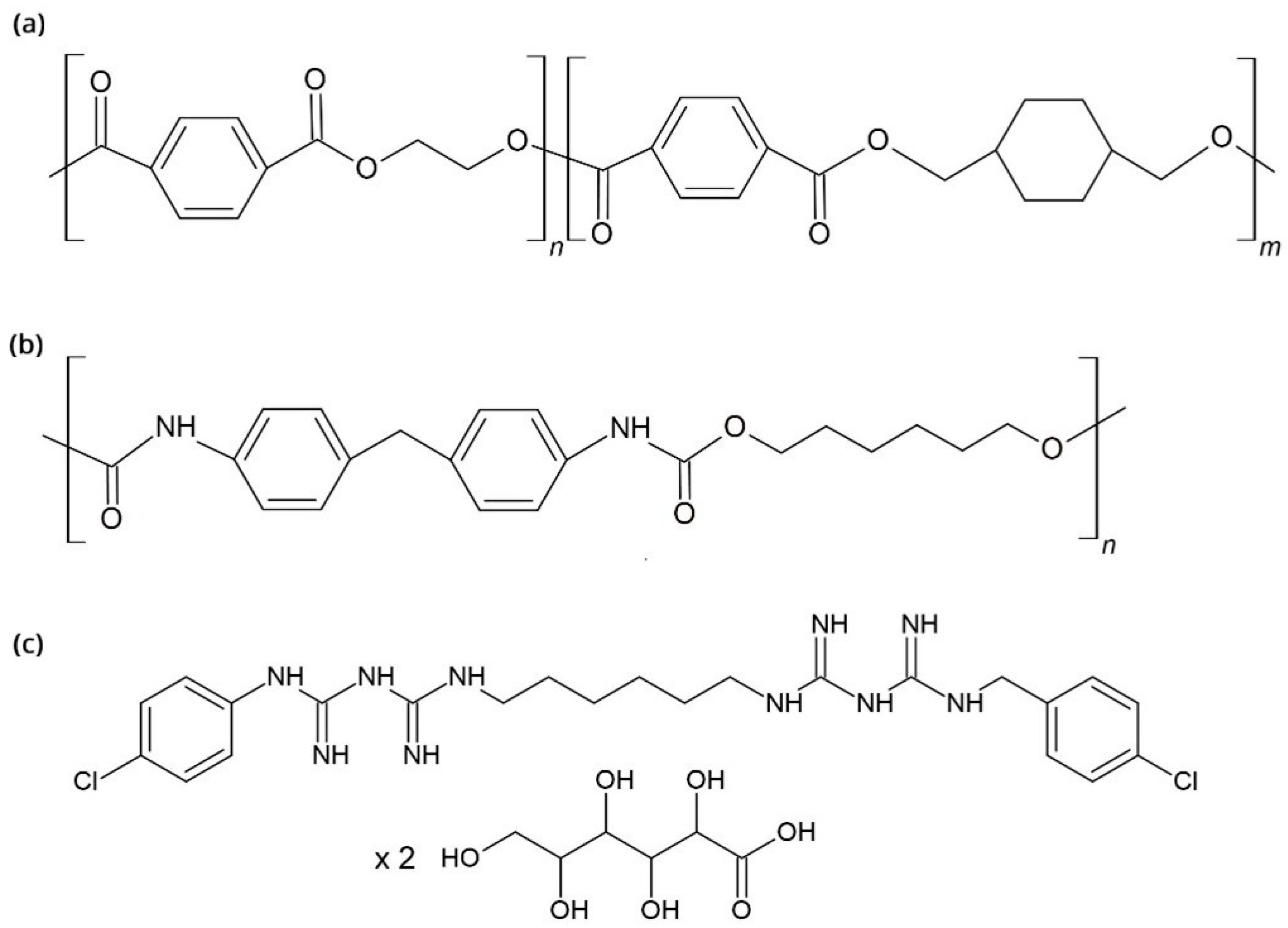
Figure 1. Molecular structure of (a) poly(ethylene terephthalate glycol); (b) polyurethane and (c) chlorhexidine digluconate salt (CHX).
Processes 13 03069 g001
Figure 2. S. mutans Biofilm treatment efficiency Time–kill curves for S. mutans for (a) pure CHX on PU surface; (b) pure CHX on PETG surface; (c) Curasept on PU surface; (d) Curasept on PETG surface; (e) Perioplus on PU surface; (f) Perioplus on PETG surface. The data show outliers, while the asterisks show extremes. Horizontal lines connect groups that differ significantly at p < 0.05.
Processes 13 03069 g002
Figure 3. Number of adhered S. mutans bacteria after mechanical dental brushing, 1-min exposure, or the combination of the two for (a) pure chlorhexidine (CHX) on polyurethane (PU) surface, (b) pure CHX on poly(ethylene terephthalate glycol) (PETG) surface, (c) Curasept on PU surface, (d) Curasept on PETG surface, (e) Perioplus on PU surface, and (f) Perioplus on PETG surface. The dots show outliers, whereas the asterisks show extremes. Horizontal lines connect groups that differ significantly at p < 0.05.
Processes 13 03069 g003
Figure 4. Comparison of the antiseptics’ efficiencies on the (a) polyurethane (PU) and (b) poly(ethylene terephthalate glycol) (PETG) surface, after mechanical brushing, 1-min exposure, or the combination of both. The dots show outliers, whereas the asterisks show extremes. Horizontal lines connect groups that differ significantly at p < 0.05.
Processes 13 03069 g004
Figure 5. Comparison of the number of adhered bacteria S. mutans after designated combined treatments using (a) pure chlorohexidine (CHX), (b) Curasept and (c) Perioplus antiseptic.
Processes 13 03069 g005
Figure 6. SEM micrographs of polyurethane (PU) aligners after 24-h exposure to S. mutans and the designated treatments. (a,b) Control; (c,d) 1 min treatment; (e,f) 5 min treatment; (g) Brushing; (h) Brushing + CHX 1 min. Short arrows on (e) and (f) show destroyed bacteria embedded in EPS. Long arrows on (h) show leakage of bacterial content from bacteria in the biofilm.
Processes 13 03069 g006
Figure 7. AFM micrographs of (a) poly(ethylene terephthalate glycol) (PETG) and (b) polyurethane (PU) clear dental aligners.
Processes 13 03069 g007
Table 1. Contact angle values of the probe liquids on poly(ethylene terephthalate glycol) (PETG) and polyurethane (PU) aligners used for surface free energy calculations.
SampleContact Angle/°
WaterFormamideDiiodomethane
PETG77.3 ± 1.057.5 ± 1.139.9 ± 2.9
PU80.4 ± 0.556.6 ± 0.442.6 ± 1.9
Table 2. Values of surface free energy components of poly(ethylene terephthalate glycol) (PETG) and polyurethane (PU) aligner materials according to the Owens-Wendt, Wu’s and Van Oss–Chaudhury–Good (OCG or acid-base) model. All components are given in mJ/m2.
Sample γ s d γ s p γ s γ s d γ s p γ s γ l L W γ l + γ l γ s
Owens-Wendt ModelWu’s ModelOCG Model
PETG36.94.9441.436.68.2044.839.70.027.9040.6
PU36.93.6540.636.47.4043.838.30.234.9040.4
Table 3. Root mean square roughness (Rq), arithmetical mean height (Ra) and maximum roughness depth (Rmax) of the poly(ethylene terephthalate glycol) (PETG) and polyurethane (PU) clear dental aligners.
PETGPU
Image 1Image 2AverageImage 1Image 2Average
Rq/nm98.016.857.441.249.645.4
Ra/nm71.09.4540.229.835.232.6
Rmax/nm1180335758561758660
Disclaimer/Publisher’s Note: The statements, opinions and data contained in all publications are solely those of the individual author(s) and contributor(s) and not of MDPI and/or the editor(s). MDPI and/or the editor(s) disclaim responsibility for any injury to people or property resulting from any ideas, methods, instructions or products referred to in the content.