Article

Comparing the Aging Processes of PLA and PE: The Impact of UV Irradiation and Water

Key Laboratory of Agro-Forestry Environmental Processes and Ecological Regulation of Hainan Province, School of Ecology and Environment, Hainan University, Haikou 570228, China
*
Author to whom correspondence should be addressed.
Processes 2024, 12(4), 635; https://doi.org/10.3390/pr12040635
Submission received: 1 March 2024 / Revised: 16 March 2024 / Accepted: 19 March 2024 / Published: 22 March 2024
(This article belongs to the Special Issue Advances in Remediation of Contaminated Sites: Volume II)

Abstract

:
The aging processes of microplastics (MPs) are prevalent in natural environments. Understanding the aging mechanisms of MPs is crucial for assessing their environmental behavior and potential risks. In this study, we selected polylactic acid (PLA) and polyethylene (PE) as representatives of biodegradable and conventional plastics, respectively, to examine changes in their physicochemical properties induced by water and UV light exposure. Laboratory aging resulted in significant fragmentation, characterized by cracks and pores on the surfaces, for both types of MPs, with PLA MPs exhibiting more severe changes, particularly under combined UV and water exposure. Notably, PLA MPs tended to become progressively smaller after aging, whereas PE MPs did not show significant size changes. Chemical analyses of aged MPs using micro-Fourier-transform infrared spectroscopy (FTIR) and X-ray photoelectron spectroscopy (XPS) revealed a substantial increase in the carbonyl index (CI) and oxygen content for PE, suggesting surface oxidation during photo-oxidation. Conversely, PLA MPs displayed a CI decrease, along with an oxygen content increase, indicating the breakdown of ester linkages in PLA and the formation of other oxidation products. Furthermore, we developed and optimized pyrolysis–gas chromatography–mass spectrometry (Py–GC–MS) methods to identify potential chemical degradation products of PE and PLA, considering their differing thermal stabilities. We observed a distinct trend regarding the peaks in the chromatogram of aged MPs and identified the typical oxidation and crosslinking products for PLA. Additionally, after the aging process, both PE and PLA exhibited a significant increase in organic carbon content, with the eluate containing submicron/nano-sized particles. This study provides a scientific foundation for a deeper understanding of the environmental aging mechanisms of various MPs, particularly in regards to the effects of UV irradiation and water exposure.

1. Introduction

Plastics are widely used and produced around the world, and plastic pollution is a global problem. Because of their stable physical and chemical properties, plastics can be stored for long periods in marine [1], terrestrial [2], atmospheric [3], riverine [4], and sewage [5] environments. Plastic waste is affected by environmental factors (light, temperature, pH, salinity, etc.). It can undergo a series of degradation processes, forming MPs with a particle size of less than 5 mm which are then released into the environment and further transformed into smaller nano plastic particles [6,7]. The degradation and aging of MPs are usually characterized by changes in the specific surface area, porosity, functional groups, chemical structure, and hydrophilic and hydrophobic properties of the MPs [8]. Further potential risks might result from the aging and degradation process, e.g., plastics are more likely to be ingested by plants and animals and retained in them as they change from larger to smaller sizes [9]. At the same time, after a series of degradations and transformations, MPs are more likely to coexist with pollutants in the environment [10], acting as carriers for the accumulation of heavy metals and hydrophobic organic pollutants from the environment, leading to bioconcentration, indirect toxicity, and thus higher biological risks [11,12].
UV irradiation is one of the main drivers of plastic aging, through the energy generated by light aging, attacking the C-H bond in the structure of MPs, generating alkyl radicals, which further react with hydroxyl and oxygen radicals to generate hydroxyl, carbonyl, and carboxyl structures and other intermediate and end products [13,14]. The generation of free radicals and the mechanism of photoreaction might vary depending on the type of plastic involved. UV irradiation leads to the formation of tertiary alkoxy and peroxyl radicals in PS, whereas PE tends to form alkoxy and carbonyl radicals [15]. PS contains a large number of aromatic ring structures, and its high sensitivity to light might produce more aromatic photodegradation products, while PE degradation produces more alkane substances [16]. These aging effects on plastics, produced by photodegradation radical reactions, are usually manifested by surface fragmentation of the polymer, leading to the structural formation of cracks, increasing the specific surface area of the MPs, which is further manifested by the generation of surface lobes and fine particles [17].
Unlike traditional petroleum-based plastic polymers, biodegradable plastics are typically prepared from biopolymers (polylactic acid, thermoplastic starch cellulose esters, and polyhydroxyalkanoate [18]. Biodegradable plastics are generally less stable and durable than conventional plastics, so they are more susceptible to physical, chemical, and biological degradation when entering the natural environment. In addition to UV irradiation, other environmental conditions, particularly the presence of water, significantly affect the degradation of different MPs [19], especially biodegradable plastics such as polylactic acid, which can be hydrolyzed in the presence of water, leading to a decrease in molecular weight and the release of low molecular weight soluble oligomers and monomers [20]. Unlike the environmental photodegradation mechanism of petroleum-based plastics, the hydrolysis process of degradable plastics usually does not start gradually from the surface (surface erosion) [19], but rather proceeds relatively homogeneously from within their structures as water molecules penetrate the plastic (bulk erosion) [21]. There are two phases, the first of which occurs within the polymer with an increase in the number of autocatalyzed carboxyl termini; the second phase is marked by the escape of soluble oligomers from the matrix [20]. Due to the different degradation mechanisms, the physical structure changes during the degradation of PLA, and the mechanical crushing process due to internal degradation will also be different from that of petroleum-based plastics. However, most of the current research on degradable plastics focuses on the improvement of the properties of the material products and composting degradation [22,23,24,25]. In addition, these degradation products usually possess more polar groups (hydroxyl and carboxyl groups) that induce and increase the affinity for water, further promoting the degradation reaction [26]. However, there are fewer studies regarding whether or not these products are further released into the water column or react with other structural substances.
In response to the different properties of petroleum-based conventional plastics and biodegradable plastics, the mechanisms by which different types of plastic particles undergo degradation and aging in different environments to produce further fragmentation, thus releasing MPs particles of smaller particle sizes or soluble organics, are not known. In addition, there are fewer studies discussing the mechanisms related to the combined effects of water and light as important influences on aging. Therefore, this study mainly focused on the combination of water and light as an important factor affecting the degradation of plastic particles and explored the degradation process and the mechanism of water and light on plastic particles. PLA and PE were selected as representatives of bio-based and petroleum-based MPs to study their environmental degradation mechanisms and the formation of small particle-size micro–nano plastics, reflecting the differences and commonalities between them after aging. This study focused on the characterization of the changes in the physicochemical properties of the particulate matter itself after aging and the analysis of the components of the eluate using scanning electron microscopy (SEM), FTIR, a total organic carbon analyzer (TOC-L), a Zetasizer Nano device, and Py–GC–MS, respectively. The purpose of this study was to explore: 1. The photodegradation and hydrolysis mechanisms of PE and PLA in aqueous and air environments; 2. The changes in particle size during the degradation of PE and PLA plastics under UV irradiation and the link between the process of generating smaller-sized micro–nano-particle plastic particles and dissolved organic matter. The results of the study will provide a scientific basis for the degradation patterns of two typical plastic polymers and their resulting environmental behaviors.

2. Materials and Methods

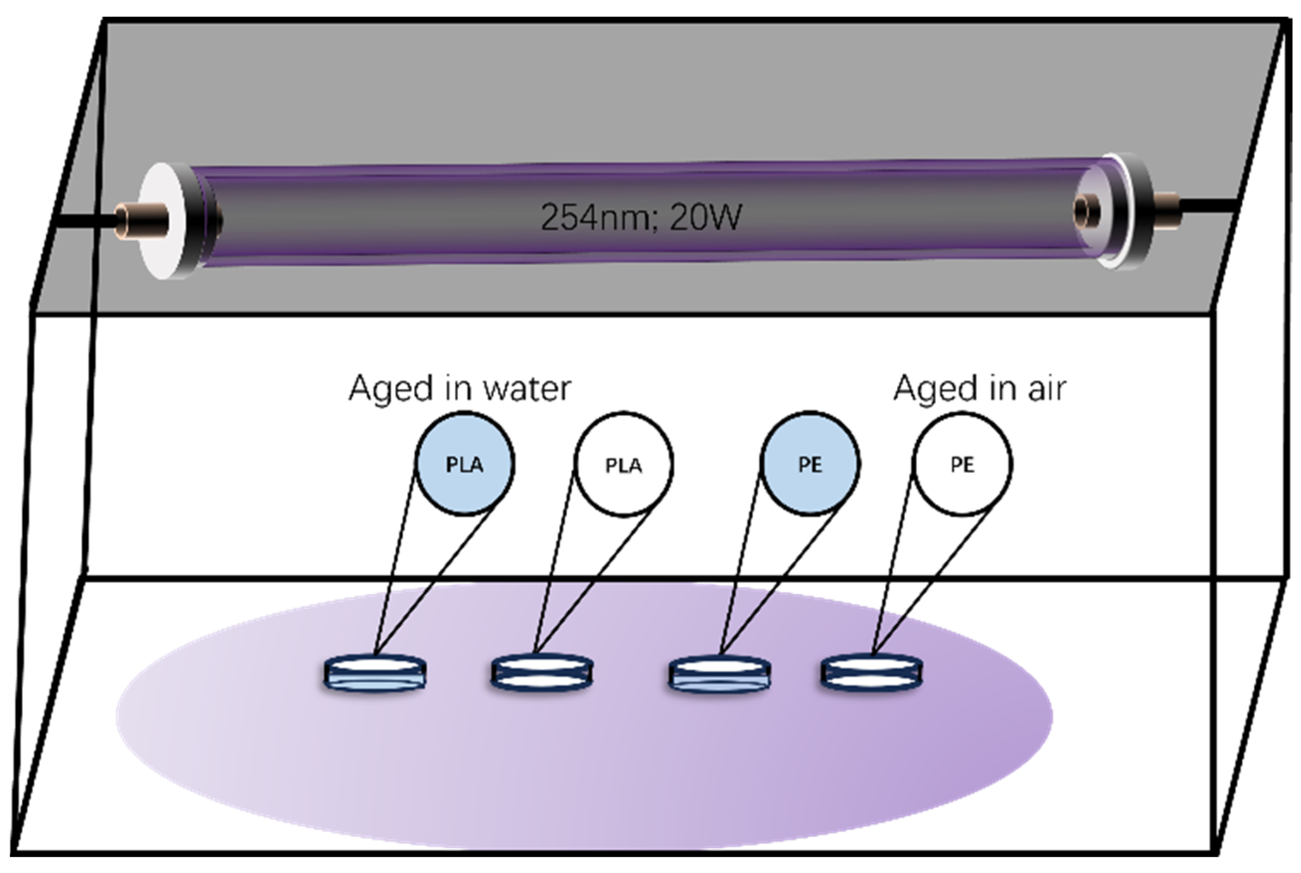
2.1. Accelerated Laboratory Aging of Plastic Particles

The materials required for the experiment, PE (CAS: 9002-88-4) and PLA (CAS: 26100-51-6), were all purchased from Shanghai Yangli Electromechanical Technology Co., Ltd., (Shanghai, China), and the detailed information regarding these products is given in Table S1. A UV lamp with a wavelength of 254 nm and a power of 20 W was employed, and the sample was maintained at a distance of about 28 cm from the UV lamp. In order to ensure the uniformity of light exposure among the samples, before the experiment, we pre-positioned a photometer in the experimental box to measure the radiation intensity received by the sample at a position of 0.4 mW/cm²; after adjusting, we found that after three days of sequential adjustment of the test paper placement, the difference between the samples was controlled to be within the range of 1%, so we followed this protocol in the placement of the actual samples. In addition, we adjusted the height, angle, and direction of the UV lamps to ensure that their position and angle could cover the entire surface of the samples and maintain as uniform an irradiation as possible. The UV aging process was carried out in a house-made sealed experimental box, which was closed with an airtight lid, assisted by aluminum foil, to avoid interference from other factors in the laboratory (Figure 1).
A sample of partially aged particles and a sample of partially virgin particles were obtained and eluted separately with pure water. The specific operations were as follows: About 0.5 g of the above dried plastic particles were added to a beaker, to which was added 40 mL of pure water, and the sample was subjected to ultrasound at 40 °C for 30 min, after which 20 mL of the solution was aspirated, and then the pure water was replenished to 40 mL, and the sample was again subjected to ultrasound for 30 min. The eluate and plastic particles were filtered through a 0.22 μm glass fiber, and the eluate was collected for subsequent analysis.

2.2. Characterization of PE and PLA MPs, before and after Aging

Plastic particles were analyzed, before and after aging, using SEM (Thermoscientific; Verios G4 UC, Waltham, MA, USA). The SEM current voltage was 10 Kv, 0.8 nA, and the samples were subjected to critical point drying and gold metallization using a sputter coater. The plastic surfaces were photographed using scanning electron microscopes of different magnifications.
FTIR, with a measuring range of 397–4000 cm−1 and a resolution of 4 cm−1, was performed, with 32 scans/sample (Nicolet is 50+ Nicolet continuum, Thermo Fisher Scientific, Waltham, MA, USA). For different areas of each sample, FTIR was performed at different locations. The sample compartment of the device was: R%T. The aged sample particles were placed on a gold mirror for observation, and the lens was adjusted and brought into focus. The energy of the optical bench was first determined to be 4–11, after which the background was scanned, followed by aiming the light source at the samples for detection, with two different areas of each sample being detected. The baselines of the atlas were corrected using the manual mode in the Omnic (Version 9.2), and the maps were smoothed (13 smoothing points). The CI was calculated as the ratio of the integrated absorbance area of the carbonyl (C=O) peak between 1850 and 1650 cm−1 to the integrated absorbance area of the methylene (-CH2) peak between 1500 and 1420 cm−1, I added theaccording to the methods of Maryam Hoseini et al. [27].
carbonyl   Index   ( C I ) = a r e a   u n d e r   b a n d   C = O a r e a   u n d e r   b a n d   C H 2
In this study, XPS (Axis Supra Axis Supra, Japan) was used to qualitatively analyze and quantify the changes in the chemical states of oxygen (O) and carbon (C) on the surface of the MPs after UV aging, and the O/C ratio was used to further analyze the degree of UV aging of the MPs. XPS scanning spectra and high-resolution spectra of virgin and UV-aged MPs were collected at an Al Kα monochromatized X-ray source under a vacuum of 10−8 Pa. The O/C ratios of the samples were derived from the percentage of O atoms compared to the percentage of C atoms using the identification of the full spectrum method.
A total of 0.3 mg of the plastic particles collected before and after aging were weighed and placed in a pyrolysis cup for analysis. A comparison of the degradation products, including the time to peak and the relative abundance between peaks, was performed. Test condition: The relevant parameters of the Py–GC–MS (PY3030D-8890/5977B, Frontier, Japan) were as follows: (1) the chromatographic columns are Rtx-5MS, 30 m × 0.25 mm × 0.25 μm; (2) the temperature was increased from 40 °C to 320 °C at a rate of 20 °C/min and held at 320 °C for 14 min; (3) the carrier gas was helium.
The filtered solution was injected into the total organic carbon analyzer for analysis (TOC-L, Shimadzu, Kyoto, Japan). A laser particle size analyzer (Zetasizer Nano S90, Malvern Instruments, Malvern, UK), was used to characterize changes in the average particle size of the soluble compounds. A total of 10 mL of the eluate was removed and added to a four-sided translucent cuvette, which was then placed in the sample cell to set the corresponding refractive index and absorbance of PE and PLA, as well as the corresponding parameters of the solution.

2.3. Data Analysis

All original data were analyzed using Excel (Microsoft Office 2021). SPSS (IBM SPSS Statistics 26) was used for statistical analysis. ANOVA was applied for the test of significant differences in data, and statistical significance was determined as statistically (* p < 0.05) and highly (** p < 0.01) significant, respectively [28].

3. Results and Discussion

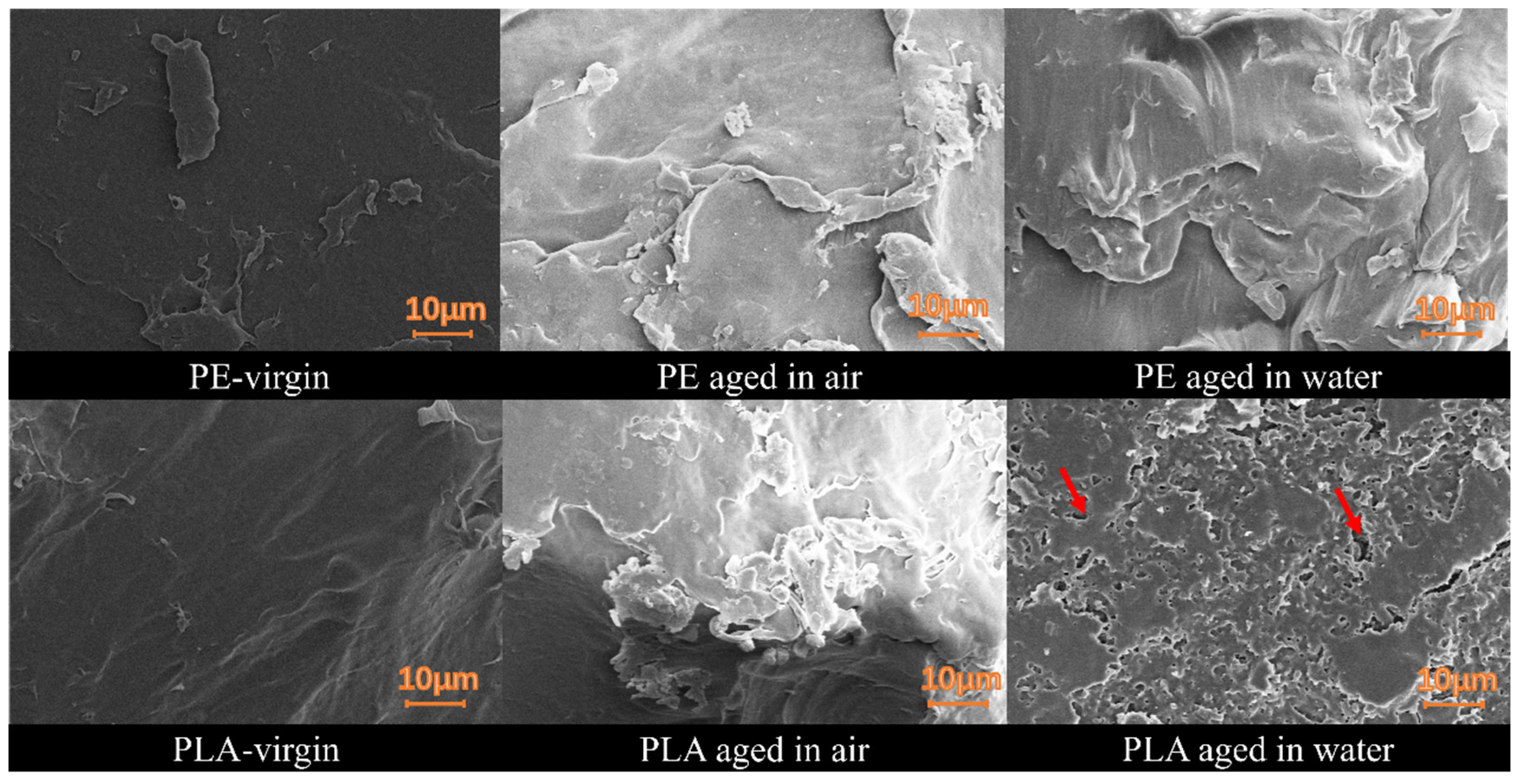
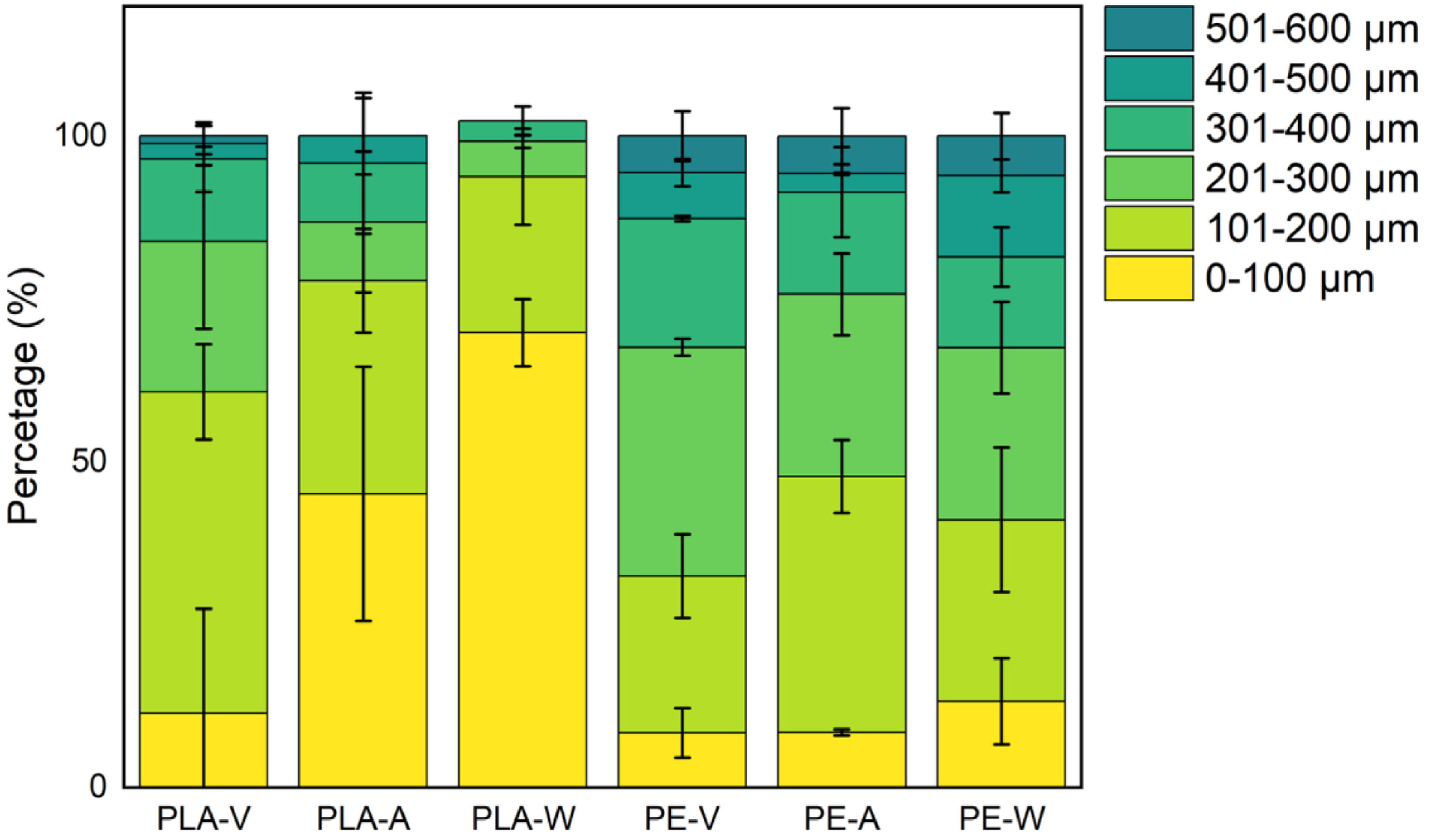
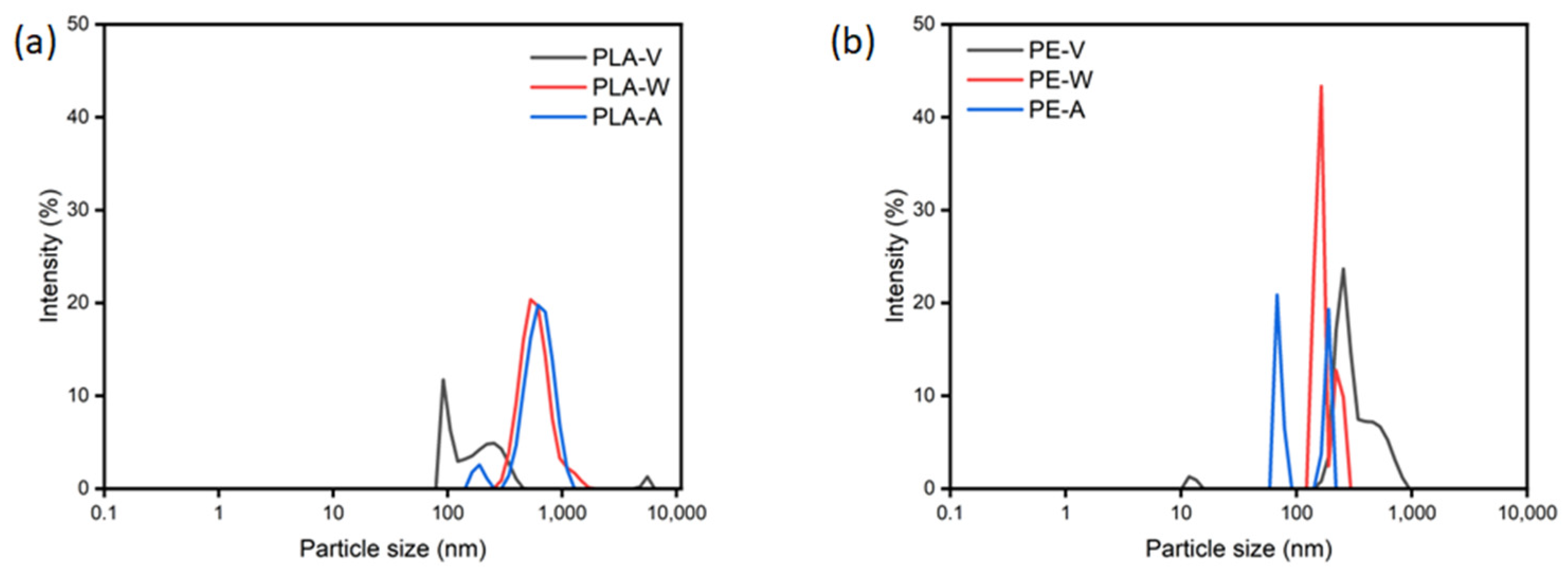
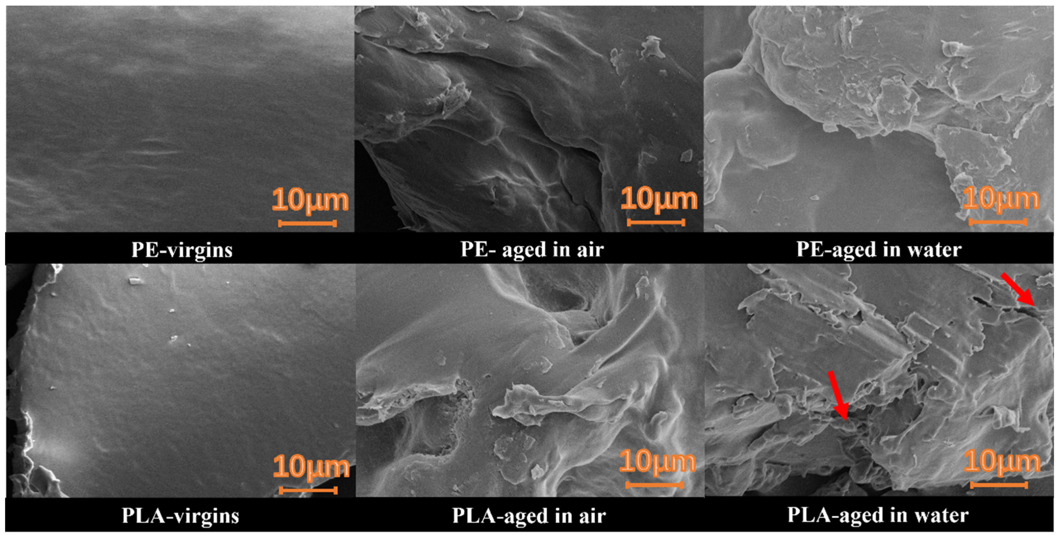
3.1. UV Aging Altered the Morphological Characteristics of the PE and PLA MPs Particles

The surface morphology of both PE and PLA plastic particles changed significantly after 30 days of UV irradiation. As shown in Figure S1, PLA plastic particles showed significant yellowing after undergoing UV aging, while PE MPs did not show significant color changes after UV aging. In addition, we noted differences in the rate of yellowing at the same time points, and the occurrence of such differences might be related to the structure of the plastic or the presence of stabilizers [29]. Carbonyls, vinyl, and quinones, as well as cyclic structures, could be conjugated via p-π and exhibit absorbance at 280 nm, ultimately resulting in the yellowing of the plastic [30,31]. The yellowing effect could be generated by conjugated double bonds generated under the excitation of UV energy with reactive oxygen species, i.e., C-H → C-OH → C═O → O-C═O [32]. For PLA, which contains rich ester bond structure, are more easily to form O-C═O structure during aging. Moreover, in the chain breaking process, it was easy to cross-link PLA to form a ring structure, which PE did not display. Thus, the yellowing phenomenon of PLA was more obvious. The rate of yellowing was slowed down in the presence of water, which might be related to the hydrolysis of PLA, leading to the breakage of double bonds or the eluate of additives into the aqueous solution.
The yellowing and weathering process of plastics usually occurs in conjunction with surface cracking [29]. Observation of the surface microstructure of the plastics using SEM showed that the texture of both initial MPs was relatively uniform and dense, with smooth and regular surfaces, no obvious faults or holes, and no other particles found to be attached (Figure 2). After 30 days of UV irradiation, fine cracks began to appear on the PE surface, while the surface appeared unchanged before and after pure water eluate (Figure 2 and Figure 3). Compared to PE, PLA formed wider intertwining, and connecting cracks after aging, creating holes, as well as signs of fracture and breaking (Figure 2). It also formed fragments of fine particles that adhered to the surface of the aging plastic. The holes of the non- eluted plastic particles were mostly distributed in the sub-surface layer. After being eluted with pure water, we found that dense holes appeared on the surface of the PLA pure water photoaging exposure group, as pointed out by the arrows in Figure 3, whereas this phenomenon did not appear in the air-exposed UV aging group, which indicated that, after the combined effects of hydrolysis and photoaging, the surface of PLA MPs could produce a large number of fine particles, which could be easily dislodged in an aqueous environment to enter into the environment (Figure 3). Dislodged microplastics could exhibit surface adsorption interactions with hydrophobic organic contaminants (e.g., cyclic hydrocarbons, as reported in the literature), and in conjunction with our findings, the increase in pore size due to UV aging could provide ideal conditions for the adsorption of cyclic hydrocarbons, along with the formation of smaller plastic particles, which could result in more potential ecotoxicity [33].
Changes in the particle size distribution of the PLA and PE MPs after different aging conditions were assessed by optical microscopy (Figure 4). The statistical methods for determining particle size distribution are detailed in Text S1 and Table S5. The PE particle size distribution before aging was mainly dominated by particles measuring 201–300 μm (35.2%), followed by those measuring 101–200 μm (24.01%), 301–400 μm (19.72%), and 0–100 μm (8.41%), respectively; After aging in both exposure modes, the particle size distribution pattern did not change significantly, with the exception that the particle size of the 201–300 μm group of PE decreased after anhydrous photoaging, while the particle size of the 101–200 μm group increased accordingly. The specific particle size distributions were: 8.47% (0–100 μm), 39.27% (101–200 μm), 27.98% (201–300 μm), 15.7% (301–400 μm), 2.82% (401–500 μm), and 5.76% (501–600 μm), respectively. This change might be due to surface degradation and the gradual disappearance of the main components of 201–300 μm during the aging process achieved by UV irradiation, resulting in a slow change in particle size to the next level. For PLA, the trend was quite different from that exhibited by PE. The particle size distribution of PLA before aging was mainly 101–200 μm (49.37%), and the percentage content gradually decreased with increasing particle size: 201–300 μm (23.1%), 301–400 μm (12.69%), 401–500 μm (2.33%), and 501–600 μm (1.17%), respectively. After aging, a large number of MPs particles with small particle sizes (0–100 μm) appeared under both exposure conditions, which was more obvious in the pure water UV aging group, with particles in the 201–600 μm particle size range decreasing, and the large-size particles (401–500 μm and 501–600 μm) actually disappearing completely. The specific distribution was the particle size distribution of PLA after anhydrous photoaging, i.e., 45.08% (0–100 μm), 32.72% (101–200 μm), 9.02% (201–300 μm), 9.02% (301–400 μm), 4.17% (401–500 μm), and 0% (501–600 μm), respectively. The particle size distribution of PLA after aging with hydrophobic light was 69.8% (0–100 μm), 23.99% (101–200 μm), 5.42% (201–300 μm), 3.12% (301–400 μm), 0% (401–500 μm), and 0% (501–600μm), respectively. This pattern of change indicated that the PLA aging degradation process was accompanied by a large number of internal fragmentation processes, in addition to surface degradation. In the presence of water, due to the hydrolysis processes, in addition to surface aging, PLA also exhibited inward erosion, accelerating its rate of fragmentation from the inside; thus, the particle size became smaller. Under air exposure conditions, PLA only absorbed moisture in the air, the rate of hydrolysis was greatly reduced, the plastic was less fragmented, and the relative content of small-sized particles was lower than that observed in the pure water exposure group. The formation of small-sized particles due to crumbling caused by internal fragmentation simultaneously exposed a larger specific surface area, further accelerating their degradation and creating a positive feedback effect along with the formation of additional small-sized fragments. According to Isadounene et al., the appearance of significant pores and the fragmentation of PLA was related to its water absorption, which led to the disruption of the chain structure, resulting in higher porosity [34]. This result was consistent with SEM observations that revealed a large number of holes and deep grooves. Smaller-sized particles might have been detached from the plastic particles, thus forming the porous areas. Hydrolysis and photolysis produced a large number of small-sized particles, which on the one hand, entered the environment to be ingested by organisms, increasing the probability of retention in the body [35], and on the other hand, might act as “carriers” for other sorbed organic pollutants, ultimately leading to more serious ecotoxicity effects [36,37].

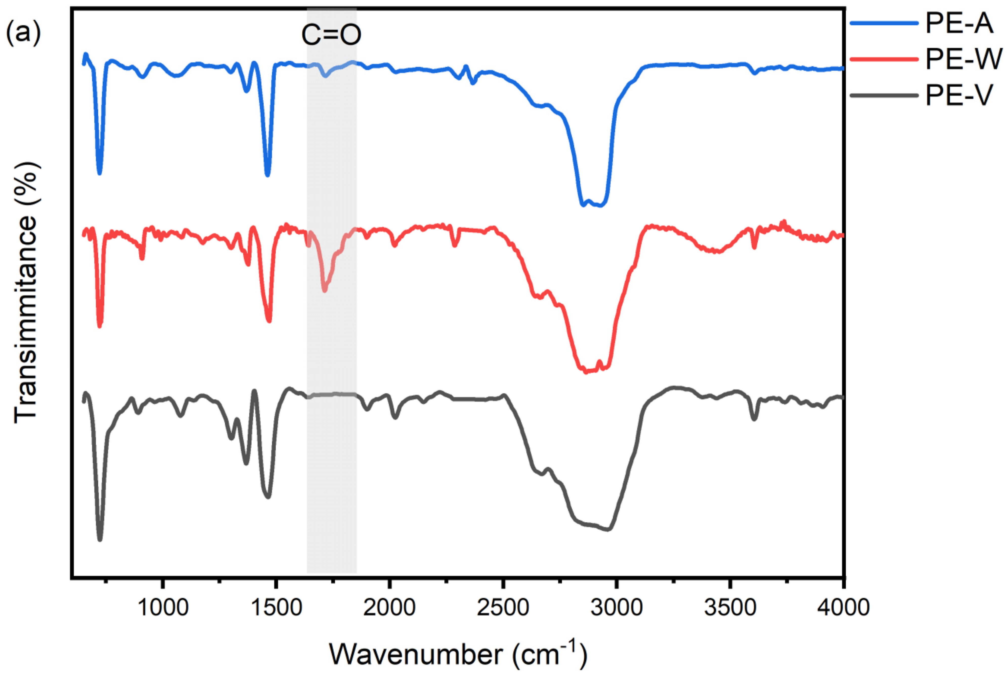
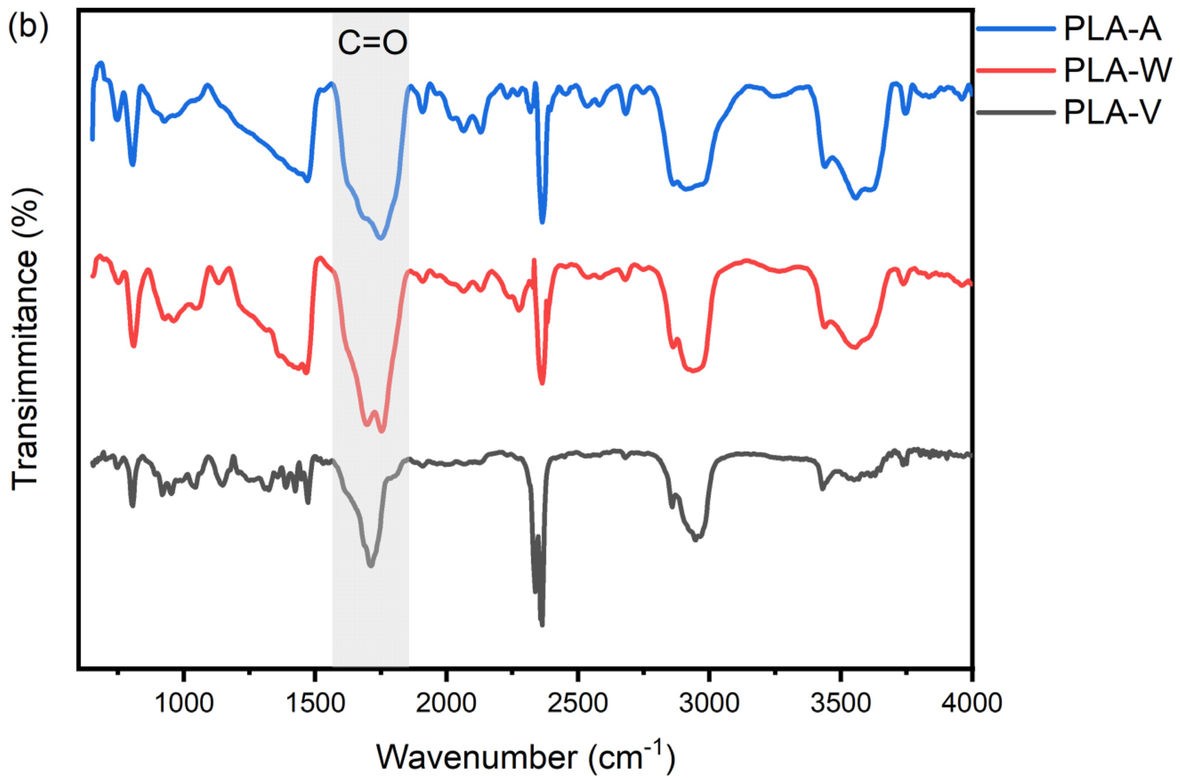
3.2. UV Aging Altered the Chemical Functional Groups of PE and PLA MPs Particles

FTIR characterization showed that after UV aging under air and water exposure, the PE MPs suffered a significant surface oxidation process. This was mainly characterized by an increase in the vibration of the carbonyl peak and band broadening at 1750 cm−1, and the rate of carbonyl formation was related to the exposure environment—carbonyl formation was faster under pure water exposure conditions (Figure 5a). Correspondingly, after UV aging for 30 days, the CI of PE MPs significantly increased (p < 0.01), and the degree of increase was greater in the condition with water, 0.31 ± 0.05, than without water, 0.09 ± 0.05 (Table 1). Under UV irradiation, alkyl radicals underwent cleavage to combine with hydroxyl radicals to form carbon–oxygen bonds, which then formed carbonyl groups with hydroxyl radicals [14]. At the same time, liquid water in this process was exposed to UV irradiation to produce volatile hydroxyl radicals, which contained a higher content of hydroxyl radicals than that of air, and the reaction of the radicals with water molecules could be used as a source of hydroxyl radicals [38]. Therefore, in our experiments, we found that the CI of the photoreaction in combination with water was higher than that of the photo-oxidation without the participation of water, and the scanning electron microscope results showed that the degree of change in the surface morphology of the former was slightly larger than that of the latter, i.e., more peeling and cracking phenomena were produced, which was similar to the results of previous research, and the higher the CI of PE, the greater the degree of aging to which it was subjected [39]. The carbonyl peaks were associated with lactones, esters, alcohols, aldehydes, and ketones [40], and the degradation products were subsequently further resolved based on the Py–GC–MS results. This trend was also reflected in the increase in oxygen content and oxygen-to-carbon ratio in the XPS analysis—the oxygen-to-carbon ratio of PE plastic particles before and after aging showed an sightly increasing trend from 0.13 ± 0.03 before aging to 0.15 ± 0.03 (photoaging under aqueous conditions) and 0.17 ± 0.03 (photoaging under anhydrous conditions) (Table 1). This trend was consistent with the findings of Brandon et al. [41].
Unlike PE, the structure of PLA is mainly connected by ester bonds, and its structure contains a large number of C=O structures (Figure 5b); therefore, the CI of the original unaged PLA was 19.66 ± 9.62, which was much higher than that of the PE MPs. The changes in the oxidized functional groups on the surface of the PLA MPs before and after UV aging showed an opposite trend to that of the PE MPs—the intensity of the C=O peaks and the CI decreased significantly after photoaging, and the decrease was more pronounced under the anhydrous condition. Based on their opposite trends, we analyzed the mechanisms behind this differing behavior. The hydrolysis mechanism of PLA is shown in Figure 6. During hydrolysis, the polymer backbone breaks, resulting in a portion of the carbonyls in the backbone breaking away from the backbone, and due to the presence of an aqueous medium, this portion of the shedding carbonyls may be released into the water, as shown by a decrease in the carbonyl index, as we have observed. Specifically speaking, the photodegradation of PLA first occurs by chain breakage through photolysis and hydrolysis, followed by conversion through Norrish type I and type II reactions, in which cross-linking or oxidation reactions may also occur. The photodegradation of PE occurs by chain breakage caused by UV light and oxygen, with the generation of ester bonds [42,43]. Some studies found an increase in the CI of PLA in sludge treatment, which was contrary to our results [40]. On the one hand, the presence of sludge and microbial films might have interfered with the spectroscopic analysis. On the other hand, the large pieces of plastic that were used were difficult to degrade further because they were quite thick, so the inside of their structures could not receive UV irradiation. However, in another study, it was found that the CI of PLA in flake form also showed a decreasing trend after undergoing UV irradiation in pure water, which was consistent with the results in this study [44]. In our study, hydrolysis and photolysis occurred simultaneously on the surface and inside of the small-sized plastics, and the hydrolysis of PLA led to the breakage of ester bonds and the formation of lactic acid and carboxylic acid structures [45]; these terminal carboxylic acid structures were also easily further decomposed by UV irradiation to produce the final product CO2 [19]. More importantly, the PLA MPs used in this study possessed smaller particle sizes, and the degradation process fragmented to form a large number of smaller-sized micro- and nanoparticles, a phenomenon that was more pronounced in the presence of water (Figure 3). The creation of these fine particles made it easier for the oxygenated ends produced by hydrolysis, as well as the carbonyl groups in the main structure of the PLA, to receive the energy of the UV irradiation and to be completely degraded to CO2. This was consistent with our surface observations of the production of holes and cracks in the presence of water, as well as the particle size distribution maps, where it was observed that the involvement of water contributed to the fracture of PLA into fragments, and the absence of water resulted in the presence of relatively large-sized PLA particulate matter. In the natural environment, the effects of water and light usually co-exist, and previous studies have focused on the photolysis, hydrolysis, and biodegradation of the single-factor and large-scale degradation of thin film plastics [44]; thus, we took the lead in exploring the degradation of micron-sized PLA plastic particles under the combined effects of light and water. Due to the simultaneous presence of hydrolysis and photolysis to accelerate the complete degradation of PLA, a more pronounced decrease in the carbonyl peak area and the carbonyl index occurred after the aging of PLA in the presence of water. Interestingly, the XPS results showed that the oxygen content of PLA plastic particles before and after aging, along with the oxygen to carbon ratio, showed a small increase, with the oxygen to carbon ratios increasing from 0.37 ± 0.03 before aging to 0.46 ± 0.00 (under aqueous conditions) and 0.42 ± 0.02 (under anhydrous conditions) after photoaging. From the previous results, it can be seen that the CI decreased, but the oxygen–carbon ratio did not decrease, suggesting that the carbonyl group might be converted to other oxygen-containing functional groups during the aging process. The O/C ratio of MPs exposed to water increased faster than the ratio under photoaging without water, which might be related to the creation of pores on the surface of PLA, leading to its susceptibility to the water absorption processes [46].
To further clarify the degradation products and their chemical structures produced by different types of plastics during degradation and aging, this study examined high-temperature pyrolysis (600 °C) and low-temperature cracking (300 °C), while combining GC–MS to analyze the cracking products. For PLA, the two pyrolysis temperatures showed different trends before and after aging. At 600 °C, the pyrolysis products were the same before and after aging; at 300 °C, there were significant differences before and after aging, and these differences were concentrated in a series of degradation products found at around 14 min, 15 min, 19 min, and 20 min, which were not characteristic of the virgin PLA (Figure 7 at the red dashed box). This result suggested that low-temperature cracking better reflected the environmental processes of plastic aging in terms of chemical structure, which might be related to the fact that substances produced by aging were more susceptible to low-temperature cracking. At 300 °C, we were able to observe the appearance of oxidation products and additives in the aged PLA at around 14 min, 15 min, 19 min, and 20 min, etc. These oxidation products and additives were compared using the NIST database, respectively: 1,3-dioxepane, 2-heptyl-; Butanedioic acid, dipropyl ester; Ethyl hydrogen succinate; 1,3-dioxepane, 2-pentadecyl-; Butanamide, n-formyl-2-hydroxy-n,3-dimethyl-2-(1-methylethyl)-; 2-t-butyl-5-propyl-[1,3]dioxolan-4-one; and 1,2,4-butanetricarboxylic acid, trimethyl ester (Table S2). These products were esters and non-carbonyl oxygenated compounds with cyclic and chain structures, respectively. The mechanisms of the thermal degradation of PLA included both free radical and non-free radical reactions. We found that succinate (Figure S3, Text S4) can be a source of plastic additives and showed that succinate is prepared from succinic acid by tandem-parallel reactions that proceed sequentially, with the intermediate product being ethyl hydrogen succinate and the final product being diethyl succinate. In our experiments, we found that the intermediate monomer could be derived from succinic acid and could potentially be converted to diethyl succinate [43]. In addition, in another study, we found that PBS (poly butylenes succinate) exhibits a structural similarity to ethyl hydrogen succinate; however, this connection requires further investigation [47]. This similarity was found in studies related to PLA pyrolysis products, which may be related to the preparation process of PLA and UV radiation [48]. PLA formed a series of oxidation, fracture, and double bond products after photoaging, and these products occurred under low-temperature thermal degradation conditions, including molecular chain fracture, depolymerization, and inter- and intramolecular ester exchange reactions [49], forming a series of small molecule lipid compounds [50]. Further normalization of the mapping data revealed all the differences in the relative intensities of the major peaks, with no significant differences in the number of peaks in the aging reactions involving water. The intensity of the three main peaks was analyzed, i.e., Succinic acid, di(2-methylallyl) ester, Ethyl hydrogen succinate, and Succinic acid, 2-methylallyl undecyl ester (Figure S2). In the presence of water, the strength of the main pyrolysis products increased by 1.23, 1.86, and 1.29 times, respectively. This result may be related to the hydrolysis of PLA to form small molecule fragments, and the pyrolysis efficiency of small-size fragments was higher than that of the large-size plastic pieces [50]. Under the traditional 600 °C pyrolysing conditions, the aged PLA appeared to generate similar pyrolysis products before aging, and only individual lactones and additives, such as Butyrolactone, Butanedioic acid, and dipropyl ester, were newly generated after aging (Table S3). This phenomenon might occur because the oxidation and cracking products formed after aging were destroyed at high temperatures, and therefore, the information could not be retained.
For PE, using the same assay, the gas chromatogram at 600 °C showed cleavage information mainly for mono-olefins, dienes, and alkanes, which was similar to the results obtained in the previous study [51]. It was noteworthy that the relative intensities of the peaks changed before and after aging at 600 °C, with an increase in the relative intensities of the small molecules after aging (0–10 min) (yellow portion of Figure 8). We speculated that the increase in the intensity of this portion of the peaks might be related to the formation of relatively active functional groups, such as oxygen-containing functional groups and double bonds, during the aging process of MPs, and the sites where these functional groups were located during the pyrolysis process were more prone to cleavage, forming small-molecule cleavage products. This result was in agreement with the FTIR and XPS results (Figure 5, Table 1), as well as with the thermal cracking mass spectrometry characterization of PE plastics with different particle sizes in the natural environment [52]. In addition, the photoaging process could also occur as a result of main chain breakage, generating small molecular fragments, which was corroborated by the SEM observation of exfoliation on the surface of the PE plastics (Figure 2). Compared with PLA, PE MPs were cracked at 300 °C under cracking conditions, and the cracking product peaks were too low, which might be related to its structural stability, although the overall peak height of the aged product was still higher than that of the virgin plastic (Figure 8). It was difficult to reach the detection limit in order to identify the structural information of the product; thus, the low-temperature pyrolysis could not adequately characterize the information regarding the aging process of PE (Table S4). In this study, we pioneered the use of Py–GC–MS to characterize the degradation products and aging process of different types of MPs under UV light and aqueous environmental conditions and optimized the analytical conditions based on their thermal stability for better investigating the degradation mechanisms. The results showed that for the PLA MPs with lower thermal stability, low-temperature cracking conditions were more suitable for studying their environmental degradation behavior. For PE, high-temperature pyrolysis could produce more characteristic products, and although no obvious new degradation products were found, the aging process could be reflected in the relative peak heights of the pyrolysis spectra.

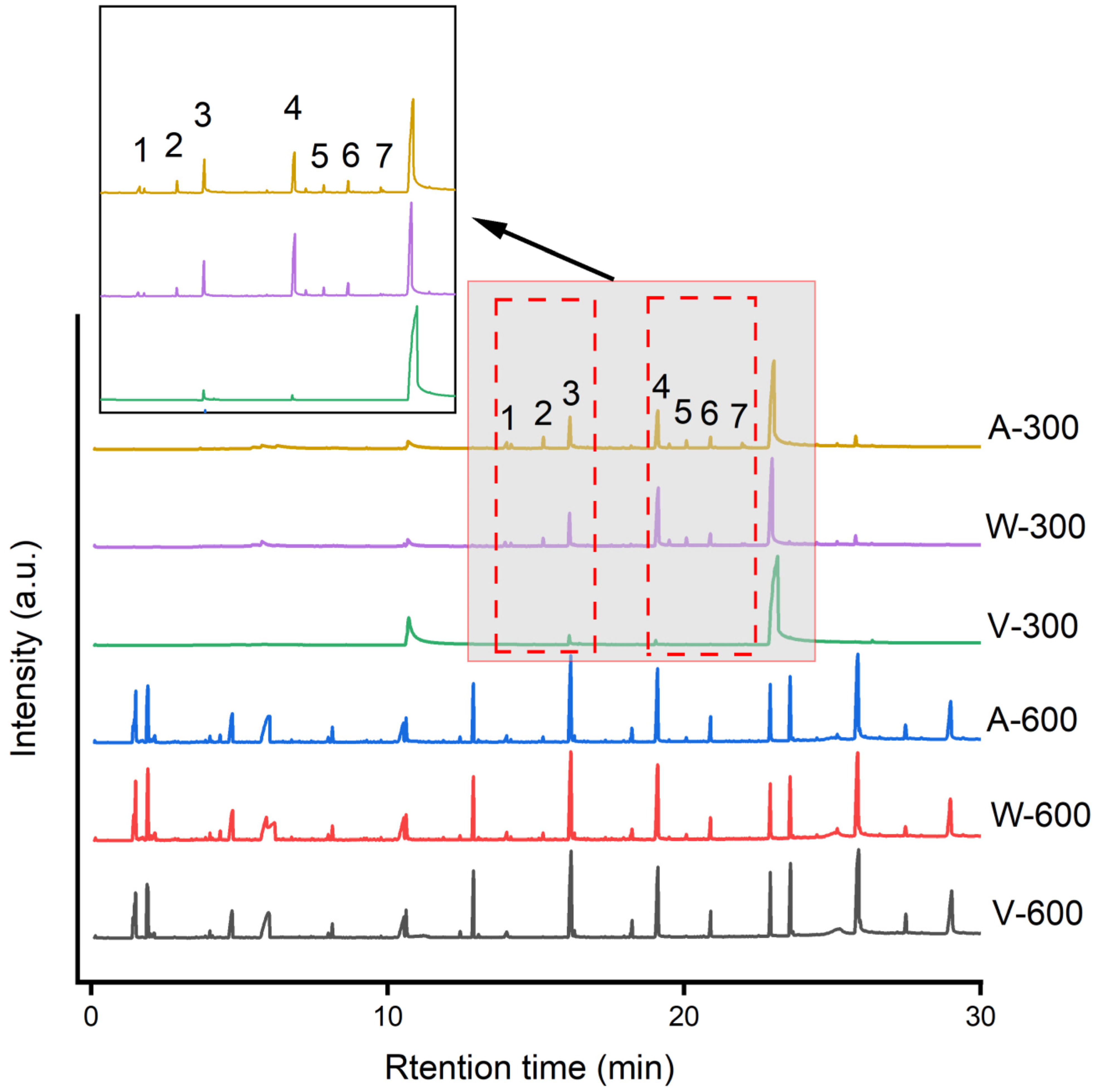
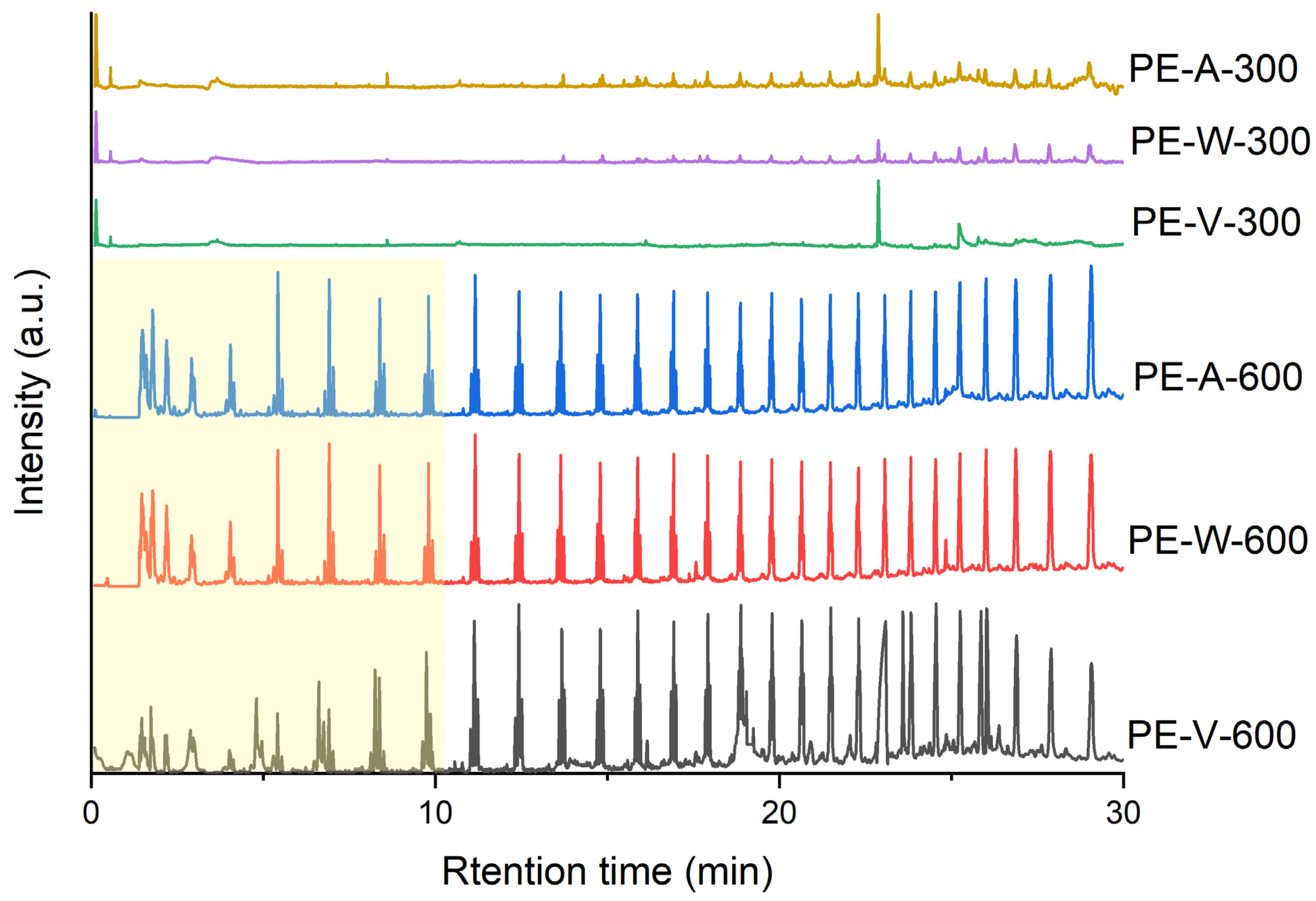
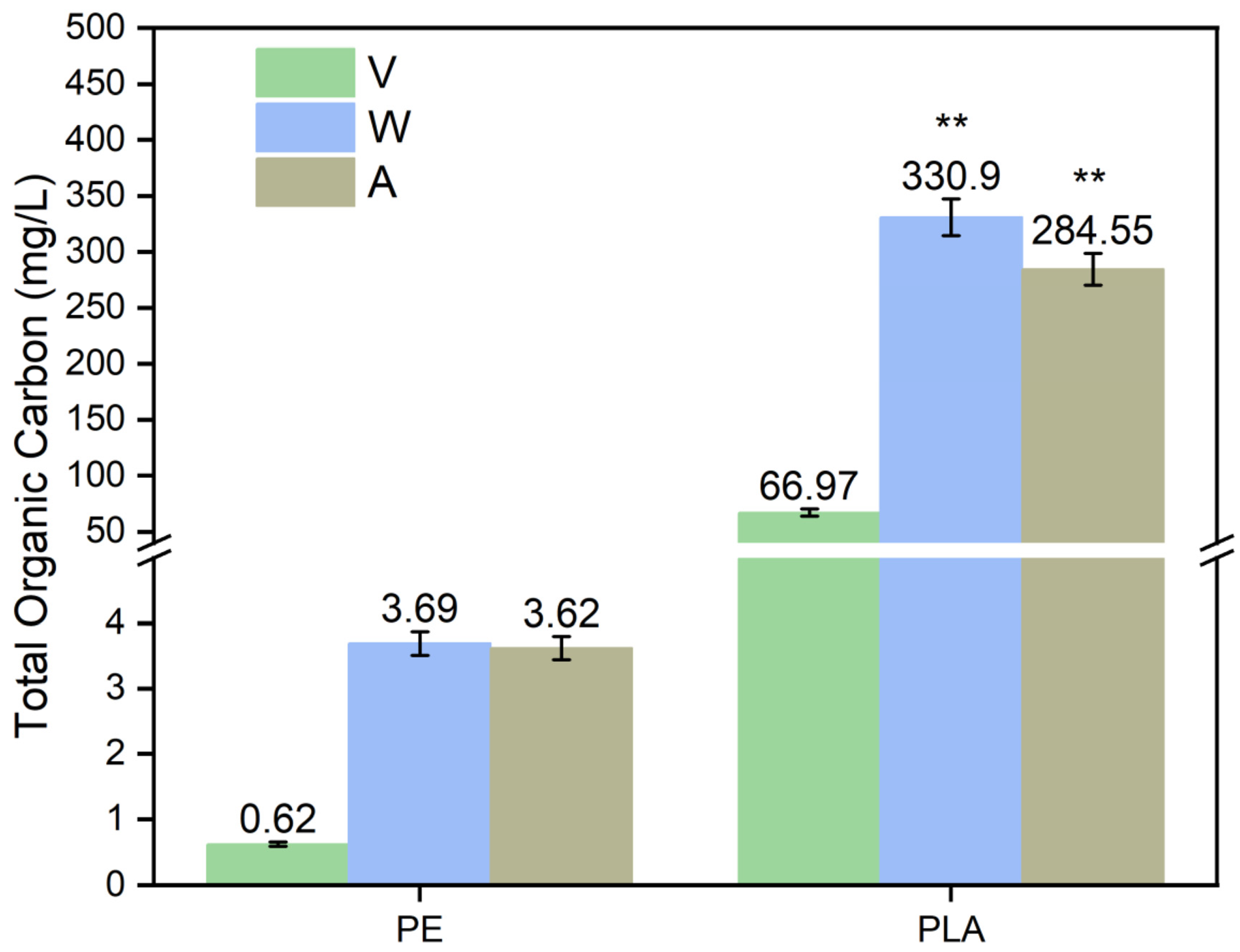
3.3. Characterization of MP Eluate after Aging

By eluting PE and PLA MP particles before and after aging and analyzing the total oxygen carbon (TOC) of the eluate (Figure 9), we found that compared to PE, the TOC eluted from PLA, with or without aging, was close to 100 times higher than that of PE, indicating that PLA MPs were more likely to release organic matter into the water column in the environment. It had been demonstrated by others that dissolved TOC could evaporate in a short period, and at the same time, MPs could adsorb TOC present in the natural environment, disrupting low trophic level processes and thus affecting microbial activity and carbon cycling in the oceans [53]. Both PE and PLA MPs particles that underwent UV aging released significantly higher organic carbon by eluate than the samples that did not undergo the aging process (p < 0.01). Specifically, the TOC of PE after aging (3.62–3.69 mg/L) was 5.84–5.95 times higher than that before aging (0.62 mg/L), and the TOC of PLA after aging (284.55–330.9 mg/L) was 4.25–4.94 times higher than that before aging (66.97 mg/L), whereas the aging process with the involvement of water produced slightly higher TOC than that of the no-water-involved conditions. Thus, our results indicated an increase in the generation and release of organic matter from the surface of MP particles after undergoing photoaging, and this was more pronounced in PLA. Combined SEM and FTIR analyses revealed that the oxidation process during photoaging and the creation of surface holes might be driving the release of more organic matter from the plastic. Our experimental results were consistent with previous findings that MPs could elute TOC under UV irradiation [54], and that the quality of organics eluted from different types of MPs varied considerably, e.g., the TOC eluted from PS and PVC was higher than that of PE and lower than that of PLA [55]. The eluted TOC might be derived from two sources: the degradation of plastic polymers and the release of additives. Combined with Py–GC–MS analysis, the plastic was irradiated by UV light to produce small molecular fragments and oligomers on the surface of the MPs, which were then eluted and released into pure water. Besides, the additives inside their structures could also release into surrounding water. In the study of Zhong et al., the related phenomenon of additive eluates released into water bodies was also found [56]. In addition, when halogens were present in the water, the eluted organic matter could react with them to further produce DBP precursors and chloroform [54].
As mentioned previously, low molecular polymers, such as plastic degradation products and additives, were released into the water column, with the potential for the release of smaller-sized submicrons and nanoparticles in addition to the dissolved material. Therefore, we used a laser particle size analyzer to further analyze the changes in the particle size of dissolved organic matter in the aqueous environment after UV aging. The results showed that 100–1000 nm sized particulate matter appeared in both aged and unaged PLA eluate, and the particle size of the aged eluted was significantly larger than that of the unaged sample, with average particle sizes of 608.3 nm, 926.6 nm, and 964.13 nm, respectively (Figure 10a). This result might be related to the fragmentation of PLA during aging degradation to form submicron particles, which was also confirmed by SEM analysis—the surface of the aged PLA plastic particles appeared to have more pronounced holes after eluting. Using ultrapure water to elute PE, it was found that the particle size of the particles in the eluate did not differ significantly in each treatment group, and the overall trend was a gradual decrease in the virgin plastic, the water photoaging, and the air photoaging groups (Figure 10b). This result indicated that there was a difference in the particle size of the substances dissolved from degradable and polyolefin plastics, and that such a difference could be linked to the difference in the carbonyl index and the surface defects, as described previously. Numerous studies have shown that small molecules at the nanoscale could be toxic to microbial communities and marine organisms [57,58]. The results of this study showed that aging in the environment produced large amounts of nanoscale polymer fragments and other dissolved small-molecule organic matter. Microplastics that have been aged may have a short-term potential to increase their hydrophilicity, which allows hydrophilic organics to adsorb to their surfaces [59]. In the long term, microplastics may interact with organic pollution in the oceans, increasing its aggregation and sedimentation, carrying it to the bottom of the oceans.

4. Conclusions

In this study, the aging process of PE and PLA particles, which were representatives of biobased and petroleum-based MPs, were investigated by laboratory UV aging simulations driven by the combined factors of water and light. It was found that under the simultaneous presence of water and light aging, the degree of fragmentation on the surface of the two MPs varied greatly, with a series of holes and a large number of fine particles appearing on the surface of the PLA MPs. Based on this, we analyzed the particle size of the crushed plastics and found that there was a tendency for the particle size of MPs to become smaller step by step after crushing, and this phenomenon was more significant with the participation of water, indicating that hydrolysis played an important role in the degradation process of PLA. Meanwhile, through FTIR characterization, we found that the carbonyl strength and index of PLA MPs decreased after aging, which might be related to the UV degradation of the carbonyl structure of the ester bonds. After XPS analysis, we found that the carbonyl group of PLA decreased, while the oxygen content increased slightly, indicating that some of the ester-bonded compounds might be converted to non-carbonyl oxygenated compounds during the aging process, and this result was also confirmed by Py–GC–MS analysis. For PE, after the same aging experiments, although the degree of aging was not as drastic as that for PLA, PE also showed the phenomena of cracking and peeling, but the particle size distribution did not change significantly. The results of FTIR showed that, contrary to PLA, the CI of PE showed an increasing trend after aging, while XPS also showed that the oxygen content on the surface of PE increased significantly after aging, and the results were similar for aging in both air and water.
In this study, Py–GC–MS, combined with different pyrolysis temperatures, was innovatively applied to investigate the environmental aging degradation of different types of MPs. For PLA MPs with lower thermal stability, low-temperature cracking conditions were more suitable for studying their environmental degradation behavior. PE, on the other hand, required high-temperature cracking due to its high thermal stability. Using Py–GC–MS analysis of PLA and PE MPs before and after aging, we found that degradation products, such as esters and non-carbonyl oxygenated compounds, were formed after the aging of PLA, and aging in water was more significant, whereas more small molecule cleavage products were formed under the high-temperature cleavage conditions of aging PE, and the trend of the change was relatively similar in both air and water. This result further illustrated that the photoaging of PLA in water was significantly affected by hydrolysis. Further analyzing the plastic eluate, we found that the MPs generated finer nano-sized particulate matter after aging, and using the combined SEM and FTIR results, we speculated that the oxidation process and the creation of surface holes during photoaging might be driving the release of additional dissolved organic matter from the plastics. Overall, this study preliminarily investigated the UV chemical aging and hydrolysis processes of polyolefin MPs (PE) and polyester MPs (PLA) in aqueous environments and analyzed and clarified their degradation trends and the characteristics of the degradation products, providing a scientific basis for further identification of their environmental processes and ecological risks. We believe that the aging effect of UV radiation is the key factor affecting the aging of microplastics in terms of strategies to mitigate UV aging. According to our experiments, it is believed that the presence of water may accelerate this phenomenon; therefore, in terms of controlling the aging of microplastics, it is necessary to store the plastics under dry conditions, and the plastics need to be stored in a way that separates the dry from the wet to mitigate the aging behavior of microplastics. On the other hand, the storage of plastics should provide protection from light, and direct exposure to UV radiation must to be minimized. In the future, we hope to continue to identify these degradation products and their derivatives in the environment and to further analyze the mechanisms by which they may undergo further transformations and to assess the potential risks they pose to the environment. It is worthwhile to further investigate whether or not these aging degradation products, as well as their nanoparticles, will further impact the aqueous ecosystem.

Supplementary Materials

The following supporting information can be downloaded at: https://www.mdpi.com/article/10.3390/pr12040635/s1, Text S1. Statistical method of particle size distribution; Text S2. PY-GC/MS data analysis methods; Text S3. Set up information; Text S4. The details of Ethyl hydrogen succinate. Table S1. Information on plastic products; Table S2. Pyrolysis products produced after PLA aging at 300 °C; Table S3. Pyrolysis products produced after PLA aging at 600 °C; Table S4. Pyrolysis products produced after PE aging at 300 °C; Table S5. particle size distributions. Figure S1. Plastic pellets before and after aging pictures. W represents photoaging in water, A represents photoaging under anhydrous conditions; Figure S2. Chromatograms of the main pyrolysis products involved in photoaging at 300 °C with and without water; Figure S3. Mass spectra of Ethyl hydrogen succinate.

Author Contributions

Conceptualization, X.W. and Y.M.; Methodology, X.W., J.C. and Y.M.; Software, X.W. and W.J.; Validation, X.W.; Formal analysis, X.W. and Y.M.; Resources, K.H and Y.M.; Data curation, X.W. and W.J.; Writing—original draft, X.W. and J.C.; Writing—review & editing, X.W., J.C. and K.H.; Supervision, Y.M.; Project administration, Y.M.; Funding acquisition, Y.M. All authors have read and agreed to the published version of the manuscript.

Funding

The support of the National Natural Science Foundation of China (No. 42277377) and the Natural Science Foundation of Hainan Province (422MS035) is acknowledged.

Data Availability Statement

Data are contained within the article and Supplementary Materials.

Conflicts of Interest

The authors declare no conflict of interest.

References

  1. Chen, R.; Zhao, X.; Wu, X.; Wang, J.; Wang, X.; Liang, W. Research progress on occurrence characteristics and source analysis of microfibers in the marine environment. Mar. Pollut. Bull. 2024, 198, 115834. [Google Scholar] [CrossRef]
  2. Rillig, M.C.; Lehmann, A. Microplastic in terrestrial ecosystems. Science 2020, 368, 1430–1431. [Google Scholar] [CrossRef] [PubMed]
  3. Zhao, C.; Liang, J.; Zhu, M.; Zheng, S.; Zhao, Y.; Sun, X. Occurrence, characteristics, and factors influencing the atmospheric microplastics around Jiaozhou Bay, the Yellow Sea. Mar. Pollut. Bull. 2023, 196, 115568. [Google Scholar] [CrossRef]
  4. Lin, L.; Zuo, L.Z.; Peng, J.P.; Cai, L.Q.; Fok, L.; Yan, Y.; Li, H.X.; Xu, X.R. Occurrence and distribution of microplastics in an urban river: A case study in the Pearl River along Guangzhou City, China. Sci. Total Environ. 2018, 644, 375–381. [Google Scholar] [CrossRef] [PubMed]
  5. Park, H.J.; Oh, M.J.; Kim, P.G.; Kim, G.; Jeong, D.H.; Ju, B.K.; Lee, W.S.; Chung, H.M.; Kang, H.J.; Kwon, J.H. National Reconnaissance Survey of Microplastics in Municipal Wastewater Treatment Plants in Korea. Environ. Sci. Technol. 2020, 54, 1503–1512. [Google Scholar] [CrossRef] [PubMed]
  6. Liu, L.; Xu, M.; Ye, Y.; Zhang, B. On the degradation of (micro)plastics: Degradation methods, influencing factors, environmental impacts. Sci. Total Environ. 2022, 806 Pt 3, 151312. [Google Scholar] [CrossRef] [PubMed]
  7. Wen, Q.; Liu, N.; Qu, R.; Ge, F. High salinity promotes the photoaging of polystyrene microplastics with humic acid in seawater. Sci. Total Environ. 2023, 901, 165741. [Google Scholar] [CrossRef] [PubMed]
  8. Xiao, C.; Zhang, M.; Ding, L.; Qiu, X.; Guo, X. New sight of microplastics aging: Reducing agents promote rapid aging of microplastics under anoxic conditions. J. Hazard. Mater. 2023, 451, 131123. [Google Scholar] [CrossRef] [PubMed]
  9. Fueser, H.; Mueller, M.T.; Weiss, L.; Hoss, S.; Traunspurger, W. Ingestion of microplastics by nematodes depends on feeding strategy and buccal cavity size. Environ. Pollut. 2019, 255 Pt 2, 113227. [Google Scholar] [CrossRef]
  10. Peng, L.; Fu, D.; Qi, H.; Lan, C.Q.; Yu, H.; Ge, C. Micro- and nano-plastics in marine environment: Source, distribution and threats—A review. Sci. Total Environ. 2020, 698, 134254. [Google Scholar] [CrossRef]
  11. Hahladakis, J.N.; Velis, C.A.; Weber, R.; Iacovidou, E.; Purnell, P. An overview of chemical additives present in plastics: Migration, release, fate and environmental impact during their use, disposal and recycling. J. Hazard. Mater. 2018, 344, 179–199. [Google Scholar] [CrossRef]
  12. Dehghanian, Z.; Asgari Lajayer, B.; Biglari Quchan Atigh, Z.; Nayeri, S.; Ahmadabadi, M.; Taghipour, L.; Senapathi, V.; Astatkie, T.; Price, G.W. Micro (nano) plastics uptake, toxicity and detoxification in plants: Challenges and prospects. Ecotoxicol. Environ. Saf. 2023, 268, 115676. [Google Scholar] [CrossRef]
  13. Gardette, M.; Perthue, A.; Gardette, J.-L.; Janecska, T.; Földes, E.; Pukánszky, B.; Therias, S. Photo- and thermal-oxidation of polyethylene: Comparison of mechanisms and influence of unsaturation content. Polym. Degrad. Stab. 2013, 98, 2383–2390. [Google Scholar] [CrossRef]
  14. Lu, Q.; Zhou, Y.; Sui, Q.; Zhou, Y. Mechanism and characterization of microplastic aging process: A review. Front. Environ. Sci. Eng. 2023, 17, 100. [Google Scholar] [CrossRef]
  15. Liu, S.; Huang, W.; Yang, J.; Xiong, Y.; Huang, Z.; Wang, J.; Cai, T.; Dang, Z.; Yang, C. Formation of environmentally persistent free radicals on microplastics under UV irradiations. J. Hazard. Mater. 2023, 453, 131277. [Google Scholar] [CrossRef]
  16. Jiang, X.; Gallager, S.; Pedrosa Pàmies, R.; Ruff, S.E.; Liu, Z. Laboratory-Simulated Photoirradiation Reveals Strong Resistance of Primary Macroplastics to Weathering. Work. Pap. 2023; ahead of print. [Google Scholar] [CrossRef]
  17. Zhong, Y.; Ding, Q.; Huang, Z.; Xiao, X.; Han, X.; Su, Y.; Wang, D.; You, J. Influence of ultraviolet-aging and adsorbed pollutants on toxicological effects of polyvinyl chloride microplastics to zebrafish. Environ. Pollut. 2023, 316 Pt 2, 120617. [Google Scholar] [CrossRef] [PubMed]
  18. Sanchez-Hernandez, J.C.; Capowiez, Y.; Ro, K.S. Potential Use of Earthworms to Enhance Decaying of Biodegradable Plastics. ACS Sustain. Chem. Eng. 2020, 8, 4292–4316. [Google Scholar] [CrossRef]
  19. Tian, L.; Chen, Q.; Jiang, W.; Wang, L.; Xie, H.; Kalogerakis, N.; Ma, Y.; Ji, R. A carbon-14 radiotracer-based study on the phototransformation of polystyrene nanoplastics in water versus in air. Environ. Sci. Nano 2019, 6, 2907–2917. [Google Scholar] [CrossRef]
  20. De Jong, S.; Arias, E.R.; Rijkers, D.; Van Nostrum, C.; Kettenes-Van den Bosch, J.; Hennink, W. New insights into the hydrolytic degradation of poly (lactic acid): Participation of the alcohol terminus. Polymer 2001, 42, 2795–2802. [Google Scholar] [CrossRef]
  21. Elsawy, M.A.; Kim, K.-H.; Park, J.-W.; Deep, A. Hydrolytic degradation of polylactic acid (PLA) and its composites. Renew. Sustain. Energy Rev. 2017, 79, 1346–1352. [Google Scholar] [CrossRef]
  22. Ohkita, T.; Lee, S.-H. Thermal degradation and biodegradability of poly (lactic acid)/corn starch biocomposites. J. Appl. Polym. Sci. 2006, 100, 3009–3017. [Google Scholar] [CrossRef]
  23. Gong, W.; Jiang, M.; Han, P.; Liang, G.; Zhang, T.; Liu, G. Comparative analysis on the sorption kinetics and isotherms of fipronil on nondegradable and biodegradable microplastics. Environ. Pollut. 2019, 254, 112927. [Google Scholar] [CrossRef]
  24. Zimmermann, L.; Dombrowski, A.; Volker, C.; Wagner, M. Are bioplastics and plant-based materials safer than conventional plastics? In vitro toxicity and chemical composition. Environ. Int. 2020, 145, 106066. [Google Scholar] [CrossRef]
  25. Chang, Q.; Zhu, D.; Hu, L.; Kim, H.; Liu, Y.; Cai, L. Rapid photo aging of commercial conventional and biodegradable plastic bags. Sci. Total Environ. 2022, 822, 153235. [Google Scholar] [CrossRef]
  26. Porfyris, A.; Vasilakos, S.; Zotiadis, C.; Papaspyrides, C.; Moser, K.; Van der Schueren, L.; Buyle, G.; Pavlidou, S.; Vouyiouka, S. Accelerated ageing and hydrolytic stabilization of poly(lactic acid) (PLA) under humidity and temperature conditioning. Polym. Test. 2018, 68, 315–332. [Google Scholar] [CrossRef]
  27. Hoseini, M.; Stead, J.; Bond, T. Ranking the accelerated weathering of plastic polymers. Environ. Sci. Process Impacts 2023, 25, 2081–2091. [Google Scholar] [CrossRef]
  28. Venkataravanappa, R.Y.; Lakshmikanthan, A.; Kapilan, N.; Chandrashekarappa, M.P.G.; Der, O.; Ercetin, A. Physico-Mechanical Property Evaluation and Morphology Study of Moisture-Treated Hemp–Banana Natural-Fiber-Reinforced Green Composites. J. Compos. Sci. 2023, 7, 266. [Google Scholar] [CrossRef]
  29. Abaroa-Perez, B.; Ortiz-Montosa, S.; Hernandez-Brito, J.J.; Vega-Moreno, D. Yellowing, Weathering and Degradation of Marine Pellets and Their Influence on the Adsorption of Chemical Pollutants. Polymers 2022, 14, 1305. [Google Scholar] [CrossRef] [PubMed]
  30. Yang, J.; Xia, Z.; Kong, F.; Ma, X. The effect of metal catalyst on the discoloration of poly(ethylene terephthalate) in thermo-oxidative degradation. Polym. Degrad. Stab. 2010, 95, 53–58. [Google Scholar] [CrossRef]
  31. Biale, G.; La Nasa, J.; Mattonai, M.; Corti, A.; Castelvetro, V.; Modugno, F. Seeping plastics: Potentially harmful molecular fragments leaching out from microplastics during accelerated ageing in seawater. Water Res. 2022, 219, 118521. [Google Scholar] [CrossRef]
  32. Su, J.; Ruan, J.; Luo, D.; Wang, J.; Huang, Z.; Yang, X.; Zhang, Y.; Zeng, Q.; Li, Y.; Huang, W.; et al. Differential Photoaging Effects on Colored Nanoplastics in Aquatic Environments: Physicochemical Properties and Aggregation Kinetics. Environ. Sci. Technol. 2023, 57, 15656–15666. [Google Scholar] [CrossRef]
  33. Wang, W.; Wang, J. Comparative evaluation of sorption kinetics and isotherms of pyrene onto microplastics. Chemosphere 2018, 193, 567–573. [Google Scholar] [CrossRef]
  34. Isadounene, S.; Hammiche, D.; Boukerrou, A.; Rodrigue, D.; Djidjelli, H. Accelerated Ageing of Alkali Treated Olive Husk Flour Reinforced Polylactic Acid (PLA) Biocomposites Physico-Mechanical Properties. Polym. Polym. Compos. 2018, 26, 223–232. [Google Scholar] [CrossRef]
  35. Rist, S.; Baun, A.; Hartmann, N.B. Ingestion of micro- and nanoplastics in Daphnia magna—Quantification of body burdens and assessment of feeding rates and reproduction. Environ. Pollut. 2017, 228, 398–407. [Google Scholar] [CrossRef]
  36. Qiu, Y.; Zhang, T.; Zhang, P. Fate and environmental behaviors of microplastics through the lens of free radical. J. Hazard. Mater. 2023, 453, 131401. [Google Scholar] [CrossRef]
  37. Joppien, M.; Westphal, H.; Stuhr, M.; Doo, S.S. Microplastics alter feeding strategies of a coral reef organism. Limnol. Oceanogr. Lett. 2022, 7, 131–139. [Google Scholar] [CrossRef]
  38. Jin, F.; Wei, M.; Liu, C.; Ma, Y. The mechanism for the formation of OH radicals in condensed-phase water under ultraviolet irradiation. Phys. Chem. Chem. Phys. 2017, 19, 21453–21460. [Google Scholar] [CrossRef] [PubMed]
  39. Prata, J.C.; Reis, V.; Paco, A.; Martins, P.; Cruz, A.; da Costa, J.P.; Duarte, A.C.; Rocha-Santos, T. Effects of spatial and seasonal factors on the characteristics and carbonyl index of (micro)plastics in a sandy beach in Aveiro, Portugal. Sci. Total Environ. 2020, 709, 135892. [Google Scholar] [CrossRef] [PubMed]
  40. Campanaro, A.L.; Simcik, M.F.; Maurer-Jones, M.A.; Penn, R.L. Sewage sludge induces changes in the surface chemistry and crystallinity of polylactic acid and polyethylene films. Sci. Total Environ. 2023, 890, 164313. [Google Scholar] [CrossRef] [PubMed]
  41. Brandon, J.; Goldstein, M.; Ohman, M.D. Long-term aging and degradation of microplastic particles: Comparing in situ oceanic and experimental weathering patterns. Mar. Pollut. Bull. 2016, 110, 299–308. [Google Scholar] [CrossRef] [PubMed]
  42. Zaaba, N.F.; Jaafar, M. A review on degradation mechanisms of polylactic acid: Hydrolytic, photodegradative, microbial, and enzymatic degradation. Polym. Eng. Sci. 2020, 60, 2061–2075. [Google Scholar] [CrossRef]
  43. Orjuela, A.; Yanez, A.J.; Evans, J.; Hassan, A.M.; Miller, D.J.; Lira, C.T. Phase equilibria in binary mixtures with monoethyl succinate. Fluid. Phase Equilibria 2011, 309, 121–127. [Google Scholar] [CrossRef]
  44. Wang, Z.; Ding, J.; Song, X.; Zheng, L.; Huang, J.; Zou, H.; Wang, Z. Aging of poly (lactic acid)/poly (butylene adipate-co-terephthalate) blends under different conditions: Environmental concerns on biodegradable plastic. Sci. Total Environ. 2023, 855, 158921. [Google Scholar] [CrossRef]
  45. Beltran-Sanahuja, A.; Casado-Coy, N.; Simo-Cabrera, L.; Sanz-Lazaro, C. Monitoring polymer degradation under different conditions in the marine environment. Environ. Pollut. 2020, 259, 113836. [Google Scholar] [CrossRef]
  46. Rahman, M.M.; Islam, M.S.; Li, G.S. Development of PLA/CS/ZnO nanocomposites and optimization its mechanical, thermal and water absorption properties. Polym. Test. 2018, 68, 302–308. [Google Scholar] [CrossRef]
  47. Mochane, M.J.; Magagula, S.I.; Sefadi, J.S.; Mokhena, T.C. A Review on Green Composites Based on Natural Fiber-Reinforced Polybutylene Succinate (PBS). Polymers 2021, 13, 1200. [Google Scholar] [CrossRef]
  48. Sun, C.; Wei, S.; Tan, H.; Huang, Y.; Zhang, Y. Progress in upcycling polylactic acid waste as an alternative carbon source: A review. Chem. Eng. J. 2022, 446, 136881. [Google Scholar] [CrossRef]
  49. Usachev, S.V.; Lomakin, S.M.; Koverzanova, E.V.; Shilkina, N.G.; Levina, I.I.; Prut, E.V.; Rogovina, S.Z.; Berlin, A.A. Thermal degradation of various types of polylactides research. The effect of reduced graphite oxide on the composition of the PLA4042D pyrolysis products. Thermochim. Acta 2022, 712, 179227. [Google Scholar] [CrossRef]
  50. Arrieta, M.P.; Parres, F.; López, J.; Jiménez, A. Development of a novel pyrolysis-gas chromatography/mass spectrometry method for the analysis of poly(lactic acid) thermal degradation products. J. Anal. Appl. Pyrolysis 2013, 101, 150–155. [Google Scholar] [CrossRef]
  51. Ainali, N.M.; Bikiaris, D.N.; Lambropoulou, D.A. Aging effects on low- and high-density polyethylene, polypropylene and polystyrene under UV irradiation: An insight into decomposition mechanism by Py-GC/MS for microplastic analysis. J. Anal. Appl. Pyrolysis 2021, 158, 105207. [Google Scholar] [CrossRef]
  52. Picó, Y.; Barceló, D. Pyrolysis gas chromatography-mass spectrometry in environmental analysis: Focus on organic matter and microplastics. TrAC Trends Anal. Chem. 2020, 130, 115964. [Google Scholar] [CrossRef]
  53. Romera-Castillo, C.; Pinto, M.; Langer, T.M.; Alvarez-Salgado, X.A.; Herndl, G.J. Dissolved organic carbon leaching from plastics stimulates microbial activity in the ocean. Nat. Commun. 2018, 9, 1430. [Google Scholar] [CrossRef]
  54. Koh, K.Y.; Chen, Z.; Lin, S.; Chandra Mohan, K.; Luo, X.; Chen, J.P. Leaching of organic matters and formation of disinfection by-product as a result of presence of microplastics in natural freshwaters. Chemosphere 2022, 299, 134300. [Google Scholar] [CrossRef] [PubMed]
  55. Lee, Y.K.; Murphy, K.R.; Hur, J. Fluorescence Signatures of Dissolved Organic Matter Leached from Microplastics: Polymers and Additives. Environ. Sci. Technol. 2020, 54, 11905–11914. [Google Scholar] [CrossRef]
  56. Zhong, X.; Yi, X.; Cheng, F.; Tong, H.; Xu, W.; Yang, X. Leaching of di-2-ethylhexyl phthalate from biodegradable and conventional microplastics and the potential risks. Chemosphere 2023, 311, 137208. [Google Scholar] [CrossRef]
  57. Sun, Y.; Zhao, X.; Sui, Q.; Sun, X.; Zhu, L.; Booth, A.M.; Chen, B.; Qu, K.; Xia, B. Polystyrene nanoplastics affected the nutritional quality of Chlamys farreri through disturbing the function of gills and physiological metabolism: Comparison with microplastics. Sci. Total Environ. 2024, 910, 168457. [Google Scholar] [CrossRef]
  58. Sharma, V.K.; Ma, X.; Lichtfouse, E.; Robert, D. Nanoplastics are potentially more dangerous than microplastics. Environ. Chem. Lett. 2022, 21, 1933–1936. [Google Scholar] [CrossRef]
  59. O’Connor, I.A.; Golsteijn, L.; Hendriks, A.J. Review of the partitioning of chemicals into different plastics: Consequences for the risk assessment of marine plastic debris. Mar. Pollut. Bull. 2016, 113, 17–24. [Google Scholar] [CrossRef] [PubMed]
Figure 1. Diagram of UV aging setup for MPs.
Processes 12 00635 g001
Figure 2. Surface morphology of MPs before and after aging, where the red arrows indicate the area of the MPs broke.
Processes 12 00635 g002
Figure 3. Surface morphology of MPs before and after aging under pure water eluate, where the red arrows indicate the area of the MPs holes.
Processes 12 00635 g003
Figure 4. Plastic particle size distribution after aging. V represents virgin plastic particles, A represents photoaging plastic particles under anhydrous conditions, and W represents photoaging plastic particles under aqueous conditions.
Processes 12 00635 g004
Figure 5. (a) μ-FTIR plot of PE after photoaging. (b) μ-FTIR plot of PLA after photoaging. W represents photoaging in water, A represents photoaging under anhydrous conditions, and V represents virgin status.
Processes 12 00635 g005aProcesses 12 00635 g005b
Figure 6. Possible reaction mechanisms of PLA and PE after UV aging. Yellow dots indicate that the MPs have been yellowing.
Processes 12 00635 g006
Figure 7. Low and high-temperature pyrolysis chromatograms of PLA before and after aging. A-300 represents chromatograms under anhydrous conditions with a pyrolysis temperature of 300 °C, W-300 represents chromatograms under aqueous conditions with a pyrolysis temperature of 300 °C, V-300 represents virgin PLA particles with a pyrolysis temperature of 300 °C, and 600 represents 600 °C. The red box marks contain the differences of PLA products before and after aging, a total of seven, respectively 1, 2, 3, 4, 5, 6, 7.
Processes 12 00635 g007
Figure 8. Low and high-temperature pyrolysis chromatograms of PE before and after aging. A-300 represents chromatograms under anhydrous conditions with a pyrolysis temperature of 300 °C, W-300 represents chromatograms under aqueous conditions with a pyrolysis temperature of 300 °C, V-300 represents virgin PLA particles with a pyrolysis temperature of 300 °C, and 600 represents 600 °C. The yellow area indicates the change in the strength of PE pyrolysis products within 10 min before and after aging at the pyrolysis temperature of 600 °C.
Processes 12 00635 g008
Figure 9. Dissolved organic carbon in eluate before and after aging of plastics. V represents virgin plastic, W represents the photoaging eluate under aqueous conditions, and A represents the photoaging eluate under anhydrous conditions. **: p-value < 0.01.
Processes 12 00635 g009
Figure 10. Particle size distribution of dissolved particles in elutes of PLA (a) and PE (b) plastics before and after aging. V represents virgin plastic, A represents the photoaging of plastic particles under anhydrous conditions, and W represents the photoaging of plastic particles under aqueous conditions.
Processes 12 00635 g010
Table 1. Chemical properties of MPs before and after aging.
MPsC Atomic %O Atomic %O/CCI
PE-V92.14 ± 2.397.86 ± 2.390.13 ± 0.030.01 ± 0.00
PE-W86.98 ± 2.0613.23 ± 2.340.15 ± 0.030.31 ± 0.05
PE-A85.31 ± 1.9714.71 ± 1.950.17 ± 0.030.09 ± 0.05
PLA-V72.95 ± 1.3427.06 ± 1.340.37 ± 0.0319.66 ± 9.62
PLA-W67.62 ± 1.1632.38 ± 1.160.46 ± 0.0114.90 ± 8.16
PLA-A71.22 ± 0.2928.79 ± 0.290.42 ± 0.0212.13 ± 5.67
Note: W represents photoaging in water, A represents photoaging under anhydrous conditions, and V represents virgin status.
Disclaimer/Publisher’s Note: The statements, opinions and data contained in all publications are solely those of the individual author(s) and contributor(s) and not of MDPI and/or the editor(s). MDPI and/or the editor(s) disclaim responsibility for any injury to people or property resulting from any ideas, methods, instructions or products referred to in the content.

