Review

Plant-Derived Extracellular Vesicles: Natural Nanocarriers for Biotechnological Drugs

1
Biochemistry and Molecular Biology Section, Department of Chemistry, Biology and Biotechnology, University of Perugia, Via del Giochetto, 06126 Perugia, Italy
2
Centro di Eccellenza Materiali Innovativi Strutturali (CEMIN), University of Perugia, Via del Giochetto, 06126 Perugia, Italy
*
Author to whom correspondence should be addressed.
Processes 2024, 12(12), 2938; https://doi.org/10.3390/pr12122938
Submission received: 25 November 2024 / Revised: 18 December 2024 / Accepted: 19 December 2024 / Published: 23 December 2024

Abstract

:
Plant-derived extracellular vesicles (PDEVs) are lipid bilayer nanoparticles, naturally produced by plant cells, with sizes ranging from 50 to 500 nm. Recent studies have highlighted their great potential in the biotechnological and medical fields, due to their natural origin, high biocompatibility and intrinsic therapeutic properties. PDEVs contain a complex biological cargo of proteins, lipids, nucleic acids and secondary metabolites, including antioxidants and anti-inflammatory molecules, making them ideal for biomedical applications such as drug delivery. These vesicles play a key role in intercellular communication and gene regulation, proving to be particularly promising in personalized medicine. Recent studies have highlighted their ability to improve drug stability and bioavailability, optimizing targeted release and minimizing side effects. Despite some challenges, such as compositional variability and the need for standardized protocols, PDEVs are at the gunsight of innovative research aimed at improving their loading capacity and therapeutic specificity. This review aims to provide a comprehensive overview of PDEVs, exploring their structure, isolation methods, functional characteristics, and applications, highlighting their advantages over synthetic nanoparticles and animal-derived extracellular vesicles, leading to an innovative and sustainable solution for the development of new therapeutic approaches.

Graphical Abstract

1. Introduction

In recent years, considerable research has supported a new and exciting concept that plant-derived extracellular vesicles (PDEVs), which are lipid bilayer nanostructures actively secreted by a wide variety of plant cells, have important bio-functional properties. These vesicles are typically between 50 and 500 nm in diameter and are found in many plant species [1,2,3]. PDEVs have been investigated for several uses, including as nutritional supplements, carriers of genetic material, drugs, and other useful biomolecules. In this review, we provide an overview of PDEVs, including their classification, characterization in terms of size and shape, purification methods, and possible uses. Extracellular vesicles have attracted much attention across various fields for over a century since they were first reported, because they play essential roles in the regulation of intercellular communication networks in multicellular organisms [4,5,6]. The recognition of PDEVs as bio-functional entities is relatively recent, ignited by the discovery of their presence in edible plants and their potential applications in biotechnology, nutrition, and medicine.
Compared to Animal-Derived Extracellular Vesicles (ADEVs), PDEVs offer significant advantages, such as greater availability, lower production costs, and reduced immunogenicity [7,8,9]. Furthermore, PDEVs are environmentally sustainable, making them promising alternatives for innovative therapeutic strategies. Recent research has highlighted their potential for enhancing the bioavailability of therapeutic agents, reducing adverse side effects through targeted delivery, and improving patient outcomes [1,10]. PDEVs serve as molecular cargo transporters, mediating the transfer of bioactive molecules like proteins, lipids, nucleic acids, and secondary metabolites. Additionally, they have shown promise as carriers for nucleic acids, including small interfering RNAs (siRNAs) and microRNAs (miRNAs), which can modulate gene expression in target cells. As more studies focus on elucidating the molecular mechanisms underlying PDEV biogenesis, cargo selection, and functional properties, it is becoming increasingly evident that these vesicles are highly adaptable and can be engineered to meet specific therapeutic needs [11,12].

2. Plant-Derived Extracellular Vesicles (PDEVs)

Plant-Derived Extracellular Vesicles (PDEVs) are lipid bilayer nanoscale structures actively secreted by plant cells into the extracellular environment, typically ranging from 50 to 500 nm in diameter. These vesicles play a crucial role in regulating plant physiology, and mediating processes such as nutrient transport, stress response, cellular signaling, and growth modulation [2,3,13,14]. PDEVs also facilitate communication between plant cells and engage in interactions with other organisms, such as bacteria, fungi, and animal cells, thereby establishing intricate networks vital for plant survival, adaptation, and defense mechanisms [15]. Structurally, PDEVs are composed of a lipid bilayer encapsulating a complex cargo of bioactive molecules, including nucleic acids, proteins, lipids, and metabolites. This diversity is central to their functional versatility, enabling PDEVs to modulate gene expression, elicit immune responses in recipient cells, and contribute to stress mitigation. They also play a pivotal role in activating defense-related genes and regulating metabolic processes in both donor and recipient cells [16]. The unique composition and multifaceted functional properties of these vesicles make them highly promising candidates for a wide range of biomedical applications, including drug delivery [17,18]. PDEVs have been shown to enhance the stability of therapeutic agents, improve bioavailability, and enable targeted delivery, thereby optimizing therapeutic efficacy [2]. Recent advancements have focused on engineering PDEVs to increase their loading capacity, specificity, and bioavailability, extending their applicability within the realm of precision medicine [19]. Their biocompatibility and natural origin position PDEVs as ideal candidates for sustainable and eco-friendly biotechnological drugs, providing significant advantages over conventional synthetic nanocarriers [19,20]. Research on PDEVs has demonstrated their role as mediators of intercellular communication within and between different species, highlighting their significance in cross-kingdom interactions [6,15]. This characteristic is particularly relevant in the field of drug delivery, as PDEVs can effectively transport therapeutic agents to target tissues, optimizing treatment outcomes and minimizing side effects [21]. Furthermore, PDEVs exhibit high biocompatibility and low immunogenicity, making them advantageous for clinical use compared to synthetic nanoparticles and animal-derived extracellular vesicles [19,20,22]. The expanding field of PDEV research continues to uncover new possibilities for harnessing the potential of these versatile structures in various biomedical applications. Through ongoing investigations and advancements, PDEVs hold great promise as a groundbreaking tool in the field of biotechnology, paving the way for innovative therapies, diagnostics, and targeted applications.

3. Biogenesis and Characterization of Plant-Derived Extracellular Vesicles

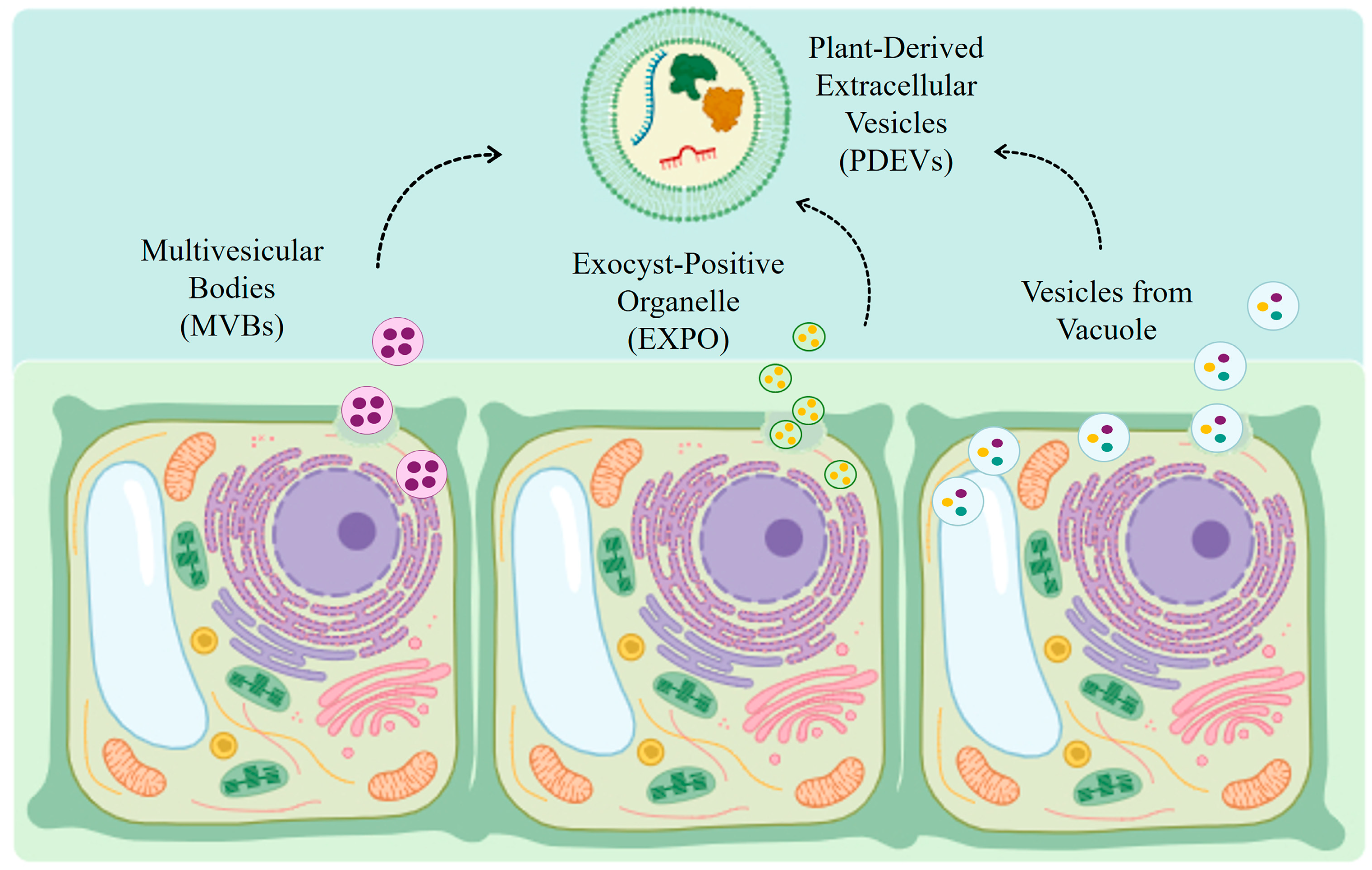
3.1. Biogenesis of Plant-Derived Extracellular Vesicles

Even though the earliest research on membrane-associated structures was conducted in the 1950s, it was not until the development of sophisticated microscopy methods like transmission electron microscopy (TEM), cryo-electron microscopy, and atomic force microscopy (AFM) that it became feasible to examine the morphology of these vesicles in great detail, which helped to clarify their biogenesis and release. Extracellular vesicles (EVs) are spherical structures that can have different origins within plant cells. The main biogenesis pathways include multivesicular bodies (MVBs), the exocyst-positive organelle (EXPO) complex, vacuoles, and autophagosomes (Figure 1) [13,23]. Fusion of MVBs with the plasma membrane is considered the most studied and best characterized mechanism for the release of extracellular vesicles. These multivesicular compartments contain small intraluminal vesicles, and in plants they act as sorting compartments, processing proteins between the trans-Golgi network and vacuoles. Extracellular vesicle biogenesis is regulated by specific proteins that mediate the transport and fusion of multivesicular compartments with the plasma membrane. Among these, Rab GTPases (such as Rab5 and Rab7) and SNARE complexes are crucial. In Arabidopsis, Rab5 and Rab7 are mainly located in multivesicular compartments and appear to facilitate fusion with the plasma membrane, thus releasing small vesicles into the extracellular space. Rab5 includes conventional subtypes (ARA7 and RHA1) and a plant-specific one (ARA6), while Rab7 includes eight protein variants [24]. Under normal conditions, the MON1–CCZ1 complex facilitates the maturation of Rab5 to Rab7 in multivesicular compartments, leading them to fuse with vacuoles for the degradation and recycling of materials. However, in response to infection by fungi such as Botrytis cinerea, an accumulation of ARA6 is observed on multivesicular compartments, colocalized with the TET8 protein at infection sites on the plasma membrane. This colocalization suggests that ARA6 may promote the fusion of the multivesicular compartment with the plasma membrane to respond to pathogenic attacks [25]. In addition to multivesicular compartments, the exocyst-positive organelle (EXPO) represents another pathway for extracellular vesicle biogenesis in plants. This complex was identified through the protein EXO70E2, which is a double-membrane structure similar to autophagosomes, but does not colocalize with known organelle markers, suggesting that they are separate entities. EXPO was observed to fuse with the plasma membrane, releasing its contents into the extracellular space [16,25,26]. This process may be crucial for the release of proteins without leader signals, such as S-adenosylmethionine synthase 2 (SAMS2), which plays a role in salt tolerance [25,27]. PDEVs can also arise from vacuoles and autophagosomes, internal structures involved in the defense response of plants. Vacuoles contain hydrolytic enzymes and defense proteins, such as Vacuolar Processing Enzymes (VPEs), which trigger the collapse of the vacuolar membrane in the event of viral infections to release cytoplasmic contents to compete with the pathogen [13,25,28]. In response to bacterial infections, vacuoles can fuse with the plasma membrane, releasing extracellular defense proteins to limit bacterial proliferation. It has also been observed that small intraluminal vesicles can form in vacuoles through fusion with multivesicular compartments, suggesting that some extracellular vesicles may arise from the fusion of vacuoles and the plasma membrane [25]. Once secreted into the extracellular space, vesicles must cross the plant cell wall, a physical barrier composed of lignin, pectin, and hemicellulose, which can hinder their passage. Recent studies have revealed the association of PDEVs with cell wall hydrolases, such as β-glucosidase and pectinesterase, which could temporarily destabilize the cell wall, facilitating the transit of vesicles [29]. In addition, the fluidity of the lipid structure of vesicles could allow them to compress to pass through the pores of the cell wall. Although these vesicles are normally spherical, it is possible that they assume tubular or distorted shapes to facilitate passage through the wall, as already observed in bacteria. Despite the progress in understanding the biogenesis of PDEVs, many aspects, such as the precise regulation of their secretion and the specific role of autophagosomes, remain unclear. Marker proteins such as TET8, TET9 and PEN1 represent promising starting points for the study of in situ localization, quantification and identification of these EVs. Deepening our knowledge of the molecular processes of extracellular vesicle sorting and release in plants may open new perspectives in biotechnology and plant defense sciences, offering tools to improve resistance to pathogens and tolerance to environmental stresses.
According to some research, plant hormones including salicylic acid and jasmonic acid have a crucial role in controlling the production of PDEVs, especially in stressful situations. In addition to the influence of plant hormones, environmental factors, including nutrient availability, temperature fluctuations, and the presence of pathogens, significantly affect the biogenesis of plant-derived extracellular vesicles [4,30,31]. For example, during a pathogen invasion, the synthesis of PDEVs often increases to provide defensive molecules to infection sites, thereby strengthening the plant’s response to biotic stresses. These adaptive mechanisms highlight the versatility of plants and their dependence on PDEVs to survive under dynamic environmental conditions.

3.2. Plant-Derived Extracellular Vesicles Molecular Cargo

The molecular cargo of PDEVs includes a wide range of bioactive molecules, such as proteins, lipids, RNAs, and metabolites. The protein content of PDEVs includes enzymes involved in plant defense, such as glucanases and proteases, as well as signaling molecules that regulate many of the physiological processes [2,32]. Recent studies have also identified the presence of chitinase in PDEVs, an enzyme capable of degrading chitin in fungal cell walls, conferring antifungal properties to PDEVs with potential applications in agriculture and clinical settings [29]. Lipids are a crucial component of PDEV membranes, ensuring vesicle stability and facilitating interactions with target cells. Among the most abundant lipids in PDEVs are phospholipids and sphingolipids, which allow vesicles to fuse with the cell membranes of recipient cells [14,33]. The presence of phytosterols, characteristic of plant vesicles, also confers anti-inflammatory effects, as evidenced by studies that have analyzed their effect on the modulation of the immune response in animal models. Glycolipids present in PDEVs improve their targeting capacity, interacting with specific receptors on mammalian cell surfaces, and increasing the precision of drug delivery [29,34]. This characteristic makes PDEVs a promising tool for the development of targeted and effective therapies against diseases such as cancer and autoimmune disorders. In addition to proteins and lipids, PDEVs contain different types of RNA, including mRNA and small non-coding RNAs such as microRNAs (miRNAs). These RNAs play key roles in gene regulation and are involved in interspecies communication, as plant RNAs can influence gene expression in mammals by binding to target mRNAs and fragmenting them. In the case of pathogen infections, plants use EVs to release miRNAs into the host, thereby silencing genes that contribute to the virulence of the pathogen. Recent studies have shown that EVs from edible plants are also able to influence the expression of genes in the mammalian genome, thanks to interkingdom interactions, through the transfer of sRNA [15,34]. In agreement with these findings, plant miRNAs have been observed to be naturally modified at the 3′ end by methylation, a process that increases their stability and protects them from degradation [24]. Plant miRNAs ingested by animals are taken up by intestinal epithelial cells and then enclosed in extracellular vesicles [35]. These vesicles protect miRNAs from degradation, allowing them to reach more distant cells, which can interact with endogenous RNAs and modulate gene expression [34]. Numerous studies indicate that these molecules also possess protective properties against some human diseases; for example, miRNA-156a, abundant in green vegetables such as cabbage, spinach, and lettuce, can slow the progression of atherosclerosis [36]. Recently, the presence of long non-coding RNAs (lncRNAs) have also been identified in PDEVs, broadening the therapeutic possibilities of these plant vesicles and offering new perspectives for treatments acting through epigenetic regulation [3,37]. The presence of secondary metabolites, such as carotenoids, polyphenols, and flavonoids, further enriches the cargo of PDEVs, making them ideal candidates for therapeutic applications by virtue of their antioxidant, antitumor, and immunomodulatory effects. Most of these compounds are packed into PDEVs during their biogenesis, providing additional health benefits [5,34]. These active metabolites are more concentrated in EVs than in plant cells; for example, ginger-derived EVs contain much higher amounts of 6-gingerol, 8-gingerol, and 10-gingerol, which are efficiently absorbed from the gut of rats [38]. Similarly, higher vitamin C content in strawberry-derived EVs has been shown to exhibit a potent antioxidant effect [39], just as broccoli-derived EVs containing high concentrations of sulforaphane were found to be effective in a mouse model of colitis [40]. It has also been demonstrated that PDEVs derived from different Aloe species carry photoinducible secondary metabolites such as anthraquinones, which have shown a phototoxic effect on human melanoma cells [41]. Table 1 reports the main metabolites characterized to date in PDEVs.

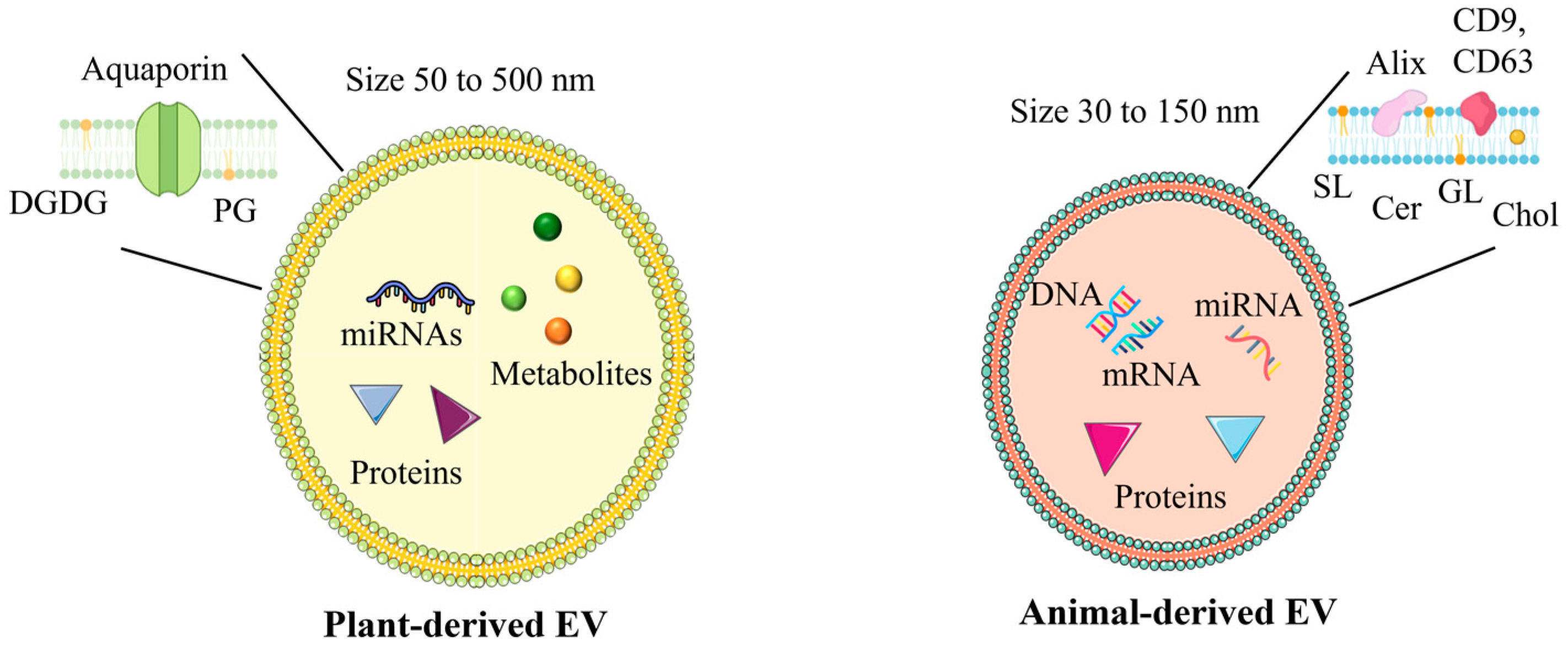
3.3. Differences Between Plant-Derived Extracellular Vesicles and Animal-Derived Extracellular Vesicles

PDEVs and ADEVs are nanoparticles of growing interest, as they play key roles in intercellular communication. However, their dimensions, molecular composition and bioavailability are significantly different. PDEVs extracted from roots, fruits and fresh plants, are composed of lipids, proteins, nucleic acids and secondary metabolites such as flavonoids, polyphenols and terpenes. Although both plant and animal types contain proteins, lipids, and RNA, there are significant differences between the two types of EVs (Figure 2). ADEVs range in size from 30 to 150 nm, while PDEVs are larger, ranging in size from 50 to 500 nm. Regarding proteins, ADEVs contain proteins such as those involved in targeted fusion and membrane transport (e.g., ALIX, TSG101, CD9, CD63), while PDEVs are characterized by the presence of proteins such as actin, proteolytic enzymes, aquaporin, and reticulin heavy chains [22]. In terms of lipids, ADEVs are mainly composed of cholesterol, sphingomyelin, glycolipids, sphingolipids, and ceramides, while PDEVs contain lipids such as digalactosyldiacylglycerol and phosphatidylglycolamine (PE) [72]. Finally, although both types of vesicles transport RNA, ADEVs contain mRNA, miRNA, and lncRNA, while PDEVs are mainly rich in miRNA [22]. This composition allows PDEVs to interact with the gut microbiome and the human immune system, showing potentially beneficial effects in modulating inflammatory and immune responses [4,33,73]. Furthermore, PDEVs are naturally predisposed to oral administration due to their high stability in gastrointestinal conditions, which allows them to deliver bioactive molecules directly into the human body. This property clearly distinguishes PDEVs from their animal counterparts, as the intrinsic stability allows a more direct and targeted absorption through the digestive system [74].

3.4. Comparison Advantages of PDEVs over Synthetic Nanoparticles

The effectiveness of many drugs is often hindered by their inability to target specific tissues, leading to toxic side effects and reduced bioavailability. To address this issue, drug delivery systems have been developed to selectively transport molecules to targeted tissues, thereby enhancing their pharmacokinetic and pharmacodynamic profiles. These systems mainly include synthetic nanoparticles such as liposomes, lipid-based, and polymeric nanoparticles [75]. Such particles encapsulate therapeutic molecules or attach them to their surface, protecting them from degradation while ensuring controlled release at the intended site [76]. However, synthetic carriers still face challenges such as toxicity, uneven distribution in the body, and high production costs. In recent years, extracellular vesicles (EVs) have gained increasing attention as potential natural drug carriers. Due to their lipid-bilayer structure, EVs can encapsulate hydrophilic drugs in their aqueous core and hydrophobic compounds within their membrane [2]. Additionally, EVs mediate intercellular communication by transferring bioactive molecules [77]. Compared to synthetic nanoparticles, EVs exhibit superior biocompatibility and reduced toxicity in preclinical studies [78]. However, EVs derived from mammals pose risks such as immunogenicity when used in heterologous species. Even autologous applications carry concerns, including the potential spread of oncogenic traits to healthy cells or the transfer of viral nucleic acids [79]. A promising alternative to animal-derived EVs is plant-derived extracellular vesicles (PDEVs). These structures share many properties of mammalian EVs, such as the ability to transport both hydrophobic and hydrophilic drugs, but without the risks of immunogenicity or transmission of animal-derived pathogens. PDEVs have demonstrated high cellular uptake efficiency, strong stability in the gastrointestinal tract, and precise tissue-targeting capabilities [22]. Furthermore, the production of PDEVs is more cost-effective and environmentally sustainable compared to synthetic or animal-based counterparts [80].

4. Isolation and Characterization Techniques for PDEVs

4.1. Isolation Methods

The isolation method of PDEVs plays a crucial role in determining the yield and purity of the obtained samples since these factors can significantly affect the quality of the isolated vesicles. Additionally, the presence of contaminants that can co-isolate with EVs—such as cellular debris, lipoproteins, protein complexes, and aggregates—increases the complexity of the isolation process [81]. It has been demonstrated that isolation techniques significantly affect the physicochemical characteristics and downstream studies of EV cargo [82]. Although there is currently little research on PDEVs, in most cases the isolation techniques for mammal EVs have been adapted for EVs of plant origin. The most common technique for the isolation of PDEVs from apoplastic fluid involves a simple protocol based on the infiltration followed by centrifugation of tissues such as seeds or leaves. A key element of this process is the composition of the infiltration buffer, which must be optimized to minimize tissue damage and prevent contamination by intracellular contents [83]. Another method instead involves the gentle disruption of the cell wall. Once the apoplastic fluids are collected, several strategies can be adopted to separate the PDEVs, but the most widespread methods are ultracentrifugation, density gradient ultracentrifugation and size exclusion chromatography (SEC).

4.1.1. Ultracentrifugation

The ultracentrifugation protocol is derived from protocols used for mammal EVs, with initial low-speed centrifugations (500× g and 10 K× g) to eliminate debris and large organelles (such as mitochondria and chloroplasts), followed by ultracentrifugation carried out at higher speeds (40 K× g or 50 K× g, and finally 100 K× g). The pellet obtained at 40–50 K is generally considered enriched in EVs with classic dimensions between 30 and 200 nm. However, the pellet resulting from centrifugation at 100 K has been described differently: for sunflower seeds, it is similar to the one at 40 K both for protein content and EV size [84]. On the contrary, in studies on the Arabidopsis leaf apoplast, the pellet at 100 K seems to contain smaller particles, ranging in size from 10 to 17 nm [85]. For this reason, some works have focused on vesicles recovered from intermediate pellets (40–50 K), while others have studied those in the final fractions (100 K). This variability, with a particular focus on intermediate pellets, is a peculiarity of the plant field, since in mammalian studies EV separation is mainly based on pellets obtained at 10–20 K and 100 K. Differential ultracentrifugation is the most used method for EV isolation, mainly due to its operational simplicity and low cost. However, this technique has significant limitations that affect its efficacy. Ultracentrifugation does not allow a selective separation of EVs, as it also sediments non-specific particles, including protein and nucleic acid aggregates. Since vegetable juices are highly heterogeneous systems, all their components can precipitate once a certain centrifugal force is exceeded. Consequently, PDEVs isolated by this methodology can be contaminated by protein aggregates and nucleic acids, compromising both their purity and quality, with a negative impact on the accuracy of functional assessments [83,86].

4.1.2. Density Gradient Ultracentrifugation

Density gradient ultracentrifugation represents an alternative method to isolate PDEVs, which relies on extracellular vesicles’ capacity to suspend in various gradient areas and separate from other molecules in biofluids. Density gradient is commonly achieved with sucrose solutions (8%, 15%, 30%, 45%, 60%) or iodixanol (20–50%) [83,87]. The latter is preferred to preserve the size of the vesicles due to its lower density and iso-osmotic properties. In the case of PDEVs, some studies have observed multiple bands in the gradient; for example, EVs derived from turmeric are positioned between 8 and 30% and 30 and 45%, with a structure similar to mammalian exosomes [88]. Other similar results have been found in plants such as grapefruit, ginger, carrot and tomato [57,58,89,90]. Lower-density EVs are also important: those from turmeric (8–30%) reduce TNF-α, IL-6, and IL-1β, increasing HO-1, suggesting anti-inflammatory and antioxidant activities [88]. EVs from ginger, also 8–30%, activate Nrf2, inhibiting hepatic ROS, with potential beneficial effects for liver diseases [50].

4.1.3. Size Exclusion Chromatography (SEC)

Size Exclusion Chromatography (SEC), or gel filtration, is a technique that separates molecules based on their size and is commonly used for protein purification. SEC is based on the principle that larger molecules cannot penetrate the pores of the filter material and pass through the column more quickly, while smaller molecules, penetrating the pores, migrate more slowly, thus allowing separation. Different polymers have been developed to be used to fill SEC columns, such as dextrans (Sephadex), agarose (Sepharose), and polyacrylamide (Biogel P or Sephacryl), and specific commercial kits have also been created for the isolation of Evs [83]. Although SEC is a widely used method for isolating EVs from biofluids, its use for the separation of PDEVs is rather limited. However, some studies have also demonstrated its applicability for EVs of plant origin; for example, SEC has been used to isolate EVs from cabbage, cucumber, tomato, and carrot [57,90,91,92].

4.2. Characterization Techniques

Currently, the most widely used characterization techniques provide information on the size, concentration, morphology, presence of protein markers, protein concentration, and biochemical composition of PDEVs, which are essential to understanding their roles in cellular processes, intercellular communication and potential therapeutic applications [93]. These methods help to determine not only the physical properties of PDEVs, but also their molecular cargo, including proteins, lipids and RNA, which are essential to understand their biological functions. Morphological analysis techniques include electron microscopy and Nanoparticle Tracking Analysis (NTA), which provide complementary data on the shape, diameter distribution and concentration of PDEVs. Flow cytometry is widely used with animal EVs as it allows the profiling of surface markers and the analysis of the heterogeneity of vesicular populations, useful to distinguish subpopulations and their biochemical differences. However, specific markers for PDEVs are still poorly known, limiting the depth of research in this field. Furthermore, biochemical techniques such as Western Blotting and mass spectrometry identify vesicular content and markers, while molecular analyses investigate the nucleic component. The combined use of these methodologies provides a detailed profile of PDEVs, favoring diagnostic and therapeutic biotechnological applications [94,95].

4.2.1. Electron Microscopy

Electron microscopy is currently used to assess the morphology of PDEVs. Scanning Electron Microscopy (SEM) is able to observe the shape, size and surface distribution of vesicles, but exhibits some limitations, such as sample preparation, that can alter the natural morphology or the inability to visualize the lipid bilayer, a distinctive feature of EVs [96]. For finer details, Transmission Electron Microscopy (TEM) or cryo-EM are preferred, with resolutions of 1–3 nm and 5 nm, respectively [2,97]. TEM, combined with gold immunolabeling, also allows for the identification of proteins present in EVs, facilitating functional studies. Cryo-EM preserves the spherical structures of EVs in a hydrated environment, reducing fixation-related artifacts and allowing the acquisition of 3D tomographic images for detailed visualization. Other advanced techniques include Atomic Force Microscopy (AFM), which uses a probe to obtain three-dimensional topographic images at high resolution (less than 1 nm), ideal for observing EVs and other nanostructures [2].

4.2.2. Nanoparticle Tracking Analysis (NTA)

Nanoparticle Tracking Analysis (NTA) is a widespread technique that provides data on particle size and concentration of EVs. NTA combines laser light scattering microscopy with a camera to visualize and track suspension nanoparticles in real-time. NTA allows one to determine the size and total concentration of particles between 30 nm and 1 μm by tracking the Brownian motion of EVs [86,98]. Specific systems, equipped with a laser and camera, also detect fluorescent particles, which is useful for analyzing subpopulations of EVs labeled with specific antibodies or fluorophores [99,100]. This capability has been exploited to study the phenotype of vesicles and to identify molecular targets, such as miR-21 in lung cancer-derived EVs, with greater sensitivity than cytometry for particles smaller than 100 nm, such as exosomes [101]. NTA has proven to be particularly useful for characterizing the size distribution and molecular content of EVs, opening new perspectives in research on biomarkers and therapeutic targets.

4.2.3. Flow Cytometry

Flow cytometry is a standard technique to characterize EV surface markers, providing information on their biological origin. This technique allows one to analyze single particles as they pass through a laser ray, detecting light scattering signals: forward scattering (FSC) indicates size, while lateral scattering (SLS) provides data on morphology and internal structure [102,103]. One of its advantages is the ability to identify vesicle subpopulations through antibody-bound fluorescent markers, facilitating the simultaneous analysis of multiple surface proteins [104]. However, the application to PDEVs has significant limitations. The small size of EVs makes it difficult to distinguish the signal from the background, and the emitted fluorescence is often weak due to the small number of antigens per particle. Furthermore, lacking knowledge of specific markers for PDEVs complicates the identification of subtypes [4].

5. Biological Functions of PDEVs

5.1. Role in Plant-Plant Communication and in Plant-Microbe Interactions

PDEVs rich in miRNAs, bioactive lipids, and proteins act as extracellular messengers, facilitating cell–cell communication in a manner similar to mammalian extracellular vesicles. In fact, PDEVs play a key role in plant cell–cell communication, enabling information exchange between cells of the same species and between different species [28]. PDEVs also participate in essential cellular processes such as proliferation and differentiation, responding to a variety of stimuli, which may include environmental signals, such as changes in light or temperature, or biological signals, such as pathogen attacks. Research conducted on EVs from coconut water has shown their contribution to the fruit ripening process, supporting this function through RNA analysis [65]. PDEVs are involved in the transportation of phytohormones such as auxins, cytokinins and gibberellins, key elements for regulating plant growth and development [105]. Auxins, for example, are essential for root elongation and the formation of lateral roots, processes that are essential for optimizing water and nutrient absorption. Cytokinins, on the other hand, promote cell division and shoot development, while gibberellins are responsible for stem elongation, seed germination and the flowering process [106]. In addition to communication, PDEVs are strongly involved in plant defense mechanisms. They help eliminate harmful compounds from cells and actively participate in immune surveillance. Bacterial or fungal infections have been shown to enhance the production of plant EVs containing defense-related proteins, sRNAs, and lipid signals, highlighting their crucial role in plant immune mechanisms [13,107,108]. For example, a study on Arabidopsis infected with Golovinomyces orontii revealed exosome-like extracellular vesicles carrying PEN1/SYP121 and the ABC transporter PEN3. These compounds accumulate around the haustoria, forming a protective barrier that hinders fungal entry [109]. PDEVs extracted from the extracellular fluids of sunflower (Helianthus annuus) seedlings showed a heterogeneous composition, including cell wall restructuring enzymes and defensive proteins identified by proteomic analyses. When spores of the phytopathogenic fungus Sclerotinia sclerotiorum were exposed to purified sunflower EVs, growth arrest, structural alterations, and cell death were observed [13,63]. Likewise, De Palma et al. showed that EVs made from tomato roots efficiently inhibited both spore germination and germ tube development in diseases such Alternaria alternata, B. cinerea, and Fusarium oxysporum [64]. Although EV-mediated plant-fungal interactions have been extensively investigated, bacterial-plant interactions remain poorly understood. It has been demonstrated that Arabidopsis plants infected with Pseudomonas syringae produced twice the amount of EVs compared to uninfected plants. This result suggests that plant EVs may play a significant role in resistance against bacterial pathogens. Furthermore, EVs isolated from P. syringae-infected plants contained numerous proteins involved in the biotic stress response, supporting the hypothesis that these vesicles may act as vectors of immune signals during bacterial infections [85].

5.2. Role in Human Health and Therapeutic Applications

PDEVs have emerged as a promising resource for therapeutic applications in human health, due to their bioactive components that exhibit anti-inflammatory, antioxidant, and anticancer properties. Numerous studies have highlighted their ability to interact with mammalian cells, contributing to positive physiological effects. Plant-based treatments have been used for centuries, exploiting their natural forms or extracts of active compounds to prevent diseases and repair damage. However, only recently, attention has also focused on the use of PDEVs which have shown interesting biological properties, including anti-inflammatory, anticancer, antibacterial, antifungal and antioxidant effects [7,14,29,110]. These effects are mediated through various mechanisms, such as gene regulation, interaction with gut microbiota, macrophage activation, gene silencing and the presence of specific active compounds [111,112]. These natural therapeutic properties offer new perspectives for innovative treatments, both alone and in combination with other drugs and nanomedicines.

5.2.1. Anti-Inflammatory Effects of PDEVs

The immunomodulating and anti-inflammatory properties of PDEVs have attracted increasing attention. These vesicles can be used in a variety of biological scenarios since they not only have positive effects on the immune system but also remain stable under harsh conditions. Because of their capacity to regulate inflammatory responses in a variety of therapeutic settings, both in vitro and in vivo, as evidenced by multiple studies, PDEVs have shown promise in the treatment of chronic inflammatory illnesses such as colitis [88]. For example, ginger-derived EVs have been able to suppress NLRP3 inflammasome activation, a key protein involved in inflammatory diseases such as inflammatory bowel disease (IBD), reducing levels of pro-inflammatory cytokines such as IL-1β and IL-18 and preventing the assembly of the inflammasome itself. These anti-inflammatory effects are mediated by mechanisms such as the induction of autophagy and the prevention of focused cell death, as demonstrated in a clinical trial examining the effectiveness of ginger-derived EVs in patients with IBD [16,113]. Mulberry bark-derived EVs have been able to prevent colitis in animal models, possibly through AhR receptor activation, mediated by HSPA8 protein [59]. Furthermore, turmeric-derived EVs have shown potent anti-inflammatory effects in murine colitis models, modulating pro-inflammatory cytokines such as TNF-α, IL-6, and IL-1β and promoting the expression of the antioxidant gene HO-1, which contributes to the resolution of intestinal inflammation. Turmeric-derived EVs have also been associated with the inhibition of the NF-κB pathway, further increasing protection against intestinal inflammation [88]. On the other hand, orange-derived EVs have been found to be effective in reducing intestinal inflammation and restoring intestinal barrier integrity by modulating the expression of key genes such as HMOX-1, ICAM1, OCLN, CLDN1, and MLCK [114].

5.2.2. Antioxidant Properties of PDEVs

Reactive Oxygen Species (ROS) are generated during physiological processes such as aerobic respiration, as well as in response to external stimuli, such as xenobiotics, cytokines and bacterial invasion. ROS play an essential role in cell survival and proliferation, but when their production exceeds the organism’s defensive capacity, oxidative stress occurs, which damages crucial biomolecules such as DNA, proteins and lipids. This oxidative damage is implicated in the onset of several common diseases, including neurodegenerative disorders, cardiovascular diseases, diabetes and cancer. Natural antioxidants, such as polyphenols, carotenoids and vitamins, play a protective role against oxidative stress and have demonstrated anti-aging and anti-inflammatory effects. In this context, PDEVs emerge as promising therapeutic agents to counteract oxidative stress and associated pathological conditions, thanks to their antioxidant and anti-inflammatory properties. PDEVs act through the upregulation of antioxidant molecules such as nuclear factor erythroid 2 (Nrf2)-related enzyme, hemazole 1 (HO-1), and NAD(P)H quinone dehydrogenase 1 (NQO1), thus strengthening cellular defense mechanisms against oxidative damage [16,90]. For example, PDEVs extracted from Aloe vera peel showed significant antioxidant activity, dose-dependently increasing the expression of key genes such as Nrf2, CAT, HO-1, and SOD [7]. Similarly, PDEVs derived from carrots were able to prevent the downregulation of Nrf2 expression, protecting H9C2 cardiomyoblastic cells and SH-SY5Y human neuroblastoma cells from oxidative stress [90]. Strawberry-derived PDEVs may contribute to protection against oxidative damage and improve cell viability, due to their high concentration of vitamin C, anthocyanins, folic acid and flavonoids. Adipose-derived mesenchymal stem cells internalize strawberry vesicles without compromising their viability and, when pretreated with these vesicles, show protection against oxidative damage in a dose-dependent manner [39]. Blueberry-derived PDEVs have also been shown to reduce oxidative stress in cellular and animal models, by affecting the functionality of the mitochondrial protein Bcl-2 and preventing apoptosis in HepG2 cells. Furthermore, administration of these PDEVs in vivo improved insulin resistance and the expression of Nrf2-regulated detoxification/antioxidant genes, showing potential in the treatment of non-alcoholic fatty liver disease (NAFLD) [115]. Additionally, other plant sources such as grape juice, rich in polyphenols, have been shown to mitigate oxidative stress by modulating ROS levels in various cell models [44]. Vesicles derived from broccoli are known to enhance antioxidant defense systems, potentially through the activation of Nrf2 and other related pathways [116,117]. Citrus-derived PDEVs, rich in vitamin C and flavonoids, contribute to reducing oxidative stress [118]. Furthermore, apple-derived EVs have been shown to act as novel anti-inflammatory compounds that modulate the extracellular matrix production in dermal fibroblasts, reducing skin aging by inhibiting the TLR4/NF-κB inflammatory pathway, enhancing collagen synthesis, and decreasing extracellular matrix degradation [119].

5.2.3. Anticancer Properties of PDEVs

In recent years, PDEVs have emerged as a promising alternative in oncology research. Numerous studies have highlighted the ability of PDEVs to inhibit tumor cell proliferation, modulate the cell cycle, induce apoptosis and influence cancer metabolism, thus offering a multifactorial approach to oncology treatment. For example, PDEVs derived from Panax ginseng were able to induce macrophage polarization toward the M1 phenotype via Toll-like receptor (TLR)-4 and myeloid differentiation antigen 88 (MyD88) signaling pathways, with direct effects on melanoma cell apoptosis. Ceramides and proteins associated with PDEVs play a crucial role in macrophage activation, increasing their antitumor potential [120]. Similarly, PDEVs derived from Moringa oleifera seeds demonstrated a targeted action against tumor cells, inhibiting their viability through the regulation of the antiapoptotic protein Bcl-2 and the impairment of mitochondrial membrane potential [121]. Bitter melon nanovesicles have been shown to be effective against breast cancer, both in vitro and in vivo, by blocking tumor cell proliferation and migration. This effect has been attributed to their ability to induce the production of ROS and to impair mitochondrial function, thus triggering oxidative stress-mediated cell death pathways [122]. Similarly, tea flower vesicles have shown significant cytotoxic activity against breast cancer cells, with the particularity of also modulating the intestinal microbiota, suggesting potential systemic effects that could counteract metastasis [49]. Lemon PDEVs have been shown to inhibit the growth of chronic myeloid leukemia tumor cells in vivo, targeting the tumor and activating the TRAIL-mediated apoptotic process, with a significant reduction in the risk of cancer progression [123]. Some PDEVs, such as those derived from ginger, have also been shown to be effective against colorectal cancer, reducing the levels of cell cycle protein D1 RNA and inhibiting the proliferation of colon cancer cells [58]. Studies in lung cancer mouse models have highlighted the potential of Artemisia PDEVs, which improved the efficacy of PD-1 blockade through the remodulation of the tumor microenvironment and the reprogramming of tumor-associated macrophages. The mechanism seems to be linked to the activation of the cGAS-STING pathway induced by mitochondrial DNA, which explains their immunomodulatory effects [124]. PDEVs derived from Aloe arborescens, Aloe barbadensis, and Aloe chinensis have been shown to inhibit melanoma cell proliferation through the photoactivation of their anthraquinone content, leading to the production of ROS [41]. PDEVs derived from grapefruit mediated the inhibition of liver metastasis that is dependent on the induction of M1 macrophages (F4/80+IFNγ+IL-12+) [125]. PDEVs represent an extremely innovative therapeutic tool as they overcome the limitations related to low bioavailability, instability and safety issues of synthetic or animal vesicles, offering new opportunities for the treatment of cancer, both through direct action and by enhancing immune responses against tumors. Table 2 reports the most widespread effects of PDEVs.

6. Enhancing PDEVs for Drug Delivery Applications

PDEVs can be used as drug delivery platforms by loading small molecules or nucleic acid-based drugs without undergoing degradation, making them an effective method to prevent drug damage [31]. PDEVs have high bioavailability, can be absorbed in the intestine, and penetrate deep into the skin, and, if administered intranasally, they are able to cross the blood–brain barrier and reach the brain [92]. Furthermore, PDEVs are considered safe due to their low toxicity and fewer side effects compared to synthetic lipid nanoparticles and animal-derived EVs. Most of the molecules used in PDEVs as cargo are nucleic acid-based drugs and small-molecule chemical drugs (Figure 3) [127].

6.1. Engineered PDEVs: Nucleic Acid Delivery

Several techniques using physical, chemical, or mechanical approaches are available to enable the introduction of genetic material into PDEVs. These methodologies allow PDEVs to be used as vehicles for the targeted delivery of nucleic acids, expanding their potential therapeutic applications. One of the most widely used techniques is electroporation, which applies an electric field to PDEVs, creating temporary pores in their lipid membrane. This process promotes the transfer of nucleic acids across the membrane and their subsequent incorporation into the vesicles. Another common method is sonication, which uses high-frequency sound waves to temporarily perturb the vesicle membrane, allowing the penetration of nucleic acids. A gentler approach is incubation, in which PDEVs are mixed with nucleic acids in a buffer solution. By varying parameters such as temperature, pH, and salt concentration, it is possible to favor the passive uptake of genetic material by the vesicles. Finally, chemical transfection involves the use of specific chemicals that increase the permeability of the PDEV membrane, making it more receptive to nucleic acids [12,127,128]. Each technique has advantages and limitations, and the choice depends on the type of nucleic acid, the nature of the PDEV, and the experimental objectives. These approaches represent versatile tools to exploit PDEVs as delivery platforms for innovative nucleic acid-based therapies. PDEVs are promising tools for the delivery of nucleic acids, such as siRNA and miRNA. A noteworthy example is grapefruit-derived PDEVs loaded with miR17, which have been shown to delay the growth of brain tumors in mice. This effect is possible due to the ability of PDEVs to reach the brain via nasal administration, allowing the transported miR17 to exert an inhibitory action. Furthermore, the addition of folic acid to PDEVs improves the targeting of folate receptor-positive brain tumors, providing a non-invasive treatment for brain diseases [129]. Different types of PDEVs lend themselves to different routes of administration. For example, acerola-derived PDEVs can deliver small RNAs to the digestive tract, with a peak of suppressive activity on target genes in the small intestine and liver within 24 h of administration, representing a valid option for the oral delivery of nucleic acids [130]. Similarly, ginger-derived PDEVs loaded with siRNA-CD98 showed effective action on colon tissue by reducing CD98 gene expression upon oral administration [131]. Broccoli-derived EVs were loaded with exogenous miRNAs with biologically active properties and then co-incubated with Caco-2 cells causing cytotoxicity. These results suggest that broccoli-derived EVs may increase the stability of therapeutic RNA, protecting it from degradation by RNases and digestive enzymes [67].

6.2. Surface Functionalization of PDEVs

In addition to carrying small therapeutic molecules, PDEVs have also been shown to improve the properties of inorganic nanoparticles by modifying their surfaces. For example, ginger-derived extracellular vesicles were used to coat nanocarriers, increasing their biocompatibility and persistence in the blood, and reducing their immunogenicity thanks to the integration of natural plant membranes [132]. Further research involved grapefruit-derived lipid nanovesicles modified with leukocyte-associated inflammatory protein receptors. This system was used for the targeted delivery of doxorubicin (DOX) to inflamed areas. The stability and detectability of the drug, assessed by spectrophotometry at 497 nm, were enhanced thanks to the coating of the vesicles [89]. In addition to carrying anticancer drugs, grapefruit-derived EVs can carry anti-inflammatory molecules such as curcumin. In mouse models of inflammation, vesicles modified with specific receptors showed a higher targeting efficiency than natural ones, optimizing drug release in inflamed areas [133]. Grapefruit-derived extracellular vesicles were functionalized with a maleimide-modified lipid moiety and conjugated with aptamers via click chemistry, enabling targeted drug delivery through enhanced selective cellular uptake in brain cell models (hCMEC/D3 and U87MG) [51]. Qiao et al. demonstrated that integrating electrodynamic platinum-palladium (Pt-Pd) nanosheets with ginger-derived extracellular vesicles improved blood circulation, promoted targeted accumulation at infection sites, and penetrated bacterial membranes. Additionally, this combination generated reactive oxygen species, enhancing synergistic electrodynamic and photothermal therapy with promising biocompatibility and in vivo applicability [134].

6.3. Oral Administration of PDEVs for Drug Delivery

PDEVs obtained from edible plant species have a high safety of use. They can efficiently encapsulate drugs and gene therapy agents, offering protection against gastrointestinal degradation and promoting absorption by the organism. Preclinical studies have shown that PDEVs do not significantly alter the physiological and biochemical parameters of laboratory animals, confirming their excellent biocompatibility [17]. In fact, oral administration is one of the preferred methods for drug intake, although it involves significant difficulties. The unfavorable environment of the gastrointestinal tract can compromise the efficacy of the drug, causing losses of active substances and hindering its absorption through the intestinal wall. In addition, orally administered drugs often show low bioavailability and may generate unwanted side effects [135]. On the other hand, the oral use of PDEVs is considered safe and promising, since these vesicles demonstrate high stability in the gastrointestinal tract. The reason why PDEVs are able to overcome the hostile environment of the gastrointestinal tract and reach target cells remains largely unknown. One possible explanation is that the lipid composition could be a determining factor. PDEVs are distinguished by their richness in specific lipids, such as PA, DGDG, MGDG, PC, PE and PI, in contrast to mammalian EVs, which are predominantly composed of CHOL, SM, PC and PE [136,137]. Previous studies suggest that lipid composition affects the formation, distribution and function of EVs in vivo, and could therefore explain their resistance to gastrointestinal digestion. Ginger-derived EVs were found to be highly stable in simulated gastrointestinal fluids, with effective uptake by intestinal epithelial cells and macrophages after oral administration [58]. Similarly, the study conducted by Wang et al. demonstrated that grapefruit-derived EVs maintained their stability both in acidic solutions and in simulated gastrointestinal fluids, showing remarkable resistance to enzymatic degradation [89]. In the same study, grapefruit PDEVs were used to encapsulate methotrexate (MTX). The authors compared the antiproliferative effect of encapsulated MTX versus the free drug, highlighting that most of the encapsulated MTX was selectively delivered to macrophages of the intestinal lamina propria. This approach significantly improved the anti-inflammatory properties of MTX while reducing its side effects, suggesting that PDEVs represent a promising system to increase the bioavailability of orally administered drugs. Similar evidence has emerged from studies on the use of PDEVs for the delivery of siRNA. Ginger-derived vesicles have been used to transport and protect siRNA from enzymatic degradation. Ginger-derived EVs have demonstrated the ability to deliver siRNA-CD98 to the intestine, effectively reducing the expression of the CD98 gene in colon cells [131].

7. Conclusions and Future Perspectives

Plant-derived extracellular vesicles represent a promising solution as biotechnological drugs. In fact, thanks to their natural origin, PDEVs are intrinsically safe, non-toxic and do not trigger immune responses. Furthermore, the production of plant material can offer a scalable alternative to the production limitations of synthetic or animal-derived EVs. Another important characteristic that distinguishes PDEVs is the presence of natural bioactive molecules cargo, such as proteins, lipids and miRNA, which play crucial functions in gene regulation and intercellular communication, giving PDEVs an intrinsic therapeutic potential. Other beneficial molecules contained in PDEVs include secondary metabolites with antioxidants and anti-inflammatory functions, which can be integrated with exogenous drugs to obtain synergistic effects. PDEVs are used in multiple contexts, ranging from the natural administration of nutrients and supplements to therapeutic support for pathologies such as cancer and inflammatory diseases. However, further studies are needed to better understand the pharmacological properties and delivery mechanism of PDEVs to fully exploit their therapeutic potential. Advances in bioengineering could further optimize their therapeutic potential, enabling more efficient and precise drug delivery. Surface modification strategies, for example, could be developed to improve targeting accuracy, allowing for personalized drug delivery systems tailored to individual patients and specific diseases. This would enable more effective treatments with fewer side effects, particularly for complex conditions such as cancer, neurodegenerative disorders, and genetic diseases. Moreover, the integration of PDEVs with cutting-edge technologies like gene editing or RNA-based therapies could open up new frontiers in precision medicine. By utilizing PDEVs as delivery vehicles for gene-editing tools or RNA molecules, it may be possible to directly correct genetic mutations or modulate gene expression in a highly targeted manner. This would enhance the therapeutic options available, offering more effective treatments for a wide range of previously untreatable diseases.

Author Contributions

Conceptualization, E.C., A.B., G.C. and C.E.; writing—original draft preparation, E.C., A.B. and G.C.; writing—review and editing, E.C., S.B. and L.U.; visualization, E.C., A.B. and G.C.; supervision, C.E. All authors have read and agreed to the published version of the manuscript.

Funding

This work has been funded by the European Union—NextGenerationEU under the Italian Ministry of University and Research (MUR) National Innovation Ecosystem grant ECS00000041—VITALITY—CUP J97G22000170005 (to Carla Emiliani).

Data Availability Statement

Data sharing is not applicable.

Acknowledgments

We acknowledge Università degli Studi di Perugia and the Italian Ministry of University and Research (MUR) for their support within the VITALITY project.

Conflicts of Interest

The authors declare no conflicts of interest.

References

  1. López de las Hazas, M.-C.; Tomé-Carneiro, J.; del Pozo-Acebo, L.; del Saz-Lara, A.; Chapado, L.A.; Balaguer, L.; Rojo, E.; Espín, J.C.; Crespo, C.; Moreno, D.A.; et al. Therapeutic Potential of Plant-Derived Extracellular Vesicles as Nanocarriers for Exogenous miRNAs. Pharmacol. Res. 2023, 198, 106999. [Google Scholar] [CrossRef]
  2. Lian, M.Q.; Chng, W.H.; Liang, J.; Yeo, H.Q.; Lee, C.K.; Belaid, M.; Tollemeto, M.; Wacker, M.G.; Czarny, B.; Pastorin, G. Plant-Derived Extracellular Vesicles: Recent Advancements and Current Challenges on Their Use for Biomedical Applications. J. Extracell. Vesicles 2022, 11, 12283. [Google Scholar] [CrossRef]
  3. Wang, X.; Xin, C.; Zhou, Y.; Sun, T. Plant-Derived Vesicle-like Nanoparticles: The Next-Generation Drug Delivery Nanoplatforms. Pharmaceutics 2024, 16, 588. [Google Scholar] [CrossRef]
  4. Kameli, N.; Dragojlovic-Kerkache, A.; Savelkoul, P.; Stassen, F.R. Plant-Derived Extracellular Vesicles: Current Findings, Challenges, and Future Applications. Membranes 2021, 11, 411. [Google Scholar] [CrossRef] [PubMed]
  5. Subha, D.; Harshnii, K.; Madhikiruba, K.G.; Nandhini, M.; Tamilselvi, K.S. Plant Derived Exosome- like Nanovesicles: An Updated Overview. Plant Nano Biol. 2023, 3, 100022. [Google Scholar] [CrossRef]
  6. Huang, J.; Wang, X.; Wang, Z.; Deng, L.; Wang, Y.; Tang, Y.; Luo, L.; Leung, E.L.-H. Extracellular Vesicles as a Novel Mediator of Interkingdom Communication. Cytokine Growth Factor Rev. 2023, 73, 173–184. [Google Scholar] [CrossRef]
  7. Kim, M.K.; Choi, Y.C.; Cho, S.H.; Choi, J.S.; Cho, Y.W. The Antioxidant Effect of Small Extracellular Vesicles Derived from Aloe Vera Peels for Wound Healing. Tissue Eng. Regen. Med. 2021, 18, 561–571. [Google Scholar] [CrossRef] [PubMed]
  8. Mao, X.; Li, T.; Qi, W.; Miao, Z.; Zhu, L.; Zhang, C.; Jin, H.; Pan, H.; Wang, D. Advances in the Study of Plant-Derived Extracellular Vesicles in the Skeletal Muscle System. Pharmacol. Res. 2024, 204, 107202. [Google Scholar] [CrossRef] [PubMed]
  9. Qiang, W.; Li, J.; Ruan, R.; Li, Q.; Zhang, X.; Yan, A.; Zhu, H. Plant-Derived Extracellular Vesicles as a Promising Anti-Tumor Approach: A Comprehensive Assessment of Effectiveness, Safety, and Mechanisms. Phytomedicine 2024, 130, 155750. [Google Scholar] [CrossRef] [PubMed]
  10. Xu, Z.; Xu, Y.; Zhang, K.; Liu, Y.; Liang, Q.; Thakur, A.; Liu, W.; Yan, Y. Plant-Derived Extracellular Vesicles (PDEVs) in Nanomedicine for Human Disease and Therapeutic Modalities. J. Nanobiotechnol. 2023, 21, 114. [Google Scholar] [CrossRef] [PubMed]
  11. Kang, S.J.; Lee, J.H.; Rhee, W.J. Engineered Plant-Derived Extracellular Vesicles for Targeted Regulation and Treatment of Colitis-Associated Inflammation. Theranostics 2024, 14, 5643. [Google Scholar] [CrossRef] [PubMed]
  12. Wu, C.; Li, J.; Huang, K.; Tian, X.; Guo, Y.; Skirtach, A.G.; You, M.; Tan, M.; Su, W. Advances in Preparation and Engineering of Plant-Derived Extracellular Vesicles for Nutrition Intervention. Food Chem. 2024, 457, 140199. [Google Scholar] [CrossRef] [PubMed]
  13. Ambrosone, A.; Barbulova, A.; Cappetta, E.; Cillo, F.; De Palma, M.; Ruocco, M.; Pocsfalvi, G. Plant Extracellular Vesicles: Current Landscape and Future Directions. Plants 2023, 12, 4141. [Google Scholar] [CrossRef] [PubMed]
  14. Rome, S. Biological Properties of Plant-Derived Extracellular Vesicles. Food Funct. 2019, 10, 529–538. [Google Scholar] [CrossRef] [PubMed]
  15. Urzì, O.; Gasparro, R.; Ganji, N.R.; Alessandro, R.; Raimondo, S. Plant-RNA in Extracellular Vesicles: The Secret of Cross-Kingdom Communication. Membranes 2022, 12, 352. [Google Scholar] [CrossRef]
  16. Zeng, Y.-B.; Deng, X.; Shen, L.-S.; Yang, Y.; Zhou, X.; Ye, L.; Chen, S.; Yang, D.-J.; Chen, G.-Q. Advances in Plant-Derived Extracellular Vesicles: Isolation, Composition, and Biological Functions. Food Funct. 2024, 15, 11319–11341. [Google Scholar] [CrossRef]
  17. Fang, Z.; Liu, K. Plant-Derived Extracellular Vesicles as Oral Drug Delivery Carriers. J. Control. Release 2022, 350, 389–400. [Google Scholar] [CrossRef] [PubMed]
  18. Kürtösi, B.; Kazsoki, A.; Zelkó, R. A Systematic Review on Plant-Derived Extracellular Vesicles as Drug Delivery Systems. Int. J. Mol. Sci. 2024, 25, 7559. [Google Scholar] [CrossRef] [PubMed]
  19. Ding, L.; Chang, C.-J.; Liang, M.-L.; Dong, K.-M.; Li, F.-R. Plant-Derived Extracellular Vesicles as Potential Emerging Tools for Cancer Therapeutics. Adv. Ther. 2024, 7, 2400256. [Google Scholar] [CrossRef]
  20. Cui, L.; Perini, G.; Palmieri, V.; De Spirito, M.; Papi, M. Plant-Derived Extracellular Vesicles as a Novel Frontier in Cancer Therapeutics. Nanomaterials 2024, 14, 1331. [Google Scholar] [CrossRef]
  21. Kathait, P.; Patel, P.K.; Sahu, A.N. Harnessing Exosomes and Plant-Derived Exosomes as Nanocarriers for the Efficient Delivery of Plant Bioactives. Nanomedicine 2024, 19, 2679–2697. [Google Scholar] [CrossRef] [PubMed]
  22. Wang, Y.; Wang, J.; Ma, J.; Zhou, Y.; Lu, R. Focusing on Future Applications and Current Challenges of Plant Derived Extracellular Vesicles. Pharmaceuticals 2022, 15, 708. [Google Scholar] [CrossRef] [PubMed]
  23. Zhao, B.; Lin, H.; Jiang, X.; Li, W.; Gao, Y.; Li, M.; Yu, Y.; Chen, N.; Gao, J. Exosome-like Nanoparticles Derived from Fruits, Vegetables, and Herbs: Innovative Strategies of Therapeutic and Drug Delivery. Theranostics 2024, 14, 4598. [Google Scholar] [CrossRef] [PubMed]
  24. Nemati, M.; Singh, B.; Mir, R.A.; Nemati, M.; Babaei, A.; Ahmadi, M.; Rasmi, Y.; Golezani, A.G.; Rezaie, J. Plant-Derived Extracellular Vesicles: A Novel Nanomedicine Approach with Advantages and Challenges. Cell Commun. Signal. 2022, 20, 69. [Google Scholar] [CrossRef]
  25. Cui, Y.; Gao, J.; He, Y.; Jiang, L. Plant Extracellular Vesicles. Protoplasma 2020, 257, 3–12. [Google Scholar] [CrossRef]
  26. Sall, I.M.; Flaviu, T.A. Plant and Mammalian-Derived Extracellular Vesicles: A New Therapeutic Approach for the Future. Front. Bioeng. Biotechnol. 2023, 11, 1215650. [Google Scholar] [CrossRef]
  27. Ma, C.; Wang, Y.; Gu, D.; Nan, J.; Chen, S.; Li, H. Overexpression of S-Adenosyl-l-Methionine Synthetase 2 from Sugar Beet M14 Increased Arabidopsis Tolerance to Salt and Oxidative Stress. Int. J. Mol. Sci. 2017, 18, 847. [Google Scholar] [CrossRef]
  28. Yang, C.; Zhang, W.; Bai, M.; Luo, Q.; Zheng, Q.; Xu, Y.; Li, X.; Jiang, C.; Cho, W.C.; Fan, Z. Edible Plant-Derived Extracellular Vesicles Serve as Promising Therapeutic Systems. Nano TransMed 2023, 2, 100004. [Google Scholar] [CrossRef]
  29. Woith, E.; Guerriero, G.; Hausman, J.-F.; Renaut, J.; Leclercq, C.C.; Weise, C.; Legay, S.; Weng, A.; Melzig, M.F. Plant Extracellular Vesicles and Nanovesicles: Focus on Secondary Metabolites, Proteins and Lipids with Perspectives on Their Potential and Sources. Int. J. Mol. Sci. 2021, 22, 3719. [Google Scholar] [CrossRef] [PubMed]
  30. Yugay, Y.; Tsydeneshieva, Z.; Rusapetova, T.; Grischenko, O.; Mironova, A.; Bulgakov, D.; Silant’ev, V.; Tchernoded, G.; Bulgakov, V.; Shkryl, Y. Isolation and Characterization of Extracellular Vesicles from Arabidopsis Thaliana Cell Culture and Investigation of the Specificities of Their Biogenesis. Plants 2023, 12, 3604. [Google Scholar] [CrossRef] [PubMed]
  31. Cong, M.; Tan, S.; Li, S.; Gao, L.; Huang, L.; Zhang, H.-G.; Qiao, H. Technology Insight: Plant-Derived Vesicles—How Far from the Clinical Biotherapeutics and Therapeutic Drug Carriers? Adv. Drug Deliv. Rev. 2022, 182, 114108. [Google Scholar] [CrossRef] [PubMed]
  32. de la Canal, L.; Pinedo, M. Extracellular Vesicles: A Missing Component in Plant Cell Wall Remodeling. J. Exp. Bot. 2018, 69, 4655–4658. [Google Scholar] [CrossRef]
  33. Ju, S.; Mu, J.; Dokland, T.; Zhuang, X.; Wang, Q.; Jiang, H.; Xiang, X.; Deng, Z.-B.; Wang, B.; Zhang, L.; et al. Grape Exosome-like Nanoparticles Induce Intestinal Stem Cells and Protect Mice From DSS-Induced Colitis. Mol. Ther. 2013, 21, 1345–1357. [Google Scholar] [CrossRef] [PubMed]
  34. Di Giulio, S.; Carata, E.; Mariano, S.; Panzarini, E. Plant Extracellular Vesicles: Investigating Their Utilization as Beneficial Nutrients in Diet. Appl. Sci. 2023, 13, 6656. [Google Scholar] [CrossRef]
  35. Zhang, L.; Hou, D.; Chen, X.; Li, D.; Zhu, L.; Zhang, Y.; Li, J.; Bian, Z.; Liang, X.; Cai, X.; et al. Exogenous Plant MIR168a Specifically Targets Mammalian LDLRAP1: Evidence of Cross-Kingdom Regulation by microRNA. Cell Res. 2012, 22, 107–126. [Google Scholar] [CrossRef]
  36. Hou, D.; He, F.; Ma, L.; Cao, M.; Zhou, Z.; Wei, Z.; Xue, Y.; Sang, X.; Chong, H.; Tian, C.; et al. The Potential Atheroprotective Role of Plant MIR156a as a Repressor of Monocyte Recruitment on Inflamed Human Endothelial Cells. J. Nutr. Biochem. 2018, 57, 197–205. [Google Scholar] [CrossRef]
  37. Ray, R.M.; Lazar, A.D.; Balahura (Stamat), L.R.; Mocanu-Dobranici, A.E.; Costache, M.; Dinescu, S. Chapter 9—Therapeutic Targeting Non-Coding RNAs. In Navigating Non-Coding RNA; Sztuba-Solinska, J., Ed.; Academic Press: Cambridge, MA, USA, 2023; pp. 349–417. ISBN 978-0-323-90406-3. [Google Scholar]
  38. Man, F.; Meng, C.; Liu, Y.; Wang, Y.; Zhou, Y.; Ma, J.; Lu, R. The Study of Ginger-Derived Extracellular Vesicles as a Natural Nanoscale Drug Carrier and Their Intestinal Absorption in Rats. AAPS PharmSciTech 2021, 22, 206. [Google Scholar] [CrossRef] [PubMed]
  39. Perut, F.; Roncuzzi, L.; Avnet, S.; Massa, A.; Zini, N.; Sabbadini, S.; Giampieri, F.; Mezzetti, B.; Baldini, N. Strawberry-Derived Exosome-Like Nanoparticles Prevent Oxidative Stress in Human Mesenchymal Stromal Cells. Biomolecules 2021, 11, 87. [Google Scholar] [CrossRef]
  40. Deng, Z.; Rong, Y.; Teng, Y.; Mu, J.; Zhuang, X.; Tseng, M.; Samykutty, A.; Zhang, L.; Yan, J.; Miller, D.; et al. Broccoli-Derived Nanoparticle Inhibits Mouse Colitis by Activating Dendritic Cell AMP-Activated Protein Kinase. Mol. Ther. 2017, 25, 1641–1654. [Google Scholar] [CrossRef]
  41. Calzoni, E.; Bertoldi, A.; Cesaretti, A.; Alabed, H.B.R.; Cerrotti, G.; Pellegrino, R.M.; Buratta, S.; Urbanelli, L.; Emiliani, C. Aloe Extracellular Vesicles as Carriers of Photoinducible Metabolites Exhibiting Cellular Phototoxicity. Cells 2024, 13, 1845. [Google Scholar] [CrossRef] [PubMed]
  42. Azizi, F.; Kazemipour-Khabbazi, S.; Raimondo, S.; Dalirfardouei, R. Molecular Mechanisms and Therapeutic Application of Extracellular Vesicles from Plants. Mol. Biol. Rep. 2024, 51, 425. [Google Scholar] [CrossRef] [PubMed]
  43. Wang, B.; Zhuang, X.; Deng, Z.-B.; Jiang, H.; Mu, J.; Wang, Q.; Xiang, X.; Guo, H.; Zhang, L.; Dryden, G.; et al. Targeted Drug Delivery to Intestinal Macrophages by Bioactive Nanovesicles Released from Grapefruit. Mol. Ther. 2014, 22, 522–534. [Google Scholar] [CrossRef] [PubMed]
  44. Shkryl, Y.; Tsydeneshieva, Z.; Menchinskaya, E.; Rusapetova, T.; Grishchenko, O.; Mironova, A.; Bulgakov, D.; Gorpenchenko, T.; Kazarin, V.; Tchernoded, G.; et al. Exosome-like Nanoparticles, High in Trans-δ-Viniferin Derivatives, Produced from Grape Cell Cultures: Preparation, Characterization, and Anticancer Properties. Biomedicines 2024, 12, 2142. [Google Scholar] [CrossRef]
  45. Liu, B.; Li, X.; Yu, H.; Shi, X.; Zhou, Y.; Alvarez, S.; Naldrett, M.J.; Kachman, S.D.; Ro, S.-H.; Sun, X.; et al. Therapeutic Potential of Garlic Chive-Derived Vesicle-like Nanoparticles in NLRP3 Inflammasome-Mediated Inflammatory Diseases. Theranostics 2021, 11, 9311. [Google Scholar] [CrossRef] [PubMed]
  46. Lee, B.-H.; Wu, S.-C.; Chien, H.-Y.; Shen, T.-L.; Hsu, W.-H. Tomato-Fruit-Derived Extracellular Vesicles Inhibit Fusobacterium Nucleatum via Lipid-Mediated Mechanism. Food Funct. 2023, 14, 8942–8950. [Google Scholar] [CrossRef] [PubMed]
  47. Zeng, L.; Wang, H.; Shi, W.; Chen, L.; Chen, T.; Chen, G.; Wang, W.; Lan, J.; Huang, Z.; Zhang, J.; et al. Aloe Derived Nanovesicle as a Functional Carrier for Indocyanine Green Encapsulation and Phototherapy. J. Nanobiotechnol. 2021, 19, 439. [Google Scholar] [CrossRef]
  48. Berger, E.; Colosetti, P.; Jalabert, A.; Meugnier, E.; Wiklander, O.P.B.; Jouhet, J.; Errazurig-Cerda, E.; Chanon, S.; Gupta, D.; Rautureau, G.J.P.; et al. Use of Nanovesicles from Orange Juice to Reverse Diet-Induced Gut Modifications in Diet-Induced Obese Mice. Mol. Ther. Methods Clin. Dev. 2020, 18, 880–892. [Google Scholar] [CrossRef]
  49. Chen, Q.; Li, Q.; Liang, Y.; Zu, M.; Chen, N.; Canup, B.S.B.; Luo, L.; Wang, C.; Zeng, L.; Xiao, B. Natural Exosome-like Nanovesicles from Edible Tea Flowers Suppress Metastatic Breast Cancer via ROS Generation and Microbiota Modulation. Acta Pharm. Sin. B 2022, 12, 907–923. [Google Scholar] [CrossRef] [PubMed]
  50. Zhuang, X.; Deng, Z.-B.; Mu, J.; Zhang, L.; Yan, J.; Miller, D.; Feng, W.; McClain, C.J.; Zhang, H.-G. Ginger-Derived Nanoparticles Protect against Alcohol-Induced Liver Damage. J. Extracell. Vesicles 2015, 4, 28713. [Google Scholar] [CrossRef] [PubMed]
  51. Moon, K.; Hur, J.; Kim, K.P.; Lee, K.; Kang, J.Y. Surface-Functionalizable Plant-Derived Extracellular Vesicles for Targeted Drug Delivery Carrier Using Grapefruit. Adv. Mater. Interfaces 2023, 10, 2300220. [Google Scholar] [CrossRef]
  52. Latella, R.; Calzoni, E.; Urbanelli, L.; Cerrotti, G.; Porcellati, S.; Emiliani, C.; Buratta, S.; Tancini, B. Isolation of Extracellular Vesicles from Agri-Food Wastes: A Novel Perspective in the Valorization of Agri-Food Wastes and By-Products. Foods 2024, 13, 1492. [Google Scholar] [CrossRef] [PubMed]
  53. Wei, Y.; Cai, X.; Wu, Q.; Liao, H.; Liang, S.; Fu, H.; Xiang, Q.; Zhang, S. Extraction, Isolation, and Component Analysis of Turmeric-Derived Exosome-like Nanoparticles. Bioengineering 2023, 10, 1199. [Google Scholar] [CrossRef] [PubMed]
  54. Martínez-Ballesta, M.d.C.; García-Gomez, P.; Yepes-Molina, L.; Guarnizo, A.L.; Teruel, J.A.; Carvajal, M. Plasma Membrane Aquaporins Mediates Vesicle Stability in Broccoli. PLoS ONE 2018, 13, e0192422. [Google Scholar] [CrossRef]
  55. Anusree, K.P.; Ashin, M.; Anusha, R.; Sijisha, K.S.; Priya, S. Optimization of the Filtration and Centrifugation Steps for the Isolation of Exosome-like Nanoparticles (ELNs) from Grapes. Sep. Sci. Technol. 2024, 59, 365–371. [Google Scholar] [CrossRef]
  56. Pocsfalvi, G.; Turiák, L.; Ambrosone, A.; del Gaudio, P.; Puska, G.; Fiume, I.; Silvestre, T.; Vékey, K. Protein Biocargo of Citrus Fruit-Derived Vesicles Reveals Heterogeneous Transport and Extracellular Vesicle Populations. J. Plant Physiol. 2018, 229, 111–121. [Google Scholar] [CrossRef] [PubMed]
  57. Bokka, R.; Ramos, A.P.; Fiume, I.; Manno, M.; Raccosta, S.; Turiák, L.; Sugár, S.; Adamo, G.; Csizmadia, T.; Pocsfalvi, G. Biomanufacturing of Tomato-Derived Nanovesicles. Foods 2020, 9, 1852. [Google Scholar] [CrossRef] [PubMed]
  58. Zhang, M.; Viennois, E.; Prasad, M.; Zhang, Y.; Wang, L.; Zhang, Z.; Han, M.K.; Xiao, B.; Xu, C.; Srinivasan, S.; et al. Edible Ginger-Derived Nanoparticles: A Novel Therapeutic Approach for the Prevention and Treatment of Inflammatory Bowel Disease and Colitis-Associated Cancer. Biomaterials 2016, 101, 321–340. [Google Scholar] [CrossRef] [PubMed]
  59. Sriwastva, M.K.; Deng, Z.; Wang, B.; Teng, Y.; Kumar, A.; Sundaram, K.; Mu, J.; Lei, C.; Dryden, G.W.; Xu, F.; et al. Exosome-like Nanoparticles from Mulberry Bark Prevent DSS-induced Colitis via the AhR/COPS8 Pathway. EMBO Rep. 2022, 23, e53365. [Google Scholar] [CrossRef]
  60. Özkan, İ.; Koçak, P.; Yıldırım, M.; Ünsal, N.; Yılmaz, H.; Telci, D.; Şahin, F. Garlic (Allium Sativum)-Derived SEVs Inhibit Cancer Cell Proliferation and Induce Caspase Mediated Apoptosis. Sci. Rep. 2021, 11, 14773. [Google Scholar] [CrossRef]
  61. Chaya, T.; Banerjee, A.; Rutter, B.D.; Adekanye, D.; Ross, J.; Hu, G.; Innes, R.W.; Caplan, J.L. The Extracellular Vesicle Proteomes of Sorghum Bicolor and Arabidopsis Thaliana Are Partially Conserved. Plant Physiol. 2024, 194, 1481–1497. [Google Scholar] [CrossRef]
  62. Yang, M.; Luo, Q.; Chen, X.; Chen, F. Bitter Melon Derived Extracellular Vesicles Enhance the Therapeutic Effects and Reduce the Drug Resistance of 5-Fluorouracil on Oral Squamous Cell Carcinoma. J. Nanobiotechnol. 2021, 19, 259. [Google Scholar] [CrossRef] [PubMed]
  63. Regente, M.; Pinedo, M.; San Clemente, H.; Balliau, T.; Jamet, E.; de la Canal, L. Plant Extracellular Vesicles Are Incorporated by a Fungal Pathogen and Inhibit Its Growth. J. Exp. Bot. 2017, 68, 5485–5495. [Google Scholar] [CrossRef]
  64. De Palma, M.; Ambrosone, A.; Leone, A.; Del Gaudio, P.; Ruocco, M.; Turiák, L.; Bokka, R.; Fiume, I.; Tucci, M.; Pocsfalvi, G. Plant Roots Release Small Extracellular Vesicles with Antifungal Activity. Plants 2020, 9, 1777. [Google Scholar] [CrossRef]
  65. Zhao, Z.; Yu, S.; Li, M.; Gui, X.; Li, P. Isolation of Exosome-Like Nanoparticles and Analysis of MicroRNAs Derived from Coconut Water Based on Small RNA High-Throughput Sequencing. J. Agric. Food Chem. 2018, 66, 2749–2757. [Google Scholar] [CrossRef] [PubMed]
  66. Leng, Y.; Yang, L.; Pan, S.; Zhan, L.; Yuan, F. Characterization of Blueberry Exosome-like Nanoparticles and miRNAs with Potential Cross-Kingdom Human Gene Targets. Food Sci. Hum. Wellness 2024, 13, 869–878. [Google Scholar] [CrossRef]
  67. del Pozo-Acebo, L.; López de las Hazas, M.-C.; Tomé-Carneiro, J.; del Saz-Lara, A.; Gil-Zamorano, J.; Balaguer, L.; Chapado, L.A.; Busto, R.; Visioli, F.; Dávalos, A. Therapeutic Potential of Broccoli-Derived Extracellular Vesicles as Nanocarriers of Exogenous miRNAs. Pharmacol. Res. 2022, 185, 106472. [Google Scholar] [CrossRef]
  68. Xiao, J.; Feng, S.; Wang, X.; Long, K.; Luo, Y.; Wang, Y.; Ma, J.; Tang, Q.; Jin, L.; Li, X.; et al. Identification of Exosome-like Nanoparticle-Derived microRNAs from 11 Edible Fruits and Vegetables. PeerJ 2018, 6, e5186. [Google Scholar] [CrossRef]
  69. Baldini, N.; Torreggiani, E.; Roncuzzi, L.; Perut, F.; Zini, N.; Avnet, S. Exosome-like Nanovesicles Isolated from Citrus limon L. Exert Anti-Oxidative Effect. Curr. Pharm. Biotechnol. 2018, 19, 877–885. [Google Scholar] [CrossRef]
  70. Dolma, L.; Damodaran, A.; Panonnummal, R.; Nair, S.C. Exosomes Isolated from Citrus Lemon: A Promising Candidate for the Treatment of Alzheimer’s Disease. Ther. Deliv. 2024, 15, 507–519. [Google Scholar] [CrossRef] [PubMed]
  71. Tajik, T.; Baghaei, K.; Moghadam, V.E.; Farrokhi, N.; Salami, S.A. Extracellular Vesicles of Cannabis with High CBD Content Induce Anticancer Signaling in Human Hepatocellular Carcinoma. Biomed. Pharmacother. 2022, 152, 113209. [Google Scholar] [CrossRef] [PubMed]
  72. Mu, N.; Li, J.; Zeng, L.; You, J.; Li, R.; Qin, A.; Liu, X.; Yan, F.; Zhou, Z. Plant-Derived Exosome-Like Nanovesicles: Current Progress and Prospects. Int. J. Nanomed. 2023, 18, 4987–5009. [Google Scholar] [CrossRef] [PubMed]
  73. Niu, G.; Jian, T.; Gai, Y.; Chen, J. Microbiota and Plant-Derived Vesicles That Serve as Therapeutic Agents and Delivery Carriers to Regulate Metabolic Syndrome. Adv. Drug Deliv. Rev. 2023, 196, 114774. [Google Scholar] [CrossRef]
  74. Yang, C.; Zhang, M.; Merlin, D. Advances in Plant-Derived Edible Nanoparticle-Based Lipid Nano-Drug Delivery Systems as Therapeutic Nanomedicines. J. Mater. Chem. B 2018, 6, 1312–1321. [Google Scholar] [CrossRef]
  75. Soprano, E.; Polo, E.; Pelaz, B.; del Pino, P. Biomimetic Cell-Derived Nanocarriers in Cancer Research. J. Nanobiotechnol. 2022, 20, 538. [Google Scholar] [CrossRef]
  76. Agrahari, V.; Burnouf, P.-A.; Burnouf, T.; Agrahari, V. Nanoformulation Properties, Characterization, and Behavior in Complex Biological Matrices: Challenges and Opportunities for Brain-Targeted Drug Delivery Applications and Enhanced Translational Potential. Adv. Drug Deliv. Rev. 2019, 148, 146–180. [Google Scholar] [CrossRef] [PubMed]
  77. Raposo, G.; Stoorvogel, W. Extracellular Vesicles: Exosomes, Microvesicles, and Friends. J. Cell Biol. 2013, 200, 373–383. [Google Scholar] [CrossRef] [PubMed]
  78. Stremersch, S.; Vandenbroucke, R.E.; Van Wonterghem, E.; Hendrix, A.; De Smedt, S.C.; Raemdonck, K. Comparing Exosome-like Vesicles with Liposomes for the Functional Cellular Delivery of Small RNAs. J. Control. Release 2016, 232, 51–61. [Google Scholar] [CrossRef]
  79. Feng, J.; Xiu, Q.; Huang, Y.; Troyer, Z.; Li, B.; Zheng, L. Plant-Derived Vesicle-Like Nanoparticles as Promising Biotherapeutic Tools: Present and Future. Adv. Mater. 2023, 35, 2207826. [Google Scholar] [CrossRef] [PubMed]
  80. Iravani, S.; Varma, R.S. Plant-Derived Edible Nanoparticles and miRNAs: Emerging Frontier for Therapeutics and Targeted Drug-Delivery. ACS Sustain. Chem. Eng. 2019, 7, 8055–8069. [Google Scholar] [CrossRef]
  81. Ramirez, M.I.; Amorim, M.G.; Gadelha, C.; Milic, I.; Welsh, J.A.; Freitas, V.M.; Nawaz, M.; Akbar, N.; Couch, Y.; Makin, L.; et al. Technical Challenges of Working with Extracellular Vesicles. Nanoscale 2018, 10, 881–906. [Google Scholar] [CrossRef] [PubMed]
  82. Sharma, S.; LeClaire, M.; Wohlschlegel, J.; Gimzewski, J. Impact of Isolation Methods on the Biophysical Heterogeneity of Single Extracellular Vesicles. Sci. Rep. 2020, 10, 13327. [Google Scholar] [CrossRef] [PubMed]
  83. Lo, K.-J.; Wang, M.-H.; Ho, C.-T.; Pan, M.-H. Plant-Derived Extracellular Vesicles: A New Revolutionization of Modern Healthy Diets and Biomedical Applications. J. Agric. Food Chem. 2024, 72, 2853–2878. [Google Scholar] [CrossRef]
  84. Regente, M.; Corti-Monzón, G.; Maldonado, A.M.; Pinedo, M.; Jorrín, J.; de la Canal, L. Vesicular Fractions of Sunflower Apoplastic Fluids Are Associated with Potential Exosome Marker Proteins. FEBS Lett. 2009, 583, 3363–3366. [Google Scholar] [CrossRef] [PubMed]
  85. Rutter, B.D.; Innes, R.W. Extracellular Vesicles Isolated from the Leaf Apoplast Carry Stress-Response Proteins. Plant Physiol. 2017, 173, 728–741. [Google Scholar] [CrossRef]
  86. Rutter, B.D.; Innes, R.W. Growing Pains: Addressing the Pitfalls of Plant Extracellular Vesicle Research. New Phytol. 2020, 228, 1505–1510. [Google Scholar] [CrossRef] [PubMed]
  87. Witwer, K.W.; Buzás, E.I.; Bemis, L.T.; Bora, A.; Lässer, C.; Lötvall, J.; Nolte-‘t Hoen, E.N.; Piper, M.G.; Sivaraman, S.; Skog, J.; et al. Standardization of Sample Collection, Isolation and Analysis Methods in Extracellular Vesicle Research. J. Extracell. Vesicles 2013, 2, 20360. [Google Scholar] [CrossRef] [PubMed]
  88. Liu, C.; Yan, X.; Zhang, Y.; Yang, M.; Ma, Y.; Zhang, Y.; Xu, Q.; Tu, K.; Zhang, M. Oral Administration of Turmeric-Derived Exosome-like Nanovesicles with Anti-Inflammatory and pro-Resolving Bioactions for Murine Colitis Therapy. J. Nanobiotechnol. 2022, 20, 206. [Google Scholar] [CrossRef] [PubMed]
  89. Wang, Q.; Ren, Y.; Mu, J.; Egilmez, N.K.; Zhuang, X.; Deng, Z.; Zhang, L.; Yan, J.; Miller, D.; Zhang, H.-G. Grapefruit-Derived Nanovectors Use an Activated Leukocyte Trafficking Pathway to Deliver Therapeutic Agents to Inflammatory Tumor Sites. Cancer Res. 2015, 75, 2520–2529. [Google Scholar] [CrossRef]
  90. Kim, D.K.; Rhee, W.J. Antioxidative Effects of Carrot-Derived Nanovesicles in Cardiomyoblast and Neuroblastoma Cells. Pharmaceutics 2021, 13, 1203. [Google Scholar] [CrossRef]
  91. You, J.Y.; Kang, S.J.; Rhee, W.J. Isolation of Cabbage Exosome-like Nanovesicles and Investigation of Their Biological Activities in Human Cells. Bioact. Mater. 2021, 6, 4321–4332. [Google Scholar] [CrossRef]
  92. Abraham, A.M.; Wiemann, S.; Ambreen, G.; Zhou, J.; Engelhardt, K.; Brüßler, J.; Bakowsky, U.; Li, S.-M.; Mandic, R.; Pocsfalvi, G.; et al. Cucumber-Derived Exosome-like Vesicles and PlantCrystals for Improved Dermal Drug Delivery. Pharmaceutics 2022, 14, 476. [Google Scholar] [CrossRef] [PubMed]
  93. Théry, C.; Witwer, K.W.; Aikawa, E.; Alcaraz, M.J.; Anderson, J.D.; Andriantsitohaina, R.; Antoniou, A.; Arab, T.; Archer, F.; Atkin-Smith, G.K.; et al. Minimal Information for Studies of Extracellular Vesicles 2018 (MISEV2018): A Position Statement of the International Society for Extracellular Vesicles and Update of the MISEV2014 Guidelines. J. Extracell. Vesicles 2018, 7, 1535750. [Google Scholar] [CrossRef]
  94. Tiwari, S.; Kumar, V.; Randhawa, S.; Verma, S.K. Preparation and Characterization of Extracellular Vesicles. Am. J. Reprod. Immunol. 2021, 85, e13367. [Google Scholar] [CrossRef] [PubMed]
  95. De Sousa, K.P.; Rossi, I.; Abdullahi, M.; Ramirez, M.I.; Stratton, D.; Inal, J.M. Isolation and Characterization of Extracellular Vesicles and Future Directions in Diagnosis and Therapy. WIREs Nanomed. Nanobiotechnol. 2023, 15, e1835. [Google Scholar] [CrossRef] [PubMed]
  96. Karamanidou, T.; Tsouknidas, A. Plant-Derived Extracellular Vesicles as Therapeutic Nanocarriers. Int. J. Mol. Sci. 2022, 23, 191. [Google Scholar] [CrossRef]
  97. Gai, C.; Pomatto, M.A.C.; Deregibus, M.C.; Dieci, M.; Piga, A.; Camussi, G. Edible Plant-Derived Extracellular Vesicles for Oral mRNA Vaccine Delivery. Vaccines 2024, 12, 200. [Google Scholar] [CrossRef] [PubMed]
  98. Crescitelli, R.; Lässer, C.; Jang, S.C.; Cvjetkovic, A.; Malmhäll, C.; Karimi, N.; Höög, J.L.; Johansson, I.; Fuchs, J.; Thorsell, A.; et al. Subpopulations of Extracellular Vesicles from Human Metastatic Melanoma Tissue Identified by Quantitative Proteomics after Optimized Isolation. J. Extracell. Vesicles 2020, 9, 1722433. [Google Scholar] [CrossRef] [PubMed]
  99. Fortunato, D.; Mladenović, D.; Criscuoli, M.; Loria, F.; Veiman, K.-L.; Zocco, D.; Koort, K.; Zarovni, N. Opportunities and Pitfalls of Fluorescent Labeling Methodologies for Extracellular Vesicle Profiling on High-Resolution Single-Particle Platforms. Int. J. Mol. Sci. 2021, 22, 10510. [Google Scholar] [CrossRef] [PubMed]
  100. Desgeorges, A.; Hollerweger, J.; Lassacher, T.; Rohde, E.; Helmbrecht, C.; Gimona, M. Differential Fluorescence Nanoparticle Tracking Analysis for Enumeration of the Extracellular Vesicle Content in Mixed Particulate Solutions. Methods 2020, 177, 67–73. [Google Scholar] [CrossRef] [PubMed]
  101. Baldwin, S.; Deighan, C.; Bandeira, E.; Kwak, K.J.; Rahman, M.; Nana-Sinkam, P.; Lee, L.J.; Paulaitis, M.E. Analyzing the miRNA Content of Extracellular Vesicles by Fluorescence Nanoparticle Tracking. Nanomed. Nanotechnol. Biol. Med. 2017, 13, 765–770. [Google Scholar] [CrossRef]
  102. Gul, B.; Syed, F.; Khan, S.; Iqbal, A.; Ahmad, I. Characterization of Extracellular Vesicles by Flow Cytometry: Challenges and Promises. Micron 2022, 161, 103341. [Google Scholar] [CrossRef] [PubMed]
  103. Lucchetti, D.; Battaglia, A.; Ricciardi-Tenore, C.; Colella, F.; Perelli, L.; De Maria, R.; Scambia, G.; Sgambato, A.; Fattorossi, A. Measuring Extracellular Vesicles by Conventional Flow Cytometry: Dream or Reality? Int. J. Mol. Sci. 2020, 21, 6257. [Google Scholar] [CrossRef] [PubMed]
  104. Zhao, Q.; Liu, G.; Liu, F.; Xie, M.; Zou, Y.; Wang, S.; Guo, Z.; Dong, J.; Ye, J.; Cao, Y.; et al. An Enzyme-Based System for Extraction of Small Extracellular Vesicles from Plants. Sci. Rep. 2023, 13, 13931. [Google Scholar] [CrossRef] [PubMed]
  105. Hao, S.; Yang, H.; Hu, J.; Luo, L.; Yuan, Y.; Liu, L. Bioactive Compounds and Biological Functions of Medicinal Plant-Derived Extracellular Vesicles. Pharmacol. Res. 2024, 200, 107062. [Google Scholar] [CrossRef]
  106. Sharma, A.; Zheng, B. Molecular Responses during Plant Grafting and Its Regulation by Auxins, Cytokinins, and Gibberellins. Biomolecules 2019, 9, 397. [Google Scholar] [CrossRef]
  107. Abubakar, Y.S.; Sadiq, I.Z.; Aarti, A.; Wang, Z.; Zheng, W. Interplay of Transport Vesicles during Plant-Fungal Pathogen Interaction. Stress Biol. 2023, 3, 35. [Google Scholar] [CrossRef] [PubMed]
  108. Cheng, A.-P.; Kwon, S.; Adeshara, T.; Göhre, V.; Feldbrügge, M.; Weiberg, A. Extracellular RNAs Released by Plant-Associated Fungi: From Fundamental Mechanisms to Biotechnological Applications. Appl. Microbiol. Biotechnol. 2023, 107, 5935–5945. [Google Scholar] [CrossRef]
  109. Cai, Q.; Qiao, L.; Wang, M.; He, B.; Lin, F.-M.; Palmquist, J.; Huang, S.-D.; Jin, H. Plants Send Small RNAs in Extracellular Vesicles to Fungal Pathogen to Silence Virulence Genes. Science 2018, 360, 1126–1129. [Google Scholar] [CrossRef] [PubMed]
  110. Wang, S.; Zhao, M.; Fan, H.; Wu, J. Emerging Proteins as Precursors of Bioactive Peptides/Hydrolysates with Health Benefits. Curr. Opin. Food Sci. 2022, 48, 100914. [Google Scholar] [CrossRef]
  111. Teng, Y.; Ren, Y.; Sayed, M.; Hu, X.; Lei, C.; Kumar, A.; Hutchins, E.; Mu, J.; Deng, Z.; Luo, C.; et al. Plant-Derived Exosomal MicroRNAs Shape the Gut Microbiota. Cell Host Microbe 2018, 24, 637–652.e8. [Google Scholar] [CrossRef]
  112. Dad, H.A.; Gu, T.-W.; Zhu, A.-Q.; Huang, L.-Q.; Peng, L.-H. Plant Exosome-like Nanovesicles: Emerging Therapeutics and Drug Delivery Nanoplatforms. Mol. Ther. 2021, 29, 13–31. [Google Scholar] [CrossRef]
  113. Chen, X.; Zhou, Y.; Yu, J. Exosome-like Nanoparticles from Ginger Rhizomes Inhibited NLRP3 Inflammasome Activation. Mol. Pharm. 2019, 16, 2690–2699. [Google Scholar] [CrossRef] [PubMed]
  114. Bruno, S.P.; Paolini, A.; D’Oria, V.; Sarra, A.; Sennato, S.; Bordi, F.; Masotti, A. Extracellular Vesicles Derived From Citrus Sinensis Modulate Inflammatory Genes and Tight Junctions in a Human Model of Intestinal Epithelium. Front. Nutr. 2021, 8, 778998. [Google Scholar] [CrossRef] [PubMed]
  115. Zhao, W.; Bian, Y.; Wang, Q.; Yin, F.; Yin, L.; Zhang, Y.; Liu, J. Blueberry-Derived Exosomes-like Nanoparticles Ameliorate Nonalcoholic Fatty Liver Disease by Attenuating Mitochondrial Oxidative Stress. Acta Pharmacol. Sin. 2022, 43, 645–658. [Google Scholar] [CrossRef] [PubMed]
  116. Hossain, M.N.; De Leo, V.; Tamborra, R.; Laselva, O.; Ingrosso, C.; Daniello, V.; Catucci, L.; Losito, I.; Sollitto, F.; Loizzi, D.; et al. Characterization of Anti-Proliferative and Anti-Oxidant Effects of Nano-Sized Vesicles from Brassica oleracea L. (Broccoli). Sci. Rep. 2022, 12, 14362. [Google Scholar] [CrossRef]
  117. Ramírez-Pavez, T.; García-Peñaranda, A.; Garcia-Ibañez, P.; Yepes-Molina, L.; Carvajal, M.; Ruiz-Alcaraz, A.J.; Moreno, D.A.; García-Peñarrubia, P.; Martínez-Esparza, M. Potential of Sulforaphane and Broccoli Membrane Vesicles as Regulators of M1/M2 Human Macrophage Activity. Int. J. Mol. Sci. 2022, 23, 11141. [Google Scholar] [CrossRef] [PubMed]
  118. Addi, M.; Elbouzidi, A.; Abid, M.; Tungmunnithum, D.; Elamrani, A.; Hano, C. An Overview of Bioactive Flavonoids from Citrus Fruits. Appl. Sci. 2022, 12, 29. [Google Scholar] [CrossRef]
  119. Trentini, M.; Zanolla, I.; Zanotti, F.; Tiengo, E.; Licastro, D.; Dal Monego, S.; Lovatti, L.; Zavan, B. Apple Derived Exosomes Improve Collagen Type I Production and Decrease MMPs during Aging of the Skin through Downregulation of the NF-κB Pathway as Mode of Action. Cells 2022, 11, 3950. [Google Scholar] [CrossRef]
  120. Cao, M.; Yan, H.; Han, X.; Weng, L.; Wei, Q.; Sun, X.; Lu, W.; Wei, Q.; Ye, J.; Cai, X.; et al. Ginseng-Derived Nanoparticles Alter Macrophage Polarization to Inhibit Melanoma Growth. J. Immunother. Cancer 2019, 7, 326. [Google Scholar] [CrossRef]
  121. Potestà, M.; Roglia, V.; Fanelli, M.; Pietrobono, E.; Gismondi, A.; Vumbaca, S.; Nguedia Tsangueu, R.G.; Canini, A.; Colizzi, V.; Grelli, S.; et al. Effect of Microvesicles from Moringa Oleifera Containing miRNA on Proliferation and Apoptosis in Tumor Cell Lines. Cell Death Discov. 2020, 6, 43. [Google Scholar] [CrossRef]
  122. Feng, T.; Wan, Y.; Dai, B.; Liu, Y. Anticancer Activity of Bitter Melon-Derived Vesicles Extract against Breast Cancer. Cells 2023, 12, 824. [Google Scholar] [CrossRef] [PubMed]
  123. Raimondo, S.; Naselli, F.; Fontana, S.; Monteleone, F.; Lo Dico, A.; Saieva, L.; Zito, G.; Flugy, A.; Manno, M.; Di Bella, M.A.; et al. Citrus limon-Derived Nanovesicles Inhibit Cancer Cell Proliferation and Suppress CML Xenograft Growth by Inducing TRAIL-Mediated Cell Death. Oncotarget 2015, 6, 19514–19527. [Google Scholar] [CrossRef] [PubMed]
  124. Liu, J.; Xiang, J.; Jin, C.; Ye, L.; Wang, L.; Gao, Y.; Lv, N.; Zhang, J.; You, F.; Qiao, H.; et al. Medicinal Plant-Derived mtDNA via Nanovesicles Induces the cGAS-STING Pathway to Remold Tumor-Associated Macrophages for Tumor Regression. J. Nanobiotechnol. 2023, 21, 78. [Google Scholar] [CrossRef] [PubMed]
  125. Teng, Y.; Mu, J.; Hu, X.; Samykutty, A.; Zhuang, X.; Deng, Z.; Zhang, L.; Cao, P.; Yan, J.; Miller, D.; et al. Grapefruit-Derived Nanovectors Deliver miR-18a for Treatment of Liver Metastasis of Colon Cancer by Induction of M1 Macrophages. Oncotarget 2016, 7, 25683. [Google Scholar] [CrossRef] [PubMed]
  126. Song, H.; Canup, B.S.B.; Ngo, V.L.; Denning, T.L.; Garg, P.; Laroui, H. Internalization of Garlic-Derived Nanovesicles on Liver Cells Is Triggered by Interaction with CD98. ACS Omega 2020, 5, 23118–23128. [Google Scholar] [CrossRef]
  127. Alzahrani, F.A.; Khan, M.I.; Kameli, N.; Alsahafi, E.; Riza, Y.M. Plant-Derived Extracellular Vesicles and Their Exciting Potential as the Future of Next-Generation Drug Delivery. Biomolecules 2023, 13, 839. [Google Scholar] [CrossRef]
  128. Wang, H.; Sui, H.; Zheng, Y.; Jiang, Y.; Shi, Y.; Liang, J.; Zhao, L. Curcumin-Primed Exosomes Potently Ameliorate Cognitive Function in AD Mice by Inhibiting Hyperphosphorylation of the Tau Protein through the AKT/GSK-3β Pathway. Nanoscale 2019, 11, 7481–7496. [Google Scholar] [CrossRef]
  129. Zhuang, X.; Teng, Y.; Samykutty, A.; Mu, J.; Deng, Z.; Zhang, L.; Cao, P.; Rong, Y.; Yan, J.; Miller, D.; et al. Grapefruit-Derived Nanovectors Delivering Therapeutic miR17 Through an Intranasal Route Inhibit Brain Tumor Progression. Mol. Ther. 2016, 24, 96–105. [Google Scholar] [CrossRef]
  130. Umezu, T.; Takanashi, M.; Murakami, Y.; Ohno, S.; Kanekura, K.; Sudo, K.; Nagamine, K.; Takeuchi, S.; Ochiya, T.; Kuroda, M. Acerola Exosome-like Nanovesicles to Systemically Deliver Nucleic Acid Medicine via Oral Administration. Mol. Ther. Methods Clin. Dev. 2021, 21, 199–208. [Google Scholar] [CrossRef] [PubMed]
  131. Zhang, M.; Wang, X.; Han, M.K.; Collins, J.F.; Merlin, D. Oral Administration of Ginger-Derived Nanolipids Loaded With siRNA As a Novel Approach for Efficient siRNA Drug Delivery to Treat Ulcerative Colitis. Nanomedicine 2017, 12, 1927–1943. [Google Scholar] [CrossRef]
  132. Mao, Y.; Han, M.; Chen, C.; Wang, X.; Han, J.; Gao, Y.; Wang, S. A Biomimetic Nanocomposite Made of a Ginger-Derived Exosome and an Inorganic Framework for High-Performance Delivery of Oral Antibodies. Nanoscale 2021, 13, 20157–20169. [Google Scholar] [CrossRef] [PubMed]
  133. Yang, L.-Y.; Li, C.-Q.; Zhang, Y.-L.; Ma, M.-W.; Cheng, W.; Zhang, G.-J. Emerging Drug Delivery Vectors: Engineering of Plant-Derived Nanovesicles and Their Applications in Biomedicine. Int. J. Nanomed. 2024, 19, 2591–2610. [Google Scholar] [CrossRef]
  134. Qiao, Z.; Zhang, K.; Liu, J.; Cheng, D.; Yu, B.; Zhao, N.; Xu, F.-J. Biomimetic Electrodynamic Nanoparticles Comprising Ginger-Derived Extracellular Vesicles for Synergistic Anti-Infective Therapy. Nat. Commun. 2022, 13, 7164. [Google Scholar] [CrossRef] [PubMed]
  135. Tibbitt, M.W.; Dahlman, J.E.; Langer, R. Emerging Frontiers in Drug Delivery. J. Am. Chem. Soc. 2016, 138, 704–717. [Google Scholar] [CrossRef] [PubMed]
  136. Skotland, T.; Sandvig, K.; Llorente, A. Lipids in Exosomes: Current Knowledge and the Way Forward. Prog. Lipid Res. 2017, 66, 30–41. [Google Scholar] [CrossRef] [PubMed]
  137. Boura, E.; Ivanov, V.; Carlson, L.-A.; Mizuuchi, K.; Hurley, J.H. Endosomal Sorting Complex Required for Transport (ESCRT) Complexes Induce Phase-Separated Microdomains in Supported Lipid Bilayers. J. Biol. Chem. 2012, 287, 28144–28151. [Google Scholar] [CrossRef] [PubMed]
Figure 1. Plant-derived extracellular vesicles (PDEVs) biogenesis pathways.
Processes 12 02938 g001
Figure 2. Difference between Plant-derived EV and Animal-derived EV in terms of size, content and membrane composition. DGDG (Digalactosyldiacylglycerol); PG (Phosphatidylglycolamine); SL (Sphingolipids); GL (Glycolipids); Cer (Ceramides); Chol (Cholesterol).
Processes 12 02938 g002
Figure 3. Engineered PDEVs with exogenous cargo such as miRNAs, siRNA, curcumin, doxorubicin (DOX), methotrexate (MTX), and 5-fluorouracil (5-FU).
Processes 12 02938 g003
Table 1. Principal lipids, proteins, nucleic acids and secondary metabolites observed in PDEVs.
PDEV-Associated ComponentTypeSourceReferences
LipidsPhosphatidic acid (PA)Ginger, Citrus, Grapefruit, Grape, Garlic, Tomato, Aloe, Orange, Tea flowers[33,42,43,44,45,46,47,48,49,50]
Phosphatidylethanolamine (PE)Citrus, Grapefruit, Ginger, Olive Vegetation Water, Garlic, Tomato, Orange, Tea flowers[42,43,46,48,49,51,52]
Phosphatidylcholine (PC)Grapefruit, Ginger, Garlic, Tomato, Aloe, Curcuma, Orange, Tea flowers[33,43,45,46,47,48,49,53]
Digalactosyldiacylglycerol (DGDG)Garlic, Aloe, Orange[45,47,48]
Cholesterol esters (CE)Tomato [46]
Diacylglycerols (DAG)Tomato[46]
ProteinsAquaporinsBroccoli, Grape, Citrus[54,55,56]
Heat shock proteinsGrape, Citrus, Tomato, Ginger, Mulberry, Garlic[44,56,57,58,59,60]
AnnexinsGrape, Citrus, Ginger, Sorghum bicolor, Arabidopsis thaliana, Bitter melon, Tea flowers [44,49,56,58,61,62]
PeroxidasesCitrus, Sunflower, Grapefruit, Tomato [43,56,63,64]
Glyceraldehyde 3 phosphate dehydrogenaseBitter melon, Citrus, Sunflower, Aloe[47,56,62,63]
Nucleic acidsmiR-156Grape, Ginger, Bitter melon, Coconut[33,58,62,65]
miR-159Coconut, Bitter melon, Ginger, Grape, Strawberry, Broccoli[33,39,54,58,62]
miR-166Coconut, Ginger, Bitter melon, Strawberry, Blueberry, Broccoli[39,58,62,65,66,67]
miR-167Grape, Broccoli, Bitter melon[33,62,67]
miR-169Grape, Hami melon[33,68]
miR-172Kiwi, Grape, Broccoli, Ginger, Bitter melon, Cabbage[33,68]
miR-394Grape, Broccoli, Bitter melon,[33,62,67]
miR-396Strawberry, Ginger, Broccoli, Blueberry, Bitter melon,[39,58,62,66,67]
miR-398Orange, Broccoli[48,67]
miT-408Grape, Broccoli[33,67]
MetabolitesVitamin CCitrus, Strawberry, Orange,[39,48,69]
NaringinGrapefruit, Citrus[43,70]
GingerolGinger[50]
CannabidiolCannabis[71]
Trans-δ-ViniferinGrape[44]
AnthraquinonesAloe sp.[41]
CurcuminoidsCurcuma[29]
SulforaphaneBroccoli[4]
Table 2. PDEVs anti-inflammatory, antioxidant and anticancer effects.
PDEVs SourceBiological ActivityIn Vitro/In Vivo EffectsReferences
GingerAnti-inflammatorySuppression of NLRP3 inflammasome activation, a key protein involved in inflammatory diseases such as inflammatory bowel disease (IBD) and reduction in pro-inflammatory cytokines levels.[113]
Mulberry barkAnti-inflammatoryStimulation of heat shock protein family A (Hsp70) member 8 (HSPA8) that mediated the activation of the AhR signaling pathway.[59]
TurmericAnti-inflammatoryRegulation of pro-inflammatory cytokines such as TNF-α, IL-6, and IL-1β and promotion of the expression of the antioxidant gene HO-1 in murine colitis models.[53]
OrangeAnti-inflammatoryReduction in intestinal inflammation and restoration of intestinal barrier integrity by modulating the expression of key genes such as HMOX-1, ICAM1, OCLN, CLDN1, and MLCK.[114]
GarlicAnti-inflammatoryReduction in the expression of pro-inflammatory factors IFN-γ and IL-6 in LPS-stimulated HepG2 cells.[126]
CarrotAnti-inflammatoryIncreased expression of the anti-inflammatory cytokine IL-10 in Raw 264.7 macrophages.[72]
CabbageAnti-inflammatoryDecreased levels of pro-inflammatory cytokines IL-6 and IL-1β, along with COX-2, in LPS-stimulated Raw 264.7 macrophages, and suppression of NLRP3 inflammasome assembly and activation in primary macrophages from C57BL/6J mice, mediated by ginger’s anti-inflammatory properties.[91]
BroccoliAnti-inflammatoryReduced expression of pro-inflammatory cytokines TNF-α, IL-17A, and IFN-γ, coupled with increased expression of the anti-inflammatory cytokine IL-10, in a DSS-induced colitis model using C57BL/6 (B6) mice.[40]
Aloe veraAntioxidantIncrease expression of key genes such as Nrf2, CAT, HO-1, and SOD.[47]
CarrotAntioxidantPrevention of downregulation of Nrf2 expression, protecting H9C2 cardiomyoblastic cells and SH-SY5Y human neuroblastoma cells from oxidative stress.[90]
StrawberryAntioxidantProtection against oxidative damage[39]
BlueberryAntioxidantReduction in oxidative stress by affecting the functionality of the mitochondrial protein Bcl-2 and preventing apoptosis in HepG2 cells.[115]
GinsengAnticancerInduction of melanoma cell apoptosis by macrophage polarization toward the M1 phenotype via Toll-like receptor (TLR)-4 and myeloid differentiation antigen 88 (MyD88) signaling pathways.[120]
Moringa oleiferaAnticancerUpregulation of the antiapoptotic protein Bcl-2 and impairment of mitochondrial membrane potential.[121]
Bitter melonAnticancerInhibition of breast cancer cells proliferation by induction of ROS production.[122]
Tea flowerAnticancerCytotoxic activity against breast cancer cells via ROS generation.[49]
LemonAnticancerGrowth inhibition of chronic myeloid leukemia tumor cells by activation of the TRAIL-mediated apoptotic process.[123]
GingerAnticancerInhibition of the proliferation of colon cancer cells by reducing the levels of cell-cycle protein D1 RNA.[58]
Aloe arborescens, Aloe barbadensis, Aloe chinensisAnticancerInhibition of melanoma cell proliferation through anthraquinones photoactivation leading to ROS production.[41]
GrapefruitAnticancerInhibition of liver metastasis dependent on the induction of M1 macrophages (F4/80+IFNγ+IL-12+).[125]
GrapeAnticancerApoptosis induction in colon-26 tumor and HT-29 adenocarcinoma cells.[33]
Disclaimer/Publisher’s Note: The statements, opinions and data contained in all publications are solely those of the individual author(s) and contributor(s) and not of MDPI and/or the editor(s). MDPI and/or the editor(s) disclaim responsibility for any injury to people or property resulting from any ideas, methods, instructions or products referred to in the content.

