Article

New Insights into Red and White Quinoa Protein Isolates: Nutritional, Functional, Thermal Properties

by
Khalid A. Alsaleem
1,*,
Rofida F. Moftah
2 and
Mennatallah M. A. El-Geddawy
2
1
Department of Food Science and Human Nutrition, College of Agriculture and Food, Qassim University, Buraydah 51452, Saudi Arabia
2
Food Science and Technology Department, Faculty of Agriculture, Assiut University, Assiut 71511, Egypt
*
Author to whom correspondence should be addressed.
Processes 2024, 12(12), 2822; https://doi.org/10.3390/pr12122822
Submission received: 23 September 2024 / Revised: 30 November 2024 / Accepted: 4 December 2024 / Published: 9 December 2024
(This article belongs to the Special Issue Separation and Extraction Techniques in Food Processing and Analysis)

Abstract

:
Quinoa (Chenopodium quinoa Willd.) seeds, renowned for their nutritional richness and balanced amino acid profile, offer promising potential as food ingredients. This study focused on extracting and characterizing the protein isolates from red and white quinoa varieties to evaluate their physicochemical and functional properties. Protein isolation involved alkaline solubilization and isoelectric precipitation, followed by characterization through amino acid analysis, phenolic profiling, scanning electron microscopy (SEM), zeta potential measurement, particle size distribution analysis, Differential Scanning Calorimetry (DSC), and rheological studies. The results showed that both the red and white quinoa protein isolates exhibited high protein content and essential amino acids, with notable differences in their amino acid compositions. The phenolic and flavonoid content varied between the red and white quinoa seeds, highlighting their potential antioxidant properties. SEM revealed distinct microstructural differences between the red and white quinoa protein isolates. Zeta potential measurements indicated the negative surface charges, influencing the stability in the solution. A particle size distribution analysis showed the monomodal distributions with minor variations in the mean particle size. The DSC profiles demonstrated multiple denaturation peaks, reflecting the complex protein compositions. Rheological studies indicated diverse gelation behaviors and mechanical properties. Overall, this comprehensive characterization underscores the potential of quinoa protein isolates as functional food ingredients with diverse applications in the food industry.

1. Introduction

Quinoa (Chenopodium quinoa Willd.) was cultivated by the Incas, and is known for its high seed yield similar to cereals. Unlike typical cereals like maize and wheat, quinoa seeds have a higher bran fraction (seed coat and embryo), resulting in elevated levels of protein and fat [1,2].
The amino acid composition of quinoa proteins is well balanced, surpassing that of common cereals, with higher lysine (5.1–6.4%) and sulfur-containing amino acids like methionine (0.4–1.0%) contents [2]. Quinoa’s seed protein content is approximately 15%, offering an excellent essential amino acid profile due to its broader spectrum compared to cereals and legumes. Unlike cereals and legumes, quinoa proteins offer promising potential plant-based supplements. Sulfur-containing amino acid occurrence is the only difference between animal-based protein and plant-based protein. It contains high amounts of sulfur amino acids that are essential amino acids and do not occur in legumes and cereals. Quinoa is rich in dietary fiber, making it valuable for gluten-free diets [3], and it provides essential minerals, vitamins, and beneficial compounds such as polyphenols, phytosterols, and flavonoids with potential nutraceutical benefits. With their balanced amino acid profile, gluten-free status, and high digestibility, red and white quinoa are great sources of plant-based protein that are perfect for functional foods catering to a range of dietary requirements. Both types of quinoa offer beneficial functional qualities such emulsification, foaming, and gelation, but red quinoa has a higher phenolic content and antioxidant capacity, offering extra health advantages. Because of their low glycemic index and bioactive ingredients, they are an ideal choice for blood sugar control and health products. Also, compared to other legumes and cereals, quinoa protein isolate has a more balanced amino acid profile, including all the essential amino acids, compared to most legume protein isolates, which are typically deficient in methionine and cysteine [4].
The primary protein fractions in quinoa grain are albumins and globulins. Abugoch et al. (2008) [5] analyzed the molecular structures of quinoa globulin and albumin, noting that both proteins are stabilized by disulfide-type bridges. The food industry has seen the increased adoption of protein isolates due to factors like high protein content, functional versatility, and low levels of anti-nutritional factors [6]. However, despite the nutritional richness of quinoa proteins, their suitability as food ingredients in the form of protein isolates hinges on their functional attributes. These properties are closely tied to their structural characteristics, which, along with their composition across various alkaline pH levels, are influenced by the extraction pH and the extent of the protein unfolding [7]. Common methods for obtaining protein isolates involve using alkaline solutions (pH 8–11) to dissolve proteins and the acidic conditions (pH 4–6) for their isoelectric precipitation [8]. The composition and nutritional properties of protein isolates vary depending on the preparation method. For instance, in studies on amaranth protein isolates using different methods like alkaline extraction followed by isoelectric precipitation and micellization, alkaline extraction resulted in higher protein yield and content compared to micellization [9]. The proportions of the protein fractions, as well as their functional and nutritional characteristics, can be adjusted by selecting specific combinations of extraction and precipitation pH levels [10]. The functional properties of food proteins play a crucial role in food processing and formulation, particularly in relation to hydration factors like water holding capacity (WHC), water imbibing capacity (WIC), and solubility. Oxidative stress impacts various biological molecules such as fatty acids, proteins, nucleic acids, and carbohydrates, leading to cellular damage and a range of physiological and pathological conditions. Conditions such as aging, neurological disorders, cardiovascular diseases, and inflammation are closely linked to oxidative stress. Addressing oxidative stress has become a significant focus in the research aimed at preventing and treating related diseases [2,11,12]. The presence of antioxidant phenols and flavonoids in a variety of fruits, vegetables, and herbs has been widely recognized for their efficacy in combating oxidative stress. These compounds are known to protect fatty acids from oxidative degradation [13,14]. As demand grows for natural antioxidants and food preservatives, interest in the antioxidant properties of medicinal plants has also increased [2]. However, the occurrence of three colors of quinoa seeds (white, red and black) highlighted the significance of comparing them. However, black quinoa contains the highest content of bioactive compounds compared to red and white quinoa [15]. In the interaction between phenolic compounds and proteins, polyphenols can combine with proteins to produce insoluble complexes that impair the functional properties of protein isolates and decrease the efficiency of protein extraction [16]. Moreover, protein functions including solubility and emulsifying ability, which are crucial for food application, can be impacted by polyphenol–protein interactions. Furthermore, black quinoa contains the highest amount of saponins comparing to red and white quinoa causing a kind of bitter component, which affects the taste of its seed [17].
Therefore, this study aimed to extract red and white quinoa protein isolates and investigate their physicochemical and functional properties. Also, the composition of amino acids, phenols, and flavonoids was analyzed.

2. Materials and Methods

2.1. Protein Isolation

Red and white quinoa seeds (Chenopodium quinoa Willd.) were sourced from an international supplier (Abu-ouf, Cairo, Egypt) that was cultivated in south Sainia, Egypt. Careful sorting was performed to remove excessively small, large, or damaged seeds. To eliminate saponins, the seeds underwent approximately fifteen washes with cold water and were subsequently dried in an oven (Wiseven Daihan Scientific WOF-105, Seoul, Republic of Korea) at 40 °C for 24 h. The quinoa seeds were then ground (IKA-M20 grinder from RSU Labsupply, Monterrey, Mexico) and passed through a number 40-mesh sieve (0.5 mm). Defatting was achieved through n-hexane (CAS 110-54-3, Thermo Fisher Scientific, Rockford, IL, USA) extraction (quinoa flour to hexane ratio of 1:5 w/v) at room temperature for 12 h, following the method described by [5]. The red and white quinoa protein isolates (RQPI and WQPI) were obtained. The ground quinoa seeds were solubilized at pH 9 and precipitated at pH 5. After stirring the suspension for 30 min at room temperature, they were centrifuged at 4000× g for 20 min. The supernatants were then adjusted to pH 5 using 2 N HCl (CAS 7647-01-0) and centrifuged again at 4000× g for 20 min at 4 °C. The resulting precipitates were freeze-dried at −80 °C, primary drying at −20 °C under low pressure, and secondary drying at 20 °C to preserve their functional properties to obtain the red quinoa protein isolate (RQPI) and white quinoa protein isolate (WQPI). The amount of protein isolate produced from the iso-electric extraction from the red and white quinoa seeds was 9 and 8 g/100 g red and white quinoa seeds, respectively [4].

2.2. Amino Acid Analysis

The amino acids were analyzed using precolumn derivatization with diethyl ethoxymethylenemalonate (CAS 87-13-8) and reversed-phase high-performance liquid chromatography (HPLC) with spectrophotometric detection at 280 nm. The HPLC setup included a Merck-Hitachi L-6200A pump (Merck, Darmstadt, Germany), a Rheodyne 7725i injector loop (Rheodyne Inc, Cotati, CA, USA) with a 20 μL sample loop, a Merck-Hitachi L-4250 UV-vis detector (Merck Hitachi, Darmstad, Germany), and a Merck-Hitachi D-2500 chromato-integrator (Merck Hitachi, Darmstad, Germany). The derivatives were separated using a Nova-Pack C18 column (300 × 3.9 mm id, 4 μm particle size; Waters, Milford, MA, USA). The sample preparation and chromatographic conditions followed Hamed et al. [4]

2.3. Phenols and Flavonoids

The identification and quantification of the phenolic compounds in each red and white quinoa were performed using HPLC (high-performance liquid chromatography) analysis according to the IOOC method [18]. Each extract (10 mg) was dissolved in 1 mL of 80% methanol and filtered through 0.45 μm filters prior to analysis. An Agilent 1260 series HPLC system equipped with an Eclipse C18 column (4.6 mm × 250 mm, 5 μm particle size) was employed for separation. The mobile phase consisted of water (A) and 0.05% trifluoroacetic acid (CAS 76-05-1) in acetonitrile (B), delivered at a flow rate of 0.9 mL/min. The gradient program started with 82% A, transitioning to 80% A over 5 min, then to 60% A from 5 to 8 min, held at 60% A until 12 min, and returned to 82% A by 15 min, maintaining this composition until 16 min before concluding at 82% A by 20 min. Detection was performed at 280 nm using a multi-wavelength detector, with each sample solution injected at a volume of 5 μL. The column temperature was maintained at 40 °C throughout the analysis [18].

2.4. Scanning Electron Microscopy (SEM)

The surface morphologies of the lyophilized RQPI and WQPI samples were characterized using scanning electron microscopy (SEM) and the mapping method (ZEISS). With double sticky tape coated with 10 nm gold, SEM was performed on freeze-dried protein samples that were stuck on aluminum stubs. The sample microstructures were captured at magnifications of 100×, 500×, and 1500× micrographs according to the methods of Hamed et al. [4].

2.5. Zeta Potential

Zeta potential measurements were conducted using a Nano Particle Zeta Potential Analyzer (NanoPIUS-3, Mike Instruments, New York, NY, USA). The analysis was performed at pH 7 with a protein concentration of 2% (w/v), following the method described by Kang et al. [19].

2.6. Particle Size Distribution

The particle size distribution of the isolated protein powder was analyzed using a laser light diffraction particle size analyzer (S3500, Microtrac Inc., Montgomeryville, PA, USA). A small amount of the powder was dispersed in methanol and stirred to ensure proper obscuration. The analyses were conducted in triplicate. The width distribution of the particle sizes was determined using the span value method as described by Tonon et al. [20].

2.7. Differential Scanning Calorimetry (DSC)

A Differential Scanning Calorimetry (DSC) analysis of the precipitate samples was conducted using a TA Instruments modulated MDSCTM 2920 (TA Instruments, New Castle, DE, USA). Sets of samples with low (4.0%) and high (50%) moisture content were prepared for analysis. Each run involved sealing a 50 mg sample in a stainless-steel pan and heating it to 250 °C at a rate of 3 °C per minute, with a nitrogen flow rate of 24 cm3 per minute, as described by Abugoch et al. [5].

2.8. Rheological Analysis

For rheological studies, protein isolates were dispersed in citrate phosphate buffer (CAS 7558-79-4) (15% w/w protein, pH 8) using a stirrer for 1 h at room temperature. Rheological measurements were conducted using a UDS 200 rheometer (PAAR PHYSICA) at 20 °C with a cone-plate system (75 mm cone diameter, 18° cone angle). The cone edge was coated with low-viscosity silicone oil to prevent water evaporation from the sample. Upon introducing the protein sample into the rheometer, it was sheared at 1 rpm for 5 min to establish a consistent sample history. A 10 min waiting period was observed between each measurement interval. Subsequently, a flow curve was generated by recording the shear rates from 0 to 50 s⁻1 and characterized by the flow index, p, calculated using the power law [21], along with the apparent viscosity at a shear rate of 50 s⁻1. The gel formation behavior was analyzed by ramping the temperature from 20 to 90 °C at a rate of 1 °C/min, measuring the loss modulus (G″) and storage modulus (G′) at a frequency of 1 Hz and strain amplitude of 5%.

2.9. Statistical Analysis

Statistical analyses were performed using SPSS version 28.0 (IBM Corp., Armonk, NY, USA). The data were presented as the average ± standard deviation (Av ± SD) of the three independent replicates (n = 3). The differences among the samples were analyzed using a one-way analysis of variance (T-test) followed by Tukey′s post hoc test to determine the significant differences. A significance level of p-value < 0.05 was considered statistically significant, and a p-value > 0.05 was considered statistically non-significant.

3. Results and Discussion

3.1. Amino Acids

Quinoa is often called a “complete protein” because it contains all the essential amino acids. Its amino acid profile, as described in Table 1, shows that white and red quinoa proteins are notably high in aspartic acid, with white quinoa scoring higher. The quinoa protein isolates contain all of the essential amino acids, namely isoleucine, leucine, methionine, lysine, valine, threonine, phenylalanine, and histidine, which have an important impact on the nutritional quality of plant-based products. Among the essential amino acids, white quinoa ranks highest in histidine, isoleucine, threonine, methionine, valine, lysine, phenylalanine, and leucine, listed from the highest to lowest content. Conversely, red quinoa contains isoleucine, methionine, valine, histidine, threonine, phenylalanine, leucine, and lysine. Both types of quinoa show adequate levels of the essential amino acids crucial for human nutrition, except for tryptophan. Quinoa’s lysine content (3.7%) exceeds that of wheat protein (2.6%) [22] and is comparable to soybean (4.32%) [23]. Quinoa protein isolate had appreciable quantities of histidine and threonine. On the other hand, animal-based protein contains high amounts of sulfur-containing amino acids that are essential compared to plant-based protein. Sulfur-containing amino acids such as methionine are essential for cell metabolism and protein uptake. RQPI had a significantly higher methionine content (9.253 ± 3.18%) compared to WQPI (4.813 ± 1.07%). Previous studies have consistently highlighted quinoa’s high lysine content [23]. Compared to other grains like corn, millet, and sorghum, quinoa shows the highest amount of distributed essential amino acids, as noted by Ogungbenle et al. [24]. Therefore, these findings suggest that quinoa could serve as an excellent plant-based protein supplement.

3.2. Phenols and Flavonoids in White Quinoa Protein Isolate (WQPI) and Red Quinoa Protein Isolate (RQPI)

Table 2 presents the free phenols and flavonoids found in white and red quinoa samples, totaling eight different types with concentrations ranging from 319.93 ± 10.86 to 510.15 ± 10.48 µg/g. Liu et al. [25] also identified these eight phenolic acids, which is consistent with our findings, while Tang et al. [26] reported six types only. Previous studies have noted caffeic acid in quinoa. Protocatechuic acid was found only in red quinoa, not in white, which aligns with prior research [2,26]. On the other hand, free phenolics from the diet are rapidly absorbed and can benefit health by preventing the oxidation of LDL cholesterol and liposomes [2]. In addition to phenolic acids, five flavonoids were detected in quinoa, with rutin and quercetin being the main types in both the white and red varieties. Gallic acid exhibited by far the highest content of gallic acid in both WQPI and RQPI. Relevant concentrations were found in ferulic acid. The more abundant compounds were dietary polyphenols with potential biological activities. Several appreciable therapeutic roles have been attributed to them for their antioxidant capacity. In fact, diabetes mellitus protection and a neuroprotective role, among other effects, have been attributed to ferulic acid in animal models [27], and Russo et al. found that high ferulic acid intake significantly reduces the risk of prostate cancer in a human study [28].
According to Gómez-Caravaca et al. [29], the average concentrations of rutin and quercetin in quinoa were 120 and 304 µg/g, respectively. However, rutin showed the highest average concentration of flavonoids in both WQPI and RQPI (30.1 + 7.52 and 39.67 ± 7.72) as determined by triplicate measurements (p < 0.05). Rutin is a potent antioxidant as it shows protective effects on the liver and blood vessels, including coronary arteries [30]. This molecule was also found to exhibit the differences between our findings and the other obtained results in the literature, which might have occurred due to the conditions of the quinoa plant growth or the procedures of the protein extraction process, such as the pH.

3.3. Scanning Electron Microscopy (SEM)

The SEM images of protein isolates provide critical insights into their structural and morphological properties, such as their particle size, surface roughness, porosity, and aggregation, which directly influence food processing and functionality. Smaller, smoother particles improve solubility and mixing in beverages, while porous structures enhance water and oil absorption for applications like meat analogs and baked goods [5]. The dense aggregates observed in SEM suggest strong gel-forming potential for texturized products, while the surface morphology impacts the emulsification, foaming, and extrusion behavior, which are key for plant-based and dairy alternatives [31]. By understanding these microstructural characteristics, food processors can optimize methods like drying, milling, enzymatic modification, or extrusion to enhance the product quality and functionality in specific applications.
Figure 1 shows that the microstructure of the red quinoa protein isolate (RQPI) and the white quinoa protein isolate (WQPI) was analyzed using scanning electron microscopy (SEM) at three different magnifications (100×, 500×, and 1500×). At a low magnification (100×), RQPI (A) exhibited irregularly shaped, large aggregates with rough surfaces, while WQPI (D) showed a comparable morphology but appeared to have slightly more fragmented particles. At 500× magnification, RQPI (B) revealed a more compact structure with smooth surface fragments, whereas WQPI (E) displayed more porous structures with visible cracks and crevices. At the highest magnification (1500×), RQPI (C) exhibited a relatively uniform surface with small granular particles, indicating a denser microstructure. In contrast, WQPI (F) showed a rougher surface with larger, uneven particles and loose aggregates, suggesting a less compact arrangement. These observations highlight the structural differences between the two protein isolates, potentially influencing their functional properties. At 100x magnification of the RQPI sample, the angular edges of the protein particles and a regular structure were visible. At 500× magnification of the same sample (Figure 1C), an isolated protein particle with a smoother surface was observed, consistent with previous studies on quinoa protein isolate [5,32]. These findings align with observations reported by Mir et al. [33].

3.4. Zeta Potential

Zeta potential measures the net charge of the droplets within the shear plane and does not directly measure the surface charges [34]. In our study, at pH 7, the isolated proteins from both the red quinoa and the white quinoa were negatively charged, consistent with the higher pH compared to the isoelectric point of most of the pseudocereal proteins, where aspartic and glutamic acids contributed to a negative charge on the protein surface [35]. Our findings confirm this observation. Due to the higher levels of aspartic and glutamic acids in WQPI compared to RQPI, the WQPI samples exhibited more negative zeta potentials (−36.1 and −22.3 mV, respectively). Aspartic and glutamic acids play a critical role in determining protein functionality due to their negatively charged side chains at a neutral pH. These amino acids contribute to the protein’s net negative charge. Furthermore, their hydrophilic nature enhances the ability of the protein to interact with water, improving the emulsification capacity and stabilizing the foams. Additionally, their presence influences the protein’s ability to form gels by promoting interactions with other proteins and stabilizing the gel network through electrostatic interactions [36]. These factors combined make aspartic and glutamic acids essential in enhancing the functionality of quinoa protein isolates in food systems. The higher concentration of these acidic amino acids in white quinoa protein isolates creates a stronger negative surface charge. Additionally, the protein structure and interactions with the surrounding ions may further amplify this difference, contributing to WQPI’s increased colloidal stability.

3.5. Particle Size Distribution

The volume-weighted particle size distributions of the WQPI and RQPI dispersions were both monomodal, with mean diameters of 701.1 ± 157.7 nm for WQPI and 671.1 ± 337.9 nm for RQPI, indicating relatively consistent particle sizes for WQPI and greater variability for RQPI (at 50% distribution). There were no significant differences in the particle diameter between WQPI and RQPI, indicating a slightly higher prevalence of smaller particles in WQPI, although the differences were minor. Meanwhile, the mean diameters at d10 and d90 of the RQPI were 354.3 ± 138.2 nm and 1986.9 ± 291.5 nm, respectively, and 496.4 ± 142.6 nm 1162.4 ± 318.6 nm for the WQPI sample, respectively. In contrast, Zhao et al. [37] demonstrated the particle size distribution of the quinoa proteins post-homogenization treatment at various levels. The treated samples showed uniform unimodal distributions, indicating even dispersion in water. Homogenization can influence particle size distribution by promoting more uniform dispersion and preventing the formation of large aggregates [38]. Higher pressure or repeated homogenization cycles typically result in smaller, more uniform particles due to the enhanced disruption of the protein structures, which directly affects the final particle size distribution observed in the study. The untreated quinoa proteins exhibited a distribution with a prominent peak around 3200 nm, which shifted towards smaller sizes with increasing homogenization pressure (30 to 150 MPa), dispersing the large protein aggregates. The spray-dried protein powders showed narrower and more uniform distributions, while freeze-dried and vacuum-dried proteins displayed similar distribution patterns [39]. Protein enrichment through air classification relies on the size and density differences between the protein-rich and protein-poor particles in legumes and pseudocereals. Effective milling is crucial for separating smaller protein bodies from larger starch granules and cell wall fibers. A practical protein content of 60 g protein/100 g dry matter has been achieved, suggesting potential for improvement through more precise milling and separation methods. In oil-rich legumes, maximum protein content is achieved at larger particle sizes compared to starch-rich legumes due to larger protein bodies. Yield losses in dry fractionation processes can occur due to material remaining in the equipment and the presence of protein in the coarse fraction negatively impacting the yield. The enhancement of the protein content in the fine fraction can be achieved through pretreatments like increased moisture content or the defatting of the oil-rich legumes prior to fractionation [38]. Moisture content influences the fracture behavior of starch and protein domains, impacting the local fracture behavior [40]. In addition to amino acid composition, factors such as protein aggregation, surface hydrophobicity, and variations in extraction methods contribute to differences in the particle size distribution between red and white quinoa protein isolates. These factors influence how the proteins interact with water and other molecules, affecting their overall structure and particle size [4].

3.6. Differential Scanning Calorimetry (DSC)

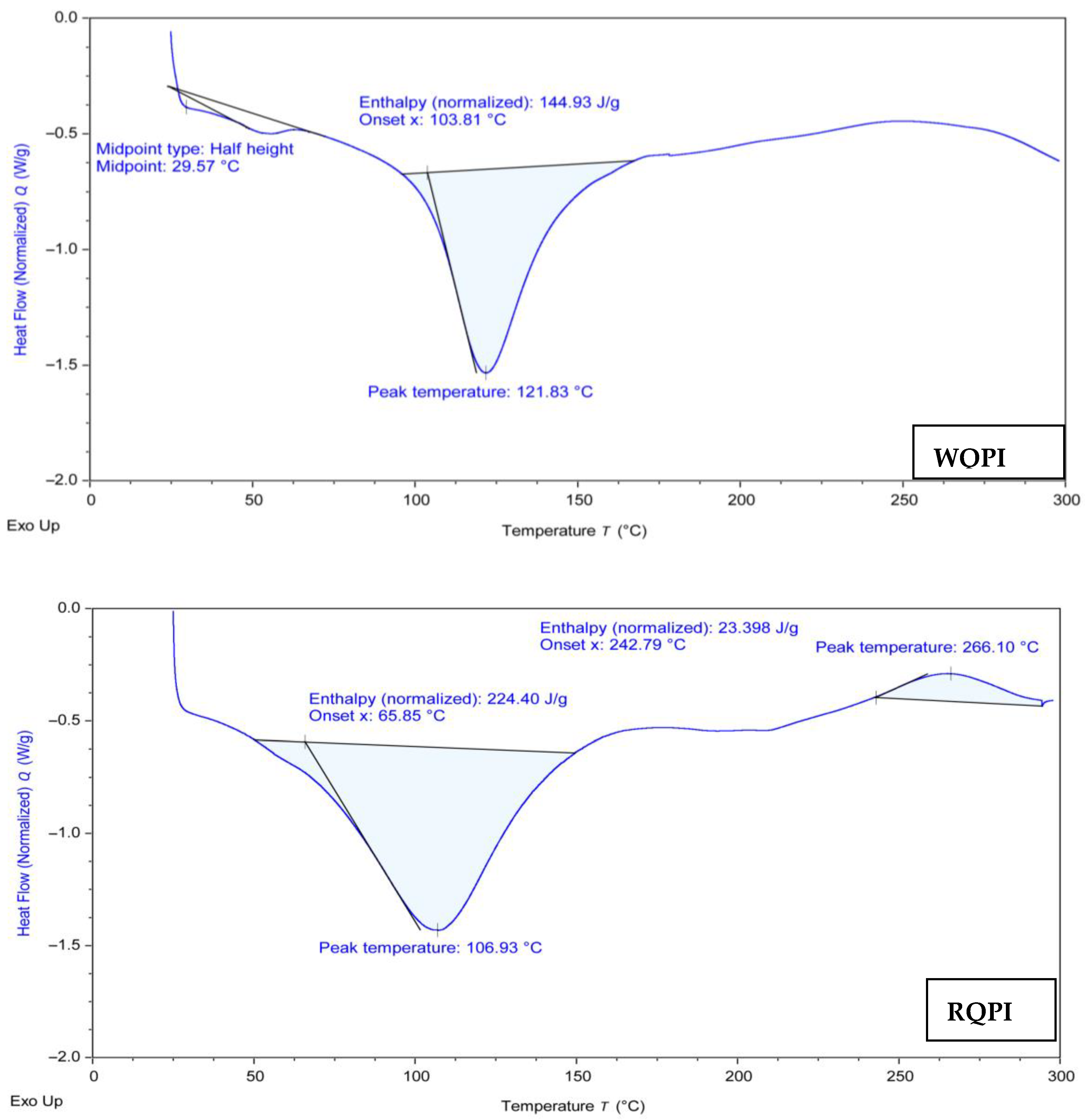
During the temperature scan from 0 to 300 °C, both RQPI and WQPI (Figure 2) showed a major endothermic peak at around 106.93 °C and 121.83 °C, respectively, indicating protein denaturation. However, RQPI exhibited an additional exothermic peak at 266.10 °C. These multiple denaturation peaks are attributed to the complex composition of quinoa albumin and globular chenopedin proteins [7]. Similar findings were reported by Shen et al. [39], who noted that the spray- and vacuum-dried proteins lacked lower temperature denaturation peaks, likely due to partial denaturation during drying. Notably, the spray- and vacuum-dried proteins showed significant exothermic peaks at 148 °C and 172 °C, respectively, attributed to protein aggregation upon heating [41]. Das et al. [31] highlighted that denaturation temperatures can be influenced by extraction methods; the proteins extracted at pH 8 to 10 exhibited an endothermic peak, whereas no peak was observed at pH 11 due to denaturation during extraction. Additionally, Shen et al. [39] stated that protein denaturation and aggregation transitions are irreversible processes. The exothermic peak observed in the red quinoa protein isolates suggests a higher level of protein aggregation compared to the white quinoa protein isolates. This difference in aggregation may imply that the red quinoa proteins undergo more extensive denaturation or unfolding, potentially due to their distinct amino acid composition and thermal stability [41]. Practically, this could affect the functional properties of the proteins, such as their solubility, emulsification, and gelation behavior, which are critical for applications in food systems [4].

3.7. Rheological Properties

3.7.1. Rotational Test

The rheological properties of protein isolates play a crucial role in their manufacturing processes, akin to polymers. Non-Newtonian behavior, especially viscoelastic effects, can lead to various processing instabilities [42]. Non-Newtonian materials, such as molten polymers, often exhibit pseudoplastic behavior, where viscosity varies with shear rate or extensional rate. Viscoelastic materials, like polymeric fluids, exhibit recoverable deformation due to the presence of polymer chains, with the stress response depending on the deformation history. Unlike purely viscous liquids, viscoelastic fluids generate normal stresses in shear flows, leading to unique phenomena like extrudate swell and vortex enhancement [43]. The higher-molecular-weight proteins or the more extensive protein–protein interactions in red quinoa could also create a denser protein matrix, thus increasing the resistance to flow and contributing to the higher viscosity observed.
Figure 3 depicts the typical flow and viscosity curves for the protein isolates. The flow behavior was modeled using the power law, and the viscosity (η [mPas]) at the shear rates (D = 50/S) is detailed in Table 3. The flow behavior index (p) showed a distinct decrease with the increasing shear rate (D = 1/S, D = 10/S, and D = 50/S), but at D = 100/S, p started to increase again, indicating significant changes. The viscosity decreased sharply with the increasing shear rates in both the WQPI and RQPI dispersions. As the shear rate increased sufficiently to overcome Brownian motion, the emulsion droplets aligned more along the flow direction, reducing the resistance and viscosity [44]. The pseudoplastic flow behavior observed in the protein dispersions is typical for proteins [2]. The higher viscosity of the RQPI and the lower values of the flow index suggest the formation of complex structures in the RQPI dispersions. The small, rigid particles of the red quinoa proteins likely contribute more effectively to viscosity [45]. In contrast, shear stress disrupts the aggregated structures in WQPI samples, resulting in pronounced shear thinning. Furthermore, structural factors that probably contribute to rheological behavior are molecular weight and protein size, the degree of protein unfolding and aggregation, and the surface hydrophobicity and charge distribution.

3.7.2. Oscillatory Test

An oscillatory test was conducted to analyze the effects of varying shear stress and strain on the storage modulus (G′) and the loss modulus (G″) of plant-based protein dispersions (WQPI and RQPI). The storage modulus (G′) (Figure 4 and Table 4) indicates the ability of a material to store energy elastically, reflecting its solid-like behavior, while the loss modulus (G″) represents the viscous component, where energy is dissipated as heat through friction [46]. All the samples exhibited linear curves showing degradation patterns in G′ and G″. The G′ values were consistently higher than the G″ across all the shear stress levels, indicating that the protein dispersions effectively stored energy. WQPI displayed the highest initial G′ values, starting at 54.863 Pa at 0.0056648 Pa shear stress, decreasing to 48.327 Pa at 0.0072065 Pa shear stress, and then increasing again to 54.029 Pa at 0.037278 Pa shear stress. Subsequently, the G′ gradually declined to a minimum of 1.2199 Pa at 2.1473 Pa shear stress. Conversely, RQPI initially showed higher G′ values, ranging from 171.07 to 99.249 Pa as the shear stress increased from 0.01719 to 0.70218 Pa, respectively. However, the RQPI’s G′ values dramatically dropped to a minimum of 1.1868 Pa at the highest shear stress (2.3458 Pa). Regarding G″ (loss modulus), both WQPI and RQPI followed similar patterns. WQPI exhibited maximum and minimum G″ values of 8.9093 Pa and 1.7479 Pa, respectively, with the median values increasing with the shear stress. In contrast, RQPI showed a peak G″ value of 31.166 Pa at a median shear stress of 0.27963 Pa, decreasing to a minimum of 2.0035 Pa at the highest shear stress (2.3458 Pa). This behavior highlights RQPI as superior in maintaining structural integrity under elevated shear stress, demonstrating favorable rheological properties compared to WQPI.
The oscillatory tests revealed that the storage modulus (G′) and loss modulus (G″) of the red and white quinoa protein isolates increased with frequency, exhibiting the typical properties of the protein. These results are consistent with earlier research showing that protein gels are viscoelastic [47,48]. However, in the whole frequency range that was examined, the red quinoa protein isolate showed greater G′ and G″ values than the white quinoa protein isolate. This implies that compared to its white counterpart, the red quinoa protein isolate formed more elastic and robust gels. The higher protein content and different amino acid composition in red quinoa could account for these differences, as proteins with higher hydrophobicity tend to form stronger networks through hydrophobic interactions [49,50].
The rheological properties of quinoa protein isolates have significant implications for food processing, as higher viscosity proteins can enhance the texture and stability of products like sauces, while facilitating better emulsification in dairy products alternatives [34]. Understanding the shear-thinning behavior of these proteins allows for optimized processing conditions, improving the efficiency of mixing and pumping during production [44].

4. Conclusions

In conclusion, the current work has shed light on the distinct characteristics and possible uses of red and white quinoa protein isolates in the food sector. The two varieties of quinoa protein isolates have advantageous functional characteristics that allow them to be used in a variety of dietary applications. A balanced profile of essential and non-essential amino acids, including those containing sulfur-like cysteine and methionine, is provided by red and white protein isolates. They are complete proteins because they include all nine of the required amino acids. They offer a high concentration of protein, which is rather uncommon for plant-based protein sources, and makes them a potent source of plant-based protein. Also, the results of this study indicate that the distinct functional properties of red and white quinoa protein isolates, such as their viscosity characteristics, can be strategically utilized in product formulations to enhance their texture, stability, and mouth-feel in various food applications, thereby improving the overall product quality. This understanding allows food manufacturers to tailor formulations more effectively, aligning product attributes with consumer preferences and functional requirements. Future research directions could focus on exploring the impact of various processing techniques, such as enzymatic treatments or different drying methods, on the functional properties of quinoa protein isolates, aiming to enhance their solubility and emulsification capacity for diverse food applications. Additionally, investigating the potential health benefits and bioactive properties of these isolates, as well as their interactions with other ingredients in formulations, could provide valuable insights for developing novel functional foods.

Author Contributions

Conceptualization, K.A.A., R.F.M. and M.M.A.E.-G.; methodology, K.A.A., M.M.A.E.-G. and R.F.M.; software, K.A.A., M.M.A.E.-G. and R.F.M.; validation, K.A.A., M.M.A.E.-G. and R.F.M.; formal analysis, K.A.A., M.M.A.E.-G. and R.F.M.; investigation, K.A.A., M.M.A.E.-G. and R.F.M.; resources K.A.A., M.M.A.E.-G. and R.F.M.; data curation, K.A.A., M.M.A.E.-G. and R.F.M.; writing—original draft preparation, K.A.A., M.M.A.E.-G. and R.F.M.; writing—review and editing, K.A.A., M.M.A.E.-G. and R.F.M.; visualization, K.A.A., M.M.A.E.-G. and R.F.M.; supervision, K.A.A., M.M.A.E.-G. and R.F.M.; project administration, K.A.A., M.M.A.E.-G. and R.F.M.; funding acquisition, K.A.A., M.M.A.E.-G. and R.F.M. All authors have read and agreed to the published version of the manuscript.

Funding

The researchers would like to thank the Deanship of Graduate Studies and Scientific Research at Qassim University for their financial support (QU-APC-2024-9/1).

Data Availability Statement

Data are contained within the article.

Conflicts of Interest

The author declares no conflicts of interest.

References

  1. Valencia-Chamorro, S.A. Quinoa: Overview. Encycl. Food Grains 2016, 1, 341–348. [Google Scholar]
  2. Abugoch, L.E.; Romero, N.; Tapia, C.A.; Silva, J.; Rivera, M.; Hamed, H.A.; Kobacy, W.; Mahmoud, E.A.; El-Geddawy, M.M.A.; Kang, S.; et al. Quinoa Proteins (Chenopodium quinoa Willd.) Fractionated by Ultrafiltration Using Ceramic Membranes: The Role of PH on Physicochemical and Conformational Properties. Food Chem. 2011, 10, 104700. [Google Scholar]
  3. Taylor, J.R.N.; Parker, M.L. Quinoa. In Pseudocereals and Less Common Cereals; Springer: Berlin/Heidelberg, Germany, 2002; pp. 93–122. [Google Scholar]
  4. Hamed, H.A.; Kobacy, W.; Mahmoud, E.A.; El-Geddawy, M.M.A. Looking for a Novel Vegan Protein Supplement from Faba Bean, Lupine, and Soybean: A Dietary and Industrial Standpoint. Plant Foods Hum. Nutr. 2024, 79, 90–97. [Google Scholar] [CrossRef]
  5. Abugoch, L.E.; Romero, N.; Tapia, C.A.; Silva, J.; Rivera, M. Study of Some Physicochemical and Functional Properties of Quinoa (Chenopodium Quinoa Willd) Protein Isolates. J. Agric. Food Chem. 2008, 56, 4745–4750. [Google Scholar] [CrossRef] [PubMed]
  6. Mondor, M.; Aksay, S.; Drolet, H.; Roufik, S.; Farnworth, E.; Boye, J.I. Influence of Processing on Composition and Antinutritional Factors of Chickpea Protein Concentrates Produced by Isoelectric Precipitation and Ultrafiltration. Innov. Food Sci. Emerg. Technol. 2009, 10, 342–347. [Google Scholar] [CrossRef]
  7. Dakhili, S.; Abdolalizadeh, L.; Hosseini, S.M.; Shojaee-Aliabadi, S.; Mirmoghtadaie, L. Quinoa Protein: Composition, Structure and Functional Properties. Food Chem. 2019, 299, 125161. [Google Scholar] [CrossRef]
  8. Acosta, C.; Carpio, C.; Vilcacundo, R.; Carrillo, W. Identification of Proteins Isolate from Amaranth (Amaranthus caudatus) by Sodium Dodecyl Sulfate-Polyacrylamide Gel Electrophoresis with Water and NaCl 0.1 m Solvents. Asian J. Pharm. Clin. Res. 2016, 9, 331–334. [Google Scholar]
  9. Cordero-De-Los-Santos, M.Y.; Osuna-Castro, J.A.; Borodanenko, A.; Paredes-López, O. Physicochemical and Functional Characterisation of Amaranth (Amaranthus hypochondriacus) Protein Isolates Obtained by Isoelectric Precipitation and Micellisation. Food Sci. Technol. Int. 2005, 11, 269–280. [Google Scholar] [CrossRef]
  10. Martínez, E.N.; Añón, M.C. Composition and Structural Characterization of Amaranth Protein Isolates. An Electrophoretic and Calorimetric Study. J. Agric. Food Chem. 1996, 44, 2523–2530. [Google Scholar] [CrossRef]
  11. Zhao, Y.; Zhao, B. Oxidative Stress and the Pathogenesis of Alzheimer’ s Disease. Oxid. Med. Cell. Longev. 2013, 2013, 316523. [Google Scholar] [CrossRef]
  12. Deighton, N.; Brennan, R.; Finn, C.; Davies, H.V. Antioxidant Properties of Domesticated and Wild Rubus Species. J. Sci. Food Agric. 2000, 80, 1307–1313. [Google Scholar] [CrossRef]
  13. Hossain, M.I.; Hossain, M.A. Determinants of Capital Structure and Testing of Theories: A Study on the Listed Manufacturing Companies in Bangladesh. Int. J. Econ. Financ. 2015, 7, 176–190. [Google Scholar] [CrossRef]
  14. Alsaleem, K.A.; Elfaruk, M.S.; Hammam, A.R.A. Estimation of Antioxidants Activity and Capacity to Capture Free Radical Using 2, 2-Diphenyl-1-Picrylhydrazyl (DPPH) Assay. Aust. J. Basic Appl. Sci. 2020, 14, 86–93. [Google Scholar]
  15. Piñuel, L.; Boeri, P.; Zubillaga, F.; Barrio, D.A.; Torreta, J.; Cruz, A.; Vásquez, G.; Pinto, A.; Carrillo, W. Production of White, Red and Black Quinoa (Chenopodium quinoa Willd Var. Real) Protein Isolates and Its Hydrolysates in Germinated and Non-Germinated Quinoa Samples and Antioxidant Activity Evaluation. Plants 2019, 8, 257. [Google Scholar] [CrossRef] [PubMed]
  16. Nemli, E.; Ozkan, G.; Gultekin Subasi, B.; Cavdar, H.; Lorenzo, J.M.; Zhao, C.; Capanoglu, E. Interactions between Proteins and Phenolics: Effects of Food Processing on the Content and Digestibility of Phenolic Compounds. J. Sci. Food Agric. 2024, 104, 2535–2550. [Google Scholar] [CrossRef]
  17. Chen, X.; Zhang, Y.; Cao, B.; Wei, X.; Shen, Z.; Su, N. Assessment and Comparison of Nutritional Qualities of Thirty Quinoa (Chenopodium Quinoa Willd.) Seed Varieties. Food Chem. X 2023, 19, 100808. [Google Scholar] [CrossRef]
  18. Carciochi, R.A.; Manrique, G.D.; Dimitrov, K. Optimization of Antioxidant Phenolic Compounds Extraction from Quinoa (Chenopodium quinoa) Seeds. J. Food Sci. Technol. 2015, 52, 4396–4404. [Google Scholar] [CrossRef] [PubMed]
  19. Kang, S.; Zhang, J.; Guo, X.; Lei, Y.; Yang, M. Effects of Ultrasonic Treatment on the Structure, Functional Properties of Chickpea Protein Isolate and Its Digestibility In Vitro. Foods 2022, 11, 880. [Google Scholar] [CrossRef]
  20. Tonon, R.V.; Pedro, R.B.; Grosso, C.R.F.; Hubinger, M.D. Microencapsulation of Flaxseed Oil by Spray Drying: Effect of Oil Load and Type of Wall Material. Dry. Technol. 2012, 30, 1491–1501. [Google Scholar] [CrossRef]
  21. TUNG, M.A. Rheology of Protein Dispersions. J. Texture Stud. 1978, 9, 3–31. [Google Scholar] [CrossRef]
  22. Repo-Carrasco, R.; Espinoza, C.; Jacobsen, S.-E. Nutritional Value and Use of the Andean Crops Quinoa (Chenopodium quinoa) and Kañiwa (Chenopodium pallidicaule). Food Rev. Int. 2003, 19, 179–189. [Google Scholar] [CrossRef]
  23. Ranhotra, G.S.; Gelroth, J.A.; Glaser, B.K.; Lorenz, K.J.; Johnson, D.L. Composition and Protein Nutritional Quality of Quinoa. Cereal Chem. 1993, 70, 303. [Google Scholar]
  24. Ogungbenle, H.N.; Oshodi, A.A.; Oladimeji, M.O. The Proximate and Effect of Salt Applications on Some Functional Properties of Quinoa (Chenopodium quinoa) Flour. Pak. J. Nutr. 2009, 8, 49–52. [Google Scholar] [CrossRef]
  25. Liu, M.; Zhu, K.; Yao, Y.; Chen, Y.; Guo, H.; Ren, G.; Yang, X.; Li, J. Antioxidant, Anti-Inflammatory, and Antitumor Activities of Phenolic Compounds from White, Red, and Black Chenopodium Quinoa Seed. Cereal Chem. 2020, 97, 703–713. [Google Scholar] [CrossRef]
  26. Tang, Y.; Zhang, B.; Li, X.; Chen, P.X.; Zhang, H.; Liu, R.; Tsao, R. Bound Phenolics of Quinoa Seeds Released by Acid, Alkaline, and Enzymatic Treatments and Their Antioxidant and α-Glucosidase and Pancreatic Lipase Inhibitory Effects. J. Agric. Food Chem. 2016, 64, 1712–1719. [Google Scholar] [CrossRef]
  27. Son, M.J.; Rico, C.W.; Nam, S.H.; Kang, M.Y. Effect of Oryzanol and Ferulic Acid on the Glucose Metabolism of Mice Fed with a High-Fat Diet. J. Food Sci. 2011, 76, H7–H10. [Google Scholar] [CrossRef]
  28. Russo, G.I.; Campisi, D.; Di Mauro, M.; Regis, F.; Reale, G.; Marranzano, M.; Ragusa, R.; Solinas, T.; Madonia, M.; Cimino, S.; et al. Dietary Consumption of Phenolic Acids and Prostate Cancer: A Case-Control Study in Sicily, Southern Italy. Molecules 2017, 22, 2159. [Google Scholar] [CrossRef]
  29. Gómez-Caravaca, A.M.; Segura-Carretero, A.; Fernandez-Gutierrez, A.; Caboni, M.F. Simultaneous Determination of Phenolic Compounds and Saponins in Quinoa (Chenopodium quinoa Willd) by a Liquid Chromatography–Diode Array Detection–Electrospray Ionization–Time-of-Flight Mass Spectrometry Methodology. J. Agric. Food Chem. 2011, 59, 10815–10825. [Google Scholar] [CrossRef]
  30. Kazantseva, V.V.; Goncharuk, E.A.; Fesenko, A.N.; Shirokova, A.V.; Zagoskina, N.V. Features of the phenolics’ formation in seedlings of different varieties of buckwheat (Fagopyrum Esculentum Moench). Agric. Biol. 2015, 50, 611–619. [Google Scholar] [CrossRef]
  31. Das, D.; Panesar, P.S.; Saini, C.S. PH Shifting Treatment of Ultrasonically Extracted Soybean Meal Protein Isolate: Effect on Functional, Structural, Morphological and Thermal Properties. Process Biochem. 2022, 120, 227–238. [Google Scholar] [CrossRef]
  32. Li, X.; Da, S.; Li, C.; Xue, F.; Zang, T. Effects of High-intensity Ultrasound Pretreatment with Different Levels of Power Output on the Antioxidant Properties of Alcalase Hydrolyzates from Quinoa (Chenopodium quinoa Willd.) Protein Isolate. Cereal Chem. 2018, 95, 518–526. [Google Scholar] [CrossRef]
  33. Mir, N.A.; Riar, C.S.; Singh, S. Physicochemical, Molecular and Thermal Properties of High-Intensity Ultrasound (HIUS) Treated Protein Isolates from Album (Chenopodium album) Seed. Food Hydrocoll. 2019, 96, 433–441. [Google Scholar] [CrossRef]
  34. Sun, C.; Gunasekaran, S. Effects of Protein Concentration and Oil-Phase Volume Fraction on the Stability and Rheology of Menhaden Oil-in-Water Emulsions Stabilized by Whey Protein Isolate with Xanthan Gum. Food Hydrocoll. 2009, 23, 165–174. [Google Scholar] [CrossRef]
  35. Malik, A.M.; Singh, A. Pseudocereals Proteins-A Comprehensive Review on Its Isolation, Composition and Quality Evaluation Techniques. Food Chem. Adv. 2022, 1, 100001. [Google Scholar] [CrossRef]
  36. Mapiour, M.; Amira, A. Critical Influences of Plasma PH on Human Protein Properties for Modeling Considerations: Size, Charge, Conformation, Hydrophobicity, and Denaturation. J. Compos. Sci. 2023, 7, 28. [Google Scholar] [CrossRef]
  37. Zhao, Y.; Yuan, Y.; Yuan, X.; Zhao, S.; Kang, Z.; Zhu, M.; He, H.; Ma, H. Physicochemical, Conformational and Functional Changes of Quinoa Protein Affected by High-Pressure Homogenization. LWT 2023, 173, 114343. [Google Scholar] [CrossRef]
  38. Pelgrom, P.J.M.; Boom, R.M.; Schutyser, M.A.I. Method Development to Increase Protein Enrichment During Dry Fractionation of Starch-Rich Legumes. Food Bioprocess Technol. 2015, 8, 1495–1502. [Google Scholar] [CrossRef]
  39. Shen, Y.; Tang, X.; Li, Y. Drying Methods Affect Physicochemical and Functional Properties of Quinoa Protein Isolate. Food Chem. 2021, 339, 127823. [Google Scholar] [CrossRef]
  40. Schutyser, M.A.I.; Pelgrom, P.J.M.; Van der Goot, A.J.; Boom, R.M. Dry Fractionation for Sustainable Production of Functional Legume Protein Concentrates. Trends Food Sci. Technol. 2015, 45, 327–335. [Google Scholar] [CrossRef]
  41. Goyal, M.; Chaudhuri, T.K.; Kuwajima, K. Irreversible Denaturation of Maltodextrin Glucosidase Studied by Differential Scanning Calorimetry, Circular Dichroism, and Turbidity Measurements. PLoS ONE 2014, 9, e115877. [Google Scholar] [CrossRef]
  42. Sánchez, H.A.C.; Jovanović, M.R.; Kumar, S.; Morozov, A.; Shankar, V.; Subramanian, G.; Wilson, H.J. Understanding Viscoelastic Flow Instabilities: Oldroyd-B and Beyond. J. Nonnewton. Fluid Mech. 2022, 302, 104742. [Google Scholar] [CrossRef]
  43. Schuller, T.; Fanzio, P.; Galindo-Rosales, F.J. Analysis of the Importance of Shear-Induced Elastic Stresses in Material Extrusion. Addit. Manuf. 2022, 57, 102952. [Google Scholar] [CrossRef]
  44. Zhu, Y.; Gao, H.; Liu, W.; Zou, L.; McClements, D.J. A Review of the Rheological Properties of Dilute and Concentrated Food Emulsions. J. Texture Stud. 2020, 51, 45–55. [Google Scholar] [CrossRef] [PubMed]
  45. Kierulf, A.; Whaley, J.; Liu, W.; Enayati, M.; Tan, C.; Perez-Herrera, M.; You, Z.; Abbaspourrad, A. Protein Content of Amaranth and Quinoa Starch Plays a Key Role in Their Ability as Pickering Emulsifiers. Food Chem. 2020, 315, 126246. [Google Scholar] [CrossRef]
  46. Kimbell, G.; Azad, M.A. 3D Printing: Bioinspired Materials for Drug Delivery. In Bioinspired and Biomimetic Materials for Drug Delivery; Elsevier: Amsterdam, The Netherlands, 2021; pp. 295–318. [Google Scholar]
  47. Kinsella, J.E.; Morr, C.V. Milk Proteins: Physicochemical and Functional Properties. Crit. Rev. Food Sci. Nutr. 1984, 21, 197–262. [Google Scholar] [CrossRef] [PubMed]
  48. Clark, A.H.; Kavanagh, G.M.; Ross-Murphy, S.B. Globular Protein Gelation—Theory and Experiment. Food Hydrocoll. 2001, 15, 383–400. [Google Scholar] [CrossRef]
  49. Damodaran, S. Amino Acids, Peptides and Proteins. Fennema’s Food Chem. 2008, 4, 425–439. [Google Scholar]
  50. Boye, J.; Zare, F.; Pletch, A. Pulse Proteins: Processing, Characterization, Functional Properties and Applications in Food and Feed. Food Res. Int. 2010, 43, 414–431. [Google Scholar] [CrossRef]
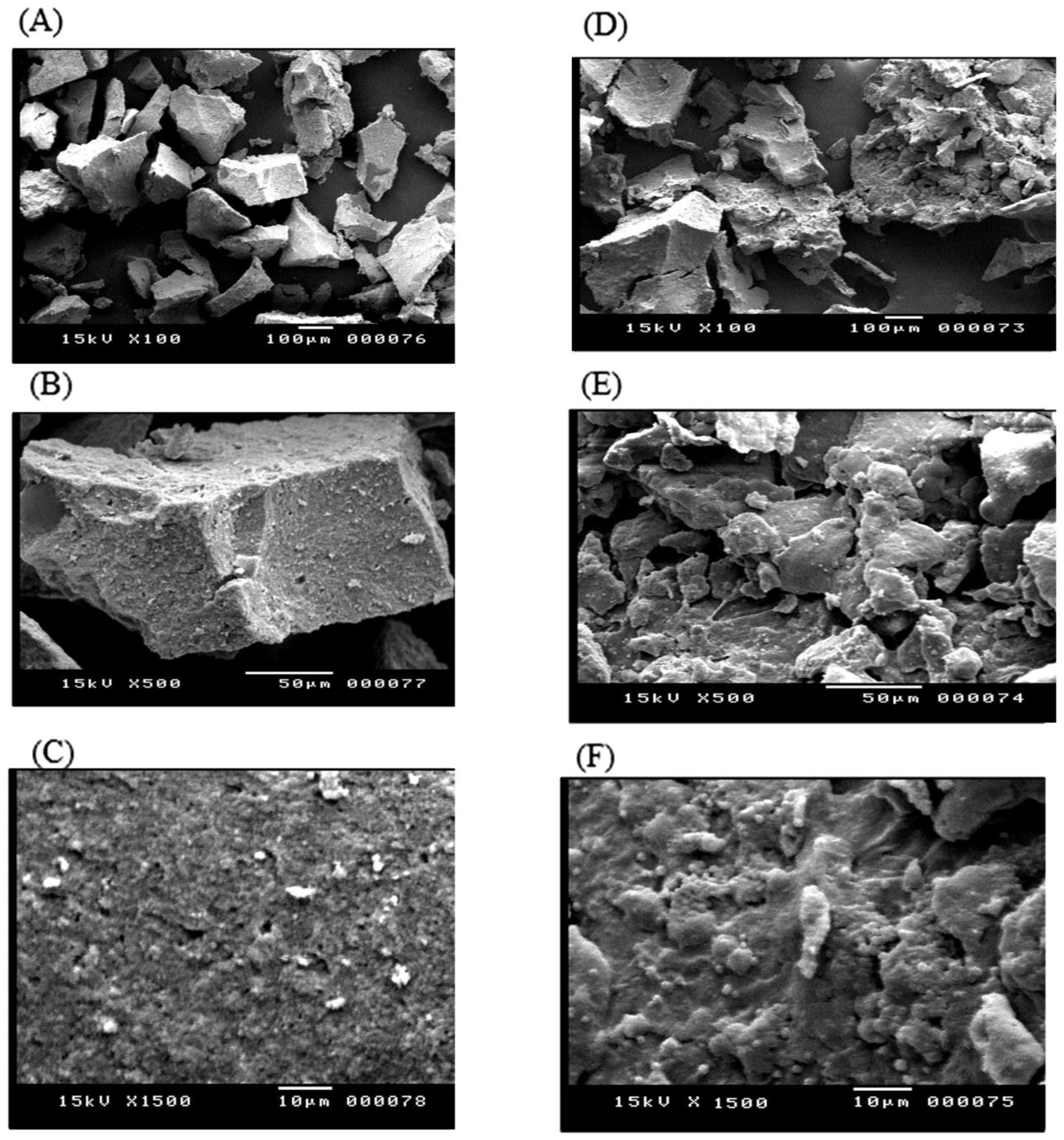
Figure 1. Microstructure of red quinoa protein isolate (RQPI) (AC) and white quinoa protein isolate (WQPI) (DF); (A,D), magnification of 100; (B,E), magnification of 500; (C,F), magnification of 1500.
Processes 12 02822 g001
Figure 2. Differential Scanning Calorimetry (DSC) of white quinoa protein isolate (WQPI) and red quinoa protein isolate (RQPI).
Processes 12 02822 g002
Figure 3. Flow and viscosity curves for white quinoa protein isolate (WQPI) and red quinoa protein isolate (RQPI).
Processes 12 02822 g003
Figure 4. Amplitude sweeps (G′ and G″) for white quinoa protein isolate (WQPI) and red quinoa protein isolate (RQPI).
Processes 12 02822 g004
Table 1. Amino acids (Av ± SD) of isolated protein from white quinoa protein isolate (WQPI) and red quinoa protein isolate (RQPI) (g/100 g) (based on dry weight).
Amino AcidsWQPI (g/100 g)RQPI (g/100 g)p-Value
Essential amino acidsAspartic13.244 ± 3.7312.685 ± 3.290.6
Phenylalanine2.825 ± 1.383.815 ± 1.150.47
Isoleucine6.871 ± 3.209.898 ± 3.080.03 *
Leucine2.750 ± 1.593.627 ± 1.230.52
Histidine10.520 ± 3.144.747 ± 1.630.00 **
Methionine4.813 ± 1.079.253 ± 3.180.00 **
Threonine5.903 ± 2.263.956 ± 1.240.16
Valine4.311 ± 1.417.957 ± 2.190.01 *
Lysine3.767 ± 1,563.380 ± 1.330.77
Non-essential amino acidsProline6.616 ± 1.353.160 ± 1.120.01 *
Tyrosine5.417 ± 2.618.777 ± 2.060.02 *
Cysteine5.59 ± 2.731.273 ± 0.220.00 **
Alanine3.811 ± 1.274.576 ± 1.570.57
Glutamic6.937 ± 1.116.210 ± 1.350.59
Arginine9.027 ± 2.715.917 ± 2.060.03 *
Glycine3.299 ± 1.082.126 ± 0.520.39
Serine6.849 ± 1.413.797 ± 1.310.03 *
TryptophanNot determined
The values in this table represent the average ± standard deviation of three independent replicates (n = 3). * p < 0.05, indicates significant differences, and ** p < 0.01, indicates highly significant differences.
Table 2. Phenols and flavonoids content (Av ± SD) in white quinoa protein isolate (WQPI) and red quinoa protein isolate (RQPI) (based on dry weight).
CompoundsWQPI Conc. (µg/g)RQPI
Conc. (µg/g)
p-Value
PhenolsGallic acid155.50 ± 12.97243.40 ± 12.650.00 **
Protocatechuic acidND17.83 ± 3.89
Cinnamic acid0.49 ± 0.121.28 ± 0.250.38
p-Hydroxybenzoic acid28.47 ± 4.7435.34 ± 5.290.000 **
Syringic acid5.27 ± 2.3223.99 ± 8.470.00 **
Ellagic acid0.000.000.00
Coumaric acid28.93 ± 5.2154.70 ± 9.350.00 **
Vanillin12.15 ± 3.1717.07 ± 3.110.00 **
Ferulic acid89.12 + 10.83116.54 ± 10.920.00 **
Total 319.93 ± 10.86510.15 ± 10.480.00 **
FlavonoidsNaringenin6.27 ± 8210.19 ± 2.560.00 **
Daidzein2.09 ± 1.622.49 ± 1.670.38
Catechin11.40 ± 5.476.59 ± 2.130.00 **
Rutin(30.1 + 7.52)39.67 ± 7.720.00 **
Quercetin8.86 ± 5.4820.08 ± 5.440.00 **
Total 58.72 ± 8.2270.02 ± 7.380.00 **
The values in this table represent the average ± standard deviation of three independent replicates (n = 3), and ** p < 0.01, indicates highly significant differences.
Table 3. Parameters that characterize the viscosity and the flow curve of white quinoa protein isolate (WQPI) and red quinoa protein isolate (RQPI).
Sampleη [m·Pa·s]pp2
WQPI47.7−0.6380.407044
RQPI99.8−0.4010.160801
η [m·Pa·s] = apparent viscosity at D50; p = flow behavior index.
Table 4. Parameters for the linear viscoelastic range of white quinoa protein isolate (WQPI) and red quinoa protein isolate (RQPI).
WQPIRQPI
G′ point (storage modulus) PaMaximum 54.863171.07
Minimum1.21991.1868
G″ point (loss modulus) PaMaximum8.909331.166
Minimum1.74792.0035
Disclaimer/Publisher’s Note: The statements, opinions and data contained in all publications are solely those of the individual author(s) and contributor(s) and not of MDPI and/or the editor(s). MDPI and/or the editor(s) disclaim responsibility for any injury to people or property resulting from any ideas, methods, instructions or products referred to in the content.