Article

Evaluation of the In Vitro Disinfection Potential of the Phytochemicals Linalool and Citronellal Against Biofilms Formed by Escherichia coli and Staphylococcus aureus

Faculty of Health Sciences, University of Ljubljana, Zdravstvena pot 5, 1000 Ljubljana, Slovenia
*
Author to whom correspondence should be addressed.
Processes 2024, 12(12), 2743; https://doi.org/10.3390/pr12122743
Submission received: 17 September 2024 / Revised: 22 November 2024 / Accepted: 25 November 2024 / Published: 3 December 2024
(This article belongs to the Special Issue Microbial Biofilms: Latest Advances and Prospects)

Abstract

:
This study aimed to analyze the potential of phytochemicals linalool and citronellal against E. coli and S. aureus biofilms and to compare the results to sodium hypochlorite. We tested the minimal inhibitory concentration, bacterial cell reduction, respiratory chain dehydrogenase activity, cell membrane integrity, and biomass reduction. The results show the lowest inhibitory concentration for both E. coli and S. aureus for sodium hypochlorite, followed by a combination of linalool and citronellal, the sole use of linalool, and the sole use of citronellal, respectively. Furthermore, we found that linalool was effective in biofilm cell reduction, cell respiratory inhibition, membrane integrity, and biomass reduction, while citronellal was less effective. Overall, this indicates that linalool has some benefits in biofilm management, especially with a focus on reducing toxic sodium hypochlorite consumption.

Graphical Abstract

1. Introduction

The use of biocides is the fundamental approach to infection control and general hygiene. However, their overuse, misuse, and abuse represent a risk for the emergence of antimicrobial resistance [1]. The typical representatives of multi-resistant bacteria are a group of ESKAPEE bacteria, or more specifically, Enterococcus faecium, Staphylococcus aureus, Klebsiella pneumoniae, Acinetobacter baumannii, Pseudomonas aeruginosa, Enterobacter spp., and Escherichia coli that are the major cause of nosocomial infections. Immunocompromised patients and those in intensive care units are at the highest risk for lung, blood, skin, and urinary tract infections [2]. The problem of antimicrobial resistance to disinfectants is not solely limited to hospitals but is due to the use of these compounds in everyday cleaning products, personal hygiene products, and laundry detergents shifted to the household environment [3]. Furthermore, disinfectants based on chlorine, glutaraldehyde, benzotriazole, and ammonia represent an important hazard to the aquatic environment [4,5,6]. In the disinfection process, chemicals react with water organic matter to form disinfection by-products, e.g., trihalomethanes, and haloacetic acids that are toxic to aquatic organisms and carcinogens [7]. Since 2020, due to the COVID-19 pandemic, disinfectant sales have increased globally, without proof of their effects, as a potential concern for environmental health [8]. Additionally, bacterial cells in the form of biofilms are much less susceptible to disinfectants like planktonic cells, which increases the risk of building bacterial resistance [9].
Therefore, in recent years, natural products based on plant extracts have gained significant interest among scientists and disinfectant professionals, especially as a safe, efficient, and non-resistant approach to biofilm management [10,11]. Due to the chemical and structural diversity of natural antimicrobials, it is difficult for bacteria to develop resistance to treatment [12]. Moreover, such chemicals with low molecular weight can easily penetrate the biofilm exopolysaccharide matrix [13].
Phytochemicals are compounds from plants that generally have biological activity, e.g., carotenoids, terpenoids, polyphenols, isoprenoids, and phytosterols, and most of them also possess antibacterial properties [14]. Generally, the antibacterial potential of natural compounds cannot be attributed to only one mechanism but to a combination of several of them [15]. For example, terpenoids’ main antibacterial effect is damage to fatty acids in the cell wall, alcohols cause cell protein denaturation and increase lipids solubility, while phenols damage the structure and permeability of cell membranes [16]. The most abundant and well-known terpenoids are linalool, citronellal, carvacrol, borneol, menthol, and others [17]. For instance, linalool (2,6-dimethyl-2,7-octadien-6-ol) is a terpene alcohol and is a major constituent of the essential oils of coriander (Coriandrum sativum), lavender (Lavandula officinalis), sweet basil (Ocimum basilicus), and others [18].
Linalool is reported to possess the function of inhibiting several bacterial strains, e.g., E. coli, S. enterica, S. aureus, and P. aeruginosa. It inhibits bacterial growth via the disruption of both the cell membrane and wall, releasing nucleic acids [19]. Linalool has been approved and is generally recognized as safe (GRAS) by the Food and Drug Administration [20]. Contrary to that, citronellal (3,7-Dimethyloct-6-enal) is a terpenoid aldehyde commonly found in lemongrass (Cymbopogon flexuosus), lemon-scented tea tree (Leptospermum petersonii), and lemon-scented gum (Corymbia citriodora) [21]. It shows the impact on bacterial cell change of the hydrophobicity, surface charge, and disruption of membrane integrity [22]. As a matter of fact, studies show that a combination of active components from different groups of phytochemicals can have additive or synergistic antibacterial potential [23,24], but less is known about how these phytochemicals affect bacterial biofilms.
Accordingly, the aim of this paper is to first (i) assess the minimum inhibitory concentration (MIC) of linalool, citronellal, and sodium hypochlorite (NaOCl) against E. coli and S. aureus; secondly (ii), to evaluate the effects of increasing the MICs on linalool, citronellal, and NaOCl on biofilm cell reduction and total biomass reduction; and finally (iii), to explain the mechanism of the anti-biofilm mode of action with cell dehydrogenase activity and integrity of cell membrane.

2. Materials and Methods

2.1. Materials

For the assessment, the reference strains of Escherichia coli ATCC 35218 and Staphylococcus aureus ATCC 25923 were obtained from the collection of the Faculty of Health Sciences. Bacteria were transferred on Tryptic Soy Agar (Merck Millipore, Darmstadt, Germany) and incubated at 37 °C for 24 h. After 24 h of incubation, a second subculture was prepared from the first in the same way and used for the experiments. The linalool, citronellal, dimethyl sulfoxide, resazurin, and iodonitrotetrazolium chloride were purchased at Sigma-Aldrich, Burlington, MO, USA. NaOCl, sodium chlorite, and crystal violet were obtained from Merck Millipore, Darmstadt, Germany. Hydrogen peroxide was obtained from Belinka, Slovenia. The BacLight® kit was obtained from Invitrogen®, Carlsbad, CA, USA, and the nutrient broth was purchased at Biolife, Milan, Italy.

2.2. Minimum Inhibitory Concentration (MIC)

The MIC of linalool, citronellal, and sodium hypochlorite against E. coli and S. aureus was analyzed using a microdilution method based on ISO 20776-1:2020 [25]. The overnight cultures of bacteria were harbored from Tryptic Soy Agar and suspended in 0.9% NaCl solution to achieve a turbidity of 0.5 McFarland. Subsequently, 0.5 mL of this bacterial solution was mixed with 9.5 mL of Nutrient broth. From this mixture, 100 μL was added to sterile flat-bottomed 96-well microplates.
Next, twofold dilutions of linalool and citronellal were prepared at concentrations ranging from 150 mg/mL to 0.15 mg/mL. The compounds were diluted in 2% (v/v) dimethyl sulfoxide in accordance with Clinical and Laboratory Standards Institute guidelines [26], while NaOCl was diluted in sterile distilled water. The positive control was 3% hydrogen peroxide, while the negative control was 2% dimethyl sulfoxide. The linalool and citronellal were combined in a 1:1 (v/v) ratio. The plates were incubated at 37 °C for 24 h. After incubation, 10 μL of 0.015% resazurin solution was added, and the plates were incubated again at 37 °C for 4 h. The MIC was determined as the lowest concentration of the linalool or citronellal that did not convert resazurin to resorufin [27].

2.3. Biofilm Cell and Biomass Reduction Assessment

Biofilm formation was assessed using both the plate counting method and the crystal violet assay [28]. Bacterial cultures of E. coli and S. aureus were diluted in a 0.9% NaCl solution to achieve a turbidity of 0.5 McFarland. Next, 0.5 mL of the bacterial suspension was combined with 9.5 mL of nutrient broth. Following this, 100 μL of the bacterial mixture was pipetted to sterile flat-bottomed 96-well microtiter plates and incubated at 37 °C for 24 h.
After incubation, the bacterial suspension aspired, and the biofilms that adhered to the microtiter plate surfaces were rinsed three times with 100 μL of PBS. The biofilms were then exposed for 60 min at room temperature to 1 MIC, 2 MIC, and 3 MIC concentrations of linalool and citronellal. The samples were washed thrice with 100 μL PBS to neutralize the active components and eliminate loosely adhering cells.
Biofilm biomass was quantified using a crystal violet assay. The cells on the surface of the microplate were stained with 2% crystal violet and washed with PBS to remove excess dye. The dye from the cells was then solubilized with 100 μL of 96% ethanol. The optical density of the resulting solution was measured at 620 nm using the Infinite 200 PRO microplate reader (Tecan, Gröding, Austria). The relative reduction for each concentration (in %) was calculated regarding the non-treated samples.
Simultaneously, the viability was evaluated by counting bacterial colonies. First, the linalool and citronellal were neutralized with PBS; after that, 100 μL of 0.9% NaCl was added to each well, and the samples were sonicated at 37 kHz and 200 W for 3 min to dislodge the cells from the well surfaces. Following serial dilutions, the samples were plated onto Tryptic Soy Agar and incubated at 37 °C for 24 h. The colonies were counted, and the results were expressed as log CFU/mL. The reduction in log CFU mL−1 was calculated regarding the non-treated samples.

2.4. Biofilm Cell Respiratory Chain Dehydrogenase Activity

Iodonitrotetrazolium chloride is a colorless stain that changes to red formazan dye in the presence of electrons of the respiratory chain and the coenzyme NADH in bacteria [29]. For this study, 100 μL of 0.5 mg/mL of iodonitrotetrazolium chloride was added into the treated and non-treated microtiter wells and incubated for 1 h at 37 °C. After that, 50 μL of dimethyl sulfoxide was added to dissolve the formazan crystal, and optical density at 492 nm [30] was measured using a Sunrise microplate reader (Tecan, Austria). The higher optical density of the formazan complex is considered a higher biofilm respiratory activity. The relative reduction for each concentration (in %) was calculated regarding the non-treated samples.

2.5. The Integrity of Bacterial Cell Membrane

The samples of biofilms exposed to linalool and citronellal were stained using a live/dead BacLight® kit [31]. The stain allows for the differentiation between cells with disrupted (red) and intact (green) membranes. The samples were stained and incubated at room temperature for 15 min. Next, a dye not bound to the bacteria was washed with PBS. Microscopy was performed using a Motic AE31 Elite inverted fluorescence microscope (Motic, San Antonio, TX, USA) at 200× magnification with fluorescence filters (TRIC, MB).

2.6. Statistical Analysis

Statistical analysis was conducted using R software version 4.1.1 (R Core Team, Vienna, Austria). Normality was assessed using the Shapiro–Wilk test (p > 0.05). One-way analysis of variance (ANOVA) and the Duncan test were employed to identify significant differences, with a significance level set at p < 0.05. All experiments were performed in triplicate, and the results are expressed as a mean +/− SD.

3. Results

3.1. Minimum Inhibitory Concentration

The results in Table 1 show the lowest MIC for NaOCl at 4.7 mg mL−1 for both E. coli and S. aureus, followed by linalool (9.4 mg mL−1 for E. coli and 18.8 mg mL−1 for S. aureus) and citronellal (37.5 mg mL−1 for E. coli and 18.8 mg mL−1 for S. aureus), respectively. However, we found that a combination of linalool and citronellal resulted in lower MICs for both tested strains at 4.7 mg mL−1.

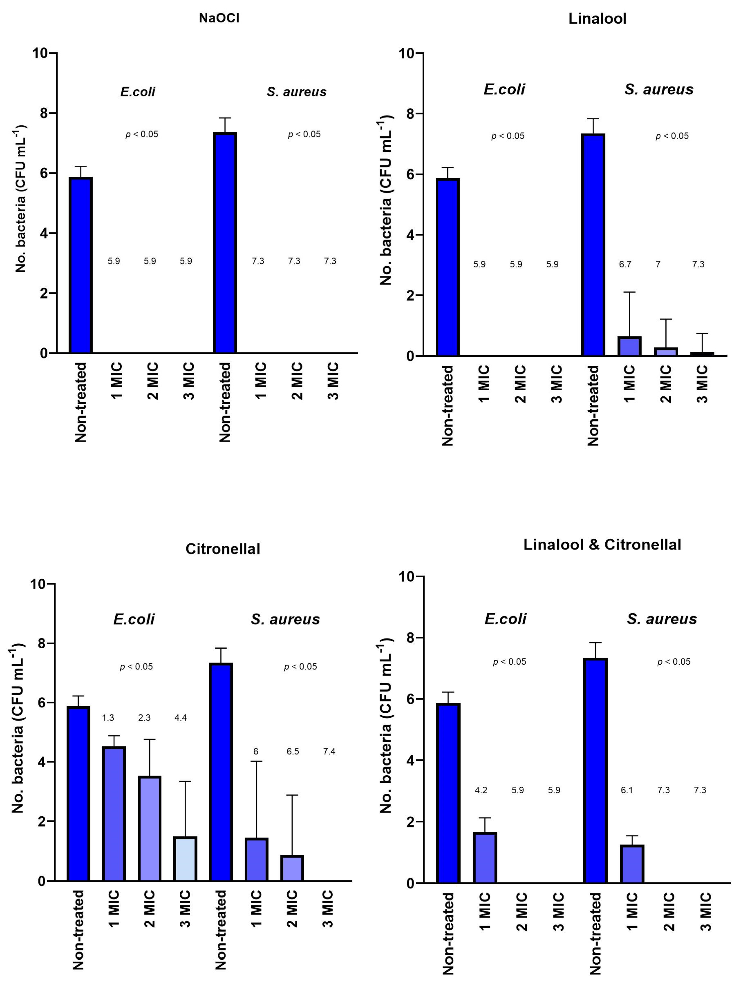
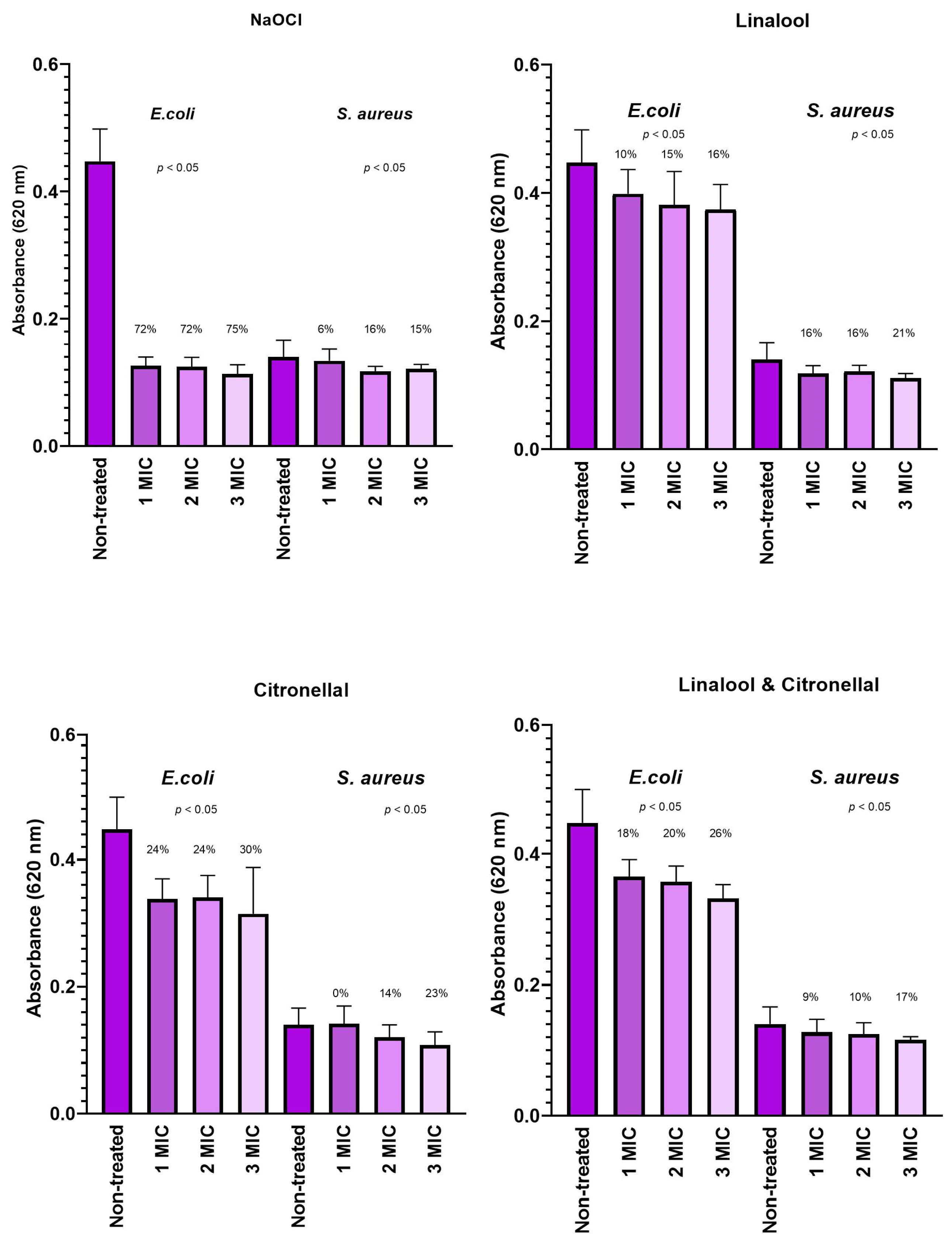
3.2. Biofilm Cell and Biomass Reduction Assessment

The results are displayed in Table S1. More detailed results of biofilm cell reduction demonstrate that NaOCl already at 1 MIC (4.7 mg mL−1) causes total reduction of both E. coli and S. aureus. A similar effect can be observed in the case of linalool, where total reduction was achieved for E. coli at 1 MIC (9.4 mg mL−1). Contrary to that, some S. aureus cells survived treatment with linalool, but the efficacy is increased by increasing concentration (Figure 1). Additionally, the results indicate that citronellal is less efficacious, reaching up to 4.4 log CFU mL−1 reduction for E. coli. In further detail, increasing the concentration of citronellal will increase the E. coli reduction (Figure 1). In the case of S. aureus, we observed a similar trend for linalool. The combination of linalool and citronellal did not significantly affect biofilm cell reduction (Figure 1).
The assessment of total biomass reduction using the crystal violet assay revealed that NaOCl can remove up to 75% (3 MIC) of total biofilm biomass in the case of E. coli, while the effect is less pronounced in the case of S. aureus (up to 15% for 3 MIC). The linalool can remove up to 16% of the total biomass of E. coli and up to 21% of S. aureus, respectively. Furthermore, citronellal can remove up to 30% of E. coli biomass and 23% of S. aureus biomass. A combination of linalool and citronellal can contribute to a 26% reduction in E. coli biomass and 17% of S. aureus biomass (Figure 2).

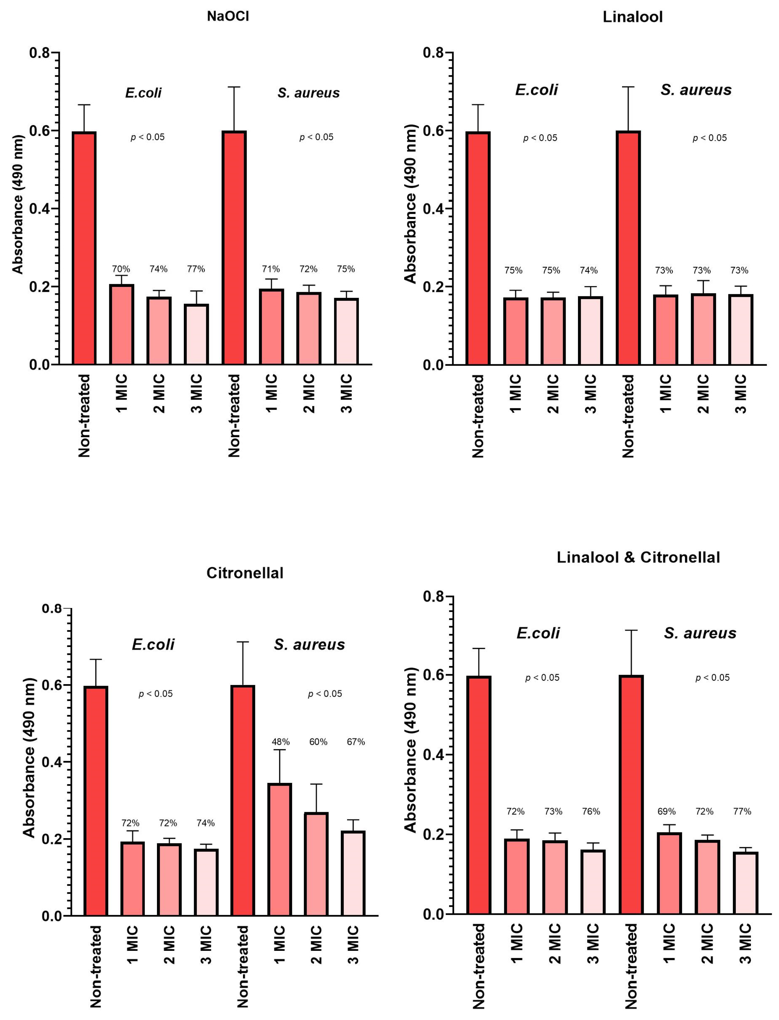
3.3. Biofilm Cell Respiratory Chain Dehydrogenase Activity

The results of bacteria cell respiratory chain dehydrogenase activity shown in the case of NaOCl demonstrate a reduction in activity up to 77% for E. coli and 75% for S. aureus, respectively. Also, the results indicate that increasing the concentration will decrease the respiratory chain activity (Figure 3). Similar effects and trends can be observed for linalool in the case of E. coli, where a reduction in respiratory chain activity of up to 75% can be observed. Citronellal treatment of E. coli and S. aureus shows similar results as for linalool, while less effect can be observed in the case of S. aureus. Combining linalool and citronellal will result in a similar trend (E. coli 76% and S. aureus 77%) as NaOCl or linalool (Figure 3).

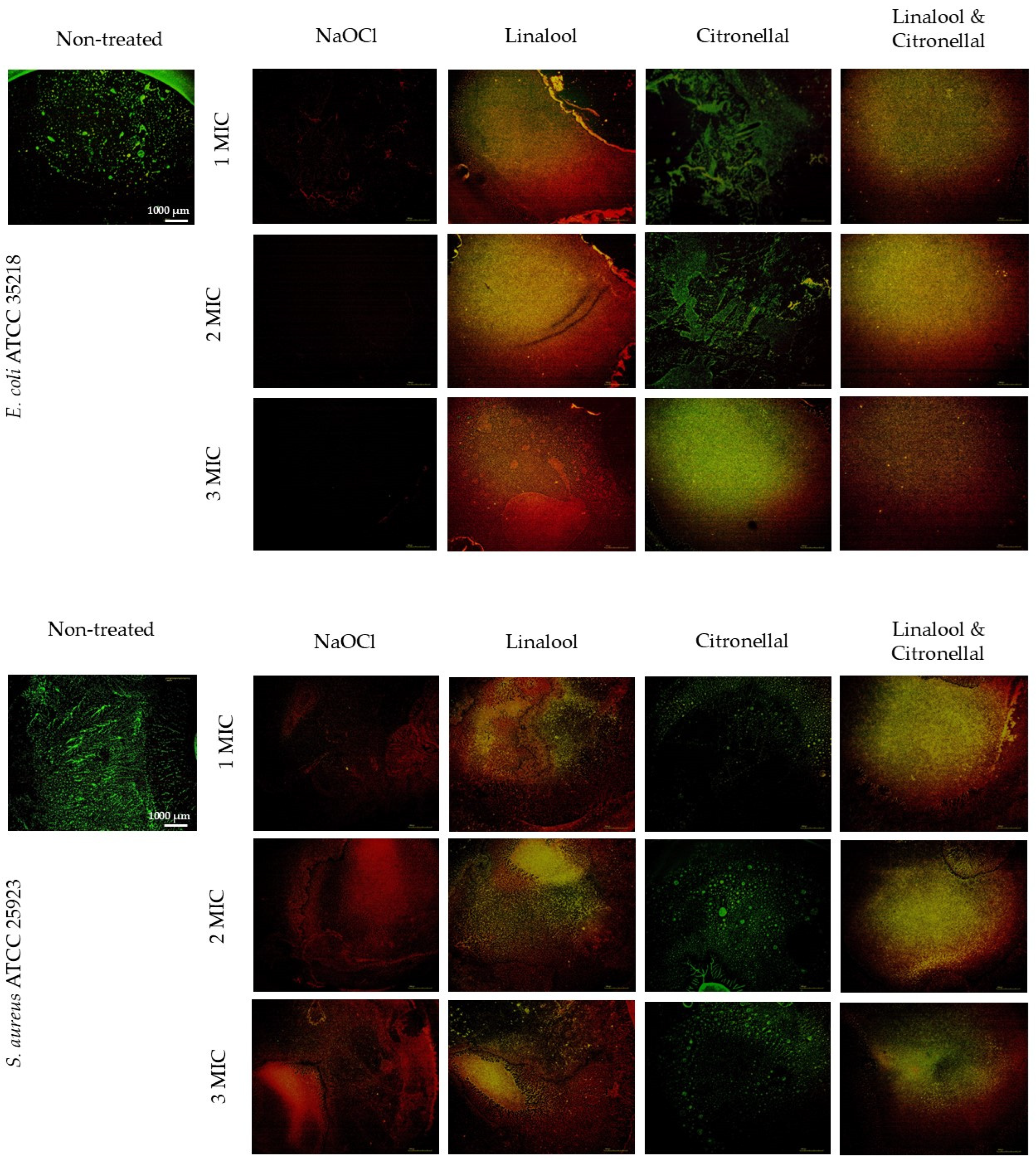
3.4. The Integrity of the Bacterial Cell Membrane

In addition, the membrane integrity assessment shows increased red (dead) cells relative to the control (green—live) by increasing the concentration of NaOCl, linalool, citronellal, and a combination of linalool and citronellal. More detailed results demonstrate the efficacy of NaOCl, linalool, and a combination of linalool and citronellal, while the sole use of citronellal is less effective. A similar trend can be observed for both tested strains (Figure 4).

4. Discussion

The management of bacterial populations on surfaces and the prevention of cross-resistance are fundamental components of the One Health strategy [32]. Therefore, approaches that are efficient in bacterial reduction while not increasing bacterial resistance gain much scientific attention [33]. Our study focused on E. coli as a typical representative of Gram-negative coliform bacteria that can form biofilms on vast materials in medicine, technology, and the domestic environment. Meanwhile, S. aureus is a Gram-positive bacteria that occupies human skin and can be transmitted via contaminated surfaces [13], making those two strains relevant models from a hygiene point of view.

4.1. Minimum Inhibitory Concentration

Our research demonstrated the lowest MIC for both E. coli and S. aureus for NaOCl at 4.7 mg mL−1. Similar to our study, researchers Fu et al. [34] tested the NaOCl of E. coli for cantaloupe disinfection and found the MIC was 3.2 mg mL−1, which is complementary to our findings. Moreover, Kim et al. [35] tested linalool against Methicillin-resistant S. aureus and found the MIC was 12.8, which is a bit low compared to our result of reference strain S. aureus (18.8 mg mL−1). We found the MIC of citronellal for E. coli was 37.5 mg mL−1, which is a bit higher than the results reported by Yang et al. [36] of citronellal MIC at 20 mg mL−1. One of the reasons for this could be that the authors diluted the citronellal in 0.2% bovine serum albumin, which can affect the antimicrobial efficiency [37].

4.2. Biofilm Cell and Biomass Reduction Assessment

The biofilm cell reduction approach is the fundamental information on the efficacy of the disinfection process. Our research showed NaOCl could effectively eradicate E. coli and S. aureus biofilms even at 1 MIC; a similar effect was observed in the case of E. coli for linalool, while some S. aureus cells survived the disinfection (6.7 and 7.3 log CFU mL−1 reduction, respectively). A study by Eriksson et al. [38] revealed that NaOCl at a concentration of 1.6 mg mL−1 can effectively eradicate S. aureus clinical strains isolated from the skin of patients. Our research demonstrated that NaOCl can eradicate reference strain biofilm on polystyrene at 1 MIC of 4.7 mg mL−1. Furthermore, Vázquez-Sánchez et al. [39] tested essential oils for S. aureus biofilm disinfection on various materials and suggested that linalool could be one of the important active components to possess an antibacterial impact. Contrary to linalool, our research revealed that citronellal is less effective against both E. coli and S. aureus. Exposing up to 3 MIC of citronellal on E. coli biofilm will result in a reduction of up to 4.4 CFU mL−1 and 3 MIC for S. aureus to achieve 7.4 log CFU mL−1. Results of combined linalool and citronellal show a good reduction in E. coli biofilm, although the sole use of linalool shows better results. A similar trend was also observed for S. aureus biofilm. The advantage of the crystal violet assay is that it assesses if the antimicrobials also remove the biomass of the biofilm, not only to destroy the cells. Results on biofilm biomass reduction show a good reduction in biofilm biomass in the case of NaOCl for E. coli (up to 75%), while for S. aureus, it was less effective, indicating the Gram-positive bacteria are less susceptive to NaOCl. Interestingly, both linalool, citronellal, and their combination were less effective for both E. coli and S. aureus biofilm biomass removal. In further detail, the results indicate that citronellal was more effective against E. coli than against S. aureus. Similar findings were reported by Borges et al. [40], pointing out that Gram-negative bacteria are more susceptible to citronellal than Gram-positive bacteria.

4.3. Biofilm Cell Respiratory Chain Dehydrogenase Activity

This approach allows us to study mechanisms of the anti-biofilm mode of action on the level of cell metabolism. The results of the respiratory chain assay demonstrate that both NaOCl and linalool cause a substantial decrease in cell respiration (>70%). In further detail, the results show that increasing the concentration of NaOCl or linalool will result in an increased reduction of respiratory activity. Guo et al. [41] tested linalool against P. fluorescens and found that 1 MIC causes a 76% reduction in respiratory chain activity, which is comparable to our results. Interestingly, citronellal had a similar effect on the E. coli (>70% reduction in activity) biofilm respiratory chain, while this effect was less predominant in the case of the S. aureus (up to 67% for 3 MIC) biofilm.

4.4. The Integrity of Bacterial Cell Membrane

Sousa et al. [42] also analyzed cell membrane integrity with dead/live staining and found that increasing the concentration of linalool will result in an increased fluorescence signal for damaged cells, which is in line with our dead/live assessment results [42]. Analyzing the damage to the cell membrane allows us to understand how antimicrobials act on bacterial cells. The results of the cell membrane integrity of our study also support the findings on biofilm biomass removal. The results show that NaOCl not only destroyed the membrane of E. coli but also removed the cells from the surface. However, this effect was less predominant in the case of S. aureus (Figure 4). Furthermore, Prakash and Vadivel [43] found that a combination of linalool and citronellal was more effective against L. monocitogenes biofilm than a single component. The efficacy of citronella’s component citronellal was also confirmed by Olszewska et al. [44], who conducted the live/dead staining and concluded that 30–50% of E. coli cells had moderately damaged cell membranes. Aelenei et al. [45] tested coriander essential oil with an abundance of linalool (>70%) and reported that linalool can alter the structural integrity of both Gram-negative and Gram-positive by increasing the permeability of the cell membranes. This finding also confirms our findings regarding biofilm cell reduction, the decrease in respiratory chain dehydrogenase activity, and membrane integrity.

5. Conclusions

This study demonstrated that terpene alcohol linalool and terpenoid aldehyde citronellal have an impact on E. coli and S. aureus biofilms. Moreover, we demonstrated that linalool reduces the number of E. coli comparable to NaOCl, while this effect is less predominant for S. aureus. Also, citronellal and a combination of citronellal and linalool was less effective. Furthermore, biomass reduction was most effectively achieved with NaOCl, followed by linalool and citronellal. The respiratory chain dehydrogenase activity and integrity of the cell membrane show that linalool disturbs the bacterial membrane and impairs cell metabolism. Above all, the results indicate that linalool has some benefits in biofilm management for both tested bacterial strains in the context of reduction, respiratory cell assessment, and membrane integrity. Overall, this indicates that linalool has potential for some applications in the food industry, medicine, and hygiene where less hazardous substances have advantages to more toxic sodium hypochlorite.

Supplementary Materials

The following supporting information can be downloaded at https://www.mdpi.com/article/10.3390/pr12122743/s1. Table S1: Results of statistical analysis.

Author Contributions

Conceptualization, R.F., M.O. and M.L.; methodology, R.F. and M.O.; software, R.F.; validation, R.F., P.K. and M.L.; formal analysis, P.K., M.L. and R.F.; investigation, P.K.; resources, R.F. and M.O.; data curation, P.K.; writing—original draft preparation, R.F., M.O., M.L. and P.K.; writing—review and editing, R.F., M.O., M.L. and P.K.; visualization, P.K.; supervision, R.F.; project administration, R.F.; funding acquisition, R.F. All authors have read and agreed to the published version of the manuscript.

Funding

This research received no external funding.

Data Availability Statement

The original contributions presented in this study are included in the article/Supplementary Materials. Further inquiries can be directed to the corresponding author.

Conflicts of Interest

The authors declare no conflicts of interest. The funders had no role in the design of the study; in the collection, analyses, or interpretation of data; in the writing of the manuscript; or in the decision to publish the results.

References

  1. Maillard, J.-Y.; Pascoe, M. Disinfectants and Antiseptics: Mechanisms of Action and Resistance. Nat. Rev. Microbiol. 2023, 22, 4–17. [Google Scholar] [CrossRef] [PubMed]
  2. Mulani, M.S.; Kamble, E.E.; Kumkar, S.N.; Tawre, M.S.; Pardesi, K.R. Emerging Strategies to Combat ESKAPE Pathogens in the Era of Antimicrobial Resistance: A Review. Front. Microbiol. 2019, 10, 539. [Google Scholar] [CrossRef] [PubMed]
  3. Fink, R.; Wang, Z.; Oder, M.; Brooks, B.W. Balancing Chemical Function with Reduced Environmental Health Hazards: A Joint Probability Approach to Examine Antimicrobial Product Efficacy and Mammalian Toxicity. J. Clean. Prod. 2020, 262, 121323. [Google Scholar] [CrossRef]
  4. Arya, S.; Gupta, S.; Thakur, S.; Mishra, A.K. Impacts of Sodium Hypochlorite on Humans and Environment. Int. J. Environ. Clim. Chang. 2023, 13, 3200–3217. [Google Scholar] [CrossRef]
  5. Di Marzio, W.D.; Cifoni, M.; Sáenz, M.E.; Galassi, D.M.; Di Lorenzo, T. The Ecotoxicity of Binary Mixtures of Imazamox and Ionized Ammonia on Freshwater Copepods: Implications for Environmental Risk Assessment in Groundwater Bodies. Ecotoxicol. Environ. Saf. 2018, 149, 72–79. [Google Scholar] [CrossRef]
  6. McGinley, H.R.; Enzien, M.; Hancock, G.; Gonsior, S.; Miksztal, M. Glutaraldehyde: An Understanding of Its Ecotoxicity Profile and Environmental Chemistry. Presented at the CORROSION 2009, Atlanta, GA, USA, 22–26 March 2009. NACE-09405. [Google Scholar]
  7. Zhang, H.; Tang, W.; Chen, Y.; Yin, W. Disinfection Threatens Aquatic Ecosystems. Science 2020, 368, 146–147. [Google Scholar] [CrossRef]
  8. Dewey, H.M.; Jones, J.M.; Keating, M.R.; Budhathoki-Uprety, J. Increased Use of Disinfectants during the COVID-19 Pandemic and Its Potential Impacts on Health and Safety. ACS Chem. Health Saf. 2021, 29, 27–38. [Google Scholar] [CrossRef]
  9. Ballén, V.; Cepas, V.; Ratia, C.; Gabasa, Y.; Soto, S.M. Clinical Escherichia coli: From Biofilm Formation to New Antibiofilm Strategies. Microorganisms 2022, 10, 1103. [Google Scholar] [CrossRef]
  10. Barbieri, R.; Coppo, E.; Marchese, A.; Daglia, M.; Sobarzo-Sánchez, E.; Nabavi, S.F.; Nabavi, S.M. Phytochemicals for Human Disease: An Update on Plant-Derived Compounds Antibacterial Activity. Microbiol. Res. 2017, 196, 44–68. [Google Scholar] [CrossRef]
  11. Mittal, R.P.; Rana, A.; Jaitak, V. Essential Oils: An Impending Substitute of Synthetic Antimicrobial Agents to Overcome Antimicrobial Resistance. Curr. Drug Targets 2019, 20, 605–624. [Google Scholar] [CrossRef]
  12. Álvarez-Martínez, F.J.; Barrajón-Catalán, E.; Micol, V. Tackling Antibiotic Resistance with Compounds of Natural Origin: A Comprehensive Review. Biomedicines 2020, 8, 405. [Google Scholar] [CrossRef] [PubMed]
  13. Fink, R. Good Hygiene Practices and Their Prevention of Biofilms in the Food Industry; Cambridge Scholars Publishing: Newcastle upon Tyne, UK, 2019; ISBN 1-5275-3677-7. [Google Scholar]
  14. Kumar, A.; P, N.; Kumar, M.; Jose, A.; Tomer, V.; Oz, E.; Proestos, C.; Zeng, M.; Elobeid, T.; K, S. Major Phytochemicals: Recent Advances in Health Benefits and Extraction Method. Molecules 2023, 28, 887. [Google Scholar] [CrossRef] [PubMed]
  15. da Silva, B.D.; Bernardes, P.C.; Pinheiro, P.F.; Fantuzzi, E.; Roberto, C.D. Chemical Composition, Extraction Sources and Action Mechanisms of Essential Oils: Natural Preservative and Limitations of Use in Meat Products. Meat Sci. 2021, 176, 108463. [Google Scholar] [CrossRef] [PubMed]
  16. Ju, J.; Chen, X.; Xie, Y.; Yu, H.; Guo, Y.; Cheng, Y.; Qian, H.; Yao, W. Application of Essential Oil as a Sustained Release Preparation in Food Packaging. Trends Food Sci. Technol. 2019, 92, 22–32. [Google Scholar] [CrossRef]
  17. Guimarães, A.C.; Meireles, L.M.; Lemos, M.F.; Guimarães, M.C.C.; Endringer, D.C.; Fronza, M.; Scherer, R. Antibacterial Activity of Terpenes and Terpenoids Present in Essential Oils. Molecules 2019, 24, 2471. [Google Scholar] [CrossRef]
  18. Herman, A.; Tambor, K.; Herman, A. Linalool Affects the Antimicrobial Efficacy of Essential Oils. Curr. Microbiol. 2016, 72, 165–172. [Google Scholar] [CrossRef]
  19. An, Q.; Ren, J.-N.; Li, X.; Fan, G.; Qu, S.-S.; Song, Y.; Li, Y.; Pan, S.-Y. Recent Updates on Bioactive Properties of Linalool. Food Funct. 2021, 12, 10370–10389. [Google Scholar] [CrossRef]
  20. Api, A.; Belsito, D.; Botelho, D.; Bruze, M.; Burton, G.; Buschmann, J.; Cancellieri, M.; Dagli, M.; Date, M.; Dekant, W. Update to RIFM Fragrance Ingredient Safety Assessment, Linalool, CAS Registry Number 78-70-6. Food Chem. Toxicol. 2022, 159, 112687. [Google Scholar] [CrossRef]
  21. Lenardao, E.J.; Botteselle, G.V.; de Azambuja, F.; Perin, G.; Jacob, R.G. Citronellal as Key Compound in Organic Synthesis. Tetrahedron 2007, 63, 6671–6712. [Google Scholar] [CrossRef]
  22. Lopez-Romero, J.C.; González-Ríos, H.; Borges, A.; Simões, M. Antibacterial Effects and Mode of Action of Selected Essential Oils Components against Escherichia coli and Staphylococcus aureus. Evid. Based Complement. Alternat. Med. 2015, 2015, 795435. [Google Scholar] [CrossRef]
  23. Gilling, D.H.; Ravishankar, S.; Bright, K.R. Antimicrobial Efficacy of Plant Essential Oils and Extracts against Escherichia coli. J. Environ. Sci. Health Part A 2019, 54, 608–616. [Google Scholar] [CrossRef] [PubMed]
  24. Yammine, J.; Chihib, N.-E.; Gharsallaoui, A.; Dumas, E.; Ismail, A.; Karam, L. Essential Oils and Their Active Components Applied as: Free, Encapsulated and in Hurdle Technology to Fight Microbial Contaminations. A Review. Heliyon 2022, 8, e12472. [Google Scholar] [CrossRef] [PubMed]
  25. EN ISO 20776-1:2020; Susceptibility Testing of Infectious Agents and Evaluation of Performance of Antimicrobial Susceptibility Test Devices—Part 1: Broth Micro-Dilution Reference Method for Testing the In Vitro Activity of Antimicrobial Agents Against Rapidly Growing Aerobic Bacteria Involved in Infectious Diseases (ISO 20776-1:2019, Including Corrected Version 2019-12). CEN-CENELEC Management Centre: Brussels, Belgium, 2020.
  26. Weinstein, M.P. Performance Standards for Antimicrobial Susceptibility Testing; Clinical and Laboratory Standards Institute: Wayne, PA, USA, 2021; ISBN 1-68440-104-6. [Google Scholar]
  27. Sarker, S.D.; Nahar, L.; Kumarasamy, Y. Microtitre Plate-Based Antibacterial Assay Incorporating Resazurin as an Indicator of Cell Growth, and Its Application in the in Vitro Antibacterial Screening of Phytochemicals. Methods 2007, 42, 321–324. [Google Scholar] [CrossRef] [PubMed]
  28. Bohinc, K.; Dražić, G.; Fink, R.; Oder, M.; Jevšnik, M.; Nipič, D.; Godič-Torkar, K.; Raspor, P. Available Surface Dictates Microbial Adhesion Capacity. Int. J. Adhes. Adhes. 2014, 50, 265–272. [Google Scholar] [CrossRef]
  29. Stiefel, P.; Schneider, J.; Amberg, C.; Maniura-Weber, K.; Ren, Q. A Simple and Rapid Method for Optical Visualization and Quantification of Bacteria on Textiles. Sci. Rep. 2016, 6, 39635. [Google Scholar] [CrossRef]
  30. Benov, L. Improved Formazan Dissolution for Bacterial MTT Assay. Microbiol. Spectr. 2021, 9, e01637-21. [Google Scholar] [CrossRef]
  31. Berney, M.; Hammes, F.; Bosshard, F.; Weilenmann, H.-U.; Egli, T. Assessment and Interpretation of Bacterial Viability by Using the LIVE/DEAD BacLight Kit in Combination with Flow Cytometry. Appl. Environ. Microbiol. 2007, 73, 3283–3290. [Google Scholar] [CrossRef]
  32. Collignon, P.J.; McEwen, S.A. One Health—Its Importance in Helping to Better Control Antimicrobial Resistance. Trop. Med. Infect. Dis. 2019, 4, 22. [Google Scholar] [CrossRef]
  33. Ghanbar, S.; Fumakia, M.; Ho, E.A.; Liu, S. A New Strategy for Battling Bacterial Resistance: Turning Potent, Non-Selective and Potentially Non-Resistance-Inducing Biocides into Selective Ones. Nanomed. Nanotechnol. Biol. Med. 2018, 14, 471–481. [Google Scholar] [CrossRef]
  34. Fu, Y.; Deering, A.J.; Bhunia, A.K.; Yao, Y. Biofilm of Escherichia coli O157: H7 on Cantaloupe Surface Is Resistant to Lauroyl Arginate Ethyl and Sodium Hypochlorite. Int. J. Food Microbiol. 2017, 260, 11–16. [Google Scholar] [CrossRef]
  35. Kim, S.-G.; Choi, M.-H.; Park, S.-N.; Kook, J.-K. Antimicrobial Effects of Linalool and α-Terpineol against Methicillin-Resistant Staphylococcus aureus Isolated from Korean. Int. J. Oral. Biol. 2013, 38, 51–54. [Google Scholar] [CrossRef]
  36. Yang, Z.; He, S.; Wei, Y.; Li, X.; Shan, A.; Wang, J. Antimicrobial Peptides in Combination with Citronellal Efficiently Kills Multidrug Resistance Bacteria. Phytomedicine 2023, 120, 155070. [Google Scholar] [CrossRef] [PubMed]
  37. Klančnik, A.; Piskernik, S.; Jeršek, B.; Možina, S.S. Evaluation of Diffusion and Dilution Methods to Determine the Antibacterial Activity of Plant Extracts. J. Microbiol. Methods 2010, 81, 121–126. [Google Scholar] [CrossRef] [PubMed]
  38. Eriksson, S.; Van der Plas, M.; Mörgelin, M.; Sonesson, A. Antibacterial and Antibiofilm Effects of Sodium Hypochlorite against Staphylococcus aureus Isolates Derived from Patients with Atopic Dermatitis. Br. J. Dermatol. 2017, 177, 513–521. [Google Scholar] [CrossRef]
  39. Vázquez-Sánchez, D.; Galvão, J.A.; Mazine, M.R.; Gloria, E.M.; Oetterer, M. Control of Staphylococcus aureus Biofilms by the Application of Single and Combined Treatments Based in Plant Essential Oils. Int. J. Food Microbiol. 2018, 286, 128–138. [Google Scholar] [CrossRef]
  40. Borges, A.; Lopez-Romero, J.; Oliveira, D.; Giaouris, E.; Simões, M. Prevention, Removal and Inactivation of Escherichia coli and Staphylococcus aureus Biofilms Using Selected Monoterpenes of Essential Oils. J. Appl. Microbiol. 2017, 123, 104–115. [Google Scholar] [CrossRef]
  41. Guo, F.; Chen, Q.; Liang, Q.; Zhang, M.; Chen, W.; Chen, H.; Yun, Y.; Zhong, Q.; Chen, W. Antimicrobial Activity and Proposed Action Mechanism of Linalool against Pseudomonas Fluorescens. Front. Microbiol. 2021, 12, 562094. [Google Scholar] [CrossRef]
  42. Sousa, L.G.; Castro, J.; Cavaleiro, C.; Salgueiro, L.; Tomás, M.; Palmeira-Oliveira, R.; Martinez-Oliveira, J.; Cerca, N. Synergistic Effects of Carvacrol, α-Terpinene, γ-Terpinene, ρ-Cymene and Linalool against Gardnerella Species. Sci. Rep. 2022, 12, 4417. [Google Scholar] [CrossRef]
  43. Prakash, A.; Vadivel, V. Citral and Linalool Nanoemulsions: Impact of Synergism and Ripening Inhibitors on the Stability and Antibacterial Activity against Listeria Monocytogenes. J. Food Sci. Technol. 2020, 57, 1495–1504. [Google Scholar] [CrossRef]
  44. Olszewska, M.A.; Gędas, A.; Simões, M. The Effects of Eugenol, Trans-Cinnamaldehyde, Citronellol, and Terpineol on Escherichia coli Biofilm Control as Assessed by Culture-Dependent and-Independent Methods. Molecules 2020, 25, 2641. [Google Scholar] [CrossRef]
  45. Aelenei, P.; Rimbu, C.; Guguianu, E.; Dimitriu, G.; Aprotosoaie, A.; Brebu, M.; Horhogea, C.; Miron, A. Coriander Essential Oil and Linalool–Interactions with Antibiotics against Gram-positive and Gram-negative Bacteria. Lett. Appl. Microbiol. 2019, 68, 156–164. [Google Scholar] [CrossRef] [PubMed]
Figure 1. E. coli and S. aureus number of cells in biofilm (log CFU mL−1) after exposure to 1 MIC, 2 MIC, and 3 MIC of NaOCl, linalool, citronellal, and a combination of linalool and citronellal (reduction in log CFU mL−1 above columns).
Processes 12 02743 g001
Figure 2. E. coli and S. aureus biofilm total biomass (absorbance 620 nm) after exposure to 1 MIC, 2 MIC, and 3 MIC of NaOCl, linalool, citronellal, and a combination of linalool and citronellal (reduction in % above columns).
Processes 12 02743 g002
Figure 3. E. coli and S. aureus biofilm cell respiratory chain dehydrogenase activity (absorbance 490 nm) after exposure to 1 MIC, 2 MIC, and 3 MIC of NaOCl, linalool, citronellal, and a combination of linalool and citronellal (reduction in % above the columns).
Processes 12 02743 g003
Figure 4. E. coli and S. aureus biofilm membrane integrity assessment after exposure to 1 MIC, 2 MIC, and 3 MIC of linalool, citronellal, and a combination of linalool and citronellal.
Processes 12 02743 g004
Table 1. The minimum inhibitory concentration of citronellal, linalool, and NaOCl (mg mL−1) against E. coli and S. aureus.
BacteriaCitronellalLinaloolCitronellal/LinaloolNaOCl
E. coli37.59.44.74.7
S. aureus18.818.84.74.7
Disclaimer/Publisher’s Note: The statements, opinions and data contained in all publications are solely those of the individual author(s) and contributor(s) and not of MDPI and/or the editor(s). MDPI and/or the editor(s) disclaim responsibility for any injury to people or property resulting from any ideas, methods, instructions or products referred to in the content.

