Article

New Carrageenan/2-Dimethyl Aminoethyl Methacrylate/Gelatin/ZnO Nanocomposite as a Localized Drug Delivery System with Synergistic Biomedical Applications

Department of Physical Sciences, Chemistry Division, Collage of Science, Jazan University, P.O. Box 114, Jazan 45142, Saudi Arabia
*
Author to whom correspondence should be addressed.
Processes 2024, 12(12), 2702; https://doi.org/10.3390/pr12122702
Submission received: 1 November 2024 / Revised: 26 November 2024 / Accepted: 27 November 2024 / Published: 30 November 2024

Abstract

:
In recent years, the development of multifunctional hydrogels has gained significant attention due to their potential in various biomedical applications, including antimicrobial, antioxidant, and anticancer therapies. By integrating biocompatible polymers and nanoparticles, these hydrogels can achieve enhanced activity and targeted therapeutic effects. In this study, carrageenan/2-dimethyl aminoethyl methacrylate/gelatin (CAR/DEMA/Gelt) composite hydrogel was synthesized using microwave radiation specifically for its efficiency in enhancing cross-linking and promoting uniform nanoparticle dispersion within the matrix. Zinc oxide (ZnO) nanoparticles were incorporated into the hydrogel to form the (CAR/DEMA/Gelt/ZnO) nanocomposite. The hydrogels were characterized using FT-IR, FE-SEM, XRD, TGA, and EDX, confirming successful cross-linking and structural integrity. The nanocomposite hydrogel exhibited more enhanced antimicrobial activity than the composite hydrogel against Gram-positive Staphylococcus aureus (S. aureus) and Bacillus subtilis (B. subtilis), with inhibition zones of 15 mm and 16 mm, respectively, while in case of the Gram-negative bacteria, Klebsiella pneumoniae (K. pneumoniae) and Escherichia coli (E. coli), the inhibition zones were 29 mm and 19 mm, respectively. In addition to the unicellular fungi, Candida albicans (C. albicans), the inhibition zone was 19 mm. Moreover, the nanocomposite showed anti-inflammatory activity comparable to those of Indomethacin and antioxidant activity, with an impressive IC50 value of 33.3 ± 0.05 µg/mL. In vitro cytotoxicity assays revealed significant anticancer activity. Against the MCF-7 breast cancer cell line, the CAR/DEMA/Gelt/ZnO nanocomposite showed 72.5 ± 0.02% cell viability, which decreased to 30.8 ± 0.01% after loading doxorubicin (DOX). Similarly, against the HepG2 liver cancer cell line, the free nanocomposite displayed 59.9 ± 0.006% cell viability, which depleted to 29.9 ± 0.005% when DOX was uploaded. This CAR/DEMA/Gelt/ZnO nanocomposite hydrogel demonstrates strong potential as a multifunctional platform for targeted biomedical applications, particularly in cancer therapy.

1. Introduction

Wounds created by burns are considered one of the main causes of death reported annually. Microbial infections on the injured site are another significant problem, as they prolong the healing of wounds and can be lethal for patients [1]. Antibiotic resistance has become an urgent global concern that requires effective treatment [2]. Thus, wound dressing must possess antibacterial or anti-fouling activities to prevent infections at the wound site. Inorganic-based nanocomposite hydrogels have exhibited promising results for the inhibition of bacteria [3].
Many biological reactions can produce toxic species, such as superoxide anions [4]. Hydroxyl radical is a dangerous reactive oxidative species (ROS) that is formed through the disintegration of the superoxide anion and can react with amino acids, proteins, and DNA [5]. Hydroxyl and amino groups in polymer chains, carrageenan, and gelatin can take part in free-radical scavenging and antioxidant activity [6].
Cancer continues to be a formidable foe in modern medicine, necessitating multifaceted treatment strategies to combat its pernicious progression [7]. In the hierarchy of cancer therapies, surgery often stands as the initial and foundational step in treatment protocols [8,9]. However, the reach towards remission extends beyond the operating room, encompassing adjuvant therapies such as chemotherapy and radiotherapy [10,11]. While these treatments exhibit potent anti-cancer effects, they are not without their drawbacks, as they inadvertently affect both malignant and healthy cells alike [12]. One of the most important challenges in the investigation of cancer is developing treatment plans that cause less damage to normal tissues and have the most effect on tumor cells [13,14]. Because of this, the idea of localized drug delivery systems has gotten a lot of interest [15,16,17]. Such systems offer the possibility of precise targeting, focusing on cancerous cells while leaving healthy ones. Also, making drug delivery systems that have more therapeutic functions, like anti-inflammatory and antioxidant actions, is an excellent approach to improving the general effectiveness of cancer treatment plans [18,19]. Doxorubicin, a potent chemotherapeutic agent, has demonstrated remarkable efficacy against cancer due to its capacity to prevent DNA replication and the development of cancer cells [20]. However, doxorubicin’s clinical utility is hindered by its dose-dependent toxicity, which causes side effects and the destruction of normal tissues [21]. The inclusion of doxorubicin in the hydrogel-based drug delivery system is a possible solution to this problem [22,23]. This strategy aims to accomplish controlled and targeted drug release, potentially enhancing the drug’s therapeutic effect while minimizing systemic adverse effects [24]. This innovation offers the potential to improve the safety and efficacy of doxorubicin as an anticancer therapy, offering cancer patients a more precise and localized approach.
Carrageenan, a polysaccharide derived from red seaweeds, has garnered considerable attention as a crucial component of drug delivery systems [25]. Its distinctive chemical structure, characterized by alternating units of galactose and 3,6-anhydrogalactose, offers a versatile matrix for encapsulating and releasing therapeutic agents [26]. Carrageenan’s biocompatibility, biodegradability, and ability to form hydrogels make it an attractive candidate for drug delivery applications [27,28]. Gelatin, a protein generated from the hydrolysis of collagen, represents another crucial component in our drug delivery system. Its unique biophysical properties, such as its ability to form stable hydrogels, make it an ideal candidate for biomedical applications [29]. Gelatin exhibits excellent biocompatibility, biodegradability, and low antigenicity, which are essential characteristics for drug delivery systems designed to interact with living organisms [30]. ZnO nanoparticles, with their unique properties such as biocompatibility, high selectivity, and enhanced cytotoxicity, may be promising anticancer agents. Zn is one of the cofactors of more than 300 mammalian enzymes involved in different cellular processes, such as oxidative stress, DNA replication, and cell cycle progression, and alterations in Zn levels in cancer cells can have deleterious effects. The selective localization of ZnO NPs towards cancer cells due to electrostatic interaction and selective cytotoxicity due to increased ROS in cancer cells show that ZnO NPs can be considered promising anticancer agents [31].
Microwave technology is used for copolymerization and crosslinking processes when making hydrogels because it can speed up reaction kinetics and make polymer networks more regular [32,33,34]. Microwaves speed up gelation and crosslinking by heating reactants in a controlled way and promoting efficient energy transfer [35]. This leads to hydrogels with controlled properties, better structural integrity, and possible uses in drug delivery fields [36] because they cut down on reaction times and improve accuracy [37,38].
Based on the previously mentioned information, we hypothesized that the combined use of CAR, DEMA, Gelatin, and ZnO NPS could have a synergistic effect on the different properties of the CAR/DEMA/Gelt/ZnO nanocomposite. To the best of our knowledge, although various efforts have been made to improve the basic properties of films, studies on the development of polysaccharide–protein-based active drug delivery systems are still limited. Therefore, in the present research, the aim was to synthesize a new biodegradable carrageenan, 2-Dimethylaminoethyl methacrylate, and gelatin hydrogel using ZnO nanoparticles (CAR/DEMA/Gelt/ZnO) as a new multifunctional material. After characterization by FT-IR, FE-SEM, XRD, TGA, and EDX analysis, the CAR/DEMA/Gelt/ZnO nanocomposite was investigated for antimicrobial, anti-inflammatory, antioxidant, and anticancer activities as well as drug delivery of the anticancer drug doxorubicin as a new platform for DOX formulation.

2. Materials and Methods

2.1. Materials

Carrageenan (CAR), specifically Gelcarin GP-812 (CAS: 9000-07-1), composed of repeating units of C12H16O15S2, was originally sourced from PhytoTechnology Labs. 2-Dimethylaminoethyl methacrylate (DEMA), denoted as C8H15NO2, stabilized with hydroquinone monomethyl ether for synthesis, was procured from Sigma-Aldrich, Massachusetts, USA (CAS: 2867-47-2) with a molar mass of 157.21 g/mol. Gelatin (Gelt) derived from porcine skin, known for its gel strength of 300 and categorized as Type A (CAS: 9000-70-8, Molecular Weight: 477.55), was also obtained from Sigma-Aldrich. 2,2′-Azobis(2-methylpropionitrile) (AIBN 98%) and (CH3)2C(CN)N=NC(CH3)2CN were procured from Sigma-Aldrich. Zinc oxide (ZnO) with nanoparticle sizes less than 50 nm, as determined by BET analysis, and purity exceeding 97%, was supplied by Sigma-Aldrich (CAS Number: 1314-13-2; Molecular Weight: 81.39). Doxorubicin (DOX), in the form of a 50 mg lyophilized powder, was acquired from Varun Medicals in Nagpur, Maharashtra. Nutrient agar and Sabouraud dextrose media, Dulbecco’s Modified Eagle’s Medium (DMEM), and Fetal Bovine Serum (FBS) were procured from Sigma-Aldrich Massachusetts, USA. The human breast cancer cell line, MCF-7, and the human liver cancer cell line, HepG2, were purchased from the National Cancer Institute (NCI), Cairo University, Egypt.

2.2. Preparation of CAR/DEMA/Gelt/ZnO

Carrageenan (2 g) and Gelt (2 g) were dissolved individually in 20 mL of distilled water. ZnO (0.01 g) was sonicated for 30 min in 10 mL of distilled water. DEMA (20 mL) was mixed with 10 mL of distilled water. All solutions were mixed in a glass beaker with continuous stirring, and the total volume adjusted to 100 mL. Dropwise addition of 4,4′-Azobis(4-cyanovaleric acid) dissolved in acetone started the reaction. The mixture was subjected to microwave heating using a Samsung MS28J5255UB/ST microwave oven operating at 2.45 GHz and an output power of 80 W. The microwave was put in a fume hood to prevent the accumulation of acetone vapor using microwave vials (20 mL Round Bottom Vials with septa). One-third of its volume was left empty. The reaction mixture was heated until boiling commenced (approximately 240 s) and then allowed to cool to ambient temperature. This cycle, lasting approximately 10 min (240 s heating and 360 s cooling), was repeated six times over a duration of one hour to ensure complete copolymerization. The formed nanocomposite was immersed in excess deionized H2O, which was refreshed every several hours to remove unreacted compounds for 24 h. The formed nanocomposite was left in the oven at 40 °C for 24 h. The above steps were repeated without adding ZnO to obtain CAR/DEMA/Gelt hydrogel as a reference hydrogel. The gel content percentages in CAR/DEMA/Gelt and CAR/DEMA/Gelt/ZnO were calculated to be 95% and 98%, respectively. The gel content was calculated using the following steps: weigh the dried hydrogel sample (Wdry, initial) and immerse the sample in bi-distilled water at 70 °C for 24 h to remove any uncrosslinked or soluble polymer fractions. After this extraction, thoroughly wash the sample with the same solvent to remove residual soluble materials, then dry it again under the same conditions as before until a constant weight (Wdry, after extraction) is achieved. Calculate gel content using the following formula:
G e l   c o n t e n t % = W d r y ,   a f t e r   e x t r a c t i o n W d r y , i n i t i a l × 100

2.3. Drug Loading to Hydrogel Nanocomposite

CAR/DEMA/Gelt/ZnO/DOX was obtained by immersing 1.0 g of CAR/DEMA/Gelt/ZnO in a solution containing 0.1 g of DOX and 100 mL of distilled H2O (1 mg/mL) for 12 h with stirring at room temperature, and the drug-loaded nanocomposite was washed well with distilled water and air dried at room temperature until a constant weight. The concentration of DOX in the supernatant was calculated spectrophotometrically at 483 nm. The loaded drug percentage in the nanocomposite was calculated using Equation (1):
D r u g   l o a d i n g %     = I n i t i a l   D O X   c o n c e n t r a t i o n D O X   c o n c e n t r a t i o n   i n   t h e   s u p e r n a t a n t I n i t i a l   D O X   c o n c e n t r a t i o n   ×   100
DOX loading was found to be 87% (0.87 mg/mL).

2.4. Fourier-Transform Infrared Spectroscopy (FT-IR)

The primary functional groups in ZnO nanoparticles, DOX, CAR/DEMA/Gelt, CAR/DEMA/Gelt/ZnO, and CAR/DEMA/Gelt/ZnO/DOX were characterized using FT-IR. The analysis was conducted using a Bruker Unicom infrared spectrometer from Germany. the spectra were generated at 4000–400 cm−1, 4 cm−1 spectral resolutions.

2.5. X-Ray Powder Diffraction

Malvern Panalytical-Model Aeris Research spectrometer (Almelo, The Netherlands) was used for X-ray powder diffraction analysis. The dried films of CAR/DEMA/Gelt and CAR/DEMA/Gelt/ZnO, as well as ZnO alone, were subjected to X-ray powder diffraction (XRD) with 2θ angle range of 10–60° and a step angle of 0.2θ.

2.6. Thermal Gravimetric Analysis (TGA)

The thermal stabilities of CAR/DEMA/Gelt and CAR/DEMA/Gelt/ZnO nanocomposite hydrogels were assessed through TGA. This analysis was conducted by a Shimadzu TGA-30 instrument (Japan) while maintaining a nitrogen atmosphere. The temperature range for the examination spanned from ambient temperature up to 600 °C, with a heating rate of 10 °C/min.

2.7. Field Emission Scanning Electron Microscopy (FE-SEM)

The surface morphologies of CAR/DEMA/Gelt, CAR/DEMA/Gelt/ZnO, and CAR/DEMA/Gelt/ZnO/DOX nanocomposite hydrogels were examined using the FE-SEM system, specifically the FEI Quanta TM 3D FEG dual beam SEM/FIB system from Hillsboro, OR, USA. To enhance sample conductivity, a thin layer of gold was applied to the samples before observation.

2.8. Energy Dispersive X-Ray Spectroscopy (EDX)

EDX mapping, conducted with a JSM-7500F EDX instrument from JEOL (Japan), was employed to analyze the elemental composition and spatial distribution within CAR/DEMA/Gelt and CAR/DEMA/Gelt/ZnO nanocomposite hydrogels.

2.9. Microbial Cultures and In Vitro Antimicrobial Assays

Both CAR/DEMA/Gelt and CAR/DEMA/Gelt/ZnO were subjected to an antimicrobial activity test using the agar diffusion technique [39]. The antibacterial activity of the samples was assessed against the Gram-positive strains S. aureus (ATCC 6538) and B. subtilis (ATCC 6633), as well as the Gram-negative strains K. pneumoniae (ATCC 13883) and E. coli (ATCC 8739), in addition to the unicellular fungi C. albicans (ATCC 10221).
Microbial cultures: Gram-positive and Gram-negative bacteria were cultured at 37 °C for 24 h in nutrient (synthetic media), while C. albicans was cultured at 37 °C for 72 h on Sabouraud dextrose medium. By appropriately diluting with sterile saline (0.9%) solution, the cultures of bacteria and C. albicans containing 105 CFU/mL were prepared and used for the antimicrobial test.
In vitro antimicrobial assay: The antimicrobial activity of the prepared hydrogel films was tested using the agar diffusion method. The nutrient agar culture plates were prepared, in which 100 µL of bacterial suspension, prepared in the previous section, were added. For C. albicans, a Sabouraud dextrose agar (SDA) culture plate was prepared, in which 100 µL of fungal suspension was added. Equal weights (discs of 1 g) of the prepared hydrogel, with an original concentration of (50 mg/mL), were subjected to UV sterilization for 20 min at a distance of 12 cm and then placed on the culture surface of the microbial plates. Blank plates without hydrogel films were prepared for both bacteria and C. albicans cultures for comparison. The Petri dishes were transferred to an incubator at 37 °C for 24 h for bacteria and 72 h for C. albicans. The diameter of the inhibition zone around each disc was measured and recorded.

2.10. Anti-Inflammatory Assay

Inhibition of hemolysis of the HRBC membrane induced by heat or hypotonicity has been associated with anti-inflammatory activity. The assay procedure, according to previous reports [40,41,42], involves the following:
(a) Preparation of Erythrocyte Suspensions: Fresh whole blood (3 mL) was collected from normal volunteers into heparinized tubes and then centrifuged at 3000 rpm for 10 min, and the red blood pellets resuspended in normal saline (0.9%) equivalent to that of the supernatant. The volume of the red blood cell pellet solution was noted, and then it was reconstituted as a 40% v/v suspension in an isotonic buffer solution of pH 7.4 made of 10 mM sodium phosphate buffer, which contained 0.2 g of NaH2PO4, 1.15 g of Na2HPO4, and 9 g of NaCl in a liter of distilled water. The reconstituted red blood cell suspension was used for the assay.
(b) Hypotonicity-Induced Hemolysis: A CAR/DEMA/Gelt/ZnO nanocomposite hydrogel sample was prepared in distilled water to prepare a known stock solution. Duplicate pairs of centrifuge tubes were prepared for each concentration of the sample (ranging from 100 to 1000 µg/mL) in both hypotonic (in water) and isotonic solutions. A similar series of Indomethacin (an anti-inflammatory drug) was also prepared as a standard. The control tube contained distilled water only. To each tube, 0.1 mL of erythrocyte suspension was added and the tubes mixed slowly. These mixtures were incubated at 37 °C for 1 h and then centrifuged for 3 min at 1300 rpm. The absorbance (OD) of the hemoglobin in the supernatant was measured at 540 nm using a spectrophotometer (Milton Roy, Golden, CO, USA). The hemolysis percent was obtained by assuming that the hemolysis produced in the presence of distilled water was 100%. The hemolysis inhibition percentage of the nanocomposite sample was measured as follows:
I n h i b i t i o n   o f   h a e m o l y s i s   ( % ) = 1 O D 2 O D 1 O D 3 O D 1 × 100
where OD1 is the absorbance of the test sample in an isotonic solution, OD2 is the absorbance of the test sample in a hypotonic solution, and OD3 is the absorbance of the control tube.

2.11. Antioxidant Assay

The antioxidant activity of CAR/DEMA/Gelt/ZnO was assessed through the DPPH radical scavenging test [43]. A 0.1 mM solution of DPPH (1,1-diphenyl-2-picryl hydrazyl) in ethanol was prepared. A stock solution (5 mg/mL) of the nanocomposite film was made in ethanol. Serial dilutions (3.9, 7.8, 15.62, 31.25, 62.5, 125, 250, 500, 1000 µg/mL) were prepared. A 15 µL volume of each hydrogel solution was mixed with 1 mL of DPPH (0.1 mM) and then the volume was made up to 3 mL with ethanol. The mixture was vigorously mixed and put in a dark chamber for 30 min at ambient temperature. The color of DPPH changed from deep violet to light yellow according to the scavenging activity of the film solutions. Following this, absorbance was measured at 517 nm using a spectrophotometer (UV-VIS Milton Roy, Golden, CO, USA). A standard ascorbic acid (5 mg/mL) was used for each experiment in parallel, which was carried out in triplicate. The IC50 value, which represents the concentration of the material that inhibits 50% of the DPPH free radicals, was determined by a logarithmic dose inhibition curve. The lower the absorbance, the higher the free radical scavenging activity. The percentage of DPPH scavenging activity was calculated as follows:
%   s c a v e n g i n g   a c t i v i t y = A 0 A 1 A 0 × 100
where A0 and A1 are the absorbances of the control reaction and the standard sample, respectively.

2.12. In Vitro Cytotoxicity on Cancer Cells

The MTT (3-[4,5-dimethylthiazol-2-yl]-2,5 diphenyl tetrazolium bromide) assay is based on the conversion of MTT into formazan crystals by living cells, which determines mitochondrial activity. Since for most cell populations, the total mitochondrial activity is related to the number of viable cells, this assay is used to measure the in vitro cytotoxic effects of the nanocomposites CAR/DEMA/Gelt/ZnO and CAR/DEMA/Gelt/ZnO/DOX on two cell lines: human breast cancer cell line (MCF-7) and human liver cancer cell line HepG2). Initially, CAR/DEMA/Gelt/ZnO and CAR/DEMA/Gelt/ZnO/DOX (6 mm diameter, 2 mm thick) were subjected to UV sterilization for 20 min at a distance of 12 cm. Subsequently, the cancer cells (1 × 10⁵ cells/mL) were seeded on the hydrogel surface, 1 mL per well, in 12-well tissue culture plates with DMEM supplemented with 10% FBS and incubated at 37 °C for 48 h in 5% CO2 to develop a complete monolayer sheet. Upon the formation of a confluent sheet of cells, the growth medium was removed and the cell monolayer underwent two washes with wash media. MTT solution was prepared (5 mg/mL in PBS) (BIO BASIC CANADA INC) and 100 µL of it was added to each well and mixed by stirring in a gyratory shaker (150 rpm) for 5 min. The plate was then incubated at 37 °C with 5% CO2 for 4 h to facilitate MTT metabolism. Finally, 200 µL of DMSO was added with stirring in a gyratory shaker for 5 min to suspend the formazan, a metabolic product of MTT. The medium with cells was considered the control (100%). Optical density measurements were taken at 560 nm, with background subtraction at 620 nm [44,45,46]. The optical density (OD) was measured at 560 nm to determine cell viability according to the following equation:
C e l l   v i a b i l i t y   % = O D t e s t O D b l a n k O D c o n t r o l O D b l a n k
where ODtest, ODcontrol, and ODblank are the optical densities of cells incubated with hydrogel, DMEM with cells, and DMEM without cells, respectively.

2.13. Statistical Analysis

The anti-inflammatory, antioxidant, and cytotoxicity results were statistically analyzed using one-way ANOVA. Differences among average values were analyzed by Duncan’s multiple range tests using IBM SPSS software version 24 as a statistical resource at p < 0.05.

3. Results and Discussion

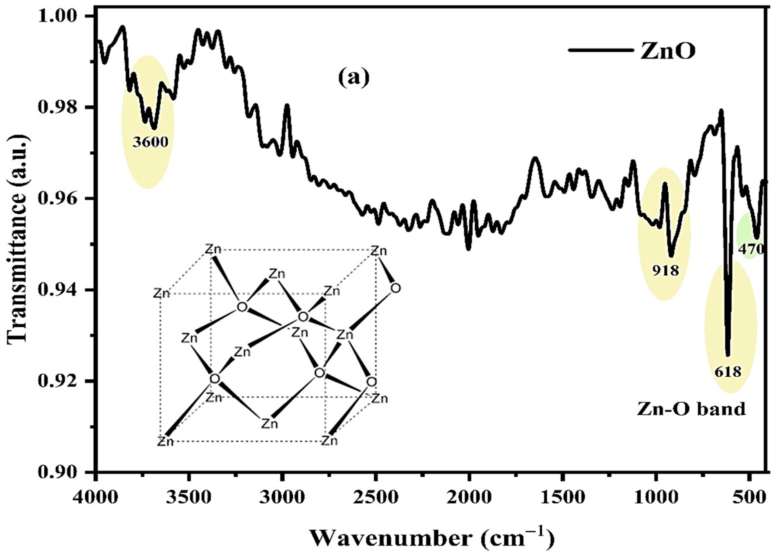
3.1. Fourier-Transform Infrared (FTIR) Analysis

FTIR analysis conducted on the pure ZnO nanoparticles, depicted in Figure 1a, yielded significant insights. Evidently, conspicuous peaks at 470 cm−1 and 618 cm−1 were identified, indicative of lattice vibration modes inherent to the ZnO crystal structure. This resonance is likely attributed to the vibrational motion of Zn–O bonds within the crystal lattice, serving as a distinct marker affirming the presence of ZnO nanoparticles [47]. Furthermore, another noteworthy peak at 918 cm−1 emerged, suggesting a plausible association with diverse vibrational modes intrinsic to the ZnO lattice. These modes may encompass vibrations linked to oxygen vacancy-related defects or surface perturbations [48]. Furthermore, the presence of a peak around 3600 cm−1 (O-H stretching) indicates the existence of residual hydroxyl groups, likely originating from atmospheric moisture [49,50]. In the case of the FTIR spectrum of pure DOX, as illustrated in Figure 1b, a series of well-defined peaks at precise wavenumbers, specifically 1622 cm−1, 1460 cm−1, 1436 cm−1, 1066 cm−1, 1014 cm−1, 926 cm−1, and 873 cm−1 are conspicuous. These distinctive peaks are due to the carbonyl groups within DOX, encompassing both quinone and ketone carbonyls [51]. However, delineating these spectral bands into their respective quinone and ketone constituents is a challenge, given the shared carbonyl moieties of these functional groups, leading to spectral overlap [52]. Additionally, the spectrum exhibits a broad band of the hydroxyl group at 3500 cm−1. Peaks at 3262 cm−1 and 3172 cm−1 are attributed to N–H stretching vibrations, while peaks at 2935 cm−1 are ascribed to C–H vibrations originating from alkane groups. Turning to Figure 1c, which presents the FTIR spectra of CAR/DEMA/Gelt hydrogel, CAR/DEMA/Gelt/ZnO nanocomposite, and CAR/DEMA/Gelt/ZnO/DOX, intriguing observations emerged. In the CAR/DEMA/Gelt hydrogel spectrum, distinct peaks were discernible at 3336 cm−1 (signifying the O–H stretching mode), 2972 cm−1 (indicative of C–H stretching vibrations), 1719 cm−1 (associated with C=O stretching), 1579 cm−1 (representative of COO-carboxyl anion vibrations), and 1149 cm−1 (linked to the SO2 group). These spectral features are attributed to the constituents of carrageenan, dimethyl aminoethyl methacrylate, and gelatin present within the hydrogel matrix. Notably, the intensity of these peaks increased in the CAR/DEMA/Gelt/ZnO nanocomposite spectrum, with a new peak at 910 cm−1 indicating the presence of ZnO nanoparticles. Additionally, in the CAR/DEMA/Gelt/ZnO/DOX spectrum, a new peak emerged at 1050 cm−1, attributed to doxorubicin’s carbonyl groups, reflecting changes in vibrational characteristics due to its incorporation. Notably, a noteworthy trend was observed, where the intensity of peaks increased in the sequence CAR/DEMA/Gelt hydrogel < CAR/DEMA/Gelt/ZnO nanocomposite < CAR/DEMA/Gelt/ZnO/DOX. This intensity variation was due to a combination of factors, including the incorporation of ZnO nanoparticles, the heightened surface area, chemical interactions, and the introduction of DOX into the composite [53].

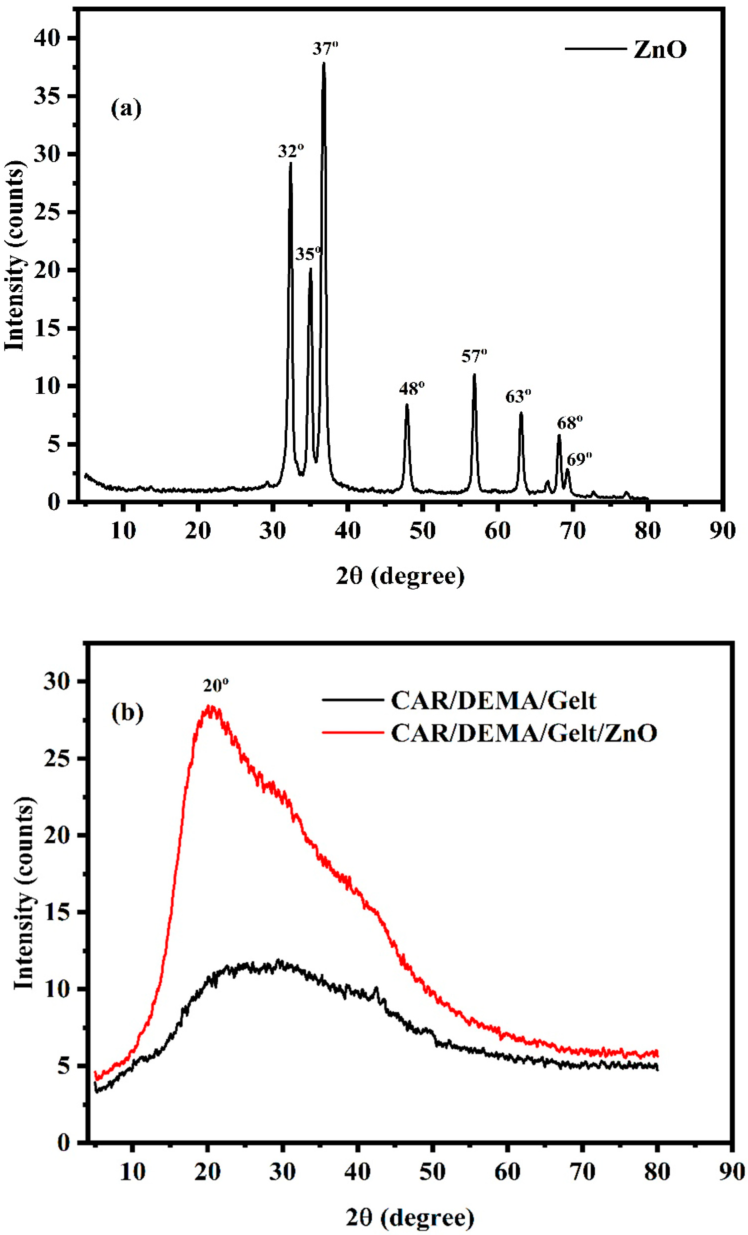
3.2. X-Ray Diffraction (XRD)

Figure 2a presents the XRD pattern of pure ZnO nanoparticles, revealing a distinctive pattern marked by diffraction peaks at 32°, 35°, 37°, 48°, 57°, 63°, 68°, and 69° corresponding to polycrystalline nature possessing planes along (100), (002), (101), (102), (110), (103), and (112) [54]. These peaks have been meticulously characterized as belonging to the hexagonal wurtzite phase of ZnO [55]. Intriguingly, when examining the XRD patterns of CAR/DEMA/Gelt hydrogel and CAR/DEMA/Gelt/ZnO nanocomposite showcased in Figure 2b, notable differences emerged. For CAR/DEMA/Gelt hydrogel, a broad XRD diffraction pattern lacking crystalline peaks was observed, centered on 2θ = 30°. The absence of distinct peaks indicates the amorphous nature of the hydrogel, in agreement with the intermolecular forces present between its constituent molecules, which influence the deformation and crystallinity of polymer chains [56,57]. The XRD pattern of CAR/DEMA/Gelt/ZnO nanocomposite displays a remarkable increase in peak intensity. The presence of ZnO nanoparticles within the biopolymer matrix augments the overall crystalline character of the resultant nanocomposite [58]. These findings not only shed light on the structural features of our materials but also provide valuable insights into the compatibility between ZnO nanoparticles and the polymer matrix, helping our understanding of their properties and potential applications [59].

3.3. Thermal Stability of the Synthesized Hydrogel and the Nanocomposite

TGA and DTA were utilized to evaluate the thermal stability of CAR/DEMA/Gelt hydrogel and CAR/DEMA/Gelt/ZnO nanocomposite hydrogel, as shown in Figure 3. Notably, both CAR/DEMA/Gelt hydrogel and CAR/DEMA/Gelt/ZnO nanocomposite hydrogel exhibited nearly the same thermograms with a three-stage profile of thermal degradation. At 216 °C, the first decomposition stage was due to the elimination of humidity and bonded water [60]. At 288 °C, the second decomposition stage, which corresponds to the elimination of side groups, emerged [61]. At 435 °C, the third and most important decomposition stage marked the degradation of the polymer matrix backbone [62]. It should be mentioned that the percentage of ZnO nanoparticles represents 0.01% of the total concentration of the CAR/DEMA/Gelt/ZnO nanocomposite. In terms of thermal stability, this bit concentration is ineffective. Interestingly, the final residues of CAR/DEMA/Gelt/ZnO nanocomposite were greater than CAR/DEMA/Gelt hydrogel by 5%, primarily as a result of the incorporation of ZnO NPs [63]. These data revealed that including ZnO nanoparticles in the hydrogel matrix may result in a more cohesive and stable structure, with some hydrogel components remaining as a weight residue.

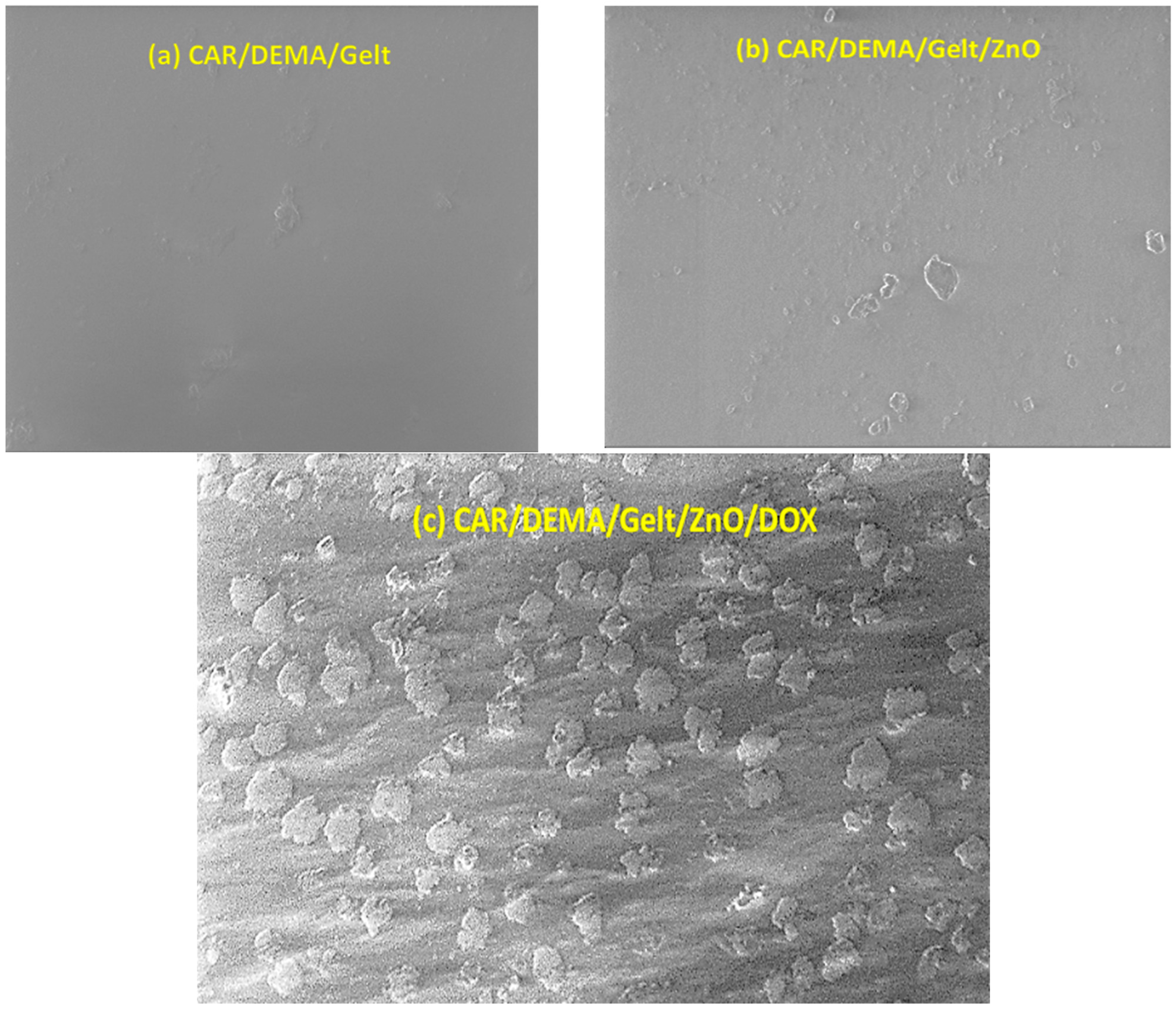
3.4. Surface Morphology and Structure of the Hydrogel and the Nanocomposite

Figure 4 displays FE-SEM images of CAR/DEMA/Gelt hydrogel, CAR/DEMA/Gelt/ZnO nanocomposite, and CAR/DEMA/Gelt/ZnO/DOX nanocomposite. These images provide valuable insights into the surface morphology and structure of the nanocomposites. In Figure 4a of the CAR/DEMA/Gelt hydrogel, the SEM image reveals a smooth and relatively uniform surface lacking any significant roughness or agglomerated structures. This characteristic surface morphology is attributed to the composition of the hydrogel components, which create a cohesive and homogeneous matrix. For the CAR/DEMA/Gelt/ZnO nanocomposite (Figure 4b), the surface remains smooth, similar to the CAR/DEMA/Gelt hydrogel with some spots of ZnO. This suggests that the incorporation of ZnO nanoparticles into the hydrogel matrix does not significantly alter the surface morphology, maintaining a relatively smooth and uniform appearance. However, a notable transformation in surface texture is observed in Figure 4c of the CAR/DEMA/Gelt/ZnO/DOX nanocomposite. The SEM image of this nanocomposite shows a distinct rough surface with evident agglomerated structures. This surface roughness is a consequence of the incorporation of doxorubicin (DOX) into the nanocomposite. DOX, with its crystalline structure and potential chemical interactions with other components, leads to the formation of rough and agglomerated regions on the surface [64]. The transition from a smooth surface on the CAR/DEMA/Gelt hydrogel and the CAR/DEMA/Gelt/ZnO nanocomposite to a rough and agglomerated surface on the CAR/DEMA/Gelt/ZnO/DOX nanocomposite underscores the significant impact of DOX on the nanocomposite’s surface morphology. The presence of these surface irregularities can enhance the surface area available for drug release, allowing for a more efficient interaction between the nanocomposite and its surrounding environment. In drug delivery systems, surface roughness often plays a pivotal role, enabling controlled and sustained drug release by providing additional binding sites and facilitating diffusion processes [65,66].

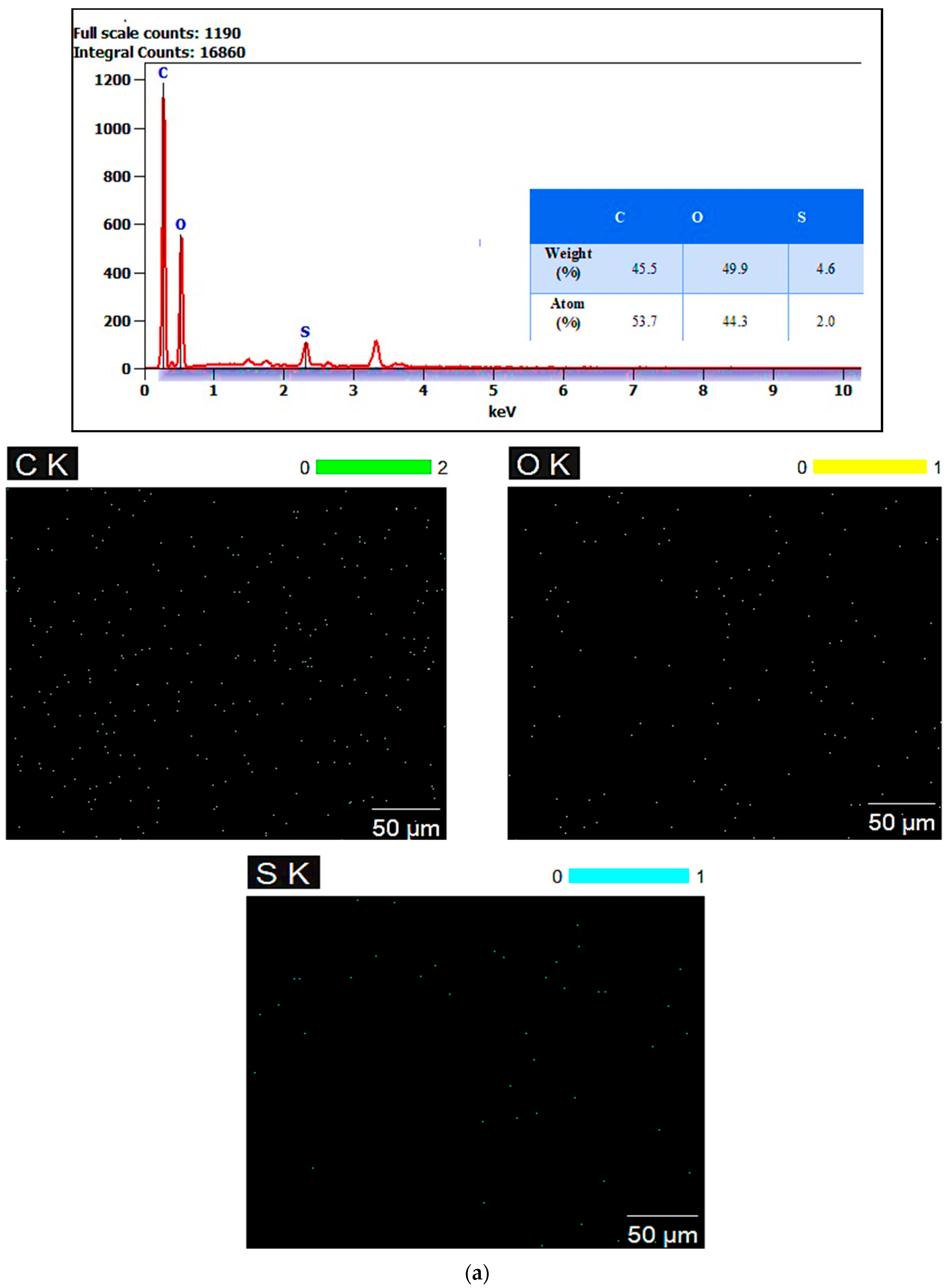
3.5. Energy Dispersive X-Ray (EDX) Mapping Analysis

The implementation of EDX and mapping tools provides indispensable insights for advancing the development and optimization of advanced materials, particularly in fields like biomedicine, where fine-tuning elemental composition is of paramount importance [67]. Figure 5 presents the EDX elemental analysis and mapping of the CAR/DEMA/Gelt hydrogel (Figure 5a) and the CAR/DEMA/Gelt/ZnO nanocomposite (Figure 5b), offering vital insights into the elemental composition via atom and weight percentages. In the case of the CAR/DEMA/Gelt hydrogel (Figure 5a), the EDX analysis reveals weight percentages of 45.5% carbon (C), 49.9% oxygen (O), and 4.6% sulfur (S), accompanied by atom percentages of 53.7% C, 44.3% O, and 2.0% S. These findings provide a comprehensive breakdown of elemental composition within the hydrogel. The CAR/DEMA/Gelt/ZnO nanocomposite (Figure 5b) presents distinct elemental proportions, with weight percentages of 49.7% C, 47.0% O, 2.4% S, and 0.9% zinc (Zn). Atom percentages for this nanocomposite are measured at 57.8% C, 41.0% O, 1.1% S, and 0.2% Zn. These results highlight the successful incorporation of ZnO nanoparticles into the composite, shedding light on the elemental constituents and their distribution. The utilization of EDX analysis in tandem with atom and weight percentages greatly enhances our understanding of these nanocomposites [68]. The EDX results, alongside mapping, play a pivotal role in verifying the effective integration of ZnO nanoparticles into the CAR/DEMA/Gelt matrix and elucidating their distribution across the material [69].

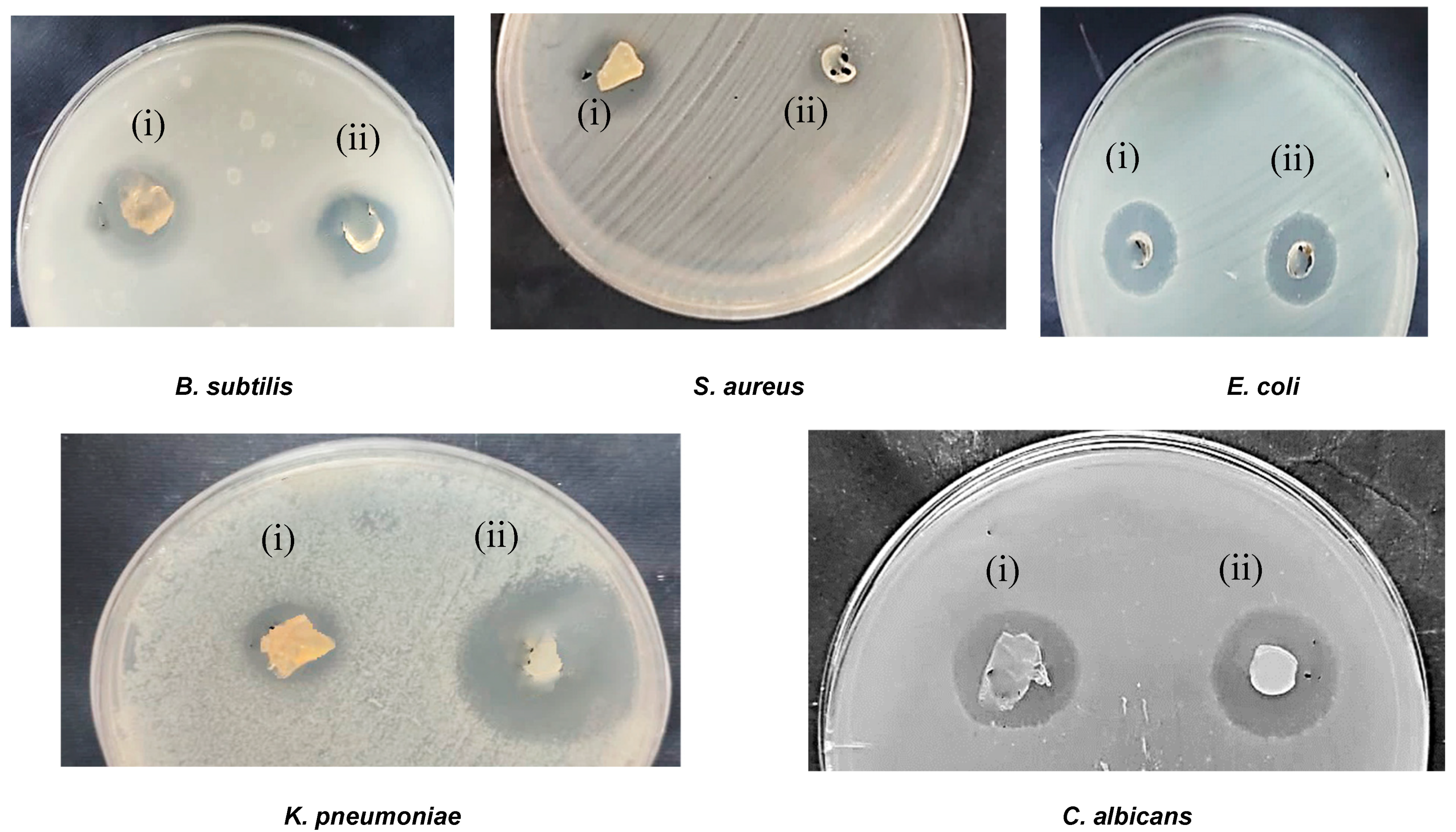
3.6. Antimicrobial Activity

The antimicrobial properties of the CAR/DEMA/Gelt hydrogel and CAR/DEMA/Gelt/ZnO nanocomposite were rigorously assessed against various pathogens, including B. subtilis, S. aureus, E. coli, K. pneumoniae, and C. albicans, using the agar diffusion method. The results, presented in Figure 6, revealed strong antimicrobial activity exhibited by both the CAR/DEMA/Gelt hydrogel and the CAR/DEMA/Gelt/ZnO nanocomposite against all tested microorganisms. Notably, the CAR/DEMA/Gelt hydrogel displayed significant antibacterial efficacy, positioning it as a promising candidate for a range of biomedical applications. Intriguingly, these findings align with previous research by Wang et al., who reported similar antibacterial effects of κ-carrageenan against E. coli and S. aureus bacteria [70]. The CAR/DEMA/Gelt/ZnO nanocomposite exhibited even more pronounced antimicrobial activity compared to the CAR/DEMA/Gelt hydrogel. This result is similar to that reported before with the CMC/GEL/ZnO composite [71]. This inhibitory effect can be attributed to the incorporation of ZnO nanoparticles within the polymeric matrix. ZnO is a transition metal oxide and semiconductor with a high binding energy that allows a highly oxidative character [72]. This reaction leads to the formation of reactive oxygen species (ROS) as the pathway of bactericidal action. In addition, another bactericidal mode of action occurs through the release of zinc ions (Zn2+), which damage the cell membrane and may interrupt some metabolic pathways [73]. In a previous study, SEM and TEM analyses of the bacterial cells showed that ZnO NPs damage the cell membrane and, right after, go to the cytoplasm, where they interact with other cell structures as a secondary effect [74].

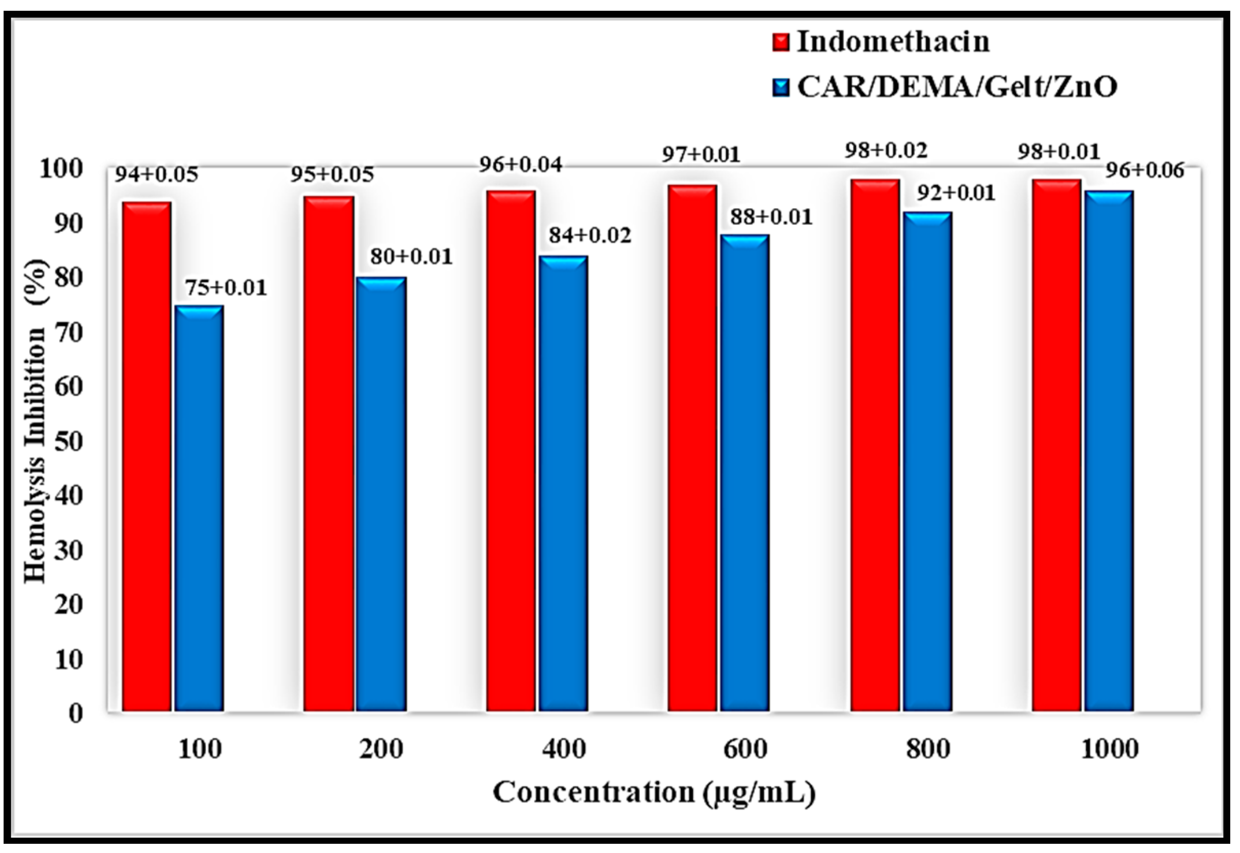
3.7. Anti-Inflammatory Activity

Hypotonicity-Induced Hemolysis test is one of several tests used to assess the ability of a substance to inhibit red blood cell hemolysis. The evaluation of the anti-inflammatory activity of the CAR/DEMA/Gelt/ZnO nanocomposite compared with that of Indomethacin, a well-recognized anti-inflammatory drug, revealed noteworthy results, as shown in Figure 7. At lower concentrations of the samples (100 µg/mL), the hemolysis inhibitory effect was 94 ± 0.05% and 75 ± 0.041% for Indomethacin and ZnO nanocomposites, respectively, while at 400 µg/mL, the inhibitory effect was 96 ± 0.04% and 84 ± 0.02% for Indomethacin and ZnO nanocomposites, respectively. At 1000 µg/mL, the hemolysis inhibitory effect was comparable, at 98 ± 0.01% and 96 ± 0.006% for Indomethacin and ZnO nanocomposites, respectively. These results indicate that the CAR/DEMA/Gelt/ZnO nanocomposite may serve as a promising candidate for applications in the biomedical domain [75,76].

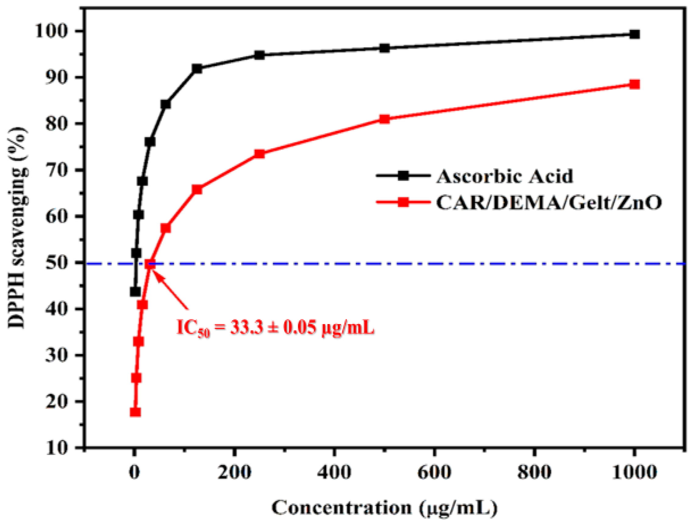
3.8. Antioxidant Activity

The antioxidant activity of the CAR/DEMA/Gelt/ZnO nanocomposite was evaluated by its DPPH (1,1-diphenyl-2-picrylhydrazyl) free radical scavenging activity. The nanocomposite radical scavenging activity is measured at 517 nm by a decrease in the molar absorptivity of DPPH. The antioxidant activity rises with a low IC50 (i.e., 50% inhibitory concentration). The obtained results are shown in Figure 8. The data indicate that the prepared CAR/DEMA/Gelt/ZnO nanocomposite possesses a potent antioxidant capacity, with an impressive IC50 value of 33.3 ± 0.05 µg/mL. This result is in agreement with that reported before for the CMC/GEL/ZnO composite [28]. This low IC50 value indicates the high antioxidant potential of the CAR/DEMA/Gelt/ZnO nanocomposite, signifying its ability to effectively neutralize harmful free radicals and oxidative stress [77]. The profound antioxidant activity demonstrated by the nanocomposite showcases its significant promise in the field of biomedical and pharmaceutical applications [78]. The capacity to combat oxidative damage is paramount in various aspects of healthcare, from disease prevention to pharmaceutical formulations. These findings open up exciting avenues for the integration of the CAR/DEMA/Gelt/ZnO nanocomposite into advanced therapeutic and pharmaceutical solutions, thereby contributing to the ever-evolving landscape of biomedical research and innovation [79].

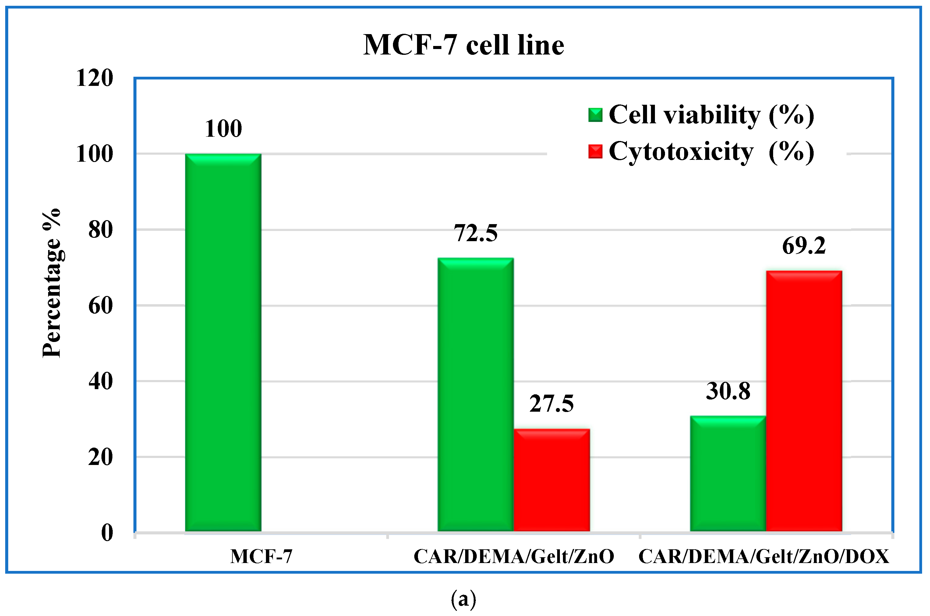
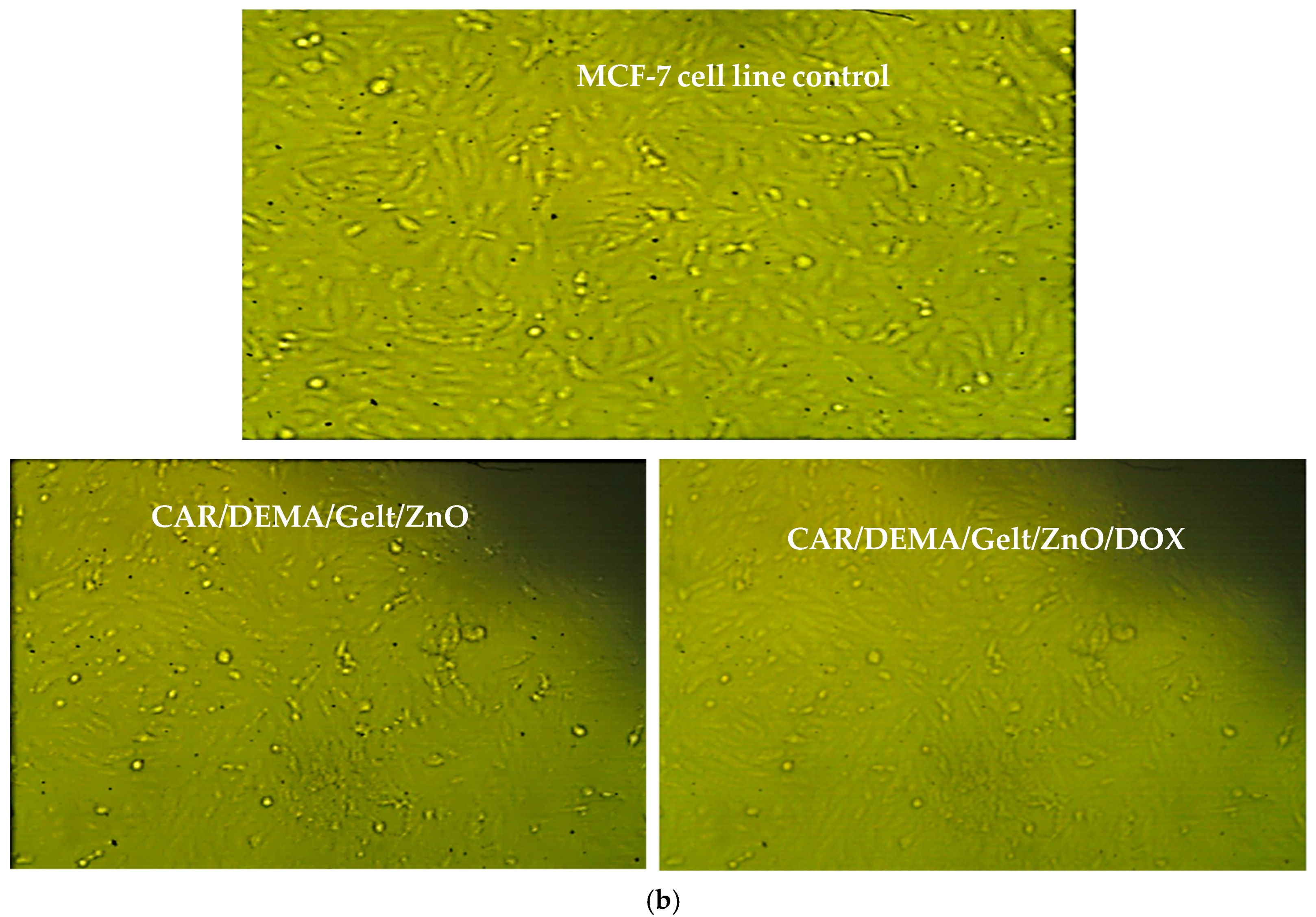
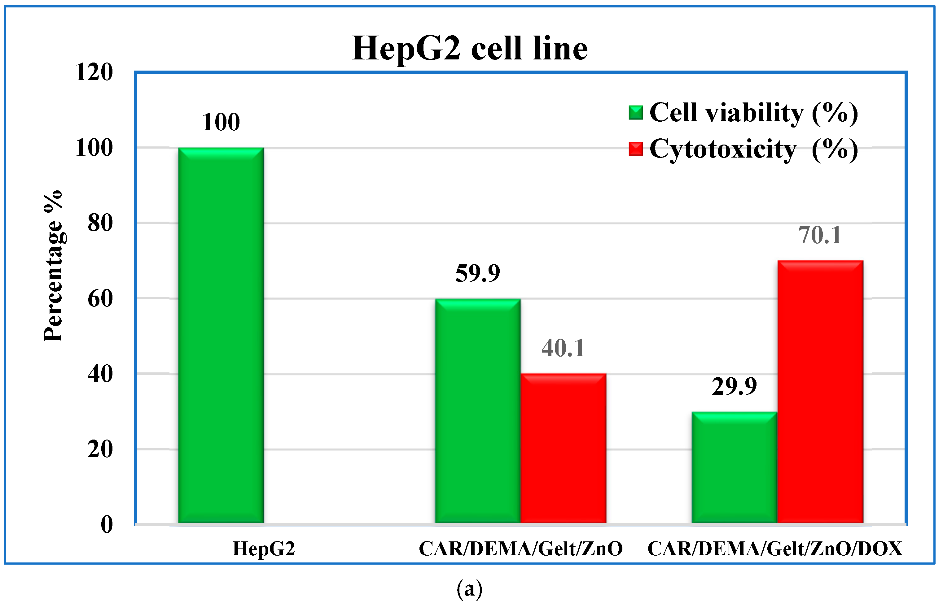
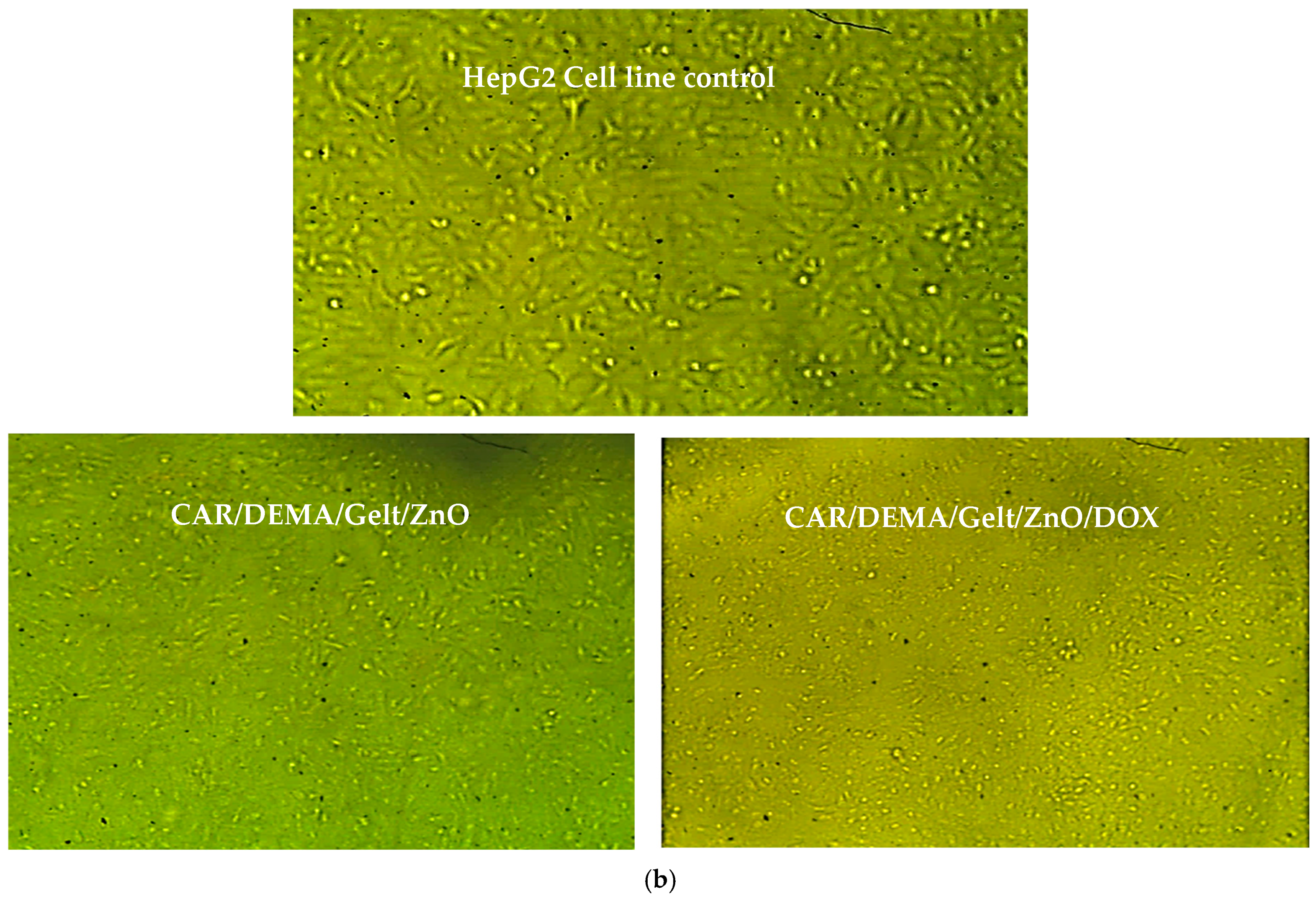
3.9. In Vitro Cytotoxic Effect Against Cancer Cells

The in vitro cytotoxicity of the nanocomposite hydrogel alone, CAR/DEMA/Gelt/ZnO, and that loaded with the anticancer drug doxorubicin (CAR/DEMA/Gelt/ZnO/DOX) were tested using the MTT assay. The cell viability of the human breast cancer cell line (MCF-7) and the human liver cancer cell line (HepG2) are shown in Figure 9a, b and Figure 10a, b, respectively. The potential cytotoxicity of the two nanocomposite hydrogels was estimated and compared with the control. We observed that the CAR/DEMA/Gelt/ZnO nanocomposite has a distinct effect on both MCF-7 and HepG2 cells after incubation for 48 h. The cell viability was 72.5 ± 0.02% and 59.9 ± 0.006% for MCF-7 and HepG2 cells, respectively. These results can be explained based on the cytotoxicity of ZnO NPS. The basic mechanism behind the cytotoxicity of ZnO NPs is the intracellular release of dissolved Zn ions followed by ROS induction, which eventually kills the cells [80]. ROS include superoxide radical (O2), which further reacts with hydrogen ions, producing HO2 radicals that further react to create H2O2, which causes damage to cellular components, including DNA and proteins [81]. ROS is induced by ZnO NPs in two ways. One is due to the pro-inflammatory response of the cell against nanoparticles [82], and the other is due to the characteristic surface property of ZnO NPs that makes them a redox reaction system producing ROS [81,83]. It was reported that ZnO NPs at a concentration of 15 µL/mL decrease HepG2 cell viability to 39%, leading to the higher activity of caspase 3 enzyme along with DNA fragmentation in liver cancer cells treated with ZnO NPs. Higher production of intracellular ROS and membrane LPO in ZnO NP-treated cancer cells may be the primary mechanism for the toxicity of ZnO nanocomposites against the human liver cancer cell HepG2 [84]. The cytotoxicity of the DOX-loaded nanocomposite, CAR/DEMA/Gelt/ZnO/DOX, was higher than that of free nanocomposites due to drug release from the carrier and its internalization rate. Cell viability sharply decreased to 30.8 ± 0.01% and 29.9 ± 0.005% for MCF-7 and HepG2 cells, respectively; however, it did not exceed 70%. It was reported in a previous study that drug internalization inside the cell was time-dependent and almost completed after 2 h of exposure. However, DOX enters the cytoplasm by diffusion and quickly binds to the proteasome’s 20S subunit. This complex can diffuse into the nucleus and is observed in the cytoplasm and nucleus of the cell in 1 h; this was confirmed by the results of free DOX uptake (99.9%) after 1 h [85]. On the other hand, it was reported that the cytotoxicity of the DOX-loaded nanocarrier against osteosarcoma cells (IC50 = 0.293 µg/mL) and early apoptosis rate (33.8%) were higher in comparison to free DOX (IC50 = 0.472 µg/mL) and early apoptosis rate (8.31%) after 48 h. The study concluded that the cell undergoes apoptosis if the period of exposure to DOX nanocarrier increases 84]. Based on the above-mentioned points, we can suggest that the newly prepared CAR/DEMA/Gelt/ZnO/DOX nanocarrier could be considered a good delivery system for DOX. More investigations are required in future studies on the in vivo release of DOX from the carrier and apoptosis rates of both MCF-7 and HepG2 cancer cell lines.

4. Conclusions

Burn wound healing continues to pose significant challenges due to the risk of infection, excessive inflammation, and impaired tissue regeneration. In this regard, we developed a new nanocomposite based on carrageenan polymer, 2-dimethyl aminoethyl methacrylate, gelatin, and ZnO nanoparticles (CAR/DEMA/Gelt/ZnO). Various analyses, including FTIR, FE-SEM, XRD, TGA, and EDX, were employed to characterize the nanocomposite. The CAR/DEMA/Gelt/ZnO nanocomposite exhibited robust antibacterial activity against both Gram-negative and Gram-positive bacteria, as well as the unicellular fungi, C. albicans. During the initial stage of wound healing, the nanocomposite can release ZnO from its surface to suppress microbial activity. Additionally, the nanocomposite exhibits anti-inflammatory activity comparable to that of the recognized anti-inflammatory drug Indomethacin. Moreover, the antioxidant activity, which is indicated by the impressive IC50 value of 33.3 µg/mL, can help scavenge overexpressed reactive oxygen species (ROS) in the infected microenvironment, thus reducing the upregulation of pro-inflammatory cytokines. Finally, the nanocomposite alone and the DOX uploaded form (CAR/DEMA/Gelt/ZnO/DOX) showed variable cytotoxicities against the cancer cells MCF-7 and HepG2. The CAR/DEMA/Gelt/ZnO nanocomposite showed cell viability of 72.5 ± 0.02% and 59.5 ± 0.006% for MCF-7 and HepG2 cells, respectively, while with CAR/DEMA/Gelt/ZnO/DOX, cell viability was 30.8 ± 0.01% % and 29.9 ± 0.005% % for MCF-7 and HepG2 cells, respectively. This cytotoxicity is related to drug release and its internalization rate into the cancer cells. It can be concluded that CAR/DEMA/Gelt/ZnO, with its drug delivery capacity and its synergistic antibacterial, anti-inflammatory, and antioxidant activities, shows great promise as an effective multifunctional nanoplatform for wound dressings and DOX formulation. We recommend conducting in vivo studies in the future to investigate the efficiency of CAR/DEMA/Gelt/ZnO as a new DOX-formulating candidate.

Author Contributions

Conceptualization, methodology, formal analysis, investigation, resources, data curation, writing—original draft: A.A.A.; Conceptualization, methodology, writing—review and editing: S.F.M. All authors have read and agreed to the published version of the manuscript.

Funding

The authors gratefully acknowledge funding from the Deanship of Graduate Studies and Scientific Research, Jazan University, Saudi Arabia, through Project Number GSSRD-24.

Data Availability Statement

Data are contained within the article.

Conflicts of Interest

The authors declare that there are no conflicts of interest.

References

  1. Joshua, M.G.; Mansher, S.; David, J.M.; Elof, E.; Kristo, N. Antibiotic-Containing Agarose Hydrogel for Wound and Burn Care. J. Burn Care Res. 2019, 16, 900–906. [Google Scholar]
  2. Kirill, V.O.; Christian, K.; Tage, T.; Harald, C.; Dzung, B.D. Successful Development of Bacteriocins into Therapeutic Formulation for Treatment of MRSA Skin Infection in a Murine Model. Antimicrob. Agents Chemother. 2020, 64, e00829-20. [Google Scholar]
  3. Eid, M.; El-Arnaouty, M.; Salah, M.; Soliman, E.S.; Hegazy, E.-S.A. Radiation synthesis and characterization of poly (vinyl alcohol)/poly (N-vinyl-2-pyrrolidone) based hydrogels containing silver nanoparticles. J. Polym. Res. 2012, 19, 9835. [Google Scholar] [CrossRef]
  4. Sun, T.; Tao, H.; Xie, J.; Zhang, S.; Xu, X. Degradation and antioxidant activity of κ-carrageenans. J. Appl. Polym. Sci. 2010, 117, 194–199. [Google Scholar] [CrossRef]
  5. Cacciuttolo, M.A.; Trinh, L.; Lumpkin, J.A.; Rao, G. Hyperoxia induces DNA damage in mammalian cells. Free. Radic. Biol. Med. 1993, 14, 267–276. [Google Scholar] [CrossRef] [PubMed]
  6. Sun, T.; Zhou, D.; Xie, J.; Mao, F. Preparation of chitosan oligomers and their antioxidant activity. Eur. Food Res. Technol. 2007, 225, 451–456. [Google Scholar] [CrossRef]
  7. Choudhary, M.; bin Abdullah, A. Lipidomic Signatures in Oral Squamous Cell Carcinoma: Towards Personalized Treatment Strategies. J.Contemp. Healthc. Anal. 2023, 7, 145–162. [Google Scholar]
  8. Kashkooli, F.M.; Jakhmola, A.; Hornsby, T.K.; Tavakkoli, J.J.; Kolios, M.C. Ultrasound-mediated nano drug delivery for treating cancer: Fundamental physics to future directions. J. Control. Release 2023, 355, 552–578. [Google Scholar] [CrossRef] [PubMed]
  9. Sun, J.; Zhao, H.; Fu, L.; Cui, J.; Yang, Y. Global Trends and Research Progress of Photodynamic Therapy in Skin Cancer: A Bibliometric Analysis and Literature Review. Clin. Cosmet. Investig. Dermatol. 2023, 16, 479–498. [Google Scholar] [CrossRef]
  10. Verma, J.; Warsame, C.; Seenivasagam, R.K.; Katiyar, N.K.; Aleem, E.; Goel, S. Nanoparticle-mediated cancer cell therapy: Basic science to clinical applications. Cancer Metastasis Rev. 2023, 42, 601–627. [Google Scholar] [CrossRef]
  11. Hari, S.K.; Gauba, A.; Shrivastava, N.; Tripathi, R.M.; Jain, S.K.; Pandey, A.K. Polymeric micelles and cancer therapy: An ingenious multimodal tumor-targeted drug delivery system. Drug Deliv. Transl. Res. 2023, 13, 135–163. [Google Scholar] [CrossRef] [PubMed]
  12. Vangijzegem, T.; Lecomte, V.; Ternad, I.; Van Leuven, L.; Muller, R.N.; Stanicki, D.; Laurent, S. Superparamagnetic iron oxide nanoparticles (SPION): From fundamentals to state-of-the-art innovative applications for cancer therapy. Pharmaceutics 2023, 15, 236. [Google Scholar] [CrossRef] [PubMed]
  13. Marín, A.; Martín, M.; Liñán, O.; Alvarenga, F.; López, M.; Fernández, L.; Büchser, D.; Cerezo, L. Bystander effects and radiotherapy. Rep. Pract. Oncol. Radiother. 2015, 20, 12–21. [Google Scholar] [CrossRef] [PubMed]
  14. Nasiri, H.; Valedkarimi, Z.; Aghebati-Maleki, L.; Majidi, J. Antibody-drug conjugates: Promising and efficient tools for targeted cancer therapy. J. Cell. Physiol. 2018, 233, 6441–6457. [Google Scholar] [CrossRef] [PubMed]
  15. Wani, S.U.D.; Ali, M.; Masoodi, M.H.; Khan, N.A.; Zargar, M.I.; Hassan, R.; Mir, S.A.; Gautam, S.P.; Gangadharappa, H.V.; Osmani, R.A.M. A review on nanoparticles categorization, characterization and applications in drug delivery systems. Vib. Spectrosc. 2022, 121, 103407. [Google Scholar] [CrossRef]
  16. Chen, Y.; Qin, D.; Zou, J.; Li, X.; Guo, X.D.; Tang, Y.; Liu, C.; Chen, W.; Kong, N.; Zhang, C.Y.; et al. Living Leukocyte-Based Drug Delivery Systems. Adv. Mater. 2023, 35, 2207787. [Google Scholar] [CrossRef]
  17. Wang, H.; Zhou, Y.; Sun, Q.; Zhou, C.; Hu, S.; Lenahan, C.; Xu, W.; Deng, Y.; Li, G.; Tao, S. Update on nanoparticle-based drug delivery system for anti-inflammatory treatment. Front. Bioeng. Biotechnol. 2021, 9, 630352. [Google Scholar] [CrossRef]
  18. Ghoshal, G. Role of Nutraceutical-Based Functional Foods In Human Health. In Plant-Based Bioactive Compounds and Food Ingredients; Apple Academic Press: Palm Bay, FL, USA, 2024; pp. 113–142. [Google Scholar]
  19. Thakur, K.; Zhu, Y.Y.; Feng, J.Y.; Zhang, J.G.; Hu, F.; Prasad, C.; Wei, Z.J. Morin as an imminent functional food ingredient: An update on its enhanced efficacy in the treatment and prevention of metabolic syndromes. Food Funct. 2020, 11, 8424–8443. [Google Scholar] [CrossRef]
  20. Pang, Y.; Shi, R.; Chan, L.; Lu, Y.; Zhu, D.; Liu, T.; Yan, M.; Wang, Y.; Wang, W. The combination of the HDC1 inhibitor SAHA and doxorubicin has synergic efficacy in triple negative breast cancer in vivo. Pharmacol. Res. 2023, 196, 106926. [Google Scholar] [CrossRef]
  21. Li, S.; Li, F.; Wan, D.; Chen, Z.; Pan, J.; Liang, X.J. A micelle-based stage-by-stage impelled system for efficient doxorubicin delivery. Bioact. Mater. 2023, 25, 783–795. [Google Scholar] [CrossRef]
  22. Xiao, Y.; Gu, Y.; Qin, L.; Chen, L.; Chen, X.; Cui, W.; Li, F.; Xiang, N.; He, X. Injectable thermosensitive hydrogel-based drug delivery system for local cancer therapy. Colloids Surf. B 2021, 200, 111581. [Google Scholar] [CrossRef] [PubMed]
  23. Kesharwani, P.; Bisht, A.; Alexander, A.; Dave, V.; Sharma, S. Biomedical applications of hydrogels in drug delivery system: An update. J. Drug Deliv. Sci. Technol. 2021, 66, 102914. [Google Scholar] [CrossRef]
  24. Jahanban-Esfahlan, R.; Soleimani, K.; Derakhshankhah, H.; Haghshenas, B.; Rezaei, A.; Massoumi, B.; Farnudiyan-Habibi, A.; Samadian, H.; Jaymand, M. Multi-stimuli-responsive magnetic hydrogel based on Tragacanth gum as a de novo nanosystem for targeted chemo/hyperthermia treatment of cancer. J. Mater. Res. 2021, 36, 858–869. [Google Scholar] [CrossRef]
  25. Pacheco-Quito, E.M.; Ruiz-Caro, R.; Veiga, M.D. Carrageenan: Drug delivery systems and other biomedical applications. Mar. Drugs 2020, 18, 583. [Google Scholar] [CrossRef] [PubMed]
  26. Qureshi, D.; Nayak, S.K.; Maji, S.; Kim, D.; Banerjee, I.; Pal, K. Carrageenan: A wonder polymer from marine algae for potential drug delivery applications. Curr. Pharm. Des. 2019, 25, 1172–1186. [Google Scholar] [CrossRef]
  27. Zhang, S.; Qamar, S.A.; Junaid, M.; Munir, B.; Badar, Q.; Bilal, M. Algal Polysaccharides-Based Nanoparticles for Targeted Drug Delivery Applications. Starch 2022, 74, 2200014. [Google Scholar] [CrossRef]
  28. Mokhtari, H.; Tavakoli, S.; Safarpour, F.; Kharaziha, M.; Bakhsheshi-Rad, H.R.; Ramakrishna, S.; Berto, F. Recent advances in chemically-modified and hybrid carrageenan-based platforms for drug delivery, wound healing, and tissue engineering. Polymers 2021, 13, 1744. [Google Scholar] [CrossRef]
  29. Zhang, K.; Feng, Q.; Fang, Z.; Gu, L.; Bian, L. Structurally dynamic hydrogels for biomedical applications: Pursuing a fine balance between macroscopic stability and microscopic dynamics. Chem. Rev. 2021, 121, 11149–11193. [Google Scholar] [CrossRef]
  30. Mushtaq, F.; Raza, Z.A.; Batool, S.R.; Zahid, M.; Onder, O.C.; Rafique, A.; Nazeer, M.A. Preparation, properties, and applications of gelatin-based hydrogels (GHs) in the environmental, technological, and biomedical sectors. Int. J. Biol. Macromol. 2022, 218, 601–633. [Google Scholar] [CrossRef]
  31. Gunjan, B.; Sagar, R. ZnO Nanoparticles: A Promising Anticancer Agent. Nanobiomedicine 2016, 3, 9. [Google Scholar]
  32. Wang, Y.; Zhang, X.; Qiu, D.; Li, Y.; Yao, L.; Duan, J. Ultrasonic assisted microwave synthesis of poly (Chitosan-co-gelatin)/polyvinyl pyrrolidone IPN hydrogel. Ultrason. Sonochem. 2018, 40, 714–719. [Google Scholar] [CrossRef] [PubMed]
  33. El Sayed, M.M. Production of Polymer Hydrogel Composites and Their Applications. J. Polym. Environ. 2023, 31, 2855–2879. [Google Scholar] [CrossRef]
  34. Zhao, C.; Liu, G.; Tan, Q.; Gao, M.; Chen, G.; Huang, X.; Xu, X.; Li, L.; Wang, J.; Zhang, Y.; et al. Polysaccharide-based biopolymer hydrogels for heavy metal detection and adsorption. J. Adv. Res. 2023, 44, 53–70. [Google Scholar] [CrossRef] [PubMed]
  35. Bao, C.; Serrano-Lotina, A.; Niu, M.; Portela, R.; Li, Y.; Lim, K.H.; Liu, P.; Wang, W.J.; Bañares, M.A.; Wang, Q. Microwave-associated chemistry in environmental catalysis for air pollution remediation: A review. Chem. Eng. J. 2023, 466, 142902. [Google Scholar] [CrossRef]
  36. Basavarajappa, G.M.; Priyanka, K.M.; Goudanavar, P.; Narasimha, L.G.; Naveen, N.R.; Gowthami, B.; Fattepur, S.; Shiroorkar, P.N.; Nagaraja, S.; Telsang, M.; et al. A spotlight on application of microwave-assisted modifications of plant derived polymers in designing novel drug delivery systems. Des. Monomers Polym. 2023, 26, 106–116. [Google Scholar] [CrossRef]
  37. Ganguly, S.; Margel, S. Fluorescent quantum dots-based hydrogels: Synthesis, Fabrication and multimodal biosensing. Talanta Open 2023, 8, 100243. [Google Scholar] [CrossRef]
  38. Amini, E.; Valls, C.; Roncero, M.B. Ionic liquid-assisted bioconversion of lignocellulosic biomass for the development of value-added products. J. Clean. Prod. 2021, 326, 129275. [Google Scholar] [CrossRef]
  39. Soliman, E.A.; Khalil, A.A.; Deraz, S.F.; El-Fawal, G.; Abd Elrahman, S. Synthesis, characterization and antibacterial activity of biodegradable films prepared from Schiff bases of zein. J. Food Sci. Technol. 2014, 51, 2425–2434. [Google Scholar] [CrossRef]
  40. Fatimah, A.A.; Sahera, F.M. Synthesis and Characterization of a Self-Crosslinked Organic Copolymer Kappa-Carrageenan/Polyacrylamide/Cetrimide (κ-CAR/PAAm/CI) Hydrogel with Antimicrobial and Anti-Inflammatory Activities for Wound Healing. Chemistry 2023, 5, 2273–2287. [Google Scholar] [CrossRef]
  41. Zhou, J.; Sun, Y.Y.; Sun, M.Y.; Mao, W.A.; Wang, L.; Zhang, J.; Zhang, H. Prim-O-glucosylcimifugin Attenuates Lipopolysaccharide Induced Inflammatory Response in RAW 264.7 Macrophages. Pharmcogn. Mag. 2017, 13, 378–384. [Google Scholar]
  42. Fabian, I.E.; Philip, F.U.; Peter, I.; Bonaventure, C.O.; Patience, O.O. In vitro and In vivo Models for Anti-inflammation: An Evaluative Review. Theragnostic Pharmacol. Sci. 2019, 2, 3–15. [Google Scholar]
  43. Venkatesan, M.; Arumugam, V.; Pugalendi, R.; Ramachandran, K.; Sengodan, K.; Vijayan, S.R.; Sundaresan, U.; Ramachandran, S.; Pugazhendhi, A. Antioxidant, anticoagulant and mosquitocidal properties of water-soluble polysaccharides (WSPs) from Indian seaweeds. Process Biochem. 2019, 84, 196–204. [Google Scholar] [CrossRef]
  44. Santosh, K.B.Y.; Arun, M.I.; Mohan, K.G.C.; Inamuddin; Abdullah, M.A. Nanohydroxyapatite Reinforced chitosan composite Hydrogel with tunable Mechanical and Biological properties for cartilage Regeneration. Sci. Rep. 2019, 9, 15957. [Google Scholar]
  45. Phuong, L.T.; Yunki, L.; Thai, T.H.T.; Kyung, M.P.; Ki, D.P. Catechol-rich gelatin hydrogels in situ hybridizations with silver nanoparticle for enhanced antibacterial activity. Mater. Sci. Eng. C 2018, 92, 52–60. [Google Scholar]
  46. Maneesh, J.; Amit, K.D.; Asheesh, G.; Veena, K. Polycaprolactone diacrylate crosslinked biodegradable semi-interpenetrating networks of polyacrylamide and gelatin for controlled drug delivery. Biomed. Mater. 2010, 5, 065014. [Google Scholar]
  47. Zeferino, R.S.; Flores, M.B.; Pal, U. Photoluminescence and Raman scattering in Ag-doped ZnO nanoparticles. J. Appl. Phys. 2011, 109, 014308. [Google Scholar] [CrossRef]
  48. Nagaraju, G.; Prashanth, S.A.; Shastri, M.; Yathish, K.V.; Anupama, C.; Rangappa, D.J.M.R.B. Electrochemical heavy metal detection, photocatalytic, photoluminescence, biodiesel production and antibacterial activities of Ag–ZnO nanomaterial. Mater. Res. Bull. 2017, 94, 54–63. [Google Scholar] [CrossRef]
  49. Sun, N.; Lei, R.; Xu, J.; Kundu, S.C.; Cai, Y.; Yao, J.; Ni, Q. Fabricated porous silk fibroin particles for pH-responsive drug delivery and targeting of tumor cells. J. Mater. Sci. 2019, 54, 3319–3330. [Google Scholar] [CrossRef]
  50. Banerjee, S.S.; Chen, D.-H. Multifunctional pH-sensitive magnetic nanoparticles for simultaneous imaging, sensing and targeted intracellular anticancer drug delivery. Nanotechnology 2008, 19, 505104. [Google Scholar] [CrossRef]
  51. Ahmadi, A.; Ahmadi, P.; Sani, M.A.; Ehsani, A.; Ghanbarzadeh, B. Functional biocompatible nanocomposite films consisting of selenium and zinc oxide nanoparticles embedded in gelatin/cellulose nanofiber matrices. Int. J. Biol. Macromol. 2021, 175, 87–97. [Google Scholar] [CrossRef]
  52. Rasoulzadehzali, M.; Namazi, H. Facile preparation of antibacterial chitosan/graphene oxide-Ag bio-nanocomposite hydrogel beads for controlled release of doxorubicin. Int. J. Biol. Macromol. 2018, 116, 54–63. [Google Scholar] [CrossRef] [PubMed]
  53. Rana, S.; Gallo, A.; Srivastava, R.S.; Misra, R.D.K. On the suitability of nanocrystalline ferrites as a magnetic carrier for drug delivery: Functionalization, conjugation and drug release kinetics. Acta Biomater. 2007, 3, 233–242. [Google Scholar] [CrossRef] [PubMed]
  54. Abdulrahman, A.F.; Abd-Alghafour, N.M.; Ahmed, S.M. Optimization and characterization of SILAR synthesized ZnO nanorods for UV photodetector sensor. Sens. Actuator A Phys. 2021, 323, 112656. [Google Scholar] [CrossRef]
  55. Akgul, G.; Akgul, F.A. Impact of cobalt doping on structural and magnetic properties of zinc oxide nanocomposites synthesized by mechanical ball-milling method. Colloids Interface Sci. Commun. 2022, 48, 100611. [Google Scholar] [CrossRef]
  56. Lopez-Sanchez, P.; Martinez-Sanz, M.; Bonilla, M.R.; Wang, D.; Walsh, C.T.; Gilbert, E.P.; Stokes, J.R.; Gidley, M.J. Pectin impacts cellulose fibre architecture and hydrogel mechanics in the absence of calcium. Carbohydr. Polym. 2016, 153, 236–245. [Google Scholar] [CrossRef]
  57. Guan, Y.; Yu, C.; Zang, Z.; Wan, X.; Naeem, A.; Zhang, R.; Zhu, W. Chitosan/xanthan gum-based (Hydroxypropyl methylcellulose-co-2-Acrylamido-2-methylpropane sulfonic acid) interpenetrating hydrogels for controlled release of amorphous solid dispersion of bioactive constituents of Pueraria lobatae. Int. J. Biol. Macromol. 2023, 224, 380–395. [Google Scholar] [CrossRef]
  58. Joseph, B.; Mavelil Sam, R.; Balakrishnan, P.; Maria, H.J.; Gopi, S.; Volova, T.; Fernandes, S.C.M.; Thomas, S. Extraction of nanochitin from marine resources and fabrication of polymer nanocomposites: Recent advances. Polymers 2020, 12, 1664. [Google Scholar] [CrossRef] [PubMed]
  59. Jumaidin, R.; Sapuan, S.M.; Jawaid, M.; Ishak, M.R.; Sahari, J. Seaweeds as renewable sources for biopolymers and its composites: A review. Curr. Anal. Chem. 2018, 14, 249–267. [Google Scholar] [CrossRef]
  60. Dave, P.N.; Macwan, P.M.; Kamaliya, B. Drug release and thermal properties of magnetic cobalt ferrite (CoFe2O4) nanocomposite hydrogels based on poly (acrylic acid-gN-isopropyl acrylamide) grafted onto gum ghatti. Int. J. Biol. Macromol. 2023, 224, 358–369. [Google Scholar] [CrossRef]
  61. Torğut, G.; Yazdıç, F.C.; Gürler, N. Synthesis, characterization, pH-sensitive swelling and antimicrobial activities of chitosan–graft-poly (hydroxyethyl methacrylate) hydrogel composites for biomedical applications. Polym. Eng. Sci. 2022, 62, 2552–2559. [Google Scholar] [CrossRef]
  62. Mota, H.P.; Fajardo, A.R. Development of superabsorbent hydrogel based on Gum Arabic for enhanced removal of anxiolytic drug from water. J. Environ. Manage. 2021, 288, 112455. [Google Scholar] [CrossRef] [PubMed]
  63. Oun, A.A.; Rhim, J.-W. Carrageenan-based hydrogels and films: Effect of ZnO and CuO nanoparticles on the physical, mechanical, and antimicrobial properties. Food Hydrocoll. 2017, 67, 45–53. [Google Scholar] [CrossRef]
  64. Jayaraman, P.; Gandhimathi, C.; Venugopal, J.R.; Becker, D.L.; Ramakrishna, S.; Srinivasan, D.K. Controlled release of drugs in electrosprayed nanoparticles for bone tissue engineering. Adv. Drug Deliv. Rev. 2015, 94, 77–95. [Google Scholar] [CrossRef] [PubMed]
  65. Arakelova, E.R.; Grigoryan, S.G.; Arsenyan, F.G.; Babayan, N.S.; Grigoryan, R.M.; Sarkisyan, N.K. In vitro and in vivo anticancer activity of nanosize zinc oxide composites of doxorubicin. Int. J. Med. Pharma. Sci. Eng. 2014, 8, 38–43. [Google Scholar]
  66. Arakelova, E.R.; Farmazyan, Z.M.; Khachatryan, A.M.; Avjyan, K.E.; Babayan, N.S.; Grigoryan, R.M.; Sarkisyan, N.K. In vitro study of anticancer activity of polymer-based DOX-ZnO compositions: The case of vinyl alcohol (co) polymers. J. Nanogen. Nanomed. 2014, 2, 1–11. [Google Scholar]
  67. Gleason, K.K. Designing Organic and Hybrid Surfaces and Devices with Initiated Chemical Vapor Deposition (iCVD). Adv. Mater. 2024, 36, 2306665. [Google Scholar] [CrossRef]
  68. Javed, H.M.A.; Qureshi, A.A.; Mustafa, M.S.; Que, W.; Mahr, M.S.; Shaheen, A.; Iqbal, J.; Saleem, S.; Jamshaid, M.; Mahmood, A. Advanced Ag/rGO/TiO2 ternary nanocomposite based photoanode approaches to highly-efficient plasmonic dye-sensitized solar cells. Opt. Commun. 2019, 453, 124408. [Google Scholar] [CrossRef]
  69. Tian, N.; Giannakis, S.; Akbarzadeh, L.; Hasanvandian, F.; Dehghanifard, E.; Kakavandi, B. Improved catalytic performance of ZnO via coupling with CoFe2O4 and carbon nanotubes: A new, photocatalysis-mediated peroxymonosulfate activation system, applied towards Cefixime degradation. J. Environ. Manage. 2023, 329, 117022. [Google Scholar] [CrossRef]
  70. Wang, F.F.; Yao, Z.; Wu, H.G.; Zhang, S.X.; Zhu, N.N.; Gai, X. Antibacterial activities of kappa-carrageenan oligosaccharides. Appl. Mech. Mater. 2012, 108, 194–199. [Google Scholar] [CrossRef]
  71. Aqsa, Z.; Muhammad, K.K.; Awal, N.; Sadaf, Q.; Muhammad, J.S. Carboxymethyl Cellulose/Gelatin Hydrogel Films Loaded with Zinc Oxide Nanoparticles for Sustainable Food Packaging Applications. Polymers 2022, 14, 5201. [Google Scholar] [CrossRef]
  72. Agarwal, H.; Menon, S.; Kumar, S.V.; Rajeshkumar, S. Mechanistic study of the antibacterial action of zinc oxide nanoparticles synthesized using green route. Chem.-Biol. Inter. 2018, 286, 60–70. [Google Scholar] [CrossRef]
  73. Burman, U.; Saini, M.; Kumar, P. Effect of zinc oxide nanoparticles on growth and antioxidant system of chickpea seedlings. Toxicol. Environ. Chem. 2013, 95, 605–612. [Google Scholar] [CrossRef]
  74. Siddiqi, K.S.; Rahman, A.; Husen, A. Properties of zinc oxide nanoparticles and their activity against microbes. Nanoscale Res. Lett. 2018, 13, 141. [Google Scholar] [CrossRef]
  75. Moohan, J.; Stewart, S.A.; Espinosa, E.; Rosal, A.; Rodríguez, A.; Larrañeta, E.; Donnelly, R.F.; Domínguez-Robles, J. Cellulose nanofibers and other biopolymers for biomedical applications. A review. Appl. Sci. 2019, 10, 65. [Google Scholar] [CrossRef]
  76. Azadikhah, F.; Karimi, A.R.; Yousefi, G.H.; Hadizadeh, M. Dual antioxidant-photosensitizing hydrogel system: Cross-linking of chitosan with tannic acid for enhanced photodynamic efficacy. Int. J. Biol. Macromol. 2021, 188, 114–125. [Google Scholar] [CrossRef]
  77. Li, K.; Zhong, W.; Li, P.; Ren, J.; Jiang, K.; Wu, W. Recent advances in lignin antioxidant: Antioxidant mechanism, evaluation methods, influence factors and various applications. Int. J. Biol. Macromol. 2023, 251, 125992. [Google Scholar] [CrossRef]
  78. Yasmeen, F.; Karamat, H.; Rehman, R.; Akram, M.; Ghfar, A.A.; Abdelghani, H.T.M.; Dar, A.; Mitu, L. Fabrication and testing of edible films incorporated with ZnO nanoparticles to enhance the shelf life of bread. Food Biosci. 2023, 56, 103111. [Google Scholar] [CrossRef]
  79. Mohd Zabidi, A.R.; Lim, W.F. Formation of cerium oxide film via post-sputter oxidation of cerium in nitrogen/oxygen/nitrogen ambient. J. Alloys Compd. 2021, 851, 156786. [Google Scholar] [CrossRef]
  80. Turney, T.W.; Duriska, M.B.; Jayaratne, V.; Elbaz, A.; O’Keefe, S.J.; Hastings, A.S.; Piva, T.J.; Wright, P.F.A.; Feltis, B.N. Formation of zinc containing nanoparticles from Zn2+ ions in cell culture media: Implications for the nanotoxicology of ZnO. Chem. Res. Toxicol. 2012, 25, 2057–2066. [Google Scholar] [CrossRef] [PubMed]
  81. Rasmussen, J.W.; Martinez, E.; Louka, P.; Wingett, D.G. Zinc oxide nanoparticles for selective destruction of tumor cells and potential for drug delivery applications. Expert Opin. Drug Deliv. 2010, 7, 1063–1077. [Google Scholar] [CrossRef]
  82. Driscoll, K.; Howard, B.; Carter, J.; Janssen, Y.W.; Mossman, B.; Isfort, R. Mitochondrial-Derived Oxidants and Quartz Activation of Chemokine Gene Expression. In Biological Reactive Intermediates VI; Dansette, P., Snyder, R., Dela-Forge, M., Gibson, G.G., Greim, H., Jollow, D., Monks, T., Sipes, I., Eds.; Advances in Experimental Medicine and Biology; Springer: New York, NY, USA, 2001; Volume 500, pp. 489–496. [Google Scholar]
  83. Wilson, M.R.; Lightbody, J.H.; Donaldson, K.; Sales, J.; Stone, V. Interactions between Ultrafine Particles and Transition Metals in Vivo and in Vitro. Toxi-Cology Appl. Pharmacol. 2002, 184, 172–179. [Google Scholar] [CrossRef] [PubMed]
  84. Alemi, F.; Masomeh, M.; Mostafa, M.; Abbas, E.K.; Mojtaba, Z.; Bahman, Y.; Nadereh, R. Synthesis, characterization, and evaluation of pH-sensitive doxorubicin-loaded functionalized graphene oxide in osteosarcoma cells. BioImpacts 2023, 13, 207–218. [Google Scholar] [CrossRef]
  85. Varela-López, A.; Battino, M.; Navarro-Hortal, M.D.; Giampieri, F.; Forbes-Hernández, T.Y.; Romero-Márquez, J.M.; Ricardo, C.; José, L.Q. An update on the mechanisms related to cell death and toxicity of doxorubicin and the protective role of nutrients. Food Chem. Toxicol. 2019, 134, 110834. [Google Scholar] [CrossRef]
Figure 1. FTIR analysis of (a) ZnO nanoparticles, (b) DOX powder, and (c) CAR/DEMA/Gelt hydrogel, CAR/DEMA/Gelt/ZnO nanocomposite, and CAR/DEMA/Gelt/ZnO/DOX.
Processes 12 02702 g001aProcesses 12 02702 g001b
Figure 2. XRD patterns of (a) ZnO nanoparticles and (b) CAR/DEMA/Gelt hydrogel and CAR/DEMA/Gelt/ZnO nanocomposite.
Processes 12 02702 g002
Figure 3. TGA (a) and DTA (b) analysis of CAR/DEMA/Gelt hydrogel and CAR/DEMA/Gelt/ZnO nanocomposite.
Processes 12 02702 g003
Figure 4. FE-SEM of CAR/DEMA/Gelt hydrogel, CAR/DEMA/Gelt/ZnO nanocomposite, and CAR/DEMA/Gelt/ZnO/DOX nanocomposite.
Processes 12 02702 g004
Figure 5. (a) EDX elemental analysis and mapping of the CAR/DEMA/Gelt hydrogel. (b) EDX elemental analysis and mapping of the CAR/DEMA/Gelt/ZnO nanocomposite hydrogel.
Processes 12 02702 g005aProcesses 12 02702 g005b
Figure 6. Antimicrobial activity of the CAR/DEMA/Gelt hydrogel (i) and the CAR/DEMA/Gelt/ZnO nanocomposite (ii).
Processes 12 02702 g006aProcesses 12 02702 g006b
Figure 7. Anti-inflammatory activity of the CAR/DEMA/Gelt/ZnO nanocomposite using Indomethacin as a control.
Processes 12 02702 g007
Figure 8. Antioxidant activity of the CAR/DEMA/Gelt/ZnO nanocomposite using ascorbic acid as a control.
Processes 12 02702 g008
Figure 9. (a) The cell viability and cytotoxicity (%) data for CAR/DEMA/Gelt/ZnO and CAR/DEMA/Gelt/ZnO/DOX against MCF-7. (b) Effects of CAR/DEMA/Gelt/ZnO and CAR/DEMA/Gelt/ZnO/DOX on MCF-7 cells.
Processes 12 02702 g009aProcesses 12 02702 g009b
Figure 10. (a) The cell viability and cytotoxicity (%) data for CAR/DEMA/Gelt/ZnO and CAR/DEMA/Gelt/ZnO/DOX against the HepG2 cell line. (b) Effects of CAR/DEMA/Gelt/ZnO and CAR/DEMA/Gelt/ZnO/DOX on HepG2 cells.
Processes 12 02702 g010aProcesses 12 02702 g010b
Disclaimer/Publisher’s Note: The statements, opinions and data contained in all publications are solely those of the individual author(s) and contributor(s) and not of MDPI and/or the editor(s). MDPI and/or the editor(s) disclaim responsibility for any injury to people or property resulting from any ideas, methods, instructions or products referred to in the content.

