Article

Formulating an Innovative Emulsion Based on Poloxamer 407 Containing Oregano and Thyme Essential Oils as Alternatives for the Control of Mastitis Caused by Staphylococcus aureus

by
Nayhara M. Guimarães
1,
Nicolly S. Ferreira
1,
Kássia V. Menezes
1,
Cleveland S. Neto
2,
Gabriel M. Cunha
2,
Luciano Menini
3,
Juliana A. Resende
1 and
Janaina C. O. Villanova
1,2,*
1
Graduate Program in Veterinary Sciences, Federal University of Espírito Santo (UFES), Alto Universitário, Alegre 29500-000, ES, Brazil
2
Pharmaceutical Product Development Laboratory, Federal University of Espírito Santo (UFES), Alto Universitário, Alegre 29500-000, ES, Brazil
3
Graduate Program in Agroecology, Federal Institute of Espírito Santo (IFES), Rodovia ES-482, km 72-Rive, Alegre 29500-000, ES, Brazil
*
Author to whom correspondence should be addressed.
Processes 2024, 12(12), 2640; https://doi.org/10.3390/pr12122640
Submission received: 7 October 2024 / Revised: 7 November 2024 / Accepted: 20 November 2024 / Published: 23 November 2024

Abstract

:
Antimicrobial resistance poses a significant challenge in human and veterinary medicine, primarily due to the overuse and misuse of antimicrobial agents. This issue is especially problematic when treating bovine mastitis, a prevalent infection in dairy cattle often caused by Staphylococcus aureus. We developed a sterile emulsion incorporating essential oils (EOs) of Origanum vulgare and Thymus vulgaris, known for their antimicrobial properties. The formulation based on poloxamer 407 was designed for intramammary or topical application on bovine teats. The most promising emulsion was subjected to preliminary stability testing at various temperature conditions over a 35-day period, during which its physicochemical characteristics, texture profile, and film-forming capacity were assessed. In vitro assays were used to evaluate its efficacy against both antimicrobial-sensitive and -resistant S. aureus strains. Thymol was identified as the predominant bioactive compound in the EOs. The formulation, containing 10% (w/w) EOs, exhibited antimicrobial activity against all tested strains and remained stable without phase separation. The emulsion demonstrated film-forming properties along with a satisfactory texture profile. These findings suggest that the emulsion has potential as an alternative therapeutic approach for the treatment of antimicrobial-resistant S. aureus infections in bovine mastitis, highlighting the potential of natural compounds in combating AMR. Further clinical studies are necessary to confirm the safety and therapeutic efficacy of the emulsion in vivo.

Graphical Abstract

1. Introduction

Dairy production, a fundamental pillar of rural economies, faces challenges that threaten both operational efficiency and product quality. Mastitis is a multifactorial inflammatory disease of the mammary glands in milk-producing animals and poses a problem with considerable economic significance in the global dairy industry, since its prevention and treatment impose a significant financial burden on both small and large producers due to losses in milk production, increased veterinary costs, increased mortality rates, and negative effects on animal welfare. Moreover, these impacts are more serious in the context of one health and food safety [1,2]. The global fight against diseases such as mastitis is aligned with the United Nations’ Agenda 2030, particularly with Sustainable Development Goals (SDGs) 3 (Health and Well-being) and 12 (Responsible Consumption and Production). Addressing mastitis and its related antimicrobial resistance (AMR) is crucial to ensure safe food production systems and protect human and animal health. Altogether, AMR compromises the efficacy of treatments and represents significant public health risks [3].
Mastitis is a recurrent pathological condition with a complex nature, involving factors related to the pathogen (virulence and quantity of invading microorganisms), inherent animal conditions (udder defense efficiency), and environmental risk factors (cleanliness and hygiene of the equipment and personnel responsible for milking) [4]. Among the etiological agents, Staphylococcus aureus, Streptococcus agalactiae, Escherichia coli, Enterococcus spp., coagulase-negative Staphylococcus, and Streptococcus uberis stand out [5,6]. In the context of infection, the inflammatory process begins after the invasion of mammary glands, followed by proliferation within milk-producing tissues. The condition can have clinical or subclinical forms. Cases of subclinical mastitis cause visible changes in the milk, which can be sold and used in the production of dairy products contaminated with microorganisms, leading to foodborne infections [7]. Thus, bovine mastitis and human health are often interrelated, since mastitis can be harmful to human health by disseminating microorganisms that cause foodborne infections, along with their toxins, in both raw and processed food products. Furthermore, due to the presence of residual amounts of antimicrobials administered for mastitis treatment in milk and its derivatives, mastitis can be characterized as a public health concern [8]. Hence, managing mastitis through innovative treatments that mitigate AMR is essential to fulfill these SDGs and promote sustainable dairy production [3].
The application of essential oils (EOs) as functional additives has shown significant progress in the food, cosmetic, and pharmaceutical industries for both human and animal use [9,10,11,12,13]. EOs and their purified bioactive compounds have documented antimicrobial efficacy against Gram-positive and Gram-negative microorganisms implicated in mastitis. These ingredients can act synergistically or additively with synthetic antimicrobials, either acting directly on microorganisms or enhancing their activity, and can be used in the development of pharmaceutical dosage forms as an alternative to conventional mastitis therapy. Moreover, they are obtained from renewable sources and can have lower toxicity. Several authors have reported promising results regarding the use of plant derivatives or their isolated bioactive compounds for mastitis treatment and prophylaxis, since microbial resistance to these active ingredients has not been reported [14,15,16,17].
Among the major constituents of pharmacological interest that can be isolated from oregano (Origanum vulgare) and thyme (Thymus vulgaris) EOs, carvacrol, thymol and p-cymene stand out, along with others such as carvone, α-pinene, β-pinene, γ-terpinene, terpinen-4-ol, β-myrcene, and linalool [18,19,20,21,22,23]. However, the potential application of EOs by the pharmaceutical industry is limited by their low water solubility and low stability, as well as their high volatility. In contrast, the incorporation of EOs in nanocarriers, micellar systems and polymeric emulsions is a pharmacotechnical strategy that can favor their incorporation in medicines [19,20,21,22,23].
Commercially available anti-mastitis medicines contain synthetic antimicrobials for intramammary (IMM) treatment, associated with both anti-inflammatory drugs and oral pharmacotherapy. However, pharmacotherapeutic resources are based on conventional liquid and semi-solid dosage forms, most often presented as oily solutions, suspensions, and ointments [24]. Few studies have reported the preparation of IMM emulsions for the treatment of bovine mastitis. In this study, we tested an innovative formulation with possible advantages over available commercial products, based on a system composed of poloxamer 407. Poloxamers are synthetic block copolymers with amphiphilic properties, consisting of a hydrophobic poly(propylene oxide) (PPO) unit between two hydrophilic poly(ethylene oxide) (PEO) units, forming a triblock structure (PEO-PPO-PEO). Their chemical structure allows for self-assembly into spherical micelles, depending on concentration and temperature. Poloxamer 407, widely used in pharmaceuticals, contains approximately 70% PEO, which imparts hydrophilicity. In addition to stabilizing aqueous emulsions, it exhibits good compatibility with various ingredients, low toxicity, and good biocompatibility, making it useful as a surfactant, self-emulsifying agent, and solubilizer of hydrophobic molecules [25,26,27,28,29,30,31,32,33].
The main advantage of using poloxamer 407, in addition to its ability to emulsify EOs, is its thermoreversible behavior, which means that formulations remain fluid at low temperatures and gel at higher temperatures. There are reports of the preparation of poloxamer 407 suppositories to test its ability to solidify at body temperature [25,26]. The mucoadhesive capacity of formulations with poloxamer 407 is another advantage that can be exploited [27]. Also noteworthy is the possibility of preparing cold emulsions when poloxamer 407 is used as an emulsifier, contributing to the stability and maintenance of EO activities, unlike demonstrative emulsification processes that occur at hot temperatures. Another advantage of using poloxamers as self-emulsifying excipients is that they cause no damage to mucous membranes, so they are considered safe under correct conditions of use [28,29,30]. Furthermore, poloxamer 407 can be sterilized in an autoclave without thermal degradation [31]. Finally, the possibility of film formation by systems formed from poloxamers should also be considered, especially for topical formulations with antiseptic action on animal teats [32,33].
The EOs of oregano and thyme can be included in the internal oil phase of emulsified systems formed by poloxamers to protect them from volatilization and oxidation. Furthermore, poloxamer-based emulsions can increase in consistency at body temperature without flowing, avoiding the need to apply tampons to animals’ teats after administration and minimizing the chances of mechanical trauma. Thus, the aim of this study was to obtain a formulation containing the associated EOs, based on poloxamer 407, tailored for IMM administration and external application to the teats. We analyzed the processes for obtaining a sterile emulsion and tested the preliminary stability of the formulation with the goal of demonstrating the feasibility of developing a new product that can be prepared on an industrial scale. There are no reports in the literature describing the preparation of similar formulations, so our results expand the therapeutic arsenal available for the management of mastitis, while also demonstrating the feasibility of alternatives to achieve the United Nations’ goal of reducing the emergence of multidrug-resistant strains of microorganisms against synthetic antimicrobials.

2. Materials and Methods

2.1. Materials

2.1.1. Reagents and Pharmaceutical Ingredients

Oregano essential oil (OEO) and thyme essential oil (TEO) were purchased from Ferquima® (São Paulo, Brazil). According to the supplier’s report, the EOs were obtained by steam distillation, using plant materials such as leaves and flowers, respectively. Dichloromethane (≥99.9%; 50–150 ppm amylene as stabilizer) for gas chromatography was acquired from Sigma-Aldrich (St. Louis, MO, USA). The culture media used in the microbiological tests, including nutrient agar, casein soy broth (CSB), Mueller–Hinton (MH) broth, thioglycolate fluid medium (TFM), peptone meat broth (PMB), and brain heart infusion (BHI) medium, were acquired from HiMedia (Mumbai, India). Methylene blue dye, essential for the confirmation test of the emulsion type, was supplied by the company Neon (São Paulo, Brazil). The ingredients used to formulate the emulsions, namely poloxamer 407 and polysorbate 80, were also acquired from Sigma-Aldrich (St. Louis, MO, USA). Other pharmaceutical ingredients such as sunflower oil, potassium sorbate and vitamin E, used as excipients to prepare the emulsions, were purchased from specific Brazilian suppliers (Emfal, Caal, and Exodo, respectively). All these excipients were of pharmaceutical grade, classified as “Generally Recognized As Safe (GRAS)”, and their usual concentrations were as described in the Handbook of Pharmaceutical Excipients [33].

2.1.2. Microorganisms

For the microbiological assays, a standard strain of S. aureus (ATCC 25923) and S. aureus strains isolated from the milk of cows infected with mastitis were provided by the Brazilian Agricultural Research Corporation (EMBRAPA—Empresa Brasileira de Pesquisa Agropecuária, Brasilia, Brazil), from its Collection of Microorganisms of Interest to Agribusiness and Livestock. According to EMBRAPA, the identification of resistant and sensitive strains was based on biochemical tests, cultivation in selective media, and morpho-staining characterization. A total of nine antimicrobials were tested: clindamycin, enrofloxacin, erythromycin, gentamicin, penicillin, sulfamethoxazole, tetracycline, cefoxitin, and sulfamethoxazole with trimethoprim. The detailed susceptibility profiles of these clinical isolates are provided in Supplementary Table S1. The standard strain of Candida albicans (ATCC 24433) was used in the sterility test.

2.2. Characterization of Essential Oils

The chemical constituents of OEO and TEO were confirmed by gas chromatography coupled with mass spectrometry (GC-MS), utilizing a selective mass detector (Shimadzu, QP-Plus-2010, Kyoto, Japan). The analysis was conducted in accordance with Adams (2007), with adaptations [34]. The chromatographic column used was a silica capillary type with Rtx-5 MS as the stationary phase (30 m × 0.25 mm × 0.25 μm) and using helium gas as the carrier. Initially, the column temperature was 60 °C, with an increase of 3 °C min−1 until reaching the maximum temperature of 240 °C. Samples of 10 mg of each EO were diluted in dichloromethane, and then 1 μL of each solution was injected into the capillaries. The identification of the components was carried out by comparing the mass spectra obtained with the spectra available in the database of the Willey7, NIST05, NIST05s, NIST12, and NIST62 libraries and by calculating the retention index (RI). For this purpose, a homologous mixture of n linear alkanes (C7 to C40) was used and the calculated value for each compound was compared with literature values. Compounds present in EOs with a relative area greater than 1% were identified.

2.3. Emulsified Systems’ Development

To carry out preliminary tests and define the best formulation, emulsified systems were prepared with and without associated EOs on a small scale using the cold emulsification method. Base formulations with and without preservative (without EOs) were prepared for control purposes during antimicrobial activity tests. The EOs were incorporated at concentrations of 2.5, 3.5, 5.0, and 10.0% (w/w). In these formulations, the proportion of EOs employed in the association was 89:11 (OEO:TEO), preliminarily determined by Beloni [35].
Initially, the excipients of the formulations were weighed individually on an analytical balance (Marte, AY220, Santa Rita do Sapucaí, Brazil) and separated into the aqueous (AP) and oily (OP) phases, which were left to rest for 12 h: the AP was kept under refrigeration (4 ± 2 °C) and the OP was kept at room temperature (25 ± 2 °C). Vitamin E was previously dissolved in polysorbate 80 before being added to the oily phase. The phases were added to the flask in a 40:60 ratio (AP:OP) and stirring (Fisatom, São Paulo, Brazil) was carried out for 10 min, at 2500 rpm, in an ice bath at 4 °C (±2 °C).
Once the best formulation was determined, preparation was carried out via an aseptic process within a laminar flow system (Class II biological safety cabinet, Lutech, São José do Rio Preto, Brazil). The preparation process was the same as previously described, preceded by the phase’s sterilization step. After resting, the AP underwent autoclave sterilization, while the OP was sterilized by filtration using a membrane filter. All utensils and equipment involved in the aseptic emulsification process were autoclaved for sterilization. The production yielded 600 g of the base formulation (FB) and 1600 g of the formulation chosen (F4). Samples were packaged in high-density polyethylene pots, sealed, and stored for further use.
Table 1 presents the qualitative and quantitative composition of the pharmaceutical formulas. For the microbiological tests and evaluation of the viability of using the aseptic process, 200 g of each formulation was prepared.

2.4. Selection of the Best Formulation Based on In Vitro Antimicrobial Activity

The best formulation was selected based on the results of antimicrobial activity tests using the S. aureus standard strain. For this experiment, the microorganisms were cultured in BHI broth at 35 ± 2 °C for 24 h and the concentration of the microbial suspension was adjusted to 0.5 on the McFarland scale (1.5 × 10⁸ CFU mL−1) using sterile saline solution (0.9% w/v). Subsequently, 100 µL of each adjusted inoculum was added into 9900 µL of sterile saline solution (1.5 × 10⁶ CFU mL−1). From this suspension, 300 µL was inoculated into 29,700 µL of MH broth, yielding a concentration of 1.5 × 10⁴ CFU mL−1.
In sterile screw-capped glass tubes, 1 g of each tested formulation (F1, F2, F3, and F4) was aseptically weighed and 3 mL of the adjusted microorganism culture was added. The tubes were incubated for 24 h at 35.5 °C. A growth control was also prepared using 3 mL of each adjusted microorganism culture and 1 mL of sterile saline solution, along with two commercially available positive controls—a suspension of gentamicin sulfate (40 mg mL−1) (Mastizone V.S.®, UCBVET, Jaboticabal, Brazil) and a suspension of sodium cefoperazone (25 mg mL−1) (Mastizone® UCBVET, Jaboticabal, Brazil)—which were inoculated into the tubes containing 1 g of formulations and 3 mL of the adjusted microorganism culture.
Following the incubation period, serial dilutions (10−1 to 10−3) were prepared in BHI broth from the test tubes containing the growth control and the positive controls. A total of 100 µL of each dilution was inoculated onto the surface of BHI agar plates in triplicate and spread using a Drigalski loop for the enumeration of viable microorganisms. The plates were incubated at 35 ± 2 °C for 72 h. After that, the number of CFUs per plate was counted and the population density was calculated according to the following formula:
C F U   m L 1 = n u m b e r   o f   C F U s × d i l u t i o n   f a c t o r / p l a t e   v o l u m e
The formulation was considered effective against the evaluated strain when it inhibited microbial growth or reduced the bacterial population density (≈2 × 10⁴ CFU mL−1). The tests were conducted in triplicate.

2.5. Formulation Sterility Research

The sterility test was conducted with the selected formulation to verify that a sterile emulsion was obtained after the formulation was prepared by an aseptic process [36]. This evaluation started by verifying the sterility of two broth media: TFM and CSB. To this end, 22.5 mL of each medium was aseptically transferred into hermetically sealed tubes and incubated at 32.5 ± 2.5 °C and 22.5 ± 2.5 °C for 14 days, respectively. Concurrently, the growth-supporting capacity of these media was confirmed by inoculating tubes with S. aureus and C. albicans standard strains, ensuring an inoculum with no more than 100 CFUs of each strain. Incubation occurred at the mentioned temperatures for 3 days.
Following the confirmation of medium sterility and growth promotion, formulation sterility was assessed. This involved diluting 2.5 g of each sample (1:10) in 1% (w/v) PMB (pH = 7.1) and adding them to the tubes with sterile broths. During the 14-day incubation period, these tubes were subjected to the specific conditions, gently agitated daily, and visually inspected for turbidity or gas production throughout the study. Simultaneously, a bacteriostasis and fungistasis validation stage was carried out following the method described for the sterility test to ensure that no component of the formulation had bacteriostatic and fungistatic capacity that could interfere with the reliability of the test. According to the Brazilian Pharmacopoeia, 6th ed. [36], sterility is determined when samples show no turbidity or gas production in tubes and no microbial growth on plates.

2.6. Antimicrobial Activity of the Chosen Formulation Against Clinical Strains

Following the selection of the most effective formulation (F4; 10% (w/w) EOs) for inhibiting the proliferation of the ATCC S. aureus strain and ensuring its sterility, the antimicrobial activity of this formulation was tested against clinical isolates of S. aureus using the same methodology outlined for the standard strain (Section 2.4). To assess the formulation’s efficacy against clinical strains, we selected 10 S. aureus isolates that were susceptible to synthetic antimicrobials and 10 isolates that were resistant.

2.7. Analysis and Preliminary Evaluation of the Stability of the Chosen Formulation

Characterization and preliminary stability assessments were conducted in F4 as outlined in the guidelines of Brazil’s National Health Surveillance Agency (ANVISA) and the scientific literature [37,38,39,40]. Unlike accelerated and long-term stability tests, which aim to assign and confirm the shelf life of pharmaceutical products, preliminary stability tests focus on detecting the need for adjustments in the formulation. The duration of 35 days for conducting the preliminary stability tests was established based on the United States Pharmacopeia (2019) [41], which specifies a usage period of 35 days for aqueous formulations containing preservatives prepared on small scales, as in the present study.

2.7.1. Macro e Microscopic Evaluation, Type of Emulsion and Size of Droplets

The formulations were visually inspected to assess their appearance and observe any signs of phase separation or other visible changes. The microstructures of the emulsions were observed based on the formation of dispersed droplets using a 3D laser scanning optical microscope (Lext, OLS 5100, Tokyo, Japan) equipped with a 20× magnification immersion lens [37,38].
The type of emulsion formed was determined by the staining method using the water-soluble dye methylene blue 2% (w/v). Approximately 3 g of each sample was placed in a watch glass and 2 drops of the dye solution were added. After mixing, the formation or not of colored droplets was visually observed. A homogeneous and continuous blue phase indicates the formation of an oil-in-water (O/W) system, whereas the formation of colored droplets suggests that the system is a water-in-oil (W/O) system [37].
A laser light diffraction instrument Cilas 1190 (OH, USA) was used to determine the particle size of the droplets: samples were previously dispersed in water (1:50; formulation/water) and the dispersion was inserted into the analysis cell until adequate obscuration (10 to 30%). Samples were kept under stirring during analysis [38].

2.7.2. Preliminary Stability Assessment

The mechanical stress test was conducted on 5 g samples, which were submitted to three cycles of centrifugation (Kasvi, K14-0815P, São José dos Pinhais, Brazil) at 3000 rpm for 30 min. At the end of each cycle, the samples were checked to observe any alteration. For the thermal stress test, 5 g of each sample was heated in a water bath (Solidsteel, SSDC-10L, Piracicaba, Brazil) and subjected to heating (40 to 60 °C, with a heating ramp of 5 °C, and 15 min cycles). Finally, samples were subjected to cooling/heating cycles for 12 days, with the following conditions: cooling under refrigeration (6 ± 2 °C) for 24 h and heating in a reciprocating agitation water bath (Marconi, MA093/1, Piracicaba, Brazil) at 45 ± 2 °C for 24 h. At the end of each cycle, the appearance, pH, and mechanical stress were evaluated again [38,39].
Briefly, the pH of the formulations was determined by direct potentiometry in samples diluted in water (1:10) using a digital potentiometer (Gehaka, PG2000, São Paulo, Brazil) [38,39].
Preliminary physical stability was investigated in samples kept in different conditions (room temperature (RT): 25 to 30 °C; under refrigeration (UR): 4 ± 2 °C; oven (O): 37 ± 2 °C), for analysis at T0 (24 h after emulsification) and T1 (after 35 days).

2.7.3. Texture Profile Analysis

The textural analysis was performed in the compression mode using a Brookfield CT3 texture analyzer (Brookfield Engineering, Middleborough, MA, USA) at room temperature (25 °C) by carrying out a penetration test using a load cell of 5 kg. Samples were penetrated by an acrylic cylindrical probe with a diameter of 2.54 cm, inserted into the samples to a depth of 30 mm at a velocity of 2 mm s−1 and a trigger force of 10 g [40]. Textural analysis was also carried out in samples kept under different conditions (RT, UR, and O) at T0 and T1.

2.7.4. Water Loss

Water loss was determined by a moisture analyzer using infrared absorption (Shimadzu, MOC63u, Kyoto, Japan). One gram of each sample was deposited in the sample holder, which was subjected to heating at 105 °C until mass equilibrium was reached. The meter displayed the weight variations, the percentage of moisture and the time required to calculate the % of mass lost [38].

2.7.5. Film-Forming Ability of EO Formulations

The film-forming capacity was evaluated by timing the period required for the formation of a transparent film on the glass slides after depositing and spreading 0.5 g of the formulation. The glass slides were left exposed at room temperature. The samples were also analyzed by polarized light microscopy (Nikon Eclipse, E200, Nikon, Tokyo, Japan) to observe changes in the microstructure of the emulsion when the samples were applied to the glass slides and after 5 min. A 40 objective was used, with magnification of 400×.

2.7.6. Statistical Analysis

When applicable, the results were obtained in triplicate and were expressed as average ± standard deviation. The comparison between the means in the antimicrobial activity and texture profile tests was performed using the t-test with Microsoft Excel (2016). The data were analyzed considering a confidence interval of 95% (p < 0.05).

3. Results

3.1. Chemical Characterization of Essential Oils

The analysis of the EOs utilized in this study identified 13 components, as detailed in Table 2. The main structures of the identified terpenes are illustrated in Figure 1.
In the OEO, the major compound was thymol, followed by p-cymene. In the TEO, thymol had the highest relative area among the identified components. Following this, prominent relative areas were observed for p-cymene, linalool, and γ-terpinene. The carvacrol relative area ranged from 3.4% to 4.1% in OEO and TEO, respectively.

3.2. Study of In Vitro Antimicrobial Activity for the Selection of the Best Formulation, Sterility Research, and Investigation of Antimicrobial Activity Against Clinical Strains of Mastitis

To determine the best proportion of EOs for the study, the antimicrobial efficacy of formulations containing EOs at 2.5, 3.75, 5.0, and 10.0% (w/w) was investigated on standard strains of S. aureus. The upper and lower limits for counting viable microorganisms were established according to population density: plates with a population density of microorganisms ranging from 25 to 250 CFUs were considered countable. Plates with colony counts greater than 250 CFUs were considered countless. Plates with colony counts below 25 CFUs were considered below the minimum counting threshold [42]. Data are expressed as the average number of CFUs per 1 g of the formulation (Table 3).
To confirm sterility, it is essential to perform media inoculation, growth promotion testing, and evaluations for any bacteriostatic or fungistatic effects caused by formulation components. In this study, all tubes containing broth showed no microbial growth, validating their sterility for subsequent testing. The media’s capacity to support the growth of S. aureus and C. albicans was confirmed. In bacteriostasis and fungistasis assessments, none of the formulation components inhibited microbial growth, as verified in tubes without inhibitors. Furthermore, tubes and plates inoculated with emulsion samples prepared under aseptic conditions displayed no microbial growth, thereby confirming both product sterility and the effectiveness of the aseptic techniques used [36].
Following the sterility confirmation of formulation F4, its antimicrobial activity was evaluated against both standard S. aureus strains and clinical isolates sensitive and resistant to synthetic antimicrobials commonly used for the treatment and prophylaxis of mastitis. Remarkably, all clinical isolates were effectively inhibited, with bacterial counts reduced to 0 CFU.

3.3. Characterization and Preliminary Stability of the Chosen Formulation

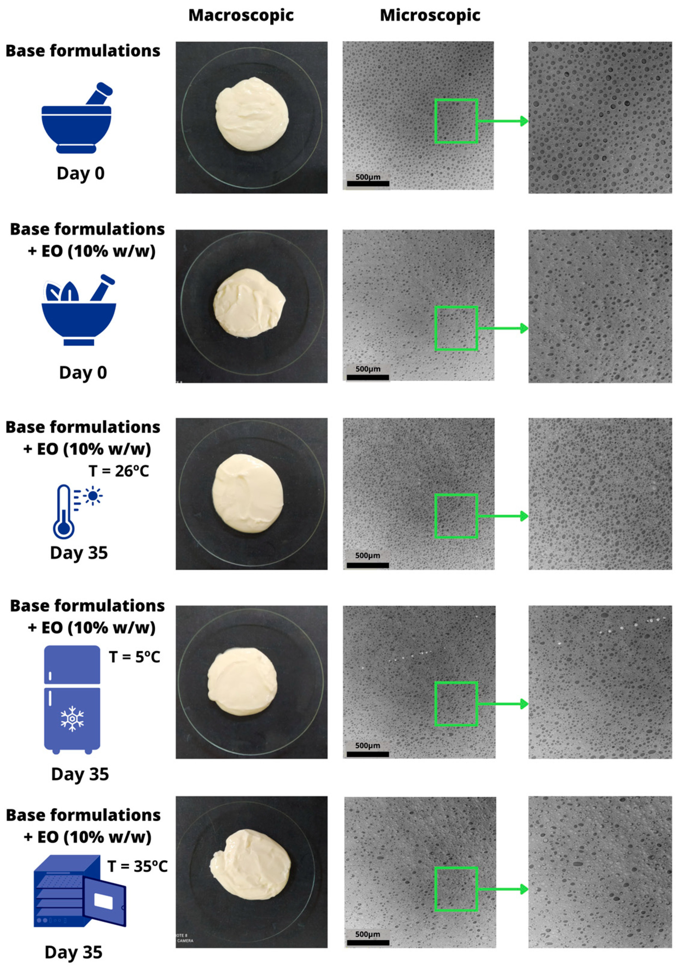
Samples of FB and F4, prepared aseptically and stored under various conditions, were analyzed at T0 and T1 to identify any changes that could indicate the need for reformulation in pharmaceutical development. Emulsion formation was confirmed by microscopic imaging, which revealed the presence of two distinct phases (Figure 2): in both FB and F4 samples, droplets of one phase dispersed within the other were observed, maintaining stability over time. Initially, FB and F4 appeared smooth, uniform, and free from clumps, and had a shiny, milky-white appearance, with F4 displaying the characteristic odor of EOs. After 35 days, minor changes were observed only in samples stored under refrigeration and in an oven, where an increase and decrease in consistency, respectively, were noted.
The emulsion type was confirmed by the appearance of a continuous blue color, indicating an oil-in-water (O/W) system (Supplementary Figure S1). The droplet diameter within the emulsion was also assessed, yielding values of 21.8 µm (±0.6) and 22.2 µm (±1.9) for FB and F4, respectively, classifying the system as coarse, consistent with droplet sizes ranging between 1 and 100 µm [37,43,44].
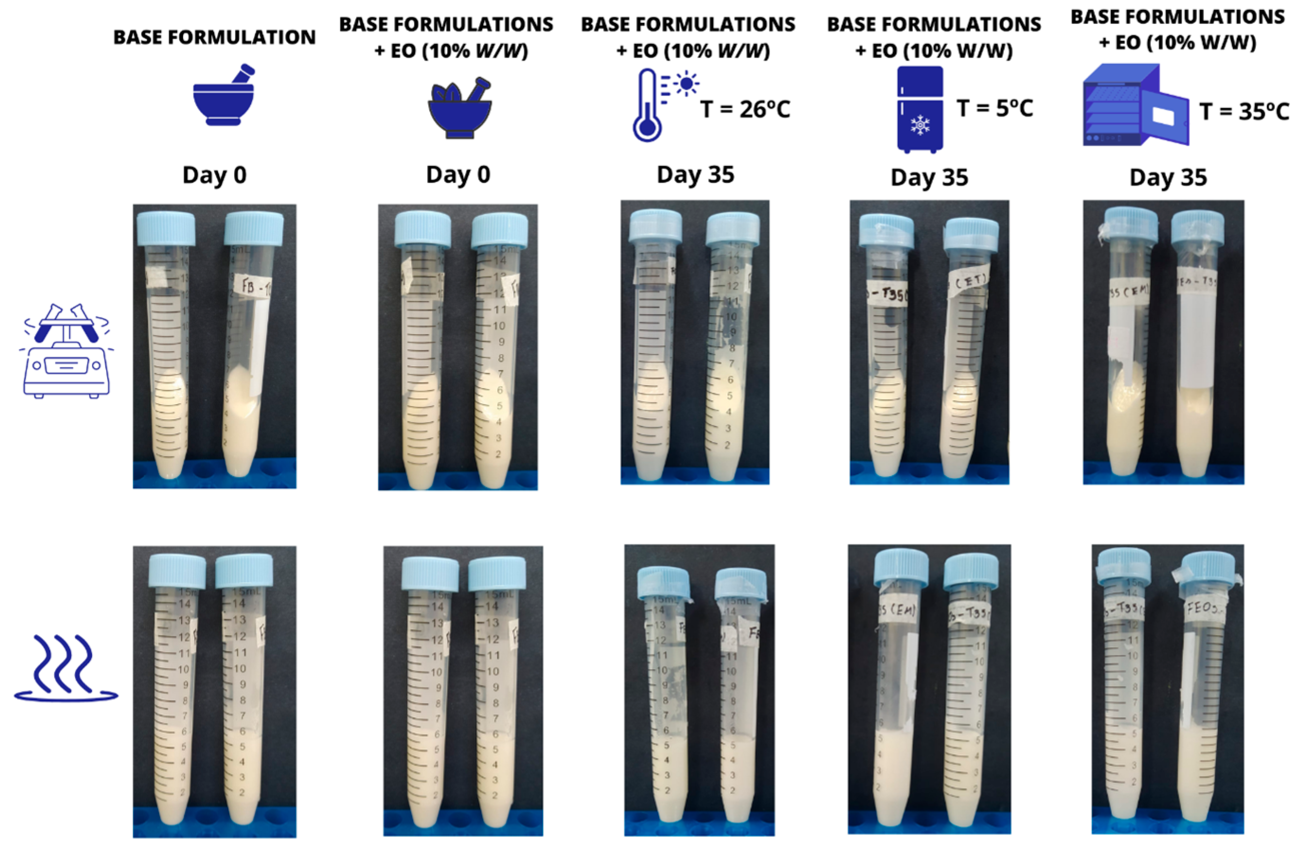
Following thermal and mechanical stress testing, no signs of instability, such as creaming, agglomeration, phase separation, or changes in color or odor, were observed (Figure 3). Similarly, after undergoing heating and cooling cycles at T0, both FB and F4 showed no detectable changes.
The pH of FB had a non-significant decrease after the addition of the EOs at T0 (Table 4). At T35, pH values showed a significant variation in comparison with the value observed for the formulation at T0, but no significant variation was observed when these values were compared to each other. Also, after each heating/cooling cycle, no significant variation was observed in the pH values of F4, which remained between 6.25 and 6.38.

3.4. Texture Profile Analysis, Water Loss and Film-Forming Capacity

The TPA results for formulations FB and F4 at T0 and T1 are presented in Table 5, detailing the parameters of hardness, adhesiveness, elasticity, and cohesiveness. Additionally, at T0, the water content in each formulation was determined using an infrared balance, yielding values of 23.42% (±0.71) for FB and 23.99% (±0.12) for F4.
Figure 4 illustrates the film formation by FB and F4 formulations, applied to glass slides and dried either at room temperature or in an oven for 5 min. When applied, the formulations formed a transparent layer, distinct from the original white emulsion layer. These films appeared thin, smooth, transparent, and soft, and could be easily removed without compromising their integrity. Confocal microscopy images of the deposited films are shown in detail.

4. Discussion

4.1. Composition and Antimicrobial Activity of the Essential Oils of Oregano and Thyme

EOs are complex mixtures of volatile bioactive compounds present in different anatomical parts of medicinal plants. They can be isolated by various processes and solvents. The main components of EOs are terpenes, aldehydes, alcohols, and esters [12,13,45]. The identification of the EO components in a phase before the pharmaceutical product formulation stage is essential to identify the presence of the bioactive compounds responsible for the pharmacological activity, since factors such as plant variety, geographic location, surrounding climate, seasonal variations, stress during growth, degree of crop growth, and drying and post-harvest storage conditions, along with the type of plant material used, the extraction and isolation method, and the type of solvent system used, all affect the chemical constitution of EOs [45,46,47].
The main components identified in the studied OEO and TEO, which are responsible for their antimicrobial activity, include thymol, carvacrol, linalool, and p-cymene [13,48,49,50,51,52,53,54,55,56,57]. The existing literature predominantly emphasizes carvacrol as the primary compound in OEO, a conclusion that contrasts with our findings. Specifically, studies by Rodrigues et al. [48], Maida et al. [52], and Waller et al. [54] have reported carvacrol concentrations of 71.8%, 73.9%, and 64.5%, respectively. In contrast, Cleff et al. [58] found that 4-terpineol accounted for 47.95% of the EO, with carvacrol and thymol present at levels of 9.43% and 8.42%, respectively. Additionally, Pensel et al. [53] reported a relative area of 20.14% for carvacrol.
The exact mechanisms by which these bioactive compounds exert their activity have not yet been fully elucidated. However, existing studies provide insights into their actions. According to some authors, these compounds interact with bacterial cell membranes primarily by hydrophobic interactions and hydrogen bonding. Such interactions disrupt the integrity of the lipid bilayer, leading to membrane distortion and permeabilization. Therefore, the fundamental barrier function of the membrane is compromised, causing the escape of intracellular contents and ions. This cascade of events substantiates their impact on bacterial homeostasis [59,60,61,62]. According to Almeida [63], carvacrol exerts its antimicrobial activity by disrupting the bacterial cell membrane, leading to the release of lipopolysaccharides, and ultimately resulting in cell lysis and death. The antimicrobial efficacy of thymol is attributed to its ability to bind to membrane proteins, thereby interfering with protein synthesis and altering membrane permeability, which facilitates the leakage of intracellular components and contributes to microbial death. Additional studies [51,61,64] indicate that carvacrol and thymol may disrupt the outer membrane of Gram-negative bacteria, increasing permeability and causing excessive release of ions and nucleic acids, ultimately leading to cell death. In addition to phenolic compounds, the monoterpenes present in both oils, such as p-cymene and γ-terpinene, are recognized for their potential to interact with essential enzymes in bacterial metabolism, resulting in inhibitory effects that imperil bacterial survival [65]. Furthermore, Silva [66] proposed that p-cymene enhances the transport of carvacrol across the cytoplasmic membrane, promoting its infiltration into bacterial cells and augmenting its antimicrobial effects.

4.2. Antimicrobial Activity of the Formulation

For the evaluation of antimicrobial activity, all dilutions assessed, including the growth control and formulations FB and FBSC, contained more than 250 CFUs, confirming the viability of the microorganisms and the suitability of the testing protocol. This result also indicates the absence of antimicrobial activity from the excipients in the formulations, specifically the preservative potassium sorbate, polysorbate 80, and poloxamer 407. In contrast, the commercially available synthetic antimicrobials, such as Mastizone® and Mastizone® V.S., exhibited complete inhibition of microbial growth. Total inhibition of growth was also observed for formulation F4, which contained 10% (w/w) essential oils, thereby justifying its selection for further study.
The selected formulation (F4) underwent an aseptic preparation process due to the requirement for sterile medicines administered via the IMM route for bovine mastitis treatment [67]. Emulsification was conducted in a laminar flow environment after sterilizing all ingredients and equipment used in the process. As previously mentioned, research by Dimitrova et al. [68] and Liu and Chu [69] highlights that poloxamer 407 and its gels maintain their properties after autoclaving, so the aqueous phase containing poloxamer 407 was sterilized by moist heat (autoclaving). Conversely, EOs are volatile, and their primary compounds are prone to oxidation, resulting in a loss of pharmacological activity. Hence, the oily phase underwent sterilization by filtration to maintain efficacy [70].
The inhibition of clinical strain proliferation by formulation F4, prepared under aseptic conditions, demonstrated a significant bactericidal effect, comparable to the efficacy of the commercially available products (Mastizone® and Mastizone® V.S.), both of which also reduced microbial load to 0 CFU. The administration of this test focused on analyzing the efficacy in inhibiting S. aureus, explained by the fact that this microorganism plays a crucial role in this pathological condition, being regarded as the primary agent responsible for mastitis cases. S. aureus is a highly significant contagious pathogen responsible for mammary infections in dairy cattle, posing a substantial threat to the dairy industry globally [6]. The virulence of S. aureus is attributed to complex biological mechanisms, including toxin production and the involvement of enzymes in tissue invasion. Additionally, it is well known for its ability to form biofilms, adhere to surfaces, and persist in various environments. Moreover, the emergence of AMR strains of S. aureus, including methicillin-resistant Staphylococcus aureus (MRSA), has intensified the challenge of effectively treating mastitis. These attributes make S. aureus a significant concern in livestock management due to its implications for the health of dairy cattle and the quality of milk production, highlighting the need to develop alternative therapeutic strategies, such as those based on natural compounds [6,71,72].
Upon comparing the composition of the EOs used in our study with the existing literature, we identified bioactive compounds known for their antimicrobial properties, specifically against bacteria associated with bovine mastitis, particularly S. aureus. This supports the incorporation of OEO and TEO in our formulations. Both OEO and TEO exhibit significant activity against multidrug-resistant microorganisms, reducing the resistance of these pathogens to synthetic antimicrobials and enhancing therapeutic efficacy when used in combination [14,15,59,71,72,73,74,75]. Moreover, the synergy between bioactive compounds from OEO and TEO offers a multifaceted approach to combating bacterial infections. Given that bacterial resistance often targets a singular action mechanism of an antimicrobial agent, the inclusion of multiple targets complicates the simultaneous development of resistance by bacteria. This strategy of employing multiple mechanisms of action has the potential to mitigate the emergence of bacterial resistance [13,35,62]. Thus, the combination of OEO and TEO in an alternative formulation represents a viable strategy for managing AMR.

4.3. Characterization and Preliminary Stability Assessment of the Chosen Formulation

Medicines for mastitis control are usually available in the form of solutions, suspensions, or ointments for IMM infusion. Information on the research of anti-mastitic emulsions for IMM use is limited. Mathur et al. [24] and Kitching et al. [76] obtained an emulsion based on liquid petrolatum and polysorbate 80 containing Lactococcus lactis DPC3147. Their results demonstrated therapeutic efficacy comparable to a commercially available suspension of cephalexin monohydrate and kanamycin for mastitis treatment, as observed in the present study for gentamicin sulfate and sodium cefoperazone. Alves et al. [77] formulated an emulsion utilizing an anionic self-emulsifying wax with soluble polypyrrole (PPS) for mastitis treatment. PPS is a polymer with conductive and antimicrobial capacity. Administering three doses of this experimental formulation via the IMM route led to greater reductions in microbial populations compared with control groups that received no treatment or gentamicin sulfate. The emulsion did not change the composition of the milk or the hematological parameters studied in the animals.
Emulsions are defined as pharmaceutical dosage forms consisting of two immiscible phases, in which one will be dispersed (internal or dispersed) in the other (external or continuous) in the form of droplets. The factors that influence the physical stability of the systems are the diameter of the droplets along with the viscosity, type and quantity of emulsifiers used [36,43,44,78]. Poloxamer is a non-ionic biocompatible block copolymer widely used in the pharmaceutical industry as an emulsifying agent due to its high solubilization capacity and good chemical compatibility [25,33]. No reports were found on the preparation of veterinary-use IMM emulsions containing natural ingredients using poloxamer 407 as the emulsifier.
Poloxamer 407 has been approved for use as an excipient by the Food and Drug Administration (FDA) [25,33,79,80]. An advantage in preparing poloxamer 407-based emulsified systems for use via IMM is that the formulations exhibit temperature-dependent gelation, with a gel state at body temperature (37 °C) and a fluid state at cold temperature (4 °C) [25,81]. The thermoreversible behavior of poloxamer 407 results from interactions between different segments of the copolymer. As the temperature increases, the copolymer molecules aggregate into micelles due to the dehydration of the hydrophobic propylene oxide blocks, marking the first step in the gelation process. Micellization is followed by gelation in sufficiently concentrated samples, attributed to the ordered packing of the micelles [25,28,81]. This behavior can be exploited for IMM administration to prevent the formulation from flowing away and increase the contact time with the application site [25,27]. Another advantage is that cold preparations favor the stability of thermosensitive ingredients such as oregano and thyme EOs [25,78].
Poloxamer 407 can play multiple roles in an emulsified system due to its amphiphilic nature and thickening capacity [82]. In the present study, we initially tested different concentrations of poloxamer 407 (5.0%, 10.0%, 20.0%, and 40.0% w/w) as an emulsifier and thickening agent. We observed that, at concentrations of 5.0% and 10.0% w/w, phase separation occurred during thermal and mechanical stress tests. At 40.0% w/w, the product consistency was very high, which could potentially compromise the administration and release of the EOs. Therefore, the concentration of 20.0% w/w was experimentally chosen for the continuation of this study.
Poloxamer 407 is an emulsifier capable of forming W/O or O/W systems [24,25]. Considering that EOs have a nonpolar nature, the system formed in the present study (O/W) favored the incorporation of the EOs in the internal phase of the oil droplets, stabilizing them against volatilization and oxidation. Similar to what was observed in the present study, Lucia et al. [83] prepared O/W emulsions of poloxamer 407 at 7.5% (w/w) containing different concentrations of thymol and carvacrol, obtaining a stable system with larvicidal activity. Ramos et al. [84] prepared micellar systems based on poloxamer 407-containing ethanolic extracts of the oiti fruit (Licania tomentosa) that originated stable O/W emulsions.
For the characterization of emulsions, the diameter and the variation in the diameter of the droplets are two parameters that must be known, as they contribute to the physical stability of systems. According to Stokes’ law, the sedimentation speed of droplets is directly proportional to their diameter. The viscosity of the system is also influenced by the diameter of the droplets [37,43,44,78]. In this sense, emulsified systems may require adjustments in the type or quantity of emulsifiers in the formulation, in the nature of the internal and external phase, or in the degree of division of the droplets [78,84,85,86]. Lucia et al. [87] prepared emulsions of monoterpenic bioactive compounds based on different proportions of poloxamer 407 (geraniol, citronellol, 1,8-cineole, linalool, α-terpineol, eugenol, thymol, menthol, and carvacrol) and observed mono- or bimodal distributions in droplet diameters, which were achieved on a nanometric scale and varied according to the included bioactive compound. In contrast, the emulsions produced in the present study were of a coarse type. However, no significant changes were observed when the formulations were subjected to preliminary stability studies, which included mechanical and thermal stress tests as well as heating and cooling cycles. These studies are instrumental in identifying the most stable formulations and provide critical insights into the potential integrity or coalescence of these dispersed systems [37,38,39,43,44,87]. Regarding the stress tests conducted on the samples stored for 35 days under varying temperature conditions, the results indicated that there was no occurrence of phase separation, creaming, agglomeration, or other alterations that would necessitate modifications to the formulations. Ferreira et al. [88] prepared emulsions of poloxamer 407 at a concentration of 15% (w/w) containing curcuminoids, and their findings suggested that the formulations maintained at 25 °C exhibited greater physical stability. After the heating/cooling cycles, no phase separation occurred in F4, contrary to what was observed for FB, indicating that the addition of EOs may contribute to stabilizing the system. Emulsions prepared by Ferreira and Bruschi [82] using poloxamer 407 at concentrations of 15.0, 17.5 and 20.0% (w/w) were subjected to freeze/thaw cycles, resulting in average droplet diameters ranging from 2.07 and 6.94 µm, depending on the type and amount of oil phase employed (sesame oil or isopropyl myristate) and the number of cycles analyzed. The droplet size analyses revealed that some formulations showed an increase in droplet size due to coalescence, indicating low stability and dependence on the composition of the polymer and the oil phase. No physical instability was observed in F4 throughout the duration of this study.
In the initial stages of pharmaceutical dosage form development, pH determination can yield valuable insights regarding the physicochemical stability prognosis of the formulations [37,38,39,43,44]. Although the pH of formulation F4 decreased after the addition of the EOs in FB, this variation was not deemed significant. However, after 35 days under different temperature conditions, a significant reduction in the pH of F4 was noted. The literature indicates that the addition of EOs in semi-solid or liquid formulations may lead to a reduction in pH due to the presence of acidic components in these ingredients [82]. Ramos et al. [84] attributed the reduction in pH in a poloxamer 407 emulsion to the rearrangement process of the micellar system, which released the incorporated extract and decreased the pH. When comparing the pH values of formulation F4 maintained under different temperature conditions, the observed variation was not considered significant. Additionally, the pH of F4 remained stable and did not exhibit significant variation when subjected to heating/cooling cycles at T0.

4.4. Texture Profile Analysis, Water Content, and Film-Forming Capacity

Texture profile analysis (TPA) is useful for defining attributes of interest of formulations related to ease of administration and removal along with the retention time of the product at the application site. Parameters such as hardness, adhesiveness, cohesiveness, elasticity and spreadability are defined based on data obtained during the insertion and removal of a probe in the samples [40,89].
According to Pandit et al. [90], hardness is related to a structure’s resistance to deformation and is represented by the maximum force required for a probe to deform a sample when penetrating it, providing information about consistency. Cohesiveness is a measure of the extent to which the sample can be deformed before its structure breaks. Adhesion measures the force necessary to completely remove the probe from the formulation, and is measured by the force necessary to overcome the force of attraction between the surface of the probe and the emulsion. Both parameters can be correlated to the application and spreadability. Elasticity denotes the ability of a formulation to stretch, which may be related to spreading. The lower its numerical value is, the higher the specification is of the formulation [90,91,92,93]. In this study, the inclusion of the EOs in the base formulation significantly increased only the hardness of the product. The texture parameters of the formulations stored at different temperatures for 35 days were compared with the initial values, and a significant increase in hardness, adhesiveness, and elasticity index was observed for the samples kept at room temperature and under refrigeration. In contrast, no parameters varied significantly for the samples kept in the oven. When the texture parameter values of the samples stored for 35 days under different conditions were compared with each other, only the hardness and adhesiveness of the sample kept in the oven showed a significant reduction.
The loss of water from emulsions can influence their mechanical parameters as well as the thixotropy and stability profile. Low water content can also result in an emulsion that is difficult to spread, whereas excess water can make it sticky and require longer drying time at the application site [37]. According to Umar et al. [94], the water content in emulsions capable of forming films will also influence the drying time, stickiness, and mechanical resistance of the films. We observed the ability of the formulations to form films. Upon application to glass slides, a transparent layer formed, distinct from the original white emulsion layer. Thin, smooth, and soft films were created, easily removable without losing integrity. Confocal microscopy analysis revealed crystalline structures in the FB and F4 films, with non-uniformly distributed organized materials, indicating structural organization after 5 min. This can be attributed to the loss of water due to the desolvation of poloxamer 407 during film formation. According to Fakhar-Ud and Khan [95], the film-forming characteristic of poloxamers occurs due to the increase in temperature and the loss of water in the system. The first factor directly influences the hydrogen bonds between the aqueous vehicle and the hydrophilic polymer chains (POE), disorganizing them. The loss of water and other solvents favors the interaction between the hydrophobic blocks (POP) in the polymer structure, resulting in micelle formation. The analysis of the images suggests that in F4, the polymer chains are organized around droplets of the oil phase, which is typical of the organization of poloxamer 407 and the formation of O/W systems. The film-forming capacity of the emulsion can be exploited to prevent the formation of microbial biofilm at the site of administration of the formulations, especially when applied externally to the teats of animals.
The veterinary product market has seen significant growth in recent years, with an anticipated increase of 7.4% from 2023 to 2024. Globally, sales of medicines intended exclusively for veterinary use are projected to exceed USD 42 billion by 2028 [96]. Despite this significant growth, particularly in the sector for farm animals, veterinary pharmacotherapy faces a critical challenge: the development of new active pharmaceutical ingredients (APIs) and pharmaceutical dosage forms tailored to diverse populations and health conditions [97,98]. This study seeks to address critical gaps within this framework, highlighting several significant aspects. Primarily, the incorporation of active ingredients derived from plant sources as antimicrobials aligns with two SDGs—specifically, Goals 3 (Good Health and Well-being) and 12 (Responsible Consumption and Production). This approach contributes to the mitigation of AMR through the use of sustainable natural resources. Upon confirming the presence of bioactive compounds with antimicrobial properties in the selected EOs, these ingredients were successfully incorporated into a formulation using poloxamer 407 as an emulsifier, resulting in an oil-in-water system characterized by the dispersion of droplets from the oil phase (including the EOs) in the aqueous phase. The cold emulsion preparation technique favored the stability of the EOs, while aseptic processing ensured the development of a sterile formulation. The formulation was effective in inhibiting S. aureus strains, the primary microorganism implicated in clinical mastitis globally. The in vitro antimicrobial screening tests conducted in this study are essential precursors to subsequent in vivo investigations, aligning with the global trend towards employing alternative models that reduce the reliance on animal testing. Furthermore, the investigation of a thermoreversible polymer capable of producing a formulation whose viscosity increases at body temperature presents a promising strategy for enhancing IMM administration and optimizing film-forming capacity. This innovative approach has the potential to improve therapeutic efficacy and compliance in veterinary practice, addressing a critical need in the management of bovine mastitis.

5. Conclusions

A sterile emulsion based on poloxamer 407 containing a 10% (w/w) blend of oregano and thyme EOs (89:11; OEO:TEO) was successfully developed using aseptic processing methods. This formulation demonstrated effective inhibition of the in vitro proliferation of standard and clinical strains of S. aureus isolated from the milk of dairy cows with mastitis, regardless of their sensitivity or resistance to synthetic antimicrobials. The successful formation of an O/W emulsion confirmed that the EOs are included in the internal phase of the system. When the EOs are incorporated into oil droplets, they are protected from volatilization, and their antimicrobial activity is preserved. No phase separation or other instability characteristics were observed in the samples stored under varying conditions throughout the study. pH and TPA results suggested that the formulation can be safely stored at room temperature or under refrigeration without significant alterations. Characterization and preliminary stability assessments of formulation F4, conducted according to ANVISA guidelines, showed no need for pharmacotechnical adjustments.
This study presents an innovative approach, since there are no existing reports of emulsions containing EOs formulated in poloxamer bases designed to deliver these active ingredients to the mammary glands and teats of animals for antimicrobial action against mastitis. While the results affirm the feasibility of this new formulation, further trials are essential to evaluate scale-up viability and conduct in vivo tests to assess the product’s safety and efficacy in reducing S. aureus counts in animals with clinical and subclinical mastitis.

Supplementary Materials

The following supporting information can be downloaded at: https://www.mdpi.com/article/10.3390/pr12122640/s1, Table S1: Description of clinical isolates from bovines with clinical mastitis [99]; Figure S1: Representative images of the formulations uniformly colored by the dye solution for observation of the type of emulsion formed (O/W): (A) FB and (B) F4.

Author Contributions

Conceptualization, J.A.R. and J.C.O.V.; methodology, J.A.R., J.C.O.V. and N.M.G.; software, L.M. and N.M.G.; validation, J.A.R. and J.C.O.V.; formal analysis, N.M.G.; investigation, N.M.G., N.S.F., K.V.M., C.S.N. and G.M.C.; resources, J.C.O.V., J.A.R. and L.M.; data curation, N.M.G., N.S.F. and C.S.N.; writing—original draft, N.M.G.; writing—review and editing, J.C.O.V.; visualization, N.M.G.; supervision, J.A.R. and J.C.O.V.; project administration, J.C.O.V.; funding acquisition, J.C.O.V. All authors have read and agreed to the published version of the manuscript.

Funding

This study was funded by the Foundation for the Support of Research and Innovation of Espírito Santo (FAPES), along with a master’s scholarship (Public Notice 03/2021; TO427/2021), and the Office to Coordinate Improvement of Higher Education Personnel (CAPES; Public Notices 13/2021; TO137/2021).

Data Availability Statement

All data are contained in the article. Complementary information can be requested from the corresponding author.

Conflicts of Interest

The authors declare no conflicts of interest.

References

  1. Kovačević, Z.; Mihajlović, J.; Mugoša, S.; Horvat, O.; Tomanić, D.; Kladar, N.; Samardžija, M. Pharmacoeconomic analysis of the different therapeutic approaches in control of bovine mastitis: Phytotherapy and antimicrobial treatment. Antibiotics 2023, 12, 11. [Google Scholar] [CrossRef] [PubMed]
  2. Krishnamoorthy, P.; Suresh, K.P.; Jayamma, K.S.; Shome, B.R.; Patil, S.S.; Amachawadi, R.G. An understanding of the global status of major bacterial pathogens of milk concerning bovine mastitis: A systematic review and meta-analysis (Scientometrics). Pathogens 2021, 10, 545. [Google Scholar] [CrossRef] [PubMed]
  3. United Nations. General Assembly Resolution A/RES/70/1. Transforming Our World, the 2030 Agenda for Sustainable Development. 2015. Available online: https://sdgs.un.org/2030agenda (accessed on 1 August 2024).
  4. Ashraf, A.; Imran, M. Causes, types, etiological agents, prevalence, diagnosis, treatment, prevention, effects on human health, and future aspects of bovine mastitis. Anim. Health Res. Rev. 2020, 21, 36–49. [Google Scholar] [CrossRef] [PubMed]
  5. Cheng, W.N.; Han, S.G. Bovine mastitis: Risk factors. therapeutic strategies and alternative treatments—A review. Asian-Australas. J. Anim. Sci. 2020, 33, 1699–1713. [Google Scholar] [CrossRef]
  6. Kerro, D.O.; Vidlund, J. Staphylococcal mastitis in dairy cows. Front. Vet. Sci. 2024, 11, 1356259. [Google Scholar] [CrossRef]
  7. Garcia, S.N.; Osburn, B.I.; Cullor, J.S. A one health perspective on dairy production and dairy food safety. One Health 2019, 7, 100086. [Google Scholar] [CrossRef]
  8. Langer, A.; Sharma, S.; Sharma, N.K.; Nauriyal, D.S. Comparative efficacy of different mastitis markers for diagnosis of sub-clinical mastitis in cows. Int. J. Appl. Sci. Biotechnol. 2014, 2, 121–125. [Google Scholar] [CrossRef]
  9. Chen, F.; Wang, Y.; Wang, K.; Chen, J.; Jin, K.; Peng, K.; Chen, X.; Liu, Z.; Ouyang, J.; Wang, Y.; et al. Effects of Litsea cubeba essential oil on growth performance, blood antioxidation, immune function, apparent digestibility of nutrients, and fecal microflora of pigs. Front. Pharmacol. 2023, 14, 1166022. [Google Scholar] [CrossRef]
  10. Wang, K.; Ma, J.; Li, Y.; Han, Q.; Yin, Z.; Zhou, M.; Luo, M.; Chen, J.; Xia, S. Effects of essential oil extracted from Artemisia argyi leaf on lipid metabolism and gut microbiota in high-fat diet-fed mice. Front. Nutr. 2022, 9, 1024722. [Google Scholar] [CrossRef]
  11. Mushtaq, S.; Shah, A.M.; Shah, A.; Lone, A.S.; Hussain, A.; Hassan, Q.P.; Ali, M.N. Bovine mastitis: An appraisal of its alternative herbal cure. Microb. Pathog. 2018, 114, 357–361. [Google Scholar] [CrossRef]
  12. Raut, J.S.; Karuppayil, S.M. A status review on the medicinal properties of essential oils. Ind. Crops Prod. 2014, 62, 250–264. [Google Scholar] [CrossRef]
  13. Chouhan, S.; Sharma, K.; Guleria, S. Antimicrobial activity of some essential oils—Present status and future perspectives. Medicines 2017, 4, 58. [Google Scholar] [CrossRef] [PubMed]
  14. Neculai-Valeanu, A.S.; Ariton, A.M.; Mădescu, B.M.; Rîmbu, C.M.; Creangă, Ş. Nanomaterials and essential oils as candidates for developing novel treatment options for bovine mastitis. Animals 2021, 11, 1625. [Google Scholar] [CrossRef] [PubMed]
  15. Caneschi, A.; Bardhi, A.; Barbarossa, A.; Zaghini, A. Plant essential oils as a tool in the control of bovine mastitis: An update. Molecules 2023, 28, 3425. [Google Scholar] [CrossRef] [PubMed]
  16. Buldain, D.; Gortari, C.L.; Buchamer, A.V.; Bandoni, A.; Marchetti, L.; Mestorino, N. In vitro synergistic interaction between Melaleuca armillaris essential oil and erythromycin against Staphylococcus aureus isolated from dairy cows. Front. Vet. Sci. 2022, 9, 1005616. [Google Scholar] [CrossRef]
  17. Arbab, S.; Ullah, H.; Bano, I.; Li, K.; Ul Hassan, I.; Wang, W.; Qadeer, A.; Zhang, J. Evaluation of in vitro antibacterial effect of essential oil and some herbal plant extract used against mastitis pathogens. Vet. Med. Sci. 2022, 8, 2655–2661. [Google Scholar] [CrossRef]
  18. Rota, C.R.; Herrera, A.; Martínez, R.M.; Sotomayor, J.A.; Jordán, M.J. Antimicrobial activity and chemical composition of Thymus vulgaris, Thymus zygis and Thymus yemalis essential oils. Food Control 2008, 19, 681–687. [Google Scholar] [CrossRef]
  19. Hussein, M.; Roby, H.; Atef, M.; Selim, K.A.; Ibrahim, K. Evaluation of antioxidant activity, total phenols and phenolic compounds in thyme (Thymus vulgaris L.), sage (Salvia officinalis L.), and marjoram (Origanum majorana L.) extracts. Ind. Crop Prod. 2013, 43, 827–831. [Google Scholar]
  20. Khodaei, M.M.; Kazemi, M.; Khaltabadi, F.A.H.; Yahyaei, M.; Rezaei, M.; De Rensis, F.; Taddei, S. Antibacterial effect of medicinal plant essence (Thymus vulgaris) on major bacterial mastitis pathogen in vitro. Int. J. Adv. Biol. Biomed. Res. 2014, 2, 286–294. [Google Scholar]
  21. Bora, L.; Burkard, T.; Juan, M.H.S.; Radeke, H.H.; Mut, A.M.; Vlaia, L.L.; Magyari-Pavel, I.Z.; Diaconeasa, Z.; Socaci, S.; Borcan, F.; et al. Phytochemical characterization and biological evaluation of Origanum vulgare L. essential oil formulated as polymeric micelles drug delivery systems. Pharmaceutics 2022, 14, 2413. [Google Scholar] [CrossRef]
  22. Sánchez-Arribas, N.; Guzmán, E.; Lucia, A.; Toloza, A.C.; Velarde, M.G.; Ortega, F.; Rubio, R.G. Environmentally friendly platforms for encapsulation of an essential oil: Fabrication, characterization and application in pests control. Colloids Surf. A Physicochem. Eng. Asp. 2018, 555, 473–481. [Google Scholar] [CrossRef]
  23. González-Reza, R.M.; Hernández-Sánchez, H.; Zambrano-Zaragoza, M.L.; Gutiérrez-López, G.F.; Del-Real, A.; Quintanar-Guerrero, D.; Velasco-Bejarano, B. Influence of stabilizing and encapsulating polymers on antioxidant capacity, stability, and kinetic release of thyme essential oil nanocapsules. Foods 2020, 9, 1884. [Google Scholar] [CrossRef] [PubMed]
  24. Mathur, H.; Linehan, K.; Flynn, J.; Byrne, N.; Dillon, P.; Conneely, M.; Grimaud, G.; Hill, C.; Stanton, C.; Ross, R.P. Emulsion-based postbiotic formulation is comparable to viable cells in eliciting a localized immune response in dairy cows with chronic mastitis. Front. Microbiol. 2020, 13, 759649. [Google Scholar] [CrossRef]
  25. Giuliano, E.; Paolino, D.; Fresta, M.; Cosco, D. Mucosal applications of poloxamer 407-based hydrogels: An overview. Pharmaceutics 2018, 10, 159. [Google Scholar] [CrossRef]
  26. Hua, S. Physiological and pharmaceutical considerations for rectal drug formulations. Front. Pharmacol. 2019, 10, 1196. [Google Scholar] [CrossRef]
  27. Lee, B.J.; Lee, T.S.; Cha, B.J.; Kim, S.H.; Kim, W.B. Percutaneous absorption, and histopathology of a poloxamer-based formulation of capsaicin analog. Int. J. Pharm. 1997, 159, 105–114. [Google Scholar] [CrossRef]
  28. Dumortier, G.; Grossiord, J.L.; Zuber, M.; Couarraze, G.; Chaumeil, J.C. Rheological study of a thermoreversible morphine gel. Drug Dev. Ind. Pharm. 1991, 17, 1255–1265. [Google Scholar] [CrossRef]
  29. Kim, C.K.; Lee, S.W.; Choi, H.G.; Lee, M.K.; Gao, Z.G.; Kim, I.S. Trials of in-situ gelling and mucoadhesive acetaminophen liquid suppository in human subjects. Int. J. Pharm. 1998, 174, 201–207. [Google Scholar] [CrossRef]
  30. Beard, M.C.; Cobb, L.H.; Grant, C.S.; Varadarajan, A.; Henry, T.; Swanson, E.A.; Kundu, S.; Priddy, L.B. Autoclaving of poloxamer 407 hydrogel and its use as a drug delivery vehicle. J. Biomed. Mater. Res. B Appl. Biomater. 2021, 109, 338–347. [Google Scholar] [CrossRef]
  31. Vigani, B.; Rossi, S.; Sandri, G.; Bonferoni, M.C.; Caramella, C.M.; Ferrari, F. Recent advances in the development of in situ gelling drug delivery systems for non-parenteral administration routes. Pharmaceutics 2020, 12, 859. [Google Scholar] [CrossRef]
  32. Bakhrushina, E.O.; Shumkova, M.M.; Sergienko, F.S.; Novozhilova, E.V.; Demina, N.B. Spray film-forming systems as promising topical in situ systems: A review. Saudi Pharm. J. 2023, 31, 154–169. [Google Scholar] [CrossRef] [PubMed]
  33. Sheskey, P.J.; Hancock, B.C.; Moss, G.P. Handbook of Pharmaceutical Excipients, 9th ed.; Libros Digitales, Pharmaceutical Press: London, UK, 2020; ISBN 978-0-8571-1375-7. [Google Scholar]
  34. Adams, R.P. Identification of Essential Oil Components by Gas Chromatography/Mass Spectrometry, 4th ed.; Allured Pub Corp.: Carol Stream, IL, USA, 2007; ISBN 978-1-932633-21-4. [Google Scholar]
  35. Beloni, M.V. Potencial Antimicrobiano e Antibiofilme da Combinação dos óleos Essenciais de Origanum vulgare e Thymus vulgaris para o Tratamento da Mastite Bovina. Master’s Thesis, Universidade Federal do Espírito Santo, Vitória, Brazil, 2022. [Google Scholar]
  36. Farmacopeia Brasileira, 6th ed.; Agência Nacional de Vigilância Sanitária: Brasília, Brasília, 2019; Volume 1.
  37. Swarbrick, J. Encyclopedia of Pharmaceutical Technology, 3rd ed.; Informa Healthcare Inc.: New York, NY, USA, 2007; ISBN 978-0-8493-9399-0. [Google Scholar]
  38. Agência Nacional de Vigilância Sanitária. Guia de Estabilidade dos Produtos Cosméticos, 1st ed.; Agência Nacional de Vigilância Sanitária: Brasília, Brazil, 2004. [Google Scholar]
  39. Pianovski, A.R.; Vilela, A.F.G.; da Silva, A.A.S.; Lima, C.G.; da Silva, K.K.; Carvalho, V.F.M.; de Musis, C.R.; Machado, S.R.P.; Ferrari, M. Use of pequi oil (Caryocar brasiliense) in cosmetics emulsions: Development and evaluate of physical stability. Braz. J. Pharm. Sci. 2008, 44, 249–259. [Google Scholar] [CrossRef]
  40. Tai, A.; Bianchini, R.; Jachowicz, J. Texture analysis of cosmetic/pharmaceutical raw materials and formulations. Int. J. Cosmet. Sci. 2014, 36, 291–304. [Google Scholar] [CrossRef] [PubMed]
  41. United States Pharmacopeia. USP Compounding Standards and Beyond-Use Dates (BUDs). 2019. Available online: https://www.usp.org/sites/default/files/usp/document/our-work/compounding/usp-bud-factsheet.pdf (accessed on 18 October 2024).
  42. Tortora, G.J.; Funke, B.R.; Case, C.L. Microbiologia, 12th ed.; Artmed: Porto Alegre, Brazil, 2017; ISBN 978-0-3219-2915-0. [Google Scholar]
  43. Aulton, M.E.; Taylor, K.M.G. Aulton’s Pharmaceutics: The Design and Manufacture of Medicines, 5th ed.; Elsevier: Toronto, ON, Canada, 2018; ISBN 978-0-7020-7005-1. [Google Scholar]
  44. Allen, L.V., Jr.; Popovich, N.G. Ansel’s: Pharmaceutical Dosage Forms and Drug Delivery Systems, 12th ed.; Lippincott Williams & Wilkins: Philadelphia, PA, USA, 2021; ISBN 978-1-9751-7177-3. [Google Scholar]
  45. Mancianti, F.; Ebani, V.V. Biological activity of essential oils. Molecules. 2020, 25, 678. [Google Scholar] [CrossRef]
  46. Hussain, A.I.; Anwar, F.; Sherazi, S.T.H.; Przybylski, R. Chemical composition, antioxidant, and antimicrobial activities of basil (Ocimum basilicum) essential oils depends on seasonal variations. Food Chem. 2008, 108, 986–995. [Google Scholar] [CrossRef]
  47. Leyva-López, N.; Gutiérrez-Grijalva, E.P.; Vazquez-Olivo, G.; Heredia, J.B. Essential oils of oregano: Biological activity beyond their antimicrobial properties. Molecules 2017, 22, 989. [Google Scholar] [CrossRef]
  48. Rodrigues, M.R.A.; Krause, L.C.; Caramão, E.B.; dos Santos, J.G.; Dariva, C.; de Oliveira, J.V. Chemical composition and extraction yield of the extract of Origanum vulgare obtained from sub-and supercritical CO2. J. Agric. Food Chem. 2004, 52, 3042–3047. [Google Scholar] [CrossRef]
  49. Pozzatti, P.; Loreto, E.S.; Mario, D.A.N.; Rossato, L.; Santurio, J.M.; Alves, S.H. Activities of essential oils in the inhibition of Candida albicans and Candida dubliniensis germ tube formation. J. Mycol. Med. 2010, 20, 185–189. [Google Scholar] [CrossRef]
  50. Escobar, P.; Leal, S.M.; Herrera, L.V.; Martinez, R.; Stashenko, E. Chemical composition and antiprotozoal activities of Colombian Lippia spp. essential oils and their major components. Mem. Inst. Oswaldo Cruz. 2010, 105, 184–190. [Google Scholar] [CrossRef]
  51. Chauhan, A.K.; Kang, S.C. Thymol disrupts the membrane integrity of Salmonella ser. typhimurium in vitro and recovers infected macrophages from oxidative stress in an ex vivo model. Res. Microbiol. 2014, 165, 559–565. [Google Scholar] [CrossRef]
  52. Maida, I.; Lo Nostro, A.; Pesavento, G.; Barnabei, M.; Calonico, C.; Perrin, E.; Chiellini, C.; Fondi, M.; Mengoni, A.; Maggini, V.; et al. Exploring the anti-burkholderia cepacia complex activity of essential oils: A preliminary analysis. Evid.-Based Complement. Altern. Med. 2014, 2014, 573518. [Google Scholar] [CrossRef] [PubMed]
  53. Pensel, P.E.; Maggiore, M.A.; Gende, L.B.; Eguaras, M.J.; Denegri, M.G.; Elissondo, M.C. Efficacy of essential oils of Thymus vulgaris and Origanum vulgare on echinococcus granulosus. Interdiscip. Perspect. Infect. Dis. 2014, 2014, 693289. [Google Scholar] [CrossRef] [PubMed]
  54. Waller, S.B.; Madrid, I.M.; Silva, A.L.; Dias de Castro, L.L.; Cleff, M.B.; Ferraz, V.; Meireles, M.C.; Zanette, R.; de Mello, J.R. In vitro susceptibility of Sporothrix brasiliensis to essential oils of Lamiaceae family. Mycopathologia 2016, 181, 857–863. [Google Scholar] [CrossRef] [PubMed]
  55. Mihaylova, S.; Tsvetkova, A.; Georgieva, E.; Vankova, D. Bioactive phyto-compounds with antimicrobial effects and AI: Results of a desk research study. Microorganisms 2024, 12, 1055. [Google Scholar] [CrossRef]
  56. Čabarkapa, I.; Čolović, R.; Duragić, O.; Popović, S.; Kokić, B.; Milanov, D.; Pezo, L. Anti-biofilm activities of essential oils rich in carvacrol and thymol against Salmonella enteritidis. Biofouling 2019, 35, 361–375. [Google Scholar] [CrossRef]
  57. Bouyahya, A.; Chamkhi, I.; Benali, T.; Guaouguaou, F.E.; Balahbib, A.; El Omari, N.; Taha, D.; Belmehdi, O.; Ghokhan, Z.; El Menyiy, N. Traditional use, phytochemistry, toxicology, and pharmacology of Origanum majorana L. J. Ethnopharmacol. 2021, 265, 113318. [Google Scholar] [CrossRef]
  58. Cleff, M.B.; Meinerz, A.R.M.; Schuch, L.F.D.; Rodrigues, M.R.A.; Meireles, M.C.A.; Mello, J.R.B. In vitro activity of the essential oil of Origanum vulgare against Sporothrix schenckii. Arq. Bras. Med. Vet. Zootec. 2008, 60, 513–516. [Google Scholar] [CrossRef]
  59. Hyldgaard, M.; Mygind, T.; Meyer, R.L. Essential oils in food preservation: Mode of action, synergies, and interactions with food matrix components. Front. Microbiol. 2012, 3, 12. [Google Scholar] [CrossRef]
  60. Mączka, W.; Twardawska, M.; Grabarczyk, M.; Wińska, K. Carvacrol—A natural phenolic compound with antimicrobial properties. Antibiotics 2023, 12, 824. [Google Scholar] [CrossRef]
  61. Pereira, A.A.; Piccoli, R.H.; Batista, N.N.; Camargos, N.G.; de Oliveira, M.M.M. Thermochemical inactivation of Escherichia coli, Staphylococcus aureus and Salmonella enterica enteritidis by essential oils. Cienc. Rural. 2014, 44, 2022–2028. [Google Scholar] [CrossRef]
  62. Gavaric, N.; Mozina, S.S.; Kladar, N.; Bozin, B. Chemical profile, antioxidant and antibacterial activity of thyme and oregano essential oils. thymol and carvacrol and their possible synergism. J. Essent. Oil-Bear. Plants. 2015, 18, 1013–1021. [Google Scholar] [CrossRef]
  63. Almeida, R.R. Mecanismos de Ação dos Monoterpenos Aromáticos: Timol e Carvacrol. Bachelor’s Thesis, Universidade Federal de São João Del-Rei, São João Del-Rei, Brazil, 2015. [Google Scholar]
  64. Nobre, M.S.V.; Camargos, L.F.M.; Soares, R.S.L.; Nogueira, T.D.A.M.C.; Freitas, M.L.A.; Oliveira, S.P.D.; Souza, C.N.D.; Almeida, A.C.D. Antibacterial activity of Origanum vulgare essential oil in front of Staphylococcus sp. isolates of minas artesanal cheese. Braz. J. Develop. 2021, 7, 38712–38720. [Google Scholar] [CrossRef]
  65. Soni, K.A.; Oladunjoye, A.; Nannapaneni, R.; Schilling, M.W.; Silva, J.L.; Mikel, B.; Bailey, R.H. Inhibition and inactivation of Salmonella typhimurium biofilms from polystyrene and stainless steel surfaces by essential oils and phenolic constituent carvacrol. J. Food Prot. 2013, 76, 205–212. [Google Scholar] [CrossRef] [PubMed]
  66. Burt, S. Essential oils: Their antibacterial properties and potential applications in foods—A review. Int. J. Food Microbiol. 2004, 94, 223–253. [Google Scholar] [CrossRef]
  67. Instrução Normativa nº 26, de 9 de julho de 2009. Regulamento Técnico para a Fabricação. o Controle de Qualidade. a Comercialização e o Emprego de Produtos Antimicrobianos de Uso Veterinário; Ministério da Agricultura. Pecuária e Abastecimento (MAPA): Brasília, Brazil, 2009; Seção1; pp. 1–972.
  68. Dimitrova, E.; Bogdanova, S.; Mitcheva, M.; Tanev, I.; Minkov, E. Development of model aqueous ophthalmic solution of indomethacin. Drug Dev. Ind. Pharm. 2000, 26, 1297–1301. [Google Scholar] [CrossRef]
  69. Liu, T.; Chu, B. Formation of homogeneous gel-like phases by mixed triblock copolymer micelles in aqueous solution: FCC to BCC phase transition. J. Appl. Cryst. 2000, 33, 727–730. [Google Scholar] [CrossRef]
  70. Guimarães, L.G.L.; Cardoso, M.G.; Zacaroni, L.M.; de Lima, R.K.; Pimentel, F.A.; Morais, A.R. Influence of light and temperature on the oxidation of lemongrass essential oil (Cymbopogon citratus (DC.) Stapf). Quím. Nova 2008, 31, 1476–1480. [Google Scholar] [CrossRef]
  71. Zhang, Z.; Chen, Y.; Li, X.; Wang, X.; Li, H. Detection of antibiotic resistance. virulence gene and drug resistance gene of Staphylococcus aureus isolates from bovine mastitis. Microbiol. Spectr. 2022, 10, e0047122. [Google Scholar] [CrossRef]
  72. Neelam, J.V.K.; Singh, M.; Joshi, V.G.; Chhabra, R.; Singh, K.; Rana, Y.S. Virulence and antimicrobial resistance gene profiles of Staphylococcus aureus associated with clinical mastitis in cattle. PLoS ONE 2022, 17, e0264762. [Google Scholar] [CrossRef]
  73. Lambert, R.J.; Skandamis, P.N.; Coote, P.J.; Nychas, G.J. A study of the minimum inhibitory concentration and mode of action of oregano essential oil. thymol and carvacrol. J. Appl. Microbiol. 2001, 91, 453–462. [Google Scholar] [CrossRef]
  74. Marchese, A.; Arciola, C.R.; Barbieri, R.; Silva, A.S.; Nabavi, S.F.; Sokeng, A.J.T.; Izadi, M.; Jafari, N.J.; Suntar, I.; Daglia, M.; et al. Update on monoterpenes as antimicrobial agents: A particular focus on p-cymene. Materials 2017, 10, 947. [Google Scholar] [CrossRef] [PubMed]
  75. Silva, J.P.L.; Duarte-Almeida, J.M.; Perez, D.V.; Franco, B.D.G.M. Oregano essential oil: Influence of the chemical composition on the inhibitory activity against Salmonella enteritidis. Food Sci. Technol. 2010, 30, 136–141. [Google Scholar] [CrossRef]
  76. Kitching, M.; Mathur, H.; Flynn, J.; Byrne, N.; Dillon, P.; Sayers, R.; Rea, M.C.; Hill, C.; Ross, R.P. A live bio-therapeutic for mastitis, containing Lactococcus lactis DPC3147 with comparable efficacy to antibiotic treatment. Front. Microbiol. 2019, 10, 2220. [Google Scholar] [CrossRef] [PubMed]
  77. Alves, A.P.P.; do Amaral, M.P.; da Silva, D.C.N.; Souza, R.F.S.; Reis, S.A.G.B.; Júnior, F.A.G.S.; de Oliveira, H.P.; Peixoto, R.M.; da Costa, M.M. Efficacy assessment of an intramammary formulation based on soluble polypyrrole in cows with experimentally induced mastites. Cienc. Rural. 2023, 53, e20220047. [Google Scholar] [CrossRef]
  78. Franzol, A.; Rezende, M.C. Emulsion stability: A case study involving anionic, cationic, and nonionic emulsifiers. Polímeros 2015, 25, 1–9. [Google Scholar] [CrossRef]
  79. Tadros, T. Viscoelastic properties of sterically stabilised emulsions and their stability. Adv. Colloid. Interface Sci. 2015, 222, 692–708. [Google Scholar] [CrossRef]
  80. Nugraha, D.H.; Anggadiredja, K.; Rachmawat, H. Mini-review of poloxamer as a biocompatible polymer for advanced drug delivery. Braz. J. Pharm. Sci. 2022, 58, e21125. [Google Scholar] [CrossRef]
  81. Jones, D.S.; Bruschi, M.L.; de Freitas, O.; Gremião, M.P.; Lara, E.H.G.; Andrews, G.P. Rheological, mechanical and mucoadhesive properties of thermoresponsive. bioadhesive binary mixtures composed of poloxamer 407 and carbopol 974P designed as platforms for implantable drug delivery systems for use in the oral cavity. Int. J. Pharm. 2009, 372, 49–58. [Google Scholar] [CrossRef]
  82. Ferreira, S.B.S.; Bruschi, M.L. Investigation of the physicochemical stability of emulgels composed of poloxamer 407 and different oil phases using the Quality by Design approach. J. Mol. Liq. 2021, 332, 115856. [Google Scholar] [CrossRef]
  83. Lucia, A.; Girard, C.; Fanucce, M.; Coviella, C.; Rubio, R.G.; Ortega, F.; Guzmán, E. Development of an environmentally friendly larvicidal formulation based on essential oil compound blend to control Aedes aegypti larvae: Correlations between physicochemical properties and insecticidal activity. ACS Sustain. Chem. Eng. 2020, 8, 10995–11006. [Google Scholar] [CrossRef]
  84. Ramos, Y.J.; da Silva, N.Q.; Costa-Oliveira, C.; Pereira, R.A.; Morra, M.D.; Cerqueira-Silva, L.C.R. Obtaining and evaluation of micellar systems containing oiti fruit extract [Licania tomentosa (Benth.) Fritsch]. Rev. Fitos 2020, 14, 88–102. [Google Scholar] [CrossRef]
  85. Gündel, S.S.; de Souza, M.E.; Quatrin, P.M.; Klein, B.; Wagner, R.; Gündel, A.; Vaucher, R.A.; Santos, R.C.V.; Ourique, A.F. Nanoemulsions containing Cymbopogon flexuosus essential oil: Development. characterization. stability study and evaluation of antimicrobial and antibiofilm activities. Microb. Pathog. 2018, 118, 268–276. [Google Scholar] [CrossRef] [PubMed]
  86. Prestes, O.S.; Peres, D.D.; de Freitas, A.Z.; Consiglieri, V.O.; Kaneko, T.M.; Velasco, M.V.R.; Baby, A.R. Particle size and morphological characterization of cosmetic emulsified systems by Optical Coherence Tomography (OCT). Braz. J. Pharm. Sci. 2016, 52, 273–280. [Google Scholar] [CrossRef]
  87. Lucia, A.; Toloza, A.C.; Guzmán, E.; Ortega, F.; Rubio, R.G. Novel polymeric micelles for insect pest control: Encapsulation of essential oil monoterpenes inside a triblock copolymer shell for head lice control. Peer J. 2017, 20, e3171. [Google Scholar] [CrossRef]
  88. Ferreira, S.B.S.; Braga, G.; de Oliveira, E.L.; Rosseto, H.C.; Hioka, N.; Caetano, W.; Bruschi, M.L. Colloidal systems composed of poloxamer 407, different acrylic acid derivatives and curcuminoids: Optimization of preparation method, type of bioadhesive polymer and storage conditions. J. Drug Deliv. Sci. Technol. 2020, 57, 101686. [Google Scholar] [CrossRef]
  89. Calixto, L.S.; Maia-Campos, P.M.B.G. Physical-mechanical characterization of cosmetic formulations and correlation between instrumental measurements and sensorial properties. Int. J. Cosmet. Sci. 2017, 39, 527–534. [Google Scholar] [CrossRef]
  90. Pandit, A.P.; Pol, V.V.; Kulkarni, V.S. Xyloglucan based in situ gel of lidocaine hcl for the treatment of periodontosis. J. Pharm. 2016, 2016, 3054321. [Google Scholar] [CrossRef]
  91. Erdem, Y.K. Effect of ultrafiltration, fat reduction and salting on textural properties of white brined cheese. J. Food Eng. 2005, 71, 366–372. [Google Scholar] [CrossRef]
  92. Gilbert, L.; Savary, G.; Grisel, M.; Picard, C. Predicting sensory texture properties of cosmetic emulsions by physical measurements. Chemom. Intell. Lab. Syst. 2013, 124, 21–31. [Google Scholar] [CrossRef]
  93. Lau, M.H.; Tang, J.; Paulson, A.T. Texture profile and turbidity of gellan/gelatina mixed gels. Food Res. Int. 2000, 33, 665–671. [Google Scholar] [CrossRef]
  94. Umar, A.K.; Butarbutar, M.; Sriwidodo, S.; Wathoni, N. Film-forming sprays for topical drug delivery. Drug Des. Devel Ther. 2020, 14, 2909–2925. [Google Scholar] [CrossRef] [PubMed]
  95. Fakhar-Ud, D.; Khan, G.M. Development and characterisation of levosulpiride-loaded suppositories with improved bioavailability in vivo. Pharm. Dev. Technol. 2019, 24, 63–69. [Google Scholar] [CrossRef] [PubMed]
  96. The Business Research Company. Veterinary Healthcare Market Reports 2024. 2024. Available online: https://www.thebusinessresearchcompany.com/reports/veterinary-healthcare-market-research (accessed on 20 October 2024).
  97. Ahmed, I. Pharmaceutical challenges in veterinary product development. Adv. Drug Del. Rev. 2002, 54, 871–882. [Google Scholar] [CrossRef] [PubMed]
  98. Rathbone, M.; Brayden, D. Controlled release drug delivery in farmed animals: Commercial challenges and academic opportunities. Curr. Drug Deliv. 2009, 6, 383–390. [Google Scholar] [CrossRef] [PubMed]
  99. M100 ED30:2020; Performance Standards for Antimicrobial Susceptibility Testing. 30th ed. Clinical and Laboratory Standards Institute: Wayne, PA, USA, 2020.
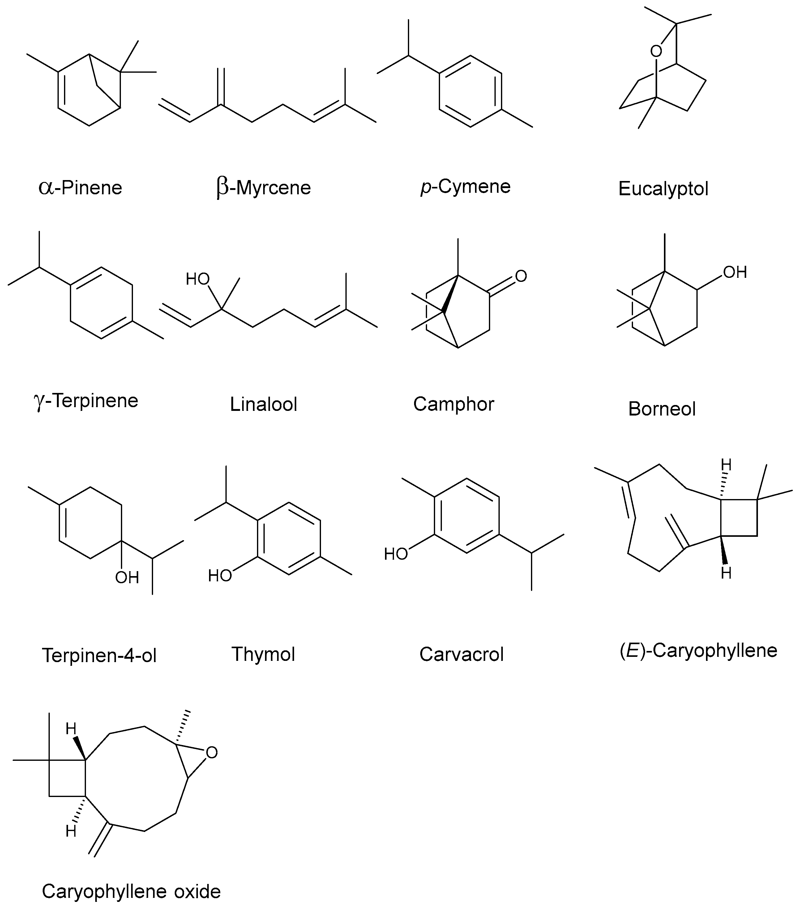
Figure 1. Chemical structure of the major constituents of the thyme and oregano EOs.
Processes 12 02640 g001
Figure 2. Representative macroscopic and microscopic images of FB and F4 maintained under different conditions over time. In microscopy, the green box highlights a region of the emulsion that was magnified for detailed analysis of its structural characteristics.
Processes 12 02640 g002
Figure 3. Representative photographic images of FB and F4, at times T0 and T35, after mechanical (centrifuge) and thermal (water bath) stress tests, showing the absence of signs of instability.
Processes 12 02640 g003
Figure 4. Representative image of films formed by FB (base formulation) and F4 (base formulation with EOs) on glass slides, dried at room temperature and in the oven for 5 min. The detail shows confocal microscopy images of regions of the films. The green box highlights a region of the emulsion that has been magnified for detailed analysis of its structural features.
Processes 12 02640 g004
Table 1. Emulsified systems’ qualitative and quantitative composition.
Components FBFBSCF1F2F3F4
Proportions % (w/w)
AP
Poloxamer 40720.020.020.020.020.020.0
Potassium sorbate0.15-0.150.150.150.15
Vitamin E0.10.10.10.10.10.1
Polysorbate 80 1.01.01.01.01.01.0
Purified water SQ100.0100.0100.0100.0100.0100.0
OP
OEO--2.243.104.488.96
TEO--0.260.400.521.04
Sunflower seed oil SQ100.0100.0100.0100.0100.0100.0
AP: aqueous phase; OP: oil phase; FB: base formulation; FBSC: preservative-free base formulation; F1: formulation with 2.5% (w/w) of EOs; F2: formulation with 3.5% (w/w) of EOs; F3: formulation with 5.0% (w/w) of EOs; F4: formulation with 10.0% (w/w) of EOs; SQ: sufficient quantity to complete the final weight.
Table 2. The main constituents identified in thyme and oregano EOs [a].
ComponentsKovat’s Index [b]Kovat’s Index [c]Area % [d]
TEOOEO
α-pinene9319322.40-
β-myrcene9909881.08-
p-cymene1021102020.367.27
Eucalyptol103310261.231.51
γ-terpinene105710547.544.84
Linalool109910958.054.24
Camphor114711411.971.15
Borneol116811651.251.30
Terpinen-4-ol119011861.331.18
Thymol1290128947.8869.22
Carvacrol129812984.063.44
(E)-caryophyllene141914171.255.85
Caryophyllene oxide158315821.60-
Hydrogenated monoterpenes31.3812.11
Oxygenated monoterpenes65.7782.04
Hydrogenated sesquiterpenes1.255.85
Oxygenated sesquiterpenes1.60-
[a] Components identified by the LTPRI index and by GC-MS with relative area > 1% using an Rtx®-5 MS column. [b] Calculated using a mixture of saturated n-alkanes (C7 to C40). [c] Indices tabulated based on Adams (2007). [d] % relative area by CG-FID.
Table 3. Evaluation of the antimicrobial activity against ATCC S. aureus.
ProductsPopulation Density (CFU g−1)
FBCountless
FBSCCountless
F1Countless
F2Countless
F3Countless
F40
Growth controlCountless
Mastizone®0
Mastizone® V.S.0
FB: base formulation; FBSC: preservative-free base formulation; F1: formulation with 2.5% (w/w) of EOs; F2: formulation with 3.5% (w/w) of EOs; F3: formulation with 5% (w/w) of EOs; F4: formulation with 10% (w/w) of EOs.
Table 4. Average pH variations throughout the study.
T0T1 (35 Days)
FBF4F4
RTURO
6.376.165.855.765.58
(±0.05) NS(±0.04) NS(±0.04) S[a](±0.07) S[a](±0.03) S[a]
FB: base formulation; F4: formulation with 10% (w/w) of EOs; RT: room temperature; UR: under refrigeration; O: oven. Comparison between F4 and FB at T0: NS refers to no significant difference and S refers to significant difference (p > 0.05). Comparison between samples kept in different conditions after 35 days: [a] refers to no significant difference (p > 0.05).
Table 5. Average values of TP parameters for FB and F4.
ParametersFB
(T0)
F4
T0T1 (35 Days)
RTURO
Hardness (g)123.33 (± 10.02) S191.33 (± 26.10) S308.33
(± 27.30) S[a]
278.00
(± 60.10) S[a]
194.33
(± 21.57) NS[b]
Adhesiveness (mJ)6.47 (± 0.76) NS6.57 (± 2.02) NS16.20
(± 2.39) S[a]
17.40
(± 4.86) S[a]
5.67
(± 2.96) NS[b]
Elasticity index (mm)7.72 (± 0.27) NS6.01 (± 1.89) NS13.80
(± 1.13) S[a]
14.37 (± 1.81) S[a]11.32
(± 4.69) NS[a]
Cohesiveness 0.77 (± 0.10) NS0.65 (± 0.19) NS0.62
(± 0.15) NS[a]
0.49
(± 0.06) NS[b]
0.64
(± 0.06) NS[a]
FB: base formulation; F4: base formulation with 10% (w/w) of EOs; RT: room temperature; UR: under refrigeration; O: oven. Comparison between F4 and FB at T0 and T35: NS refers to no significant difference and S refers to significant difference (p > 0.05). Comparison between samples kept in different conditions after 35 days: [a] refers to no significant difference and [b] to significant difference (p > 0.05).
Disclaimer/Publisher’s Note: The statements, opinions and data contained in all publications are solely those of the individual author(s) and contributor(s) and not of MDPI and/or the editor(s). MDPI and/or the editor(s) disclaim responsibility for any injury to people or property resulting from any ideas, methods, instructions or products referred to in the content.