Article

Evaluation of a Resorcinarene-Based Sorbent as a Solid-Phase Extraction Material for the Enrichment of L-Carnitine from Aqueous Solutions

by
Gabriel Ramirez
,
Nicolas Alejandro Cadavid-Montoya
and
Mauricio Maldonado
*
Facultad de Ciencias, Departamento de Química, Universidad Nacional de Colombia Sede Bogotá, Carrera 30 No. 45-03, Bogotá 111231, Colombia
*
Author to whom correspondence should be addressed.
Processes 2023, 11(6), 1705; https://doi.org/10.3390/pr11061705
Submission received: 22 April 2023 / Revised: 27 May 2023 / Accepted: 29 May 2023 / Published: 2 June 2023
(This article belongs to the Special Issue Transport Processes in Polymeric Aqueous Systems)

Abstract

:
We present five new sorbents for the evaluation of the pre-concentration of L-carnitine. The sorbents were obtained from copolymerization between butylmethacrylate (BuMA) and ethylene dimethacrylate (EDMA), which were physically modified on their surface by each one of the five synthetized resorcinarenes of variable chain length studied, with long-chain resorcinarenes exhibiting better fixation on the copolymer. The characterization of the synthesized resorcinarenes was done using FTIR-ATR, 1H NMR, 13C NMR, and mass spectrometry, while characterization of the sorbents was done using FTIR-ATR and scanning electron micrography (SEM). The molecular interaction between L-carnitine and the synthesized resorcinarenes was studied in DMSO via 1H-NMR spectroscopy and, in the gas phase, via electrospray ionization-mass spectrometry (ESI-MS). The results showed that the short-chain resorcinarenes underwent a stable interaction with the neurotransmitter. Once the sorption of resorcinarenes on the copolymer was accomplished, the best parameters for the evaluation of the L-carnitine preconcentration were established. The solution tests were carried out through LC/MS analysis, obtaining better results for L-carnitine absorption with the short-chain resorcinarenes such as tetra(ethyl)calix[4]resorcinarene and tetra(propyl)calix[4]resorcinarene.

1. Introduction

Some amino acids and compounds derived from them have the ability to participate in metabolic or cellular communication processes as neurotransmitters that chemically regulate neuronal synapses by selectively acting on receptor proteins located in the cell membrane. In general, these chemical transmitters can be classified as high molecular weight neuroactive polypeptides, as well-known amino acids and their derivatives, or as organic compounds (such as hormones) of low molecular weight [1,2], whose bioactive stage can generate any type of response, inhibition, or synaptic excitation. L-carnitine is a trimethylammonio carboxilate synthesized from methionine and lysine that is found in the human diet and plays an important role in fatty acid oxidation; consequently, quantification of carnitine levels in the human body can be used to diagnose disorders of fatty acid oxidation [1,2,3]. Several methods for the detection of carnitine are found in the literature, given its great importance in the human diet [3,4,5]. Among the various methods that have been described for detecting this organic cation, the following should be highlighted: analysis with biosensors [6], radioactive markers [6,7,8], chromatographic methods [9,10,11,12], capillary electrophoresis [13], and fluorescent methods [14,15,16]. Additionally, over the last few decades, advances have been reported in the detection of carnitine through supramolecular chemistry [17,18].
Of these methods, HPLC-MS is currently the most widely used for the analysis of carnitine; however, one of the great difficulties is the matrix effect, which prevents adequate detection of the analyte [19,20]. A possible solution for the problems stemming from the matrix effect could be the application of the solid phase extraction (SPE) technique as a sample pre-treatment. SPE is employed to enrich the analyte and to desalt the samples, but with polar analytes such as carnitine, retention in the SPE cartridge is not efficient [21]. A recent solution to this problem has been the development and application of sorbents with greater selectivity towards carnitine, using, for example, polymer surfaces modified with crown ether macrocycles [22,23] or other macrocyclic systems such as resorcinarenes [24,25].
Resorcinarenes are oligomeric macrocyclic tetramers [26] that can adopt the crown conformation [22] and whose monomer is a resorcinol that forms an upper rim of hydrophilic or polar character. These four resorcinol nuclei are joined together through binding carbons that can be covalently linked to different functional groups, depending on the precursor aldehyde used for their synthesis. These four chains form a lower rim in the molecule, normally of a hydrophobic character; however, this property can be modified with the precursors mentioned above. These compounds are interesting because they have been used in the functionalization of monolithic columns [27], as surfactants [28], for the manufacture of nanomaterials [29], as catalysts in organic chemistry [30], and also as receptors of biomolecules [31], ammonium ions [32,33,34], and neurotransmitters [7,35], among others [36,37]. Within the kinds of functionalization that have been shown, several of these types of macrocycles can be used in processes of detection of organic molecules.
Resorcinarenes, as mentioned above, have been used as modifiers of polymeric materials via physical impregnation. In this process, the macrocyclic system is fixed to the polymer by physisorption [23,24,37], which can be efficient depending on the length of the alkyl chain on the lower rim of the macrocycle. In this context, the present paper describes the use of the solid-phase extraction procedure based on the use of modified polymeric sorbents for the selective extraction of L-carnitine. For this purpose, we used methacrylate-based polymers because they exhibit a high degree of porosity, large surface area, and chemical stability, as well as low mechanical strength. In the same way, physical modification of the polymer was carried out with five synthetized resorcinarenes. L-carnitine retention efficiency was assessed for the modified surfaces by examining the effects of time of contact, pH, and volume of the sample on the effective sorption. The experimental results showed that the long-chain resorcinarenes exhibit better fixation on the copolymer; however, they are not the ones that interact best with L-carnitine. The sorbent poly(BuMA-co-EDMA) modified with tetra(ethyl)calix[4]resorcinarene exhibits excellent selectivity for L-carnitine, showing great application value for analytical methods important in the biological, biomedical, and pharmaceutical areas.

2. Materials and Methods

All chemical reagents were acquired from commercial suppliers and used without further purification. IR spectra were recorded on a Thermo Fisher Scientific Nicolet iS10 FT-IR spectrometer with the monolithic diamond ATR accessory and absorption in cm−1. 1H-NMR spectra were recorded in DMSO-d6 at 400 MHz on a Bruker Avance 400 instrument. Chemical shifts are reported in ppm, using the residual solvent signal as a reference. For liquid chromatography-mass spectrometry (LC/MS), the samples were centrifuged at 15,000 rpm for 3 min at room temperature, the supernatant was diluted 1000 times, and 2 µL was analyzed in a Bruker Impact II LC/MS Q-TOF equipped with electrospray ionization (ESI) in positive mode. The chromatographic conditions were as follows: an intensity C18 column (2.1 × 100 mm, 1.8 μm) (Bruker Daltonik, Billerica, MA, USA) at a temperature of 40 °C and a flow rate of 0.250 mL min−1. Mobile phase water (A) and acetonitrile (B), each containing 0.1% formic acid, were used. Isocratic elution B 5% was used for 2 min. The ESI source conditions were as follows: end plate offset 500 V, capillary 4500 V, nebulizer 1.8 bar, dry gas nitrogen 8.0 L/min, and dry temperature 220 °C. The morphological characterization of the copolymer and the physical modification of the resorcinarenes was done using a QUANTA 200 (FEI) scanning electron microscope with secondary electron and backscattered electron detectors and an EDAX probe. The samples, being non-conductive, were pretreated with a gold metallic coating and carbon evaporation equipment. For particle reduction of the synthesized compounds, a 0.8″ × 2″ high ASTM E11 analytical sieve with an aperture of 106 μm with 140 mesh from Endecotts was used. Melting points were taken on an Electrothermal 9100 SMP11 melting point meter with a heating rate of 1 to 10 °C per min from Stuart-Equipment.

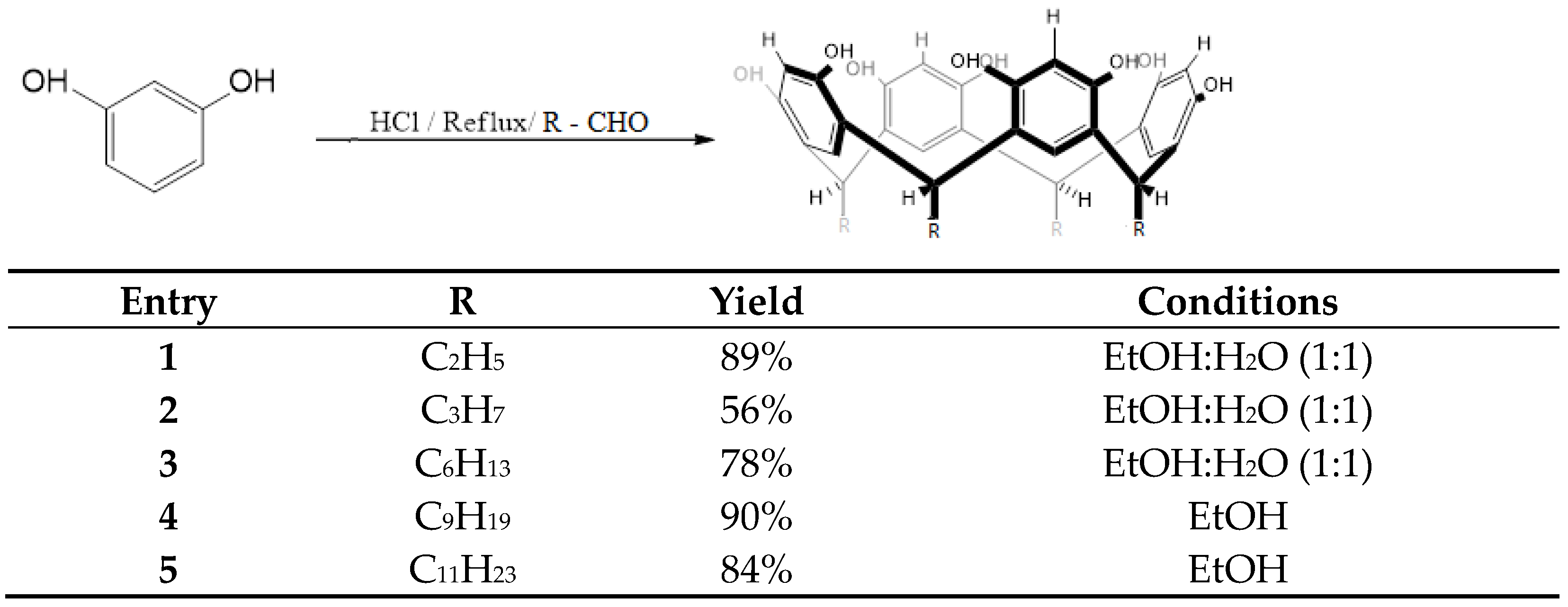
2.1. Synthesis of calix[4]resorcinarenes

The procedure for obtaining the macrocycles was adapted from previous research [24,26,38]. Hydrochloric acid (3 mL) was carefully added to a solution of resorcinol (10 mmol) and the respective aldehyde was slowly added (10 mmol), in a 1:1 ethanol/water mixture (20 mL) for short-chain-length resorcinarenes (1–3), while pure ethanol was used for long-chain-length resorcinarenes (4, 5) due to aldehydes’ insolubility. The reaction mixture was reacted at reflux with constant stirring for 8–12 h and subsequently cooled in an ice bath down to 0 °C, then filtered and washed with water. Finally, the filtrate was dried under a vacuum and was characterized via FTIR-ATR, 1H-NMR, 13C-NMR, and ESI-MS. In this way, five resorcinarenes were obtained: tetra(ethyl)calix[4]resorcinarene (1), tetra(propyl)calix[4]resorcinarene (2), tetra(hexyl)calix[4]resorcinarene (3), tetra(nonyl)calix[4]resorcinarene (4), and tetra(undecyl)calix[4]resorcinarene (5). The spectroscopic data is shown in Table 1.

2.2. Studies of Molecular Interaction between Resorcinarenes and L Carnitine
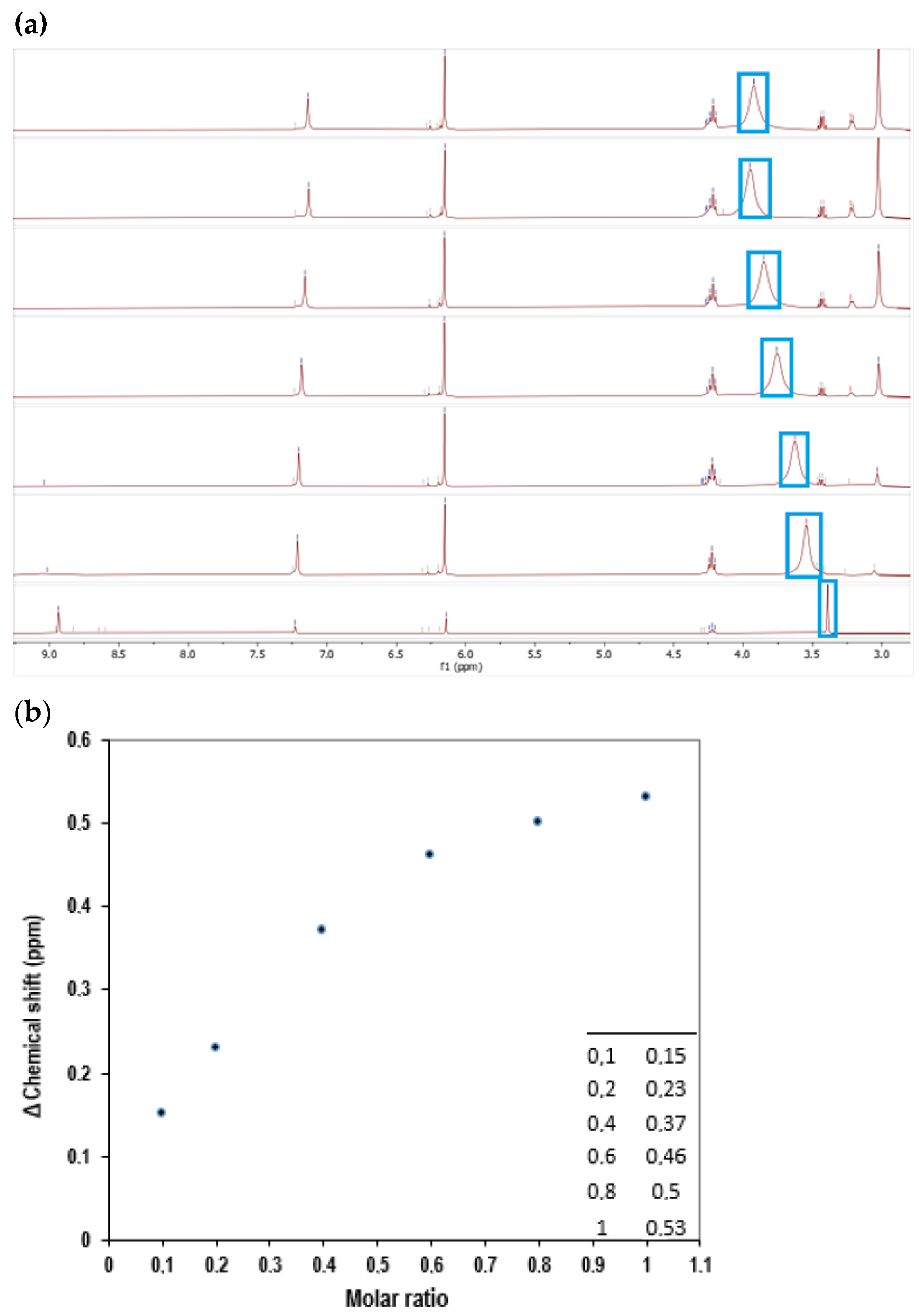
2.2.1. NMR Studies

The binding ability of macrocycles 1, 2, and 3 with L-carnitine was studied in DMSO-d6 via 1H-NMR. A solution with a concentration of 1.2–2 mg/mL of the macrocycle host was treated with various volume amounts of L-carnitine, according to different molar ratios set between 1:0,1 and 1:2. After each addition, 1H-NMR was recorded. It should be clarified that the initial concentrations of carnitine varied between 13 and 68 mg/mL, depending on the resorcinarene to be studied, and that this analysis for compounds 4 and 5 was not carried out due to their insolubility in the deuterated solvents used.

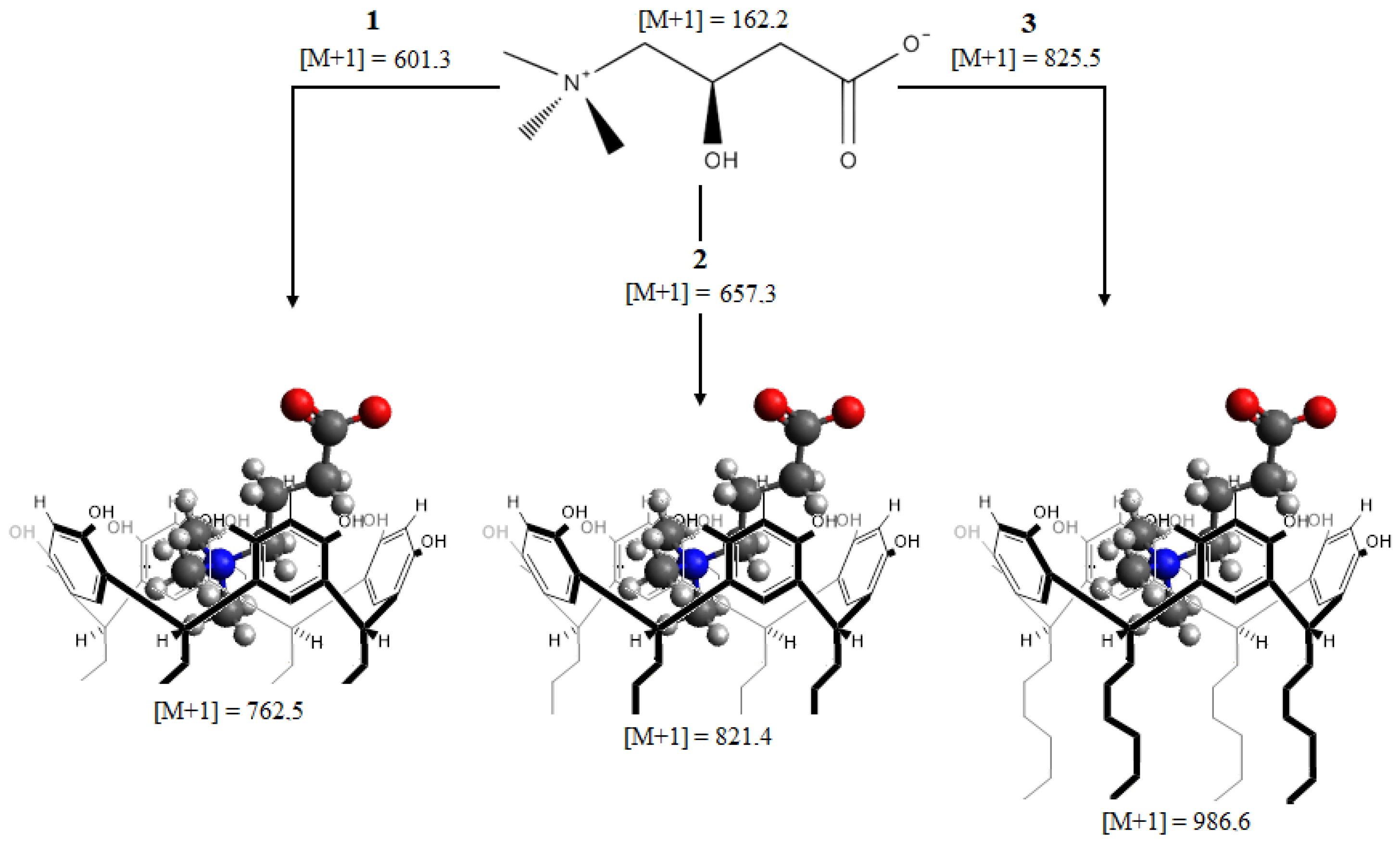
2.2.2. ESI-MS Studies

As mentioned above, the gaseous phase experiments were performed on a Bruker Impact II LC/MS Q-TOF equipped with electrospray ionization (ESI) in positive mode. Resorcinarenes 1–5 were each injected into the ion source as an ethanol solution with L-carnitine using 1:1, 2:1, and 1:2 molar ratios.

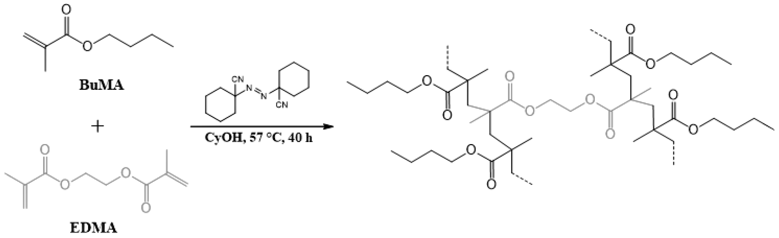
2.3. Obtention of Poly(BuMA-Co-EDMA)

Poly (BuMA-co-EDMA) polymer was synthesized following a procedure adapted from the methodology of Castillo-Aguirre [24]. The monomers were previously treated through a silica column in order to separate the polymerization inhibitor with which they are stored. Then a mixture of ethylene glycol dimethacrylate (EDMA) (12%, 570 μL) and butylmethacrylate (BMA) (18%, 1 mL) was stirred in a mixture of cyclohexanol (58.8%, 3.1 mL) and dodecanol (11.2%, 0.56 g) in the presence of 1,1 azobis(cyclohexanecarbonitrile) (ABCN) (0.2%, 10 mg). It was then homogenized via ultrasonication for 20 min at room temperature and the argon was bubbled for 15 min.
The mixture was transferred to an airtight container and heated in a water bath at 57 °C for 19–40 h, cooled to room temperature, filtered, washed with EtOH, and dried under a vacuum at 55 °C to constant mass. Finally, the polymer was crushed and sieved to a particle size of 106 μm. Chemical characterization was performed using FTIR-ATR and morphological characterization with SEM.

2.4. Physical Modification of Polymers with Resorcinarenes

Resorcinarene (0.6 mmol) was dissolved in 20 mL CHCl3 (in the case of sample 1, CO(CH3)2 was used because of insolubility in CHCl3). Then, the sieved polymer (1 g) was added. The mixture was magnetically stirred for 36 h at room temperature, and the resulting solid was filtered under a vacuum, washed with EtOH to neutral pH, and dried at 57 °C until it reached a constant mass, which was compared with the initial mass of the polymer added to determine how much resorcinarene was adsorbed on its surface. Finally, the polymeric material was crushed and sieved to a particle size of 106 µm. The procedure was carried out with a sieved polymer without resorcinarene additions.

2.5. SPE Protocol

For the optimization of the SPE procedure, the influences of various parameters on the extraction performance, including time, volume of the sample solution, elution solvent, pH, and concentration of the analyte were studied. Once the ideal extraction conditions were determined, quantitative tests were carried out using a standard solution of carnitine at 0.5 M, which was diluted in water for the preparation of an HPLC-MS calibration curve (six solutions 0.1–0.6 mg/mL) and the preparation of the sample solution 0.02 M to be eluted through a SPE cartridge to quantify the carnitine remaining in solution.
In each quantitative test performed, 200 mg of the sorbent resulting from impregnation was packed into the SPE cartridge using frits and filter paper to avoid loss. Then, the cartridge was preconditioned with methanol and water, and finally the sample solution was passed through the SPE cartridge and was collected in a volumetric flask (10 mL) to be analyzed using HPLC-MS.
The desorption process was done immediately after each sample elution. In order to carry this out, 10 mL of a solution of CH3COONH4 was eluted through the SPE cartridge and was collected in a volumetric flask (10 mL) to be analyzed using HPLC-MS.

3. Results and Discussion

3.1. Synthesis of Resorcinarenes

The synthesis of tetra(ethyl)calix[4]resorcinarene (1), tetra(propyl)calix[4] resorcinarene (2), tetra(hexyl)calix[4]resorcinarene (3), tetra(nonyl)calix[4]resorcinarene (4), and tetra(undecyl)calix[4]resorcinarene (5) was performed using the methodology described in the literature [24,25,26] (Scheme 1). In this way, the obtaining of compounds 15 was done through the acid-catalyzed cyclocondensation of the respective aldehyde with resorcinol in ethyl alcohol at reflux condition. The specific conditions are summarized in Scheme 1. In all cases, a solid was formed that was filtered, washed with water, and characterized using the spectroscopic techniques mentioned. As shown in Table 1, the characterization of the compounds was performed using ESI–TOF/MS, FT-IR, 1H-NMR, and 13C-NMR spectroscopy (Figures S1–S4). Compounds 15 had been previously synthesized [39,40,41], and our spectroscopic data agreed with those reported (Figures S5–S18); however, the following aspects should be noted: the signals of the macrocyclic compounds are described and they exhibited similar behavior. The characteristic band for OH was seen in the FT-IR spectrum, and the characteristic bands for aromatic and alkyl substituents were resolved. In 1H-NMR, the macrocycles showed similar profiles, which contained the characteristic signal of the methylene bridge between 4.09 and 4.32 ppm. For the protons corresponding to the hydroxyls, the signals were resolved between 8.93 and 8.87 ppm. Additionally, the signals corresponding to the aromatic system were observed between 6.15 and 7.23 ppm. The signals at 6.96 ppm for compound 1 and 7.35 ppm for compound 2, which are singlets, showed that the majority conformation was crown in DMSO. Finally, the 13C-NMR spectra of compounds 1 and 2 showed seven and nine signals, respectively, confirming the obtention of the products.

3.2. Molecular Interaction Studies between Resorcinarenes and L-Carnitine

Once the resorcinarenes were synthesized, molecular interaction experiments evaluating resorcinarenes 1 to 5 against carnitine were carried out in solution via 1H-NMR and, in the gas phase, by means of ESI-MS (Figures S20, S22 and S24). It was found that the complexes of compounds 1, 2, and 3 with carnitine are stable in solution and in the gas phase. For example, as shown in Figure 1, the variation in the 1H-NMR chemical shift of a single proton resonance in carnitine as a function of resorcinarene 2 (for compounds 1 or 3 it is the same behavior) in a molar ratio of 1:1 to carnitine in DMSO-d6 shows great molecular interaction in solution (Figures S19, S21 and S23), while the interaction of L-carnitine with compounds 4 and 5 was considerably lower.
The stoichiometric relationship was confirmed via electrospray ionization-mass spectrometry, which is a soft ionization technique that allows evaluating this type of interaction, as previously studied with various macrocyclic systems [42,43,44]. The experiments were carried out with different host:guest stoichiometries, producing the same signal for the 1:1 complex in all cases. Using this method, it was possible to establish that the m/z of the complex formed between carnitine and compound 1 was 762.5 (Figure S20), while the complexes formed between carnitine and compounds 2 and 3 were m/z = 821.4 (Figure S22) and m/z = 986.6 (Figure S24), respectively (Figure 2). The formation of these complexes is not surprising, since the structural arrangement of resorcinarenes in crown conformation and the tetraalkyl ammonium group of carnitine coordination is possibly dictated by π-interactions in the resorcinarene cavity. However, as observed by 1H-NMR, the interaction of carnitine with compounds 4 or 5 was very weak, and no signal could be seen for the complexes with these two resorcinarenes.
The observed results for the behavior of resorcinarenes 4 and 5 in solution were difficult to evaluate due to their low solubility in the solution of the deuterated solvents used for the study via 1H-NMR. On the other hand, in the gas phase, the interaction was not evident, despite obtaining homogeneous solutions, in the mass spectra of the signals of carnitine and resorcinarenes observed separately. These results suggest a low interaction of these resorcinarenes with L-carnitine. Despite the fact that the resorcinarenes on their upper rim are structurally equivalent, the effect of the interaction of the alkyl chains on the lower rim affects the cavity size, this effect being greater with long-chain resorcinarenes, as has previously been observed in other studies [45,46].

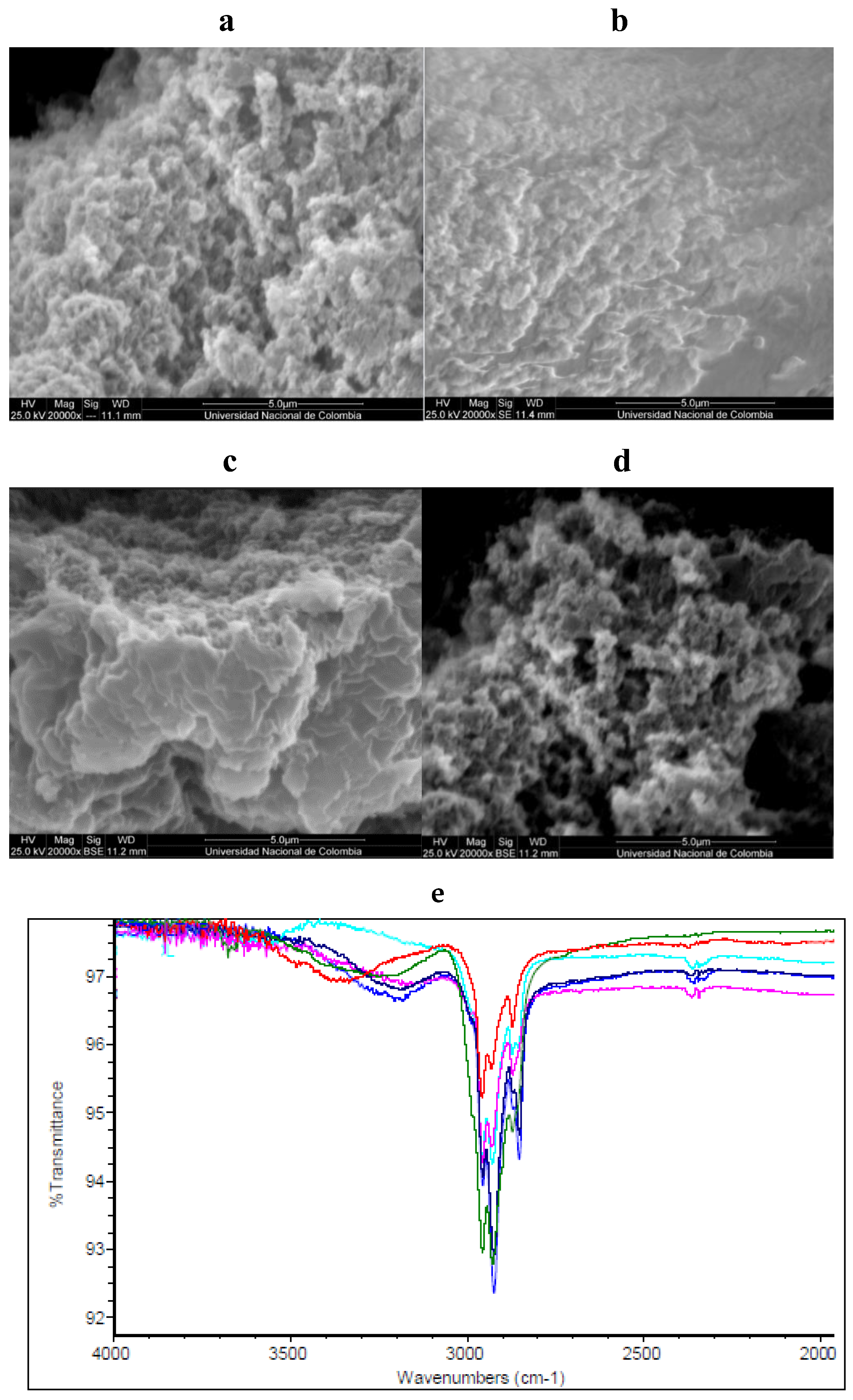
3.3. Obtention and Characterization of Sorbents

As shown in Scheme 2, the copolymer poly(BuMA-co-EDMA) was obtained from the reaction between EDMA and BuMA in cyclohexanol in the presence of 1,1-azobis (cyclohexanecarbonitrile), in accordance with previous reports [23,24]. The chemical characterization of poly (BuMA-co-EDMA) was performed via FT-IR, and the morphological characterization was performed via SEM. This copolymer had been previously synthesized, and our experimental data agreed with those reported [24,25]. The next step in the process was the physical modification of poly(BuMA-co-EDMA) with resorcinarenes 15. In this way, impregnation is a useful technique for the adsorption of molecules on the surface of a polymeric matrix, which enables more adsorption sites, rapid mass transfer, fast binding kinetics, and high selectivity copolymer modification due to lipophilic interactions between alkyl chains on the lower rim of the resorcinarenes with butyl chains in the copolymer.
The ATR-FTIR spectra of the obtained sorbents show characteristic signals. For example, poly(BuMA-co-EDMA)-1 and poly(BuMA-co-EDMA)-2 (Figure 3d) show signals corresponding to a C=O bond at 1714 cm−1. Other characteristic peaks were found at 3070 cm−1, assigned to the C-H stretching vibration of aromatic rings, 2910 cm−1 due to the C-H stretching vibration alkyl chains, 1455 cm−1 due to the C-H bending vibration, and 1155 cm−1 due to the C-O stretching vibration of the ester group. The adsorption bands at 3330 cm−1 of the copolymer modified with compounds 1 or 2 can be ascribed to the characteristic adsorption peaks of the OH stretching vibrations of resorcinarene rings. Furthermore, typical bands of benzene rings increased, as observed at 1400–1500 cm−1.
The morphologies of the obtained sorbents were analyzed via scanning microscopy images (Figure 3a–c). First, it was observed that the morphology of the copolymer is quite uniform, with continuous pores. The uniform and rich open-pore structure is suitable for mass transfer efficiency in the impregnation process with resorcinarenes 1, 2, or 3. Secondly, the morphologies of the impregnated copolymers poly(BuMA-co-EDMA)-1 poly(BuMA-co-EDMA)-2 or poly(BuMA-co-EDMA)-3 maintain their porosity characteristics, which are suitable for mass transfer efficiency as well as for carnitine adsorption.
The amount of fixed resorcinarenes 15 on the polymeric surface was determined via gravimetric analysis; these results show that the polymer poly(EDMA-co-GMA) has a high degree of substitution of the studied macrocycles on the surface (Figure 4). The best substitution was found with long-chain resorcinarenes (4 and 5), which was 352.2 µmol/g for poly(BuMA-co-EDMA)-4 and 367.5 µmol/g for poly(BuMA-co-EDMA)-5. The gravimetric data indicated that resorcinenarenes 1–3 were attached to the copolymer, but in a smaller proportion; however, it can be considered that the impregnation process was efficient. These results are in agreement with other investigations of the physical modification of poly(GMA-co-EDMA) [23,24].

3.4. Optimization of the SPE Procedure

In order to optimize the extraction conditions, the best modified polymer was chosen for these tests, in this case poly(BuMA-co-EDMA)-1, because resorcinarerene 1 exhibited the best interaction with carnitine in aqueous solution. In this way, the effect of the contact time in the extraction process for different times (30–120 min) was tested. As was evident from the results, the extraction reached a maximum at 60 min for the sorbents tested; after this time, no appreciable changes were observed. The flow rate of the sample solution was constant for all the experiments. To obtain high desorption efficiency, the selection of a suitable elution solvent was necessary. Five solvents were tested: water, methanol, acetonitrile, ethanol, and acetone. Preliminary experiments via HPLC showed that there were interfering peaks in the chromatograms when ethanol, acetone, or acetonitrile was used as the eluent, possibly because part of the macrocycle dissolves in the eluent. Between the other two solvents, using methanol as the elution solvent provided the highest extraction recoveries; however, with water the results were comparatively good. For this reason, water was chosen as the solvent in the process. The appropriate volume of water was further investigated using volumes of 1.0–4.0 mL. The extraction recoveries increased with the increasing water volume from 1.0 mL to 3.0 mL and remained nearly constant at higher volumes. Therefore, water with a volume of 3.0 mL was selected as the eluent.
The concentration of analyte played a significant role in the SPE procedure. Different concentrations of the analyte, from 0.005 to 0.1 M, were tested. The results showed that the extraction efficiency increased with increasing concentration up to 0.02 M; above this value, the results tended to be constant. When the concentration of L-carnitine was too small, most of the analyte was not extracted because there were too many unoccupied active sites, resulting in low extraction recovery. Therefore, a concentration of 0.02 M was selected for use in the subsequent experiments. Finally, the pH of the sample solution will have an influence on both L-carnitine and the sorbent; nevertheless, the structures of resrocinarenes grafted onto a copolymer play a crucial role in enriching and extracting the neurotransmitter in the proposed SPE procedure. The hydroxyl groups in resorcinarene provide hydrogen bonding and dipole-induced dipole interactions, which aid in extracting L-carnitine from the aqueous solution. Among these groups, the ionic form of the hydroxyl group is influenced by the pH of the sample solution. Therefore, very high pH values were not considered to be appropriate because they would affect the stability of the modified sorbent. To investigate the effect of the pH of the neurotransmitter solution on the extraction process, different pH values (2–8) were tested. An increase in the extraction efficiency for L-Carnitine was observed when the pH of the aqueous solution was changed from pH 2 to 6. At low pH, the hydroxyl groups interacted weakly with the tetralkylammonium group of L-carnitine, while at pH 6, some of the deprotonated hydroxyls of the macrocyclic resulted in stronger interactions with L-carnitine.

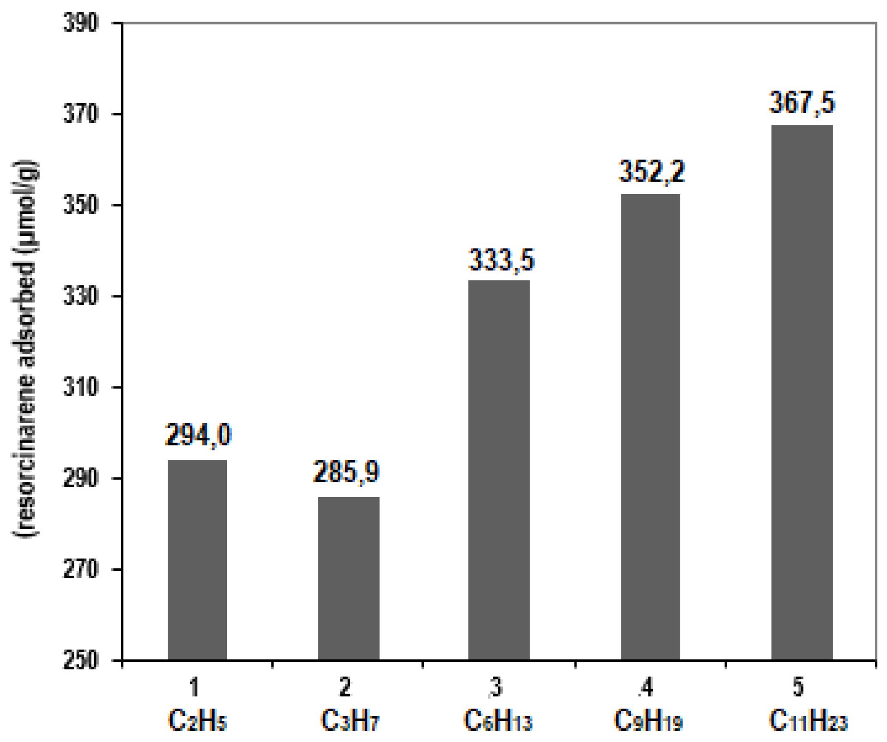
3.5. Analyses of the Extraction of L-Carnitine with the Modified Sorbents

Once the best conditions had been established for the carnitine extraction process with the modified sorbents, the final step consisted of determining the amount of carnitine recovered in an extraction-desorption cycle. For this purpose, it was decided to evaluate the process by means of a carnitine calibration curve obtained with the HPLC-MS technique, as described in the experimental section. As shown in Figure 5, the calibration curve of L-carnitine showed good linearity in the range of 0.1–0.6 mg/mL, with a correlation coefficient (R2) of 0.9984. According to the results established for the extraction with the three sorbents, it was observed that evidently poly(BuMA-co-EDMA)-1 provided higher extraction efficiency for L-carnitine (67%) than poly(BuMA-co-EDMA)-2 and poly(BuMA-co-EDMA)-3, which had efficiencies close to 35%. Finally, the stability of the sorbents was evaluated by performing adsorption–desorption cycles. The sorbents were recycled at least four times without any obvious loss of extraction recovery, indicating that the modified sorbents were stable enough to be recycled in the extraction procedure.

4. Conclusions

We have presented a copolymer modified with resorcineranes for the pre-concentration of carnitine with an acceptable efficiency in the analyte recovery process. These macrocycles were highly hydrophobic, which is essential for the modification and stability of the sorbent. The host-guest interaction between resorcinarenes and L-carnitine was confirmed via 1H-NMR and mass spectrometry, showing good affinity in both cases. Among the two macrocycles evaluated as candidates for the pre-concentration process, tetra(ethyl)calix[4]resorcinarene exhibited the highest affinity in the adsorption process on the modified sorbent. The sorbents were successfully applied to the pre-concentration of L-carnitine in a synthetic solution. This macrocycle-copolymer provides a new method for the pre-concentration of the neurotransmitter L-carnitine. While this investigation aimed to propose a new lipophilic sorbent for the pre-concentration of L-carnitine and focused more on fundamental characterization, the obtained sorbents could be useful in biological samples and for pre-concentration processes above micromolar range. However, we believe there is still room for technical improvements based on the proposed copolymer, including the fabrication of sorbents for the pre-concentration of other neurotransmitters.

Supplementary Materials

The following supporting information can be downloaded at https://www.mdpi.com/article/10.3390/pr11061705/s1: Figure S1. FT-IR spectrum of compound 1; Figure S2. 1H-NMR spectrum (400 MHz, DMSO-d6, 293 K) of compound 1; Figure S3. 13C-NMR spectrum (400 MHz, DMSO-d6, 293 K) of compound 1; Figure S4. MS spectrum of compound 1; Figure S5. FT-IR spectrum of compound 2; Figure S6. 1H-NMR spectrum (400 MHz, DMSO-d6, 293 K) of compound 2; Figure S7. 13C-NMR spectrum (400 MHz, DMSO-d6, 293 K) of compound 2; Figure S8. MS spectrum of compound 2; Figure S9. FT-IR spectrum of compound 3; Figure S10. 1H-NMR spectrum (400 MHz, DMSO-d6, 293 K) of compound 3; Figure S11. 13C-NMR spectrum (400 MHz, DMSO-d6, 293 K) of compound 3; Figure S12. MS spectrum of compound 3; Figure S13. FT-IR spectrum of compound 4; Figure S14. 1H-NMR spectrum (400 MHz, DMSO-d6, 293 K) of compound 4; Figure S15. 13C-NMR spectrum (400 MHz, DMSO-d6, 293 K) of compound 4; Figure S16. FT-IR spectrum of compound 5; Figure S17. 1H-NMR spectrum (400 MHz, DMSO-d6, 293 K) of compound 5; Figure S18. 13C-NMR spectrum (400 MHz, DMSO-d6, 293 K) of compound 5; Figure S19. 1H-NMR spectrum (400 MHz, DMSO-d6, 293 K) of complex 1; Figure S20. MS spectrum of complex 1; Figure S21. 1H-NMR spectrum (400 MHz, DMSO-d6, 293 K) of complex 2; Figure S22. MS spectrum of complex 2; Figure S23. 1H-NMR spectrum (400 MHz, DMSO-d6, 293 K) of complex 3; Figure S24. MS spectrum of complex 3.

Author Contributions

Conceptualization, M.M.; methodology, N.A.C.-M. and G.R.; validation, G.R., formal analysis, M.M., N.A.C.-M., and G.R.; investigation, M.M., N.A.C.-M., and G.R.; writing—original draft preparation, M.M., N.A.C.-M., and G.R.; writing—review and editing, M.M.; supervision, M.M.; project administration, M.M. All authors have read and agreed to the published version of the manuscript.

Funding

This research received no external funding.

Data Availability Statement

The data that support the findings of this study are not openly available due to the fact that the data form part of an ongoing study but are available from the corresponding author upon reasonable request.

Acknowledgments

We gratefully acknowledge the support of the Universidad Nacional de Colombia-Sede Bogotá.

Conflicts of Interest

The authors declare no conflict of interest.

References

  1. Wrona, M.Z.; Dryhurst, G. Low Molecular Weight Neurotransmitters. Bioelectrochemistry 1994, 267, 117–169. [Google Scholar] [CrossRef]
  2. Bartfai, T.; Iverfeldt, K.; Fisone, G.; Serfozo, P. Regulation of the Release of Coexisting Neurotransmitters. Annu. Rev. Pharmacol. Toxicol. 1988, 28, 285–310. [Google Scholar] [CrossRef] [PubMed]
  3. Dąbrowska, M.; Starek, M. Analytical approaches to determination of carnitine in biological materials, foods and dietary supplements. Food Chem. 2014, 142, 220–232. [Google Scholar] [CrossRef] [PubMed]
  4. Möder, M.; Kießling, A.; Löster, H. Current Methods for Determination of L-Carnitine and Acylcarnitines. Mon. Fur Chem. 2005, 136, 1279–1291. [Google Scholar] [CrossRef]
  5. Johnson, D.W. An acid hydrolysis method for quantification of plasma free and total carnitine by flow injection tandem mass spectrometry. Clin. Biochem. 2010, 43, 1362–1367. [Google Scholar] [CrossRef]
  6. Zhang, Z.; Niwa, O.; Shiba, S.; Tokito, S.; Nagamine, K.; Ishikawa, S.; Sugimoto, M. Electrochemical enzyme biosensor for carnitine detection based on cathodic stripping voltammetry. Sens. Actuators B Chem. 2020, 321, 128473. [Google Scholar] [CrossRef]
  7. Wang, M.; Yang, R.; Mu, H.; Zeng, J.; Zhang, T.; Zhou, W.; Wang, S.; Tang, Y.; Li, H.; Zhang, C.; et al. A simple and precise method for measurement of serum free carnitine and acylcarnitines by isotope dilution HILIC-ESI-MS/MS. Int. J. Mass Spectrom. 2019, 446, 116208. [Google Scholar] [CrossRef]
  8. Seline, K.-G.; Johein, H. The determination of l-carnitine in several food samples. Food Chem. 2007, 105, 793–804. [Google Scholar] [CrossRef]
  9. Lu, W.-H.; Chiu, H.-H.; Kuo, H.-C.; Chen, G.-Y.; Chepyala, D.; Kuo, C.-H. Using matrix-induced ion suppression combined with LC-MS/MS for quantification of trimethylamine-N-oxide, choline, carnitine and acetylcarnitine in dried blood spot samples. Anal. Chim. Acta 2021, 1149, 338214. [Google Scholar] [CrossRef]
  10. Rudolph, W.; Remane, D.; Wissenbach, D.K.; Peters, F.T. Liquid chromatography-mass spectrometry-based determination of ergocristine, ergocryptine, ergotamine, ergovaline, hypoglycin A, lolitrem B, methylene cyclopropyl acetic acid carnitine, N-acetylloline, N-formylloline, paxilline, and peramine in equine hair. J. Chromatogr. B 2019, 1117, 127–135. [Google Scholar] [CrossRef]
  11. Minkler, P.E.; Stoll, M.S.K.; Ingalls, S.T.; Kerner, J.; Hoppel, C.L. Validated Method for the Quantification of Free and Total Carnitine, Butyrobetaine, and Acylcarnitines in Biological Samples. Anal. Chem. 2015, 87, 8994–9001. [Google Scholar] [CrossRef]
  12. Morand, R.; Donzelli, M.; Haschke, M.; Krähenbühl, S. Quantification of plasma carnitine and acylcarnitines by high-performance liquid chromatography-tandem mass spectrometry using online solid-phase extraction. Anal. Bioanal. Chem. 2013, 405, 8829–8836. [Google Scholar] [CrossRef] [Green Version]
  13. Prokorátová, V.; Kvasnička, F.; Ševčík, R.; Voldřich, M. Capillary electrophoresis determination of carnitine in food supplements. J. Chromatogr. A 2005, 1081, 60–64. [Google Scholar] [CrossRef]
  14. Tan, X.; Yang, Y.; Luo, S.; Zhang, Z.; Zeng, W.; Zhang, T.; Su, F.; Zhou, L. Novel Competitive Fluorescence Sensing Platform for L-carnitine Based on Cationic Pillar[5]Arene Modified Gold Nanoparticles. Sensors 2018, 18, 3927. [Google Scholar] [CrossRef] [PubMed] [Green Version]
  15. Chen, Y.-C.; Tsai, C.-J.; Feng, C.-H. Fluorescent derivatization combined with aqueous solvent-based dispersive liquid-liquid microextraction for determination of butyrobetaine, l-carnitine and acetyl-l-carnitine in human plasma. J. Chromatogr. A 2016, 1464, 32–41. [Google Scholar] [CrossRef] [PubMed]
  16. Manjón, A.; Obón, J.; Iborra, J. Determination of -Carnitine by Flow Injection Analysis with NADH Fluorescence Detection. Anal. Biochem. 2000, 281, 176–181. [Google Scholar] [CrossRef] [PubMed]
  17. He, Q.; Vargas-Zúñiga, G.I.; Kim, S.H.; Kim, S.K.; Sessler, J.L. Macrocycles as Ion Pair Receptors. Chem. Rev. 2019, 119, 9753–9835. [Google Scholar] [CrossRef] [PubMed]
  18. Ruiz-Botella, S.; Vidossich, P.; Ujaque, G.; Vicent, C.; Peris, E. A Tetraferrocenyl-Resorcinarene Cavitand as a Redox-Switchable Host of Ammonium Salts. Chem. A Eur. J. 2015, 21, 10558–10565. [Google Scholar] [CrossRef] [Green Version]
  19. Taylor, P.J. Matrix effects: The Achilles heel of quantitative high-performance liquid chromatography-electrospray-tandem mass spectrometry. Clin. Biochem. 2005, 38, 328–334. [Google Scholar] [CrossRef]
  20. Huang, Z.; Richards, M.A.; Zha, Y.; Francis, R.; Lozano, R.; Ruan, J. Determination of inorganic pharmaceutical counterions using hydrophilic interaction chromatography coupled with a Corona® CAD detector. J. Pharm. Biomed. Anal. 2009, 50, 809–814. [Google Scholar] [CrossRef]
  21. Johnson, W.M.; Kido Soule, M.C.; Kujawinski, E.B. Extraction efficiency and quantification of dissolved metabolites in targeted marine metabolomics. Limnol. Oceanogr. Methods 2017, 15, 417–428. [Google Scholar] [CrossRef] [Green Version]
  22. Lee, M.; Oh, S.Y.; Pathak, T.S.; Paeng, I.R.; Cho, B.Y.; Paeng, K.J. Selective solid-phase extraction of catecholamines by the chemically modified polymeric adsorbents with crown ether. J. Chromatogr. A 2007, 1160, 340–344. [Google Scholar] [CrossRef]
  23. Chen, L.; Wang, H.; Xu, Z.; Zhang, Q.; Liu, J.; Shen, J.; Zhang, W. High-throughput and selective solid-phase extraction of urinary catecholamines by crown ether-modified resin composite fiber. J. Chromatogr. A 2018, 1561, 48–55. [Google Scholar] [CrossRef] [PubMed]
  24. Castillo-Aguirre, A.; Maldonado, M. Preparation of methacrylate-based polymers modified with chiral resorcinarenes and their evaluation as sorbents in norepinephrine microextraction. Polymers 2019, 11, 1428. [Google Scholar] [CrossRef] [PubMed] [Green Version]
  25. Velásquez-Silva, B.A.; Castillo-Aguirre, A.; Rivera-Monroy, Z.J.; Maldonado, M. Aminomethylated calix[4]resorcinarenes as modifying agents for glycidyl methacrylate (GMA) rigid copolymers surface. Polymers 2019, 11, 1147. [Google Scholar] [CrossRef] [Green Version]
  26. Castillo, A.; Esteso, M.A.; Maldonado, M. Resorcin[4]arenes: Generalities and their role in the modification and detection of amino acids. Curr. Org. Chem. 2020, 24, 2412–2425. [Google Scholar] [CrossRef]
  27. Li, N.; Harrison, R.G.; Lamb, J.D. Application of resorcinarene derivatives in chemical separations. J. Incl. Phenom. Macrocycl. Chem. 2014, 78, 39–60. [Google Scholar] [CrossRef]
  28. Davis, F.; Faul, C.F.J.; Higson, S.P.J. Calix[4]resorcinarene-surfactant complexes: Formulation, structure and potential sensor applications. Soft Matter. 2009, 5, 2746–2751. [Google Scholar] [CrossRef]
  29. Yao, Y.; Sun, Y.; Han, Y.; Yan, C. Preparation of resorcinarene-functionalized gold nanoparticles and their catalytic activities for reduction of aromatic nitro compounds. Chin. J. Chem. 2010, 28, 705–712. [Google Scholar] [CrossRef]
  30. Natarajan, N.; Brenner, E.; Sémeril, D.; Matt, D.; Harrowfield, J. The Use of Resorcinarene Cavitands in Metal-Based Catalysis. Eur. J. Org. Chem. 2017, 2017, 6100–6113. [Google Scholar] [CrossRef]
  31. Pinalli, R.; Pedrini, A.; Dalcanale, E. Biochemical sensing with macrocyclic receptors. Chem. Soc. Rev. 2018, 47, 7006–7026. [Google Scholar] [CrossRef]
  32. Hong, M.; Zhang, Y.M.; Liu, Y. Selective binding affinity between quaternary ammonium cations and water-soluble calix[4]resorcinarene. J. Org. Chem. 2015, 80, 1849–1855. [Google Scholar] [CrossRef]
  33. Torvinen, M.; Kalenius, E.; Sansone, F.; Casnati, A.; Jänis, J. Noncovalent complexation of monoamine neurotransmitters and related ammonium ions by tetramethoxy tetraglucosylcalix[4]arene. J. Am. Soc. Mass Spectrom. 2012, 23, 359–365. [Google Scholar] [CrossRef]
  34. Lledó, A.; Hooley, R.J.; Rebek, J. Recognition of guests by water-stabilized cavitand hosts. Org. Lett. 2008, 10, 3669–3671. [Google Scholar] [CrossRef] [PubMed] [Green Version]
  35. Guo, D.S.; Uzunova, V.D.; Su, X.; Liu, Y.; Nau, W.M. Operational calixarene-based fluorescent sensing systems for choline and acetylcholine and their application to enzymatic reactions. Chem. Sci. 2011, 2, 1722–1734. [Google Scholar] [CrossRef]
  36. Nikolelis, D.P.; Petropoulou, S.S.E.; Theoharis, G. Biosensors for the Rapid Repetitive Detection of Adrenaline Using Stabilized Bilayer Lipid Membranes (BLMs) with Incorporated Calix[4]resorcinarene Receptor. Electroanalysis 2003, 15, 1616–1624. [Google Scholar] [CrossRef]
  37. Ballester, P.; Shivanyuk, A.; Far, A.R.; Rebek, J. A synthetic receptor for choline and carnitine. J. Am. Chem. Soc. 2002, 124, 14014–14016. [Google Scholar] [CrossRef] [PubMed]
  38. Maldonado, M.; Sanabria, E.; Velasquez-Silva, A.; Casas-Hinestroza, J.L.; Esteso, M.A. Comparative study of the volumetric properties of three regioisomers of diazoted C-tetra(propyl)resorcin[4]arene in DMSO at various temperatures. J. Mol. Liq. 2021, 325, 115252. [Google Scholar] [CrossRef]
  39. Castillo-Aguirre, A.; Rivera-Monroy, Z.; Maldonado, M. Selective O-Alkylation of the Crown Conformer of Tetra(4-hydroxyphenyl)calix[4]resorcinarene to the Corresponding Tetraalkyl Ether. Molecules 2017, 22, 1660. [Google Scholar] [CrossRef]
  40. Castillo-Aguirre, A.A.; Velásquez-Silva, B.A.; Palacio, C.; Baez, F.; Rivera-Monroy, Z.J.; Maldonado, M. Surface modification of poly(GMA-co-EDMA-co-MMA) with resorcarenes. J. Braz. Chem. Soc. 2018, 29, 1965–1972. [Google Scholar] [CrossRef]
  41. Castillo-Aguirre, A.; Maldonado, M.; Esteso, M.A. Removal of Toxic Metal Ions Using Poly(BuMA–co–EDMA) Modified with C-Tetra(nonyl)calix[4]resorcinarene. Toxics 2022, 10, 204. [Google Scholar] [CrossRef] [PubMed]
  42. Casas-Hinestroza, J.L.; Bueno, M.; Ibáñez, E.; Cifuentes, A. Recent advances in mass spectrometry studies of non-covalent complexes of macrocycles—A review. Anal. Chim. Acta 2019, 1081, 32–50. [Google Scholar] [CrossRef] [PubMed]
  43. Casas-Hinestroza, J.L.; Cifuentes, A.; Ibáñez, E.; Maldonado, M. Effect of the formation of capsules of tetra(propyl) pyrogallol[4]arene on the host-guest interaction with neurotransmitters. J. Mol. Struct. 2020, 1210, 128063. [Google Scholar] [CrossRef]
  44. Casas-Hinestroza, J.L.; Pérez-Redondo, A.; Maldonado, M. Inclusion complexation between neurotransmitters with polyacetylated calix[4]pyrogallolarenes: 1H-NMR and crystallographic analysis. Res. Chem. Intermed. 2022, 48, 3091–3107. [Google Scholar] [CrossRef]
  45. Velásquez-Silva, A.; Forero, R.S.; Sanabria, E.; Pérez-Redondo, A.; Maldonado, M. Host-guest inclusion systems of tetra(alkyl)resorcin [4]arenes with choline in DMSO: Dynamic NMR studies and X-ray structural characterization of the 1:1 inclusion complex. J. Mol. Struct. 2019, 1198, 126846. [Google Scholar] [CrossRef]
  46. Velásquez-Silva, A.; Cortés, B.; Rivera-Monroy, Z.J.; Pérez-Redondo, A.; Maldonado, M. Crystal structure and dynamic NMR studies of octaacetyl-tetra(propyl)calix[4]resorcinarene. J. Mol. Struct. 2017, 1137, 380–386. [Google Scholar] [CrossRef]
Scheme 1. Synthesis of calix[4]resorcinarenes 15.
Processes 11 01705 sch001
Figure 1. (a) Compiled spectra different molar ratio tetra(propyl)calix[4] resorcinarene (2) with L-carnitine; (b) chemical shift variation in different molar ratio tetra(propyl)calix[4]resorcinarene (2) + L–carnitine.
Processes 11 01705 g001
Figure 2. Complexation process and m/z data for complexes between L-Carnitine with compounds 1, 2, or 3.
Processes 11 01705 g002
Scheme 2. Product of the copolymerization.
Processes 11 01705 sch002
Figure 3. Comparative characterization of sorbents. SEM of poly(BuMA-co-EDMA) at 5 μm. (a) and poly(BuMA-co-EDMA) + 1 (2 μm). (b) poly(BuMA-co-EDMA) + 2 (2 μm). (c) poly(BuMA-co-EDMA)-3 (d). Comparative IR spectra of poly(BuMA-co-EDMA) (light green), poly(BuMA-co-EDMA) + 1 (orange), poly(BuMA-co-EDMA) + 2 (dark green), poly(BuMA-co-EDMA)-3 (purple), poly(BuMA-co-EDMA) + 4 (Light Blue), poly(BuMA-co-EDMA) + 5 (dark blue) (e).
Processes 11 01705 g003
Figure 4. Amounts of resorcinarene adsorbed on the surface of the copolymer.
Processes 11 01705 g004
Figure 5. L-carnitine calibration curve.
Processes 11 01705 g005
Table 1. Spectroscopic information of calix[4]resorcinarenes 15.
12345
ESI–TOF/MS
(m/z) [M + H]+
601.32657.33847.51993.711105.84
IR
(KBr/cm−1)
(O–H)34003280330032013235
(ArC-H),29802953295029202918
(aliphatic C-H)29202926286028512849
(C=C)16101606161016171617
(ArC-O)12901283128012181211
13C and 1H NMR
DMSO-d6
(1, 2, 3)
CHCl3-d1
(4, 5)
δ (ppm)
(C–OH)(s, 8H) 8.93
(C) 151.6
(s, 8H) 8.94
(C) 152.0
(s, 8H) 8.87
(C) 151.6
(s, 8H) 9.45
(C) 150.6
(s, 8H) 9.63
(C) 150.4
(C–H), meta to OH(s, 4H) 7.23
(C) 102.3
(s, 4H) 7.24
(C) 102.8
(s, 4H) 7.13
(C) 102.3
(s, 4H) 7.23
(C)
(s, 4H) 7.23
(C)
(C–H), ortho to OH(s, 4H) 6.15
(C) 125.0
(s, 4H) 6.15
(C) 125.5
(s, 4H) 6.14
(C) 124.8
(s, 4H) 6.14
(C) 124.9
(s, 4H) 6.16
(C) 124.8
(C–H), CH(t, 4H) 4.09
(C) 35.1
(t, 4H) 4.23
(C) 36.15
(t, 4H) 4.21
(C) 34.0
(t, 4H) 4.32
(C) 33.24
(t, 4H) 4.32
(C) 58.5
(C–H), CH2(m, 8H)
2.11
(C)
26.6
(m, 16H)
2.07–1.19
(C)
33.0–21.1
(m, 40H)
2.00–1.21
(C)
31.4–22.1
(m, 64H)
4.15–1.30
(C)
32.0–22.7
(m, 80H)
4.14–1.25
(C)
33.3–18.3
(C–H), CH3(t, 12H) 0.79
(C) 12.6
(t, 12H) 0.90
(C) 14.4
(t, 12H) 0.83
(C) 13.8
(t, 12H) 0.91
(C) 14.1
(t, 12H) 0.90
(C) 14.1
Disclaimer/Publisher’s Note: The statements, opinions and data contained in all publications are solely those of the individual author(s) and contributor(s) and not of MDPI and/or the editor(s). MDPI and/or the editor(s) disclaim responsibility for any injury to people or property resulting from any ideas, methods, instructions or products referred to in the content.

