Article

Isolation and Characterization of Novel Butachlor-Degrading Bacteria from Rice Paddy Soils

1
Department of Biological Science, College of Biological and Environmental Sciences, Sangji University, Wonju 26339, Republic of Korea
2
Department of Agricultural Biotechnology and Research Institute for Agriculture and Life Sciences, Seoul National University, Seoul 08826, Republic of Korea
*
Author to whom correspondence should be addressed.
Processes 2023, 11(4), 1222; https://doi.org/10.3390/pr11041222
Submission received: 16 March 2023 / Revised: 10 April 2023 / Accepted: 12 April 2023 / Published: 15 April 2023
(This article belongs to the Section Environmental and Green Processes)

Abstract

:
Four different bacteria capable of degrading butachlor, as well as five different syntrophic pairs of bacteria able to break down butachlor, were isolated from rice paddy soils in Korea. Genetic and phenotypic analyses were conducted to better understand their characteristics and behavior. All single isolates and syntrophic pairs were able to utilize butachlor as a sole carbon and energy source. Analysis of the 16S rRNA sequence showed that the isolates were related to members of the genus Rhodococcus and a new type of butachlor-degrading genus Sphingobium. The chromosomal DNA fingerprinting patterns of the butachlor-degrading bacteria and syntrophic pairs were analyzed using a technique called repetitive-sequence-based PCR (REP-PCR). The results showed that there were two different REP-PCR patterns found among the four independent butachlor-degrading bacteria, and ten strains of five different syntrophic pairs produced a total of eight distinct DNA fingerprints. Through the use of gas chromatography–mass spectrometry (GC-MS) analysis, it was observed that the syntrophic pair was capable of breaking down butachlor using various chemical pathways, such as 2-chloro-N-(2,6-diethylphenyl) acetamide (CDEPA), 2,6-diethylphenyl isocyanate, 2,6-diethylaniline (DEA), and 2-ethylaniline.

1. Introduction

Chloroacetamide herbicides have been commonly employed to manage weeds growth in agricultural fields that produce crops such as rice, corn, soybean, and others. These herbicides are applied during the early stages of plant growth or after emergence to prevent lipid, protein, and lignin synthesis in plants [1,2]. Among the chloroacetanilide herbicides, butachlor [N-(butoxymethyl)-2-chloro-N-(2,6-diethylphenyl)-acetamide] is one of the commonly applied herbicides to control pre-emergent weeds in Asia and South America [1,3]. Annual use of butachlor in Asia has been reported to be approximately 4.5 × 10⁷ Kg [4,5]. Butachlor is applied to beans, rice, tea, and wheat as a systematic and selective pre-emergent herbicide. It inhibits geranygeranyl pyrophosphate cyclization enzymes and elongase, which is responsible for the elongation of very long-chain fatty acids (VLCFAs) [6]. In addition, it effects lipid biosynthesis and redox homeostasis [7].
Extensive use of butachlor has led to frequent detection of butachlor and its metabolites in various agricultural soil environments [4]. Toxicological research has revealed the harmful effects of butachlor on earthworms, microbial communities, enzyme activities, freshwater fishes, and even human health [5,6,7]. Studies have shown that butachlor can cause stomach tumors in rats and oxidative DNA damage in humans [8,9]. Butachlor has also been reported to cause aberration of the micronucleus and chromosomes, as well as being genotoxic to mammalian cells [7].
Due to concerns about the persistence and fate of butachlor in the environment, there has been a growing need to assess its detoxification from the environment. Studies have shown that both biotic and abiotic processes can contribute to the degradation of butachlor, with microbial populations playing a particularly important role in soil detoxification [10,11]. Several microorganisms have been discovered with the capability to decompose butachlor as their sole carbon source, with certain strains exhibiting the capacity to degrade 100 mg L−1 of butachlor within a span of just 5 days [12]. These include Rhodococcus sp. strain B1, Pseudomonas putida strain ER1, Novosphingobium chloroacetimidivorans sp. nov. BUT-14T, Sphingomonas chloroacetimidivorans sp. nov., and Paracoccus sp. FLY-8 [13,14,15,16,17].
Previous studies have proposed potential pathways for butachlor degradation based on chemical analysis and enzyme studies [17,18]. These pathways involve C-dealkylation of butachlor to form alachlor, which is then further converted to 2-chloro-N-(2,6-dimethylphenyl) acetamide by N-dealkylation. The resulting compound is then transformed into 2,6-diethylaniline and eventually mineralized through the intermediates of aniline and catechol [17]. However, despite these proposals, there has been limited research on microorganisms capable of degrading butachlor and, as a result, there is relatively little information available about the genetic, metabolic, and physiological properties of these bacteria.
This study aimed to use an enrichment process to isolate bacteria that are capable of breaking down butachlor from rice paddy soil. Additionally, this study aimed to examine the bacteria’s potential to break down butachlor in a mineral medium where butachlor serves as the sole carbon source. Furthermore, this study aimed to determine metabolic intermediates by gas chromatography–mass spectrometry and then propose a new biodegradation pathway for butachlor.

2. Materials and Methods

2.1. Chemicals

Alachlor (99.2% purity) and butachlor (97.7% purity), 2,6-diethylaniline (98.6% purity), aniline, and catechol were obtained from Sigma-Aldrich (St. Louis, MO, USA). 2-chloro-N-(2, 6-diethylphenyl) acetamide (CDEPA) (100% purity) was purchased from Accustandard (New Haven, CT, USA). All other chemicals used were of analytical grade.

2.2. Media and Culture Condition

The isolated butachlor-degrading bacteria were grown on mineral medium (MM) [18] containing 100 mg L−1 of butachlor. The mineral medium contained 0.13 mg (NH4)6Mo7O24·4H2O, 0.24 mg CuSO4·5H2O, 0.38 mg Co(NO3)2·6H2O, 1.70 mg MnSO4·7H2O, 1.15 mg ZnSO4·7H2O, 2.75 mg FeSO4·7H2O, 5.75 mg CaCl2·H2O, 0.05 g MgSO4·7H2O, 0.30 g (NH4)2SO4, 1.36 g KH2PO4, and 1.42 g Na2HPO4 in 1 L distilled water. To purify the bacterial strain and to conduct routine culture, modified Peptone-Tryptone-Yeast extract Glucose (PTYG) medium was used. A modified PTYG medium was used in this study, containing (g/L) 0.25 peptone, 0.25 tryptone, 0.5 yeast extract, 0.5 glucose, 0.03 MgSO4, and 0.003 CaCl2. All cultures were incubated at 28 °C and liquid cultures were shaken in an incubator (Vision Co., Daejeon, Republic of Korea) at 150 rpm and 28 °C.

2.3. Enrichment and Isolation of Butachlor-Degrading Strains

Soil samples were collected from rice fields (0–15 cm deep) in Ansan, Chungju, Jangsung, Pyeongtaek, Wonju, and Yeongju regions in Korea. These rice fields are actually rice-cultivated land and butachlor has been regularly applied to these field for several years. Soil samples were sieved (2.0 mm mesh) to remove debris and homogenize. An enrichment technique was used to isolate butachlor-degrading bacteria. To obtain a final butachlor concentration of 100 mg kg−1 soil, butachlor was added to 20 g of each soil sample and mixed thoroughly. The mixture was then incubated with periodic mixing at room temperature. After 4 weeks, 1 g of soil sample was mixed with 9 mL of mineral medium containing 100 mg L−1 of butachlor. The resulting mixture was then serially diluted in 3 mL of butachlor medium (100 mg L−1) and incubated in a shaking incubator at 28 °C for 4 weeks. The degradation of butachlor was evaluated using NanoDrop 2000c spectrophotometer (Thermo Fisher Scientific, Waltham, MA, USA) and high-performance liquid chromatography (HPLC) (Shimadzu, Kyoto, Japan). The culture medium was transferred to a new medium with butachlor when less than 20% of the initial butachlor amount remained. The above process was repeated 3–4 times. To isolate individual bacterial colonies that could degrade butachlor, the final enriched culture was streaked onto PTYG agar and incubated at 28 °C for 7 days. Distinct colonies with different morphologies were chosen and purified using PTYG agar. An individual colony was then added to 3 mL of mineral medium containing 100 mg L−1 of butachlor and incubated at 28 °C. The purpose of this step was to evaluate its capacity to utilize butachlor as the only source of carbon and energy.

2.4. Identification of Butachlor-Degrading Strain

The bacteria that were able to degrade butachlor were isolated and identified through 16S rRNA gene sequence analysis. Chromosomal DNA was extracted using the Wizard Genomic DNA Purification Kit (Promega, Madison, Wisconsin, USA). To amplify the 16S rRNA gene, polymerase chain reaction (PCR) was employed with the use of universal primers, namely 27f and 1492r [19]. PCR was carried out with the following procedure: DNA was initially denatured at 94 °C for 5 min; then, it was subjected to 30 cycles of amplification consisting of 1 min at 94 °C, 1 min at 55 °C, and 1 min at 72 °C; and finally, the reaction was extended to 72 °C for 10 min. The amplified PCR products were verified through agarose gel electrophoresis and subsequently purified using the QIAquick PCR purification kit (QIAGEN, Hilden, Germany). Afterward, the purified PCR products were sequenced using the sequencing primers 515r (5′-TTACCGCGGCTGCTGGCA-3′), 926f (5′-AAACTYAAAKGAATTGACGG-3′) [20] and 1055r (5′-AGCTGACGACAGCCAT-3′) [21]. SEQMAN software (DNASTAR) was utilized to assemble the obtained overlapping fragments. To determine the similarity of 16S rRNA gene sequence among bacterial type strains, EzTaxon server [22] was employed.

2.5. Colony REP-PCR

Repetitive-sequence-based PCR (REP-PCR) was conducted using BOXA1R primer to distinguish the isolated butachlor-degrading strains at subspecies level [23]. Each butachlor-degrading strain was cultured on PTYG agar medium for 2 days. Small amounts of bacterial cells were harvested and resuspended in 25 μL of PCR mixture. The PCR protocol began with a denaturation step at 95 °C for 7 min, followed by 35 cycles of amplification, which involved heating the sample to 92 °C for 1 min, annealing at 52 °C for 1 min, and extension at 65 °C for 8 min. After the last cycle, there was a final extension step at 65 °C for 16 min. The amplified PCR products were then resolved by electrophoresis on 1.2% agarose gels.

2.6. Analysis of Butachlor Degradation

The butachlor-degrading isolates were cultured in a PTYG medium. After collecting exponentially growing cells by centrifugation at 19,000× g for 10 min and washing them twice with mineral medium, a specific quantity of resuspended cells was introduced into a mineral medium containing butachlor (100 mg L−1) as the sole source of carbon, with a final density of OD600 = 0.005. The culture medium containing the inoculated cells was incubated at 28 ℃ in a rotary shaker (150 rpm). Samples were taken at regular intervals to measure bacterial cell growth and the concentration of butachlor. Bacterial cell growth was estimated by measuring optical density at 600 nm. To estimate the concentration of butachlor, 3 mL of the sample was mixed with an equal volume of methanol, filtered, and analyzed using reverse-phase HPLC. The mobile phase consisted of acetonitrile and water (60:40, v/v), and the flow rate was set to 1 mL/ min−1. The injection volume was 20 μL and column elution was monitored at 230 nm. The amount of butachlor present in the sample was determined quantitatively based on the calibration curve. Degradation caused by the abiotic process was estimated using the medium with only butachlor in the absence of inoculation as a control. All experiments were conducted in triplicate.

2.7. Identification of Metabolic Intermediates

Gas chromatography–mass spectrometry (GC-MS) was used to identify the metabolic intermediates of butachlor, following the same growth conditions as described previously. To extract the organic layer, the culture samples were first centrifuged to remove cells. The resulting supernatant was saturated with NaCl and then extracted with dichloromethane. The extracted organic layer was dried, evaporated, and dissolved in 1 mL of dichloromethane. The sample was then filtered with a PTFE syringe filter (0.2 µm pore size, Pall Corporation, USA) before being analyzed using Perkin-Elmer Clarus 680 GC with an EI mode (70 eV) and a DB-5MS column (length: 30 m, ID: 0.25 mm, FT: 0.25 μm). GC oven temperature was increased from 100 °C (2 min hold) to 280 °C at 10 °C min−1, held for 20 min, and finally increased to 320 °C at 20 °C min−1 for 8 min. The column temperature system was programmed from 100 °C (2 min hold) to 280 °C at 10 °C min−1 and then held for 20 min. Helium was used as the carrier gas at a constant flow of 1 mL min−1. The samples were analyzed in split mode (1:20) at an injection temperature of 280 °C and detected in the mass range from m/z 30 to 650. The identification of the chromatographic peaks was based on their mass spectra and conducted using the NIST library identification program.

2.8. Degradation of Butachlor Intermediates

Cells of the isolates were cultured in PTYG medium until the logarithmic phase. Exponentially growing cells of the isolate were collected by centrifugation at 13,000 rpm for 10 min at room temperature. Collected cells were washed twice with mineral medium and resuspended in mineral medium. Aliquots of suspended cells were inoculated into mineral medium supplemented with alachlor, 2-chloro-N-(2, 6-diethylphenyl) acetamide (CDEPA), 2,6-diethylaniline (DEA), aniline, and catechol at a concentration of 100 mg L−1. All cultures were incubated at 28 °C on a rotary shaker at 150 rpm. After 1 week of incubation, an optical density of 600 nm was determined. To determine the degradation of butachlor intermediates, the cultures were centrifuged to remove the cellular material, and the reduction in peak height was measured by UV scanning.

3. Results

3.1. Isolation of Butachlor-Degrading Bacteria

Several bacterial isolates capable of degrading butachlor were obtained from soil samples collected from different rice fields in Korea. These included four individual bacterial strains and five pairs of syntrophic bacteria. (Table 1). The isolated bacteria were able to use butachlor as a carbon source for their growth and were also capable of degrading it.
A total of 140 rice paddy soil samples were tested and butachlor was not degraded in 131 samples. Additional enrichment and purification steps were performed on nine soils in which butachlor degradation was confirmed. Finally, four bacterial strains capable of independently degrading butachlor were obtained from four distinct paddy soils. It was not possible to isolate a single bacterial strain capable of degrading butachlor from some soil samples. In this case, different types of colonies were cultured in various combinations to confirm the degradation of butachlor. Several mixed cultures were found to have the ability to degrade and use butachlor as a source of growth. These mixed cultures were used to isolate five pairs of syntrophic bacteria (designated as “a” and “b”) that could break down and utilize butachlor. Isolation was conducted using soil samples collected from five different locations.

3.2. 16S rRNA Gene Sequence and REP-PCR Analyses

The analysis of the 16S rRNA gene sequence revealed that the isolated strains were identified as members of the Sphingobium and Rhodococcus genera (Table 1). Nine isolates were identified as Sphingobium species, belonging to Alphaproteobacteria. Five isolates were identified as Rhodococcus species, belonging to Actinobacteria. Despite being isolated from various rice soils, the majority of the bacterial strains in this research were closely related to the same species, including Sphingobium species, which were not previously known to be capable of butachlor degradation. To investigate the genomic relationship between closely related isolates, a REP-PCR experiment was conducted in addition to 16S rRNA sequencing (Figure 1). REP-PCR analysis revealed that four independent butachlor-degrading bacteria and five different syntrophic bacteria pairs had two identical and five different DNA fingerprinting patterns, respectively. This suggests that the butachlor-degrading isolates from this study are closely related.

3.3. Growth Characteristics and Degradation Phenotype

Each syntrophic pair consisted of one Rhodococcus species and one Sphingobium species, and all the syntrophic pairs showed very similar growth patterns to each other on the butachlor mineral medium. All five pairs of syntrophic bacteria were able to completely degrade butachlor, but individual strains were unable to mineralize it. Therefore, each strain of the syntrophic pair was denoted by “a” and “b”. The “a” type bacteria were able to grow slightly on the butachlor minimal medium but were unable to completely mineralize butachlor. Conversely, the “b” type bacteria alone were unable to utilize butachlor as a carbon source but were capable of completely degrading the metabolites produced by the “a” type bacteria.
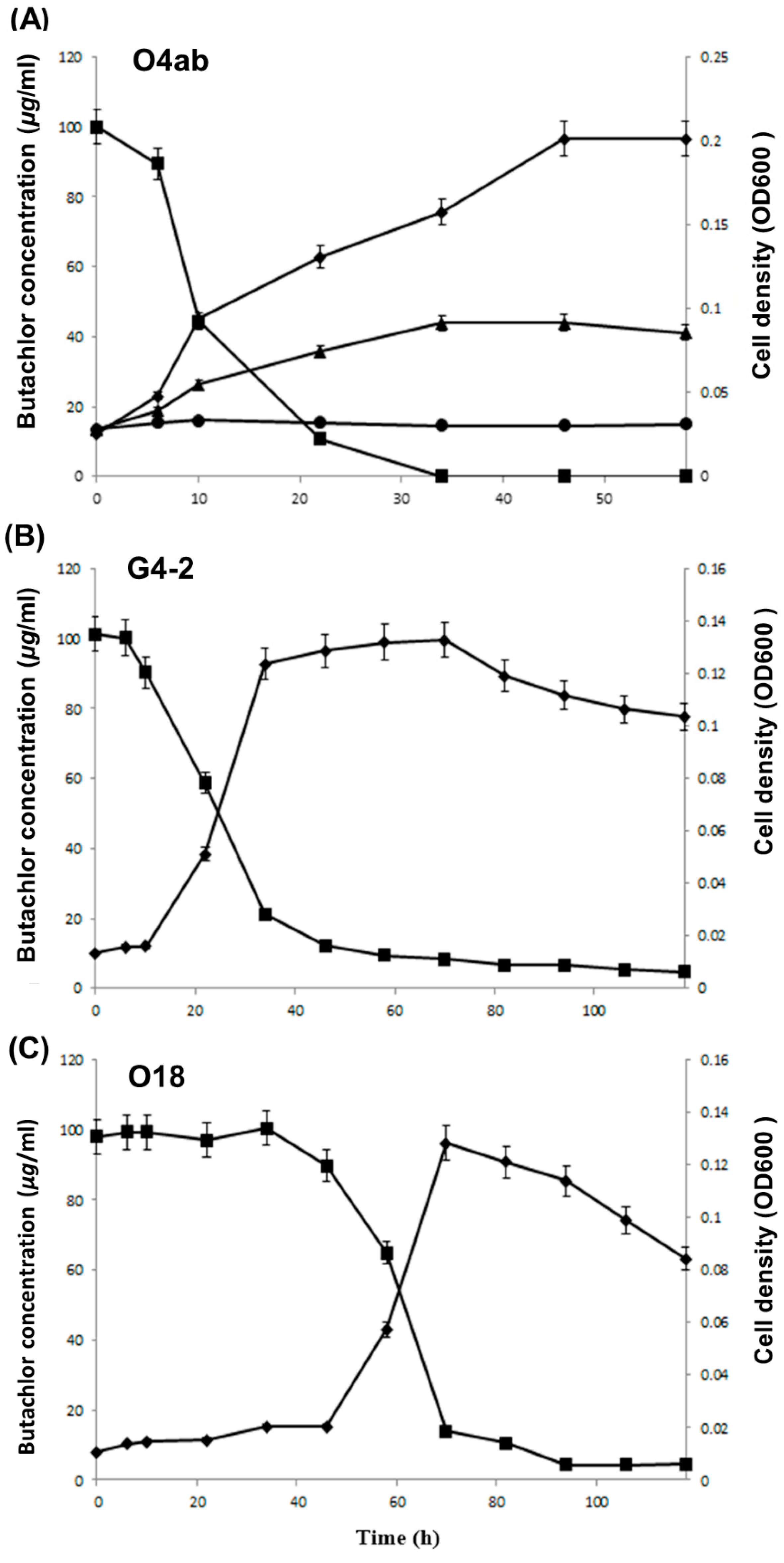
Figure 2A presents the growth and degradation curves of the syntrophic pair O4ab, which successfully degraded 100 mL L−1 of butachlor within 34 h. The cell density of this pair gradually increased from 0.035 to 0.20 at OD600, indicating their growth during the degradation process. O4ab exhibited the fastest butachlor degradation rate among the five syntrophic pairs, leading to its selection as the representative pair for further investigation of growth and metabolic pathways during butachlor biodegradation. Two representative strains, G4-2 and O18, were selected from the four isolates that were observed to degrade butachlor alone based on their REP-PCR fingerprints. These strains were then analyzed for their growth properties on the butachlor mineral medium. Figure 2B shows the growth and degradation curves of bacterial strain G4-2 on the butachlor mineral medium. This strain exhibited a quick growth response after an initial lag period of several hours, and completely utilized butachlor in approximately 40 h. On the other hand, Figure 2C shows the growth and degradation curves of bacterial strain O18, which displayed a relatively longer lag period and required around 70 h to completely degrade 100 mg L−1 of butachlor.

3.4. Identification of Metabolites Produced during Butachlor Degradation

The identification of the butachlor degradation pathway was studied by analyzing the metabolites through GC-MS. The gas chromatogram spectrum was obtained from 0 h and 12 h of incubation. GC-MS analysis of 0 h of incubation showed only one peak, which was identified as butachlor and originally added to the mineral medium. GC-MS analysis of 12 h of incubation with the syntrophic pair O4ab showed four peaks besides the butachlor peak (Figure 3A). Five peaks were observed in addition to the butachlor peak in the GC-MS analysis after 12 h of incubation with G4-2 (Figure 3B). After incubating with O18 for 12 h, GC-MS analysis revealed the presence of one peak in addition to the butachlor peak (Figure 3C). Compound (1) was identified as 2-chloro-N-(2,6-diethylphenyl) acetamide (CDEPA) by analyzing its molecular weight and fragment ion peaks. CDEPA was found to be the N-dealkylated form of butachlor (Figure 3D). CDEPA is a well-known butachlor metabolite by bacteria [16,24,25]. Based on their molecular weight and fragment ion peaks, compounds (2) and (3) were identified as 2,6-diethylaniline (DEA) and 4-amino-3,5-diethyl phenol (ADEP), respectively (Figure 3E,F). To further analyze the possible degradation pathway of butachlor by the representative isolates, the isolates were grown on various potential intermediates. Strains G4-2 and O18 could degrade and grow on butachlor, CDEPA, DEA, and catechol (Table 2). Strain O4a could grow on butachlor, DEA, and catechol, while strain O4b grew on CDEPA, DEA, and catechol (Table 2). None of the isolates could degrade and grow on alachlor and aniline.

4. Discussion

This study identified four bacterial strains and five syntrophic bacterial pairs capable of degrading butachlor in rice paddy soils. The majority of the isolates (64.3%) belonged to the genus Sphingobium, while the remaining isolates (35.7%) belonged to the genus Rhodococcus. These strains were found to be the predominant butachlor-degrading bacteria in the rice paddy soils that were tested (Table 1). Although several butachlor-degrading bacterial strains have previously been isolated [12,13,14,15,16,25], Sphingobium species isolated in this study are a novel type of butachlor-degrading bacteria that have not been reported in previous studies.
Syntrophy is a type of metabolic process where two or more organisms work together in a mutually beneficial way. In this process, one organism breaks down a particular compound, and the resulting end products are only utilized by another organism. The metabolism of the first organism only occurs when the second organism consumes the end products, thus maintaining them at low concentrations. Understanding syntrophic degradation of butachlor has clear implications for bioremediation process. In this study, novel syntrophic butachlor-degrading pairs were isolated
In terms of number and frequency of butachlor-degrading strains, it was found that the syntrophic degradation of butachlor was more dominant than the independent degradation of butachlor by a single isolate. Furthermore, butachlor degradation by syntrophic pairs was faster than that by single culture (Figure 1). Paddy soils exhibit a greater variety of microhabitats in comparison to upland soils. This diversity in microhabitats can contribute to the variation in soil bacterial communities, which may consequently result in increased metabolic diversity among soil bacteria [24]. Interestingly, in this study, all Rhodococcus species were involved in the initial attack on butachlor, while the Sphingobium species could degrade the metabolic intermediate of butachlor produced by the Rhodococcus species. The Rhodococcus species produced 2-chloro-N-(2,6-diethylphenyl) acetamide (CDEPA) as the metabolic intermediate during butachlor degradation, whose intermediate was also observed in a previous report [13]. The Rhodococcus species have been reported in numerous studies for their ability to biodegrade various pollutants, including butachlor [25,26].
The initial step of butachlor degradation involved dealkylation and dechlorination reactions, which resulted in the formation of smaller compounds, such as alachlor, CDEPA, and DEA [17]. While early-stage compounds of butachlor biodegradation have been well studied, there have been fewer reports on sub-stage compounds that are produced later during butachlor biodegradation. In a previous study, Zhang et al. [17] isolated Paracoccus sp. FLY-8 from rice field soil and found that it could mineralize butachlor. They used GC-MS analysis and enzyme activity assays to study its metabolic pathway and suggested that butachlor degradation proceeds through alachlor, CDEPA, aniline, and catechol. In contrast to the metabolic pathway proposed by Zhang et al. [17], which suggested the formation of alachlor as an intermediate during butachlor degradation, our study found no peak corresponding to alachlor. Our study found that none of the isolated bacterial strains were able to degrade and utilize alachlor as a sole carbon and energy source.
It is known that 2,6-diethylaniline is degraded into aniline, which is then further degraded into catechol. However, GC-MS analysis did not detect either aniline or catechol as metabolites in this study. Despite this, the butachlor-degrading bacterial strains O4 pairs, G4-2 and O18, were found to be unable to utilize aniline as a carbon source, but they were able to utilize catechol. This suggests that these bacterial strains transform 2,6-diethylaniline into catechol via an intermediate other than aniline. During the course of this study, a peak corresponding to 4-amino-3,5-diethyl phenol (ADEP) was detected in the butachlor biodegradation process by the O4 syntrophic pair (Figure 3C). According to previous studies, ADEP is produced through the oxidation of DEA [27]. Furthermore, when incubated with NADPH-fortified rat-liver microsomal enzymes, DEA was found to generate ADEP as the major product of oxidation. Moreover, ADEP can be further oxidized to DEBQI, which was identified as a minor metabolite during DEA oxidation [28].
In previous reports, it has been stated that the carcinogenic product 2,6-diethylaniline is present in groundwater at twice the concentration of alachlor [27]. Our study revealed that the butachlor-degrading bacteria isolated in this study were capable of utilizing 2,6-diethylaniline as the sole carbon source, indicating that the carcinogenic compound is not accumulated in the degradation process of butachlor by these isolates.
According to the results of the degradation capability test presented in Table 2 and the data obtained from GC-MS analysis shown in Figure 3, it seems that the O4 syntrophic pair can degrade butachlor using CDEPA, DEA, ADEP, and catechol as degradation pathways (Figure 4).
This study successfully isolated and characterized four bacterial strains capable of degrading butachlor, as well as five syntrophic pairs capable of breaking down butachlor from rice paddy soils in Korea. Through phenotypic and metabolic assay, it was found that the O4ab syntrophic pair degraded butachlor through a novel biodegradative pathway. The strain O4a transformed butachlor to CDEPA, while strain O4b transformed CDEPA to DEA. DEA was then transformed to ADEP, which further transformed to catechol before being mineralized. These findings provide valuable insights into the behavior and characteristics of butachlor-degrading bacteria and their potential use in bioremediation efforts.
The results of this study provide important insights into the potential for microbial biodegradation of butachlor in contaminated environments. The isolation and characterization of a new bacterial strain capable of breaking down butachlor through a previously unknown pathway suggest that there may be additional microorganisms with similar capabilities waiting to be discovered. This has implications for the development of new bioremediation strategies to remove butachlor and other chloroacetamide herbicides from contaminated soils and water sources.
The ability of this newly isolated strain to degrade butachlor in a mineral medium where butachlor serves as the sole carbon source indicates its potential as a tool for the bioremediation of contaminated sites. The identification of metabolic intermediates through gas chromatography–mass spectrometry analysis provides a more comprehensive understanding of the biodegradation pathway of butachlor, which can be useful in developing strategies to enhance the efficiency and effectiveness of microbial degradation.

5. Conclusions

In conclusion, four different bacteria and five different syntrophic pairs of bacteria capable of degrading butachlor were isolated from rice paddy soils in Korea. All single isolates and syntrophic pairs were able to utilize butachlor as a sole carbon and energy source. Among these isolates, the O4ab syntrophic pair degraded butachlor through a novel biodegradative pathway. The strain O4a transformed butachlor to CDEPA, while strain O4b transformed CDEPA to DEA. DEA was then transformed to ADEP, which further transformed to catechol before being mineralized. This study highlights the potential for microbial biodegradation as a viable strategy for removing butachlor from contaminated environments. The discovery of a new strain capable of breaking down butachlor through a novel pathway provides a promising avenue for further research and development of bioremediation technologies. This can contribute to the development of sustainable agricultural practices that minimize the negative impacts of herbicides on the environment and human health.

Author Contributions

H.L. and N.H.K. conceived, designed, and conducted all the experiments. H.L. analyzed all the data and wrote the manuscript. D.-U.K. coordinated and supervised the study. All authors have read and agreed to the published version of the manuscript.

Funding

This research was supported by Rural Development Administration (Project No. PJ014897032023).

Data Availability Statement

Not applicable.

Conflicts of Interest

The authors declare no conflict of interest.

References

  1. Yu, Y.; Chen, Y.; Luo, Y.; Pan, X.; He, Y.; Wong, M. Rapid degradation of butachlor in wheat rhizosphere soil. Chemosphere 2003, 50, 771–774. [Google Scholar] [CrossRef] [PubMed]
  2. Dwivedi, S.; Singh, B.R.; Al-Khedhairy, A.A.; Alarifi, S.; Musarrat, J. Isolation and characterization of butachlor-catabolizing bacterial strain Stenotrophomonas acidaminiphila JS-1 from soil and assessment of its biodegradation potential. Lett. Appl. Microbiol. 2010, 51, 54–60. [Google Scholar] [CrossRef] [PubMed]
  3. Lo, Y.C.; Yang, C.C.; Deng, J.F. Acute alachlor and butachlor herbicide poisoning. Clin. Toxicol. 2008, 46, 716–721. [Google Scholar] [CrossRef]
  4. Singh, J.; Kadapakkam Nandabalan, Y. Prospecting Ammoniphilus sp. JF isolated from agricultural fields for butachlor degradation. 3 Biotech 2018, 8, 164. [Google Scholar] [CrossRef]
  5. Kaur, R.; Goyal, D.; Agnihotri, S. Chitosan/PVA silver nanocomposite for butachlor removal: Fabrication, characterization, adsorption mechanism and isotherms. Carbohydra. Polym. 2021, 262, 117906. [Google Scholar] [CrossRef]
  6. Abigail, M.E.A.; Samuel, S.M.; Ramalingam, C. Addressing the environmental impacts of butachlor and the available remediation strategies: A systematic review. Int. J. Environ. Sci. Technol. 2015, 12, 4025–4036. [Google Scholar] [CrossRef] [Green Version]
  7. Götz, T.; Böger, P. The very-long-chain fatty acid synthase is inhibited by chloroacetamides. Zeitschrift fur Naturforsch. Sect. C. J. Biosci. 2004, 59, 549–553. [Google Scholar] [CrossRef] [PubMed] [Green Version]
  8. Panneerselvam, N.; Sinha, S.; Shanmugam, G. Butachlor is cytotoxic and genotoxic and induces apoptosis in mammalian cells. Environ. Mol. Mutagen. 1998, 31, 257–262. [Google Scholar]
  9. Min, H.; Ye, Y.F.; Chen, Z.Y.; Wu, W.X.; Yufeng, D. Effects of butachlor on microbial populations and enzyme activities in paddy soil. J. Environ. Sci. Health 2001, 36, 581–595. [Google Scholar] [CrossRef]
  10. Muthukaruppan, G.; Janardhanan, S.; Vijayalakshmi, G. Sublethal toxicity of the herbicide butachlor on the earthworm Perionyx sansibaricus and its histological changes. J. Soils. Sediments 2004, 5, 82–86. [Google Scholar] [CrossRef]
  11. Tilak, K.S.; Veeraiah, K.; Thathaji, P.B.; Butchiram, M.S. Toxicity studies of butachlor to the freshwater fish Channa punctata (Bloch). J. Environ. Biol. 2007, 28, 485–487. [Google Scholar] [PubMed]
  12. Coleman, S.; Linderman, R.; Hodgson, E.; Rose, R.L. Comparative metabolism of chloroacetamide herbicides and metabolites in human and rat liver microsomes. Environ. Health Perspect. 2000, 108, 1151–1157. [Google Scholar]
  13. Liu, H.; Cao, L.; Lu, P.; Ni, H.; Li, Y.X.; Yan, X.; Hong, Q.; Li, S.P. Biodegradation of butachlor by Rhodococcus sp. strain B1 and purification of its hydrolase (ChlH) responsible for N-dealkylation of chloroacetamide herbicides. J. Agric. Food. Chem. 2012, 6, 12238–12244. [Google Scholar] [CrossRef] [PubMed]
  14. Wang, J.H.; Lu, Y.T.; Chen, Y.Y. Comparative proteome analysis of butachlor-degrading bacteria. Environ. Geol. 2008, 53, 1339–1344. [Google Scholar] [CrossRef]
  15. Chen, Q.; Wang, C.H.; Deng, S.K.; Wu, Y.D.; Li, Y.; Yao, L.; Jiang, J.D.; Yan, X.; He, J.; Li, S.P. Novel three-component Rieske non-heme iron oxygenase system catalyzing the N-dealkylation of chloroacetanilide herbicides in Sphingomonads DC-6 and DC-2. Appl. Environ. Microbiol. 2014, 80, 5078–5085. [Google Scholar] [CrossRef] [PubMed] [Green Version]
  16. Chen, K.; Chen, Q.; Wang, G.X.; Ni, H.Y.; He, J.; Yan, X.; Gu, J.G.; Li, S.P. Sphingomonas chloroacetimidivorans sp. nov., a chloroacetamide herbicide-degrading bacterium isolated from activated sludge. Antonie Van Leeuwenhoek 2015, 108, 703–710. [Google Scholar] [CrossRef] [PubMed]
  17. Zhang, J.; Zheng, J.W.; Liang, B.; Wang, C.H.; Cai, S.; Ni, Y.Y.; He, J.; Li, S.P. Biodegradation of chloroacetamide herbicides by Paracoccus sp. FLY-8 in vitro. J. Agric. Food Chem. 2011, 59, 4614–4621. [Google Scholar] [CrossRef] [PubMed]
  18. Zheng, J.; Li, R.; Zhu, J.; Zhang, J.; He, J.; Li, S.; Jiang, J. Degradation of the chloroacetamide herbicide butachlor by Catellibacterium caeni sp. nov DCA-1T. Int. Biodeterior. Biodegrad. 2012, 73, 16–22. [Google Scholar] [CrossRef]
  19. Baker, G.C.; Smith, J.J.; Cowan, D.A. Review and re-analysis of domain-specific 16S primers. J. Microbiol. Methods 2003, 55, 541–555. [Google Scholar] [CrossRef] [PubMed] [Green Version]
  20. Lane, D.J. 16S/23S rRNA sequencing. In Nucleic Acid Techniques in Bacterial Systematics; Stackebrandt, E., Goodfellow, M., Eds.; Wiley: Chichester, UK, 1991; pp. 115–175. [Google Scholar]
  21. Lee, S.; Malone, C.; Kemp, P. Use of multiple 16S rRNA targeted fluorescent probes to increase signal strength and measure cellular RNA from natural planktonic bacteria. Mar. Ecol. Prog. Ser. 1993, 101, 193–201. [Google Scholar] [CrossRef]
  22. Kim, M.; Chun, J. 16S rRNA gene-based identification of bacteria and archaea using the EzTaxon server. In Methods in Microbiology; Academic Press: Cambridge, MA, USA, 2014; Volume 41, pp. 61–74. [Google Scholar]
  23. Bruijin, F.J.D. Use of repetitive (repetitive extragenic palindromic and enterobacterial repetitive intergeneric consensus) sequences and the polymerase chain reaction to fingerprint the genomes of Rhizobium meliloti isolates and other soil bacteria. Appl. Environ. Microbiol. 1992, 58, 2180–2187. [Google Scholar] [CrossRef] [Green Version]
  24. Torsvik, V.; Sorheim, R.; Goksoyr, J. Total bacterial diversity in soil and sediment communities—A review. J. Ind. Microbiol. 1996, 17, 170–178. [Google Scholar] [CrossRef]
  25. Xiong, M.; Zang, H.; Pan, J.; Xu, C.; Li, C. Isolation of a novel chlorimuron-ethyl-degrading bacterium Rhodococcus sp. D310-1. In Proceedings of the iCBBE, Wuhan, China, 10–12 May 2011; pp. 1–4. [Google Scholar]
  26. Geminia, V.; Gallegoa, A.; Oliveirab, V.D.; Gomezc, C.; Manfiob, G.; Korola, S. Biodegradation and detoxification of p-nitrophenol by Rhodococcus wratislaviensis. Int. Biodeterior. Biodegrad. 2005, 55, 103–108. [Google Scholar] [CrossRef]
  27. Osano, O.; Admiraal, W.; Klamer, H.J.C.; Pastor, D.; Bleeker, E.A.J. Comparative toxic and genotoxic effects of chloroacetanilides, formamidines and their degradation products on Vibrio fischeri and Chironomus riparius. Environ. Pollut. 2002, 119, 195–202. [Google Scholar] [CrossRef]
  28. Feng, P.C.C.; Wratten, S.J. In vitro oxidation of 2, 6-diethylaniline by rat liver microsomal enzymes. J. Agric. Food Chem. 1987, 35, 491–496. [Google Scholar] [CrossRef]
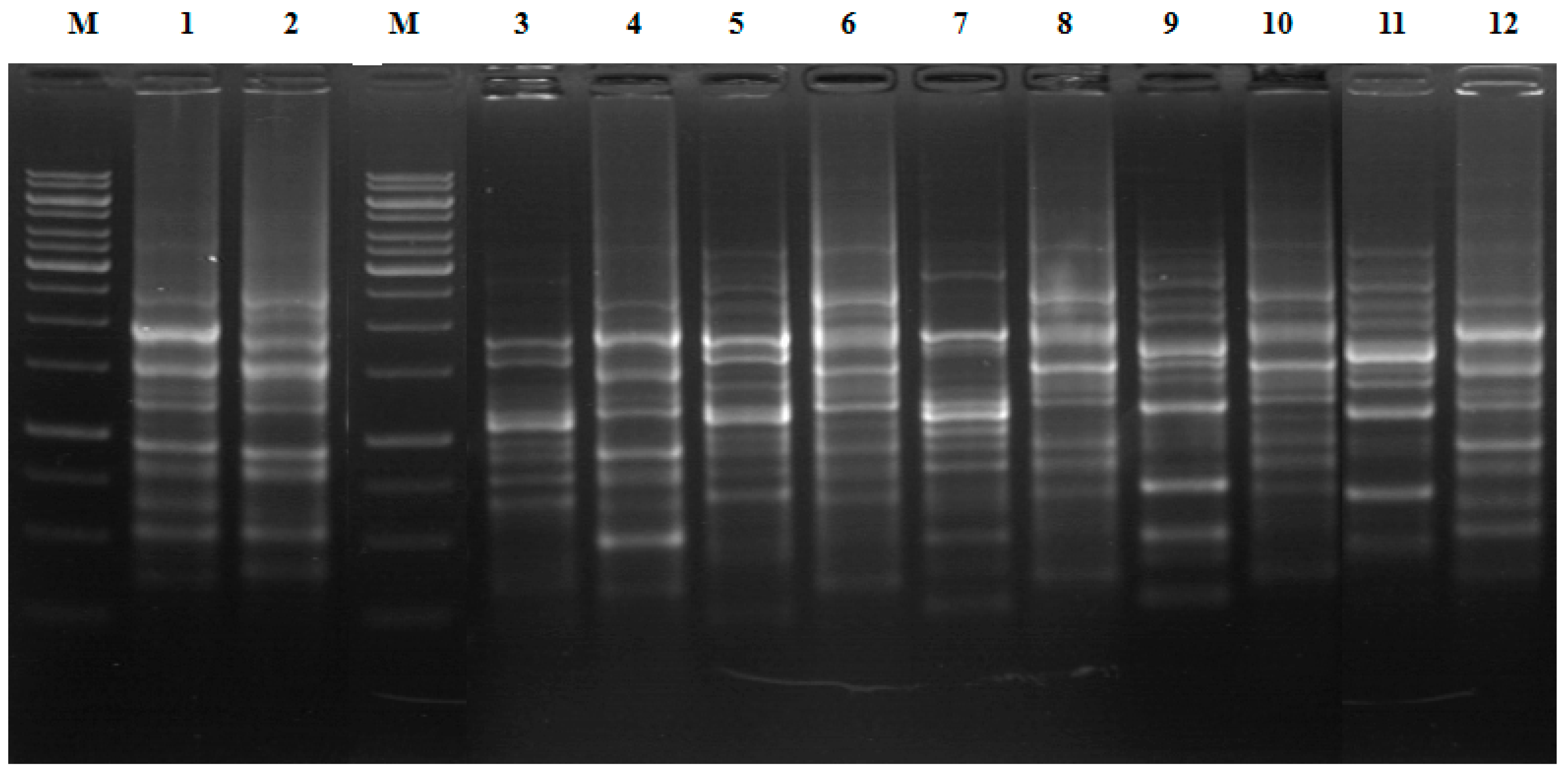
Figure 1. REP-PCR patterns of the isolates. Lanes: 1, G4-2; 2, O18; 3, H4a; 4, H4b; 5, J11a; 6, J11b; 7, J12a; 8, J12b; 9, L9a; 10, L9b; 11, O4a; 12, O4b; M, 1 kb DNA size marker.
Processes 11 01222 g001
Figure 2. Utilization and degradation of butachlor by the isolates. (A) Butachlor degradation and cell growth of syntrophic pair O4ab. ■: residual concentration of butachlor; ♦: OD600 of O4ab pair in butachlor mineral medium; ▲: slight growth of O4a; ●, no growth of O4b. (B) Butachlor degradation and cell growth of G4-2. ■: residual concentration of butachlor; ♦: OD600 of G4-2 in butachlor mineral medium. (C) Butachlor degradation and cell growth of O18. ■: residual concentration of butachlor; ♦: OD600 of O18 in butachlor mineral medium.
Processes 11 01222 g002
Figure 3. GC profiles and mass spectra of metabolite: (A) GC profile of butachlor metabolites produced by O4ab pair; (B) GC profile of butachlor metabolites produced by G4-2; (C) GC profile of butachlor metabolites produced by O18; (D) GC-MS spectra of butachlor metabolites 2-chloro-N-(2,6-diethylphenyl) acetamide (1); (E) GC-MS spectra of butachlor metabolites 2,6-diethylaniline (2); (F) GC-MS spectra of butachlor metabolites 4-amino-3,5-diethyl phenol (3).
Processes 11 01222 g003
Figure 4. Proposed biodegradation pathway of butachlor by the syntrophic pair O4ab.
Processes 11 01222 g004
Table 1. Nearest relatives of the butachlor-degrading isolates based upon 16S rRNA gene sequences.
IsolateGenebank Accession NumberSampling SiteNearest Type StrainSimilarity (%)
G4-2JX101328YeongjuSphingobium chunbukense DJ77T98.4
G3JX979142YeongjuSphingobium chunbukense DJ77T98.2
O18JX979134WonjuSphingobium chlorophenilicum ATCC 33790T98.4
K8JX979135AnsanSphingobium chunbukense DJ77T98.4
O4aJX979150JangsungRhodococcus qingshengii djl-6T100.0
O4bJX979146JangsungSphingobium chungbukense DJ77T98.6
L9aJX979140AnsanRhodococcus qingshengii djl-6T99.9
L9bJX979136AnsanSphingobium chungbukense DJ77T98.8
J12aJX979132PyeongtaekRhodococcus ruber DSM 43338T99.7
J12bJX979141PyeongtaekSphingobium chungbukense DJ77T98.8
J11aJX979145PyeongtaekRhodococcus ruber DSM 43338T100.0
J11bJX979138PyeongtaekSphingobium chungbukense DJ77T98.7
H4aJX979148ChungjuRhodococcus ruber DSM 43338T100.0
H4bJX979144ChungjuSphingobium chungbukense DJ77T98.4
Table 2. Degradation and growth of the isolates on various potential intermediates of butachlor.
CharacteristicsG4-2O18O4aO4b
Utilization of
Butachlor+++
Alachlor
CDEPA+++
DEA++++
Aniline
Catechol++++
+: over 80 % reduction in peak height as determined by UV scanning and substantial growth (OD600 >0.05); −: below 10 % reduction in peak height and scant growth (OD600 < 0.007).
Disclaimer/Publisher’s Note: The statements, opinions and data contained in all publications are solely those of the individual author(s) and contributor(s) and not of MDPI and/or the editor(s). MDPI and/or the editor(s) disclaim responsibility for any injury to people or property resulting from any ideas, methods, instructions or products referred to in the content.