Communication

Plum Pox Virus Genome-Based Vector Enables the Expression of Different Heterologous Polypeptides in Nicotiana benthamiana Plants

1
Biomedical Research Center, Institute of Virology, Slovak Academy of Sciences, 84505 Bratislava, Slovakia
2
Faculty of Natural Sciences, University of St. Cyril and Methodius, 91701 Trnava, Slovakia
*
Author to whom correspondence should be addressed.
Processes 2022, 10(8), 1526; https://doi.org/10.3390/pr10081526
Submission received: 22 June 2022 / Revised: 26 July 2022 / Accepted: 29 July 2022 / Published: 3 August 2022
(This article belongs to the Special Issue State of the Art of Protein Expression Systems)

Abstract

:
Plant viral vectors have become a promising tool for the rapid and cost-effective production of recombinant proteins in plants. Among the numerous genera of viruses that have been used for heterologous expression, potyviruses offer several advantages, such as polyprotein expression strategy or a broad host range. In our work, the expression vectors pAD/pAD-agro based on the plum pox virus (PPV) genome were used for the heterologous expression of different foreign polypeptides: alfalfa mosaic virus capsid protein (AMV CP), zucchini yellow mosaic virus capsid protein (ZYMV CP), the small heat-shock protein of Cronobacter sakazakii fused with hexahistidine (sHSP-his), a fragment of influenza A virus hemagglutinin (HA2-2), influenza A virus protein PB1-F2, SARS-CoV-2 nucleocapsid protein (CoN2-his), and its N- and C-terminal fragments (CoN-1-his and CoN3-his, respectively), each fused with a hexahistidine anchor. Particular proteins differed in their accumulation, tissue localization, stability, and solubility. The accumulation rate of produced polypeptides varied from low (N, hemagglutinin fragment) to relatively high (plant viral CPs, N-terminal fragment of N, PB1-F2). Some proteins preferentially accumulated in roots (sHSP, hemagglutinin fragment, PB1-F2), showing signs of proteolytic degradation in leaf tissues. Thus, each expression requires an individual approach and optimization. Here, we summarize our several-year experiments and discuss the usefulness of the pAD/pADep vector system.

1. Introduction

Plant viruses are infectious particles capable of autonomous replication and systemic spreading within their hosts. Given these unique features, plant viral replicons have been widely used for the expression of heterologous proteins in plants. Over the decades of the development of plant viral vectors, different plant viruses have been modified and used for this purpose, including ssDNA geminiviruses, dsDNA caulimoviruses, and several taxa of ssRNA viruses [1]. Genomes of plant RNA viruses can be easily manipulated by a generation of infectious cDNA clones. Depending on the strategy of final host transfection, infectious transcripts or cDNA may be applied [2]. Resultant infectious transcripts can be readily delivered to the target host plant either by direct mechanical inoculation or biolistic transfection. The biolistic method is applicable also for the infectious cDNA; however, Agrobacterium-mediated gene delivery is more favorable, allowing fast, simple, and efficient transient expression in plant tissues without the need for stable nuclear transformation. Although transient expression in plants may also occur directly from agrobacterial T-DNA, viral vectors provide the systemic translocation of their replicons within the plant during infection, thus increasing the expression efficiency. Recently, the new generation of deconstructed viral vectors has proven to be highly effective in recombinant protein production while being environmentally safe [3]. However, good results can also be obtained using nearly wild-type full-length viral genomes [4].
The genus Potyvirus (family Potyviridae) comprises (+)ssRNA viruses with a genome of approximately 10 kb and a polyprotein expression strategy. Despite the lack of knowledge about the in vivo functions of all 11 viral proteins (one of them produced from an alternative ORF), it is likely that each of them plays an essential role during viral infection. Therefore, no larger deletions are feasible to maintain infectivity, which limits the construction and design of potyvirus-based vectors. Nevertheless, potyviruses offer several advantages that make them favorable for heterologous gene expression. The expression of the potyviral genome through a single polyprotein ensures the equimolar production of all its products, including foreign genes inserted into it. Moreover, filamentous rod-shaped potyviral particles generally do not show genome packaging restrictions compared to icosahedral virions, thus allowing larger genetic insertions. Finally, a large number of known potyvirus species offers a wide range of potential plant hosts [5]. Several different potyviruses have already been engineered to express foreign genes, including tobacco etch virus [6,7,8], soybean mosaic virus [9], tobacco vein banding mosaic virus [10], zucchini yellow mosaic virus [11,12], turnip mosaic virus [13], lettuce mosaic virus [14], wheat streak mosaic virus [15], clover yellow vein mosaic virus [16], potato virus A [17], or plum pox virus [18].
Here we summarize and discuss our several years of experience with the expression of various foreign genes of bacterial or viral origin in Nicotiana benthamiana using the plum pox virus (PPV) genome-based vectors pAD/pAD-agro in order to compare the production efficiency and limits of particular proteins.

2. Materials and Methods

2.1. PPV-Based Viral Vectors

The vector pAD (originally described as pBOR-VT [19]) consists of a full-length PPV-Rec cDNA under the control of the 35S CaMV promoter, cloned in the plasmid vector pGEM3 (Figure 1). A cloning linker comprising EagI/KpnI restriction sites was introduced between the genes for viral replicase (NIb) and capsid protein (CP). To ensure its environmental safety, the DAG motif-coding region within the CP essential for the aphid transmissibility of the virus [20] was modified by site-directed mutagenesis [19].
The commercial Agrobacterium binary vector pCambia 1304 (Abcam, Cambridge, UK), shortened by the deletion of the β-glucuronidase gene (SphI cleavage and religation), was used as a backbone for the construction of pAD-agro. The cDNA of PPV-Rec together with upstream 35S CaMV promoter was recloned from pAD to the shortened pCambia 1304 via unique PvuII/BamHI sites.

2.2. Cloning Procedures

Several complete or partial genes of prokaryotic and viral origin were chosen for heterologous expression in plants, encoding alfalfa mosaic virus (AMV) and zucchini yellow mosaic virus (ZYMV) CP, Cronobacter sakazakii small heat-shock protein (sHSP), influenza A virus (IAV) hemagglutinin (HA), IAV nonstructural protein PB1-F2, and severe acute respiratory syndrome coronavirus 2 (SARS-CoV-2) nucleoprotein. Particular genes of interest were inserted into the pAD/pAD-agro vectors either by restriction cloning or using In-Fusion HD Cloning Kit (Takara Bio, Shiga, Japan). Target genes were amplified by PCR using Ex Taq DNA polymerase (Takara Bio, Shiga, Japan) from source DNA/cDNA. Primers used for amplification were designed to include EagI/KpnI sites (for restriction cloning), or the overlaps with the KpnI-linearized vector (for In-Fusion cloning). All cloning steps were performed in E. coli JM109 according to standard protocols. The primers are listed in Table 1 together with PCR amplification details. PCR products and linearized vectors were purified from agarose gels by Wizard SV Gel and PCR Clean-Up System (Promega, Madison, WI, USA) and subsequently used for cloning. Resultant plasmid constructs were isolated from E. coli using QIAprep Spin Miniprep Kit (Qiagen, Hilden, Germany).

2.3. Plant Transfection and Expression

The pAD constructs were introduced into N. benthamiana plants by biolistics using a common airgun [21] and 1 μm Gold Microcarrier (Bio-Rad, Hercules, CA, USA). The pAD-agro constructs were electroporated into Agrobacterium tumefaciens EHA105. An overnight culture of agrobacteria was diluted to OD600 0.1 in the infiltration solution (10 mM MES/10 mM MgCl2/200 μM acetosyringone, pH 5.6) and the suspension was infiltrated to the underside of leaves using a needleless syringe. At least 40 plants were included in each infiltration experiment and each experiment was repeated five times. The plants were grown under controlled conditions (22 ± 2 °C, photoperiod 12 h) in an insect-free room. The symptoms of PPV infection were visually monitored and leaf or root samples (each mixed from two plants) taken 2–20 days post-inoculation (dpi) were analyzed by Western blotting using PPV- and foreign protein-specific antibodies (Table 2). If no or very weak signal was detected, the analysis was repeated after tissue extraction by 8 M urea. Singular samples from plants surviving for a long time (up to 130 dpi) were also analyzed. For RT-PCR, the total RNA was isolated from upper noninoculated (systemically infected) leaves by the kit NucleoSpin RNA Plant (Macherey-Nagel, Düren, Germany) and cDNA was prepared by AMV reverse transcriptase and random primers (Promega, Madison, WI, USA). PCR with primers NCuniFor/NCuniRev spanning the insertion site [22] was performed to prove the genetic stability of each construct, followed by Sanger sequencing (Eurofins Genomics, Ebersberg, Germany). Polypeptides fused with hexahistidine tags were purified from plant tissues by batch-immobilized metal ion affinity chromatography (IMAC) using HisPur Cobalt Resin (Thermo Fisher Scientific, Waltham, MA, USA) under native or denaturing conditions in the presence of 6 M guanidinium chloride (GuHCl) or 8 M urea according to the manufacturer‘s recommendation. The protein concentration in each fraction was estimated by measuring absorbance at 280 nm using NanoDrop 2000c (Thermo Fisher Scientific, Waltham, MA, USA). Prior to SDS-PAGE, the proteins from chaotropic IMAC fractions were precipitated by 40 volumes of 96% ethanol at −20 °C overnight [23], vacuum-dried, and resuspended in the sample buffer.

3. Results and Discussion

In total, eight sequences originating from six foreign genes (one bacterial and five viral) were cloned into the pAD/pAD-agro vectors to evaluate their expression in Nicotiana benthamiana at the protein level (Table 3, Figure 2). Each of these experiments is presented and discussed below.

3.1. Capsid Proteins of Plant Viruses

The proteins of plant pathogens should be easily produced in the cells of their hosts. For this reason, two genes encoding the capsid proteins of plant viruses were chosen, serving as expression controls to assess the ability to use pAD and pAD-agro vectors. A nearly complete CP gene sequence of AMV isolate T6 [27] and a complete CP gene sequence of ZYMV isolate Kuchyna [28] were cloned into the pAD. Following the biolistic transfection of plants, both proteins accumulated in tissues beginning at 4–5 dpi with maximum levels recorded at 11–12 dpi. In both cases, the expression kinetics (time course of expression) corresponded to that of PPV CP. The highest accumulation levels were observed in apical leaves and roots, compared to inoculated leaves. Such a picture correlates well with the highest virus replication efficiency in fast-dividing meristems, as well as with the preferential systemic spread of the infection to these tissues documented by classic experiments [29].
Product concentrations later slightly decreased, however, they were still detectable in young symptomatic leaves 30 dpi. In very old plants transfected with pAD-AMV CP (more than 120 dpi), AMV CP was present only in some leaves tested. Despite the ongoing PPV infection, RT-PCR analyses and sequencing confirmed the long-term genetic instability of the construct in vivo, causing deletions within the inserted sequence. On the other hand, both inserts were stably maintained for at least three serial mechanical passages in N. benthamiana.
Several experiments (size exclusion chromatography, Centricon filtration, ultracentrifugation) indicated that heterologous CP was not present in planta as separate molecules, but rather in the form of larger clusters or pseudovirions (data not shown). AMV naturally forms bacilliform or spheroidal virions of different sizes, depending on the encapsidated genome segment of this multipartite virus [30]. In the absence of RNA, the isolated AMV CP reassembles into icosahedral empty particles, reflecting mutual interactions between AMV CP molecules [31]. ZYMV belongs to filamentous potyviruses with a higher contribution of protein–RNA interactions to the virion assembly process [32]. However, the production of RNA-free potyvirus-like particles has also been demonstrated [33,34].

3.2. Cronobacter Sakazakii Small Heat-Shock Protein

The protein referred to as sHSP is encoded by ORF H, located in an 18 kbp long thermotolerance island within the genome of the Gram-negative bacteria Cronobacter sakazakii strain ATCC 29544. According to sequence analysis, sHSP was characterized as an ATP-independent chaperone, belonging to the Hsp20 family of small heat shock proteins. Such proteins stabilize unfolded or partially folded polypeptides and were found in nearly all living organisms [35,36].
As no specific antibody was available, the target protein was designed to include hexahistidine tags (his-tags) on both termini (sHSP-his), thus enabling its detection with commercial anti-his antibodies and purification by affinity chromatography.
The prepared construct pAD-sHSP-his was infectious for N. benthamiana and remained stable even after prolonged cultivation periods. The protein accumulated in plants mainly between 7 and 14 dpi with a maximum recorded 9 dpi. However, low levels of sHSP-his could be detected even at 75 dpi. Similar infection kinetics were observed following the agroinfection of plants with the construct pAD-agro-sHSP-his. Interestingly, the detection signal was much weaker in leaves compared to the relatively high sHSP-his accumulation in roots. Both the leaves and roots of infected plants were subsequently used for product purification by IMAC. sHSP-his could not be extracted from plant tissues under native conditions, but it was effectively solubilized from leaves in the presence of 6 M GuHCl. An overall sHSP-his yield from IMAC was 24 μg/g of fresh leaf tissue. The extraction efficiency from roots was lower and yielded about 8 μg/g of the tissue.
In bacteria, the thermotolerance genomic island influenced their resistance to high temperatures and osmotic stress [37]. The N. benthamiana plants producing sHSP-his showed no increased tolerance to such types of abiotic stress compared to control plants.

3.3. Fragment of Influenza A Virus Hemagglutinin

Hemagglutinin (HA) is the most abundant glycoprotein of influenza viruses, forming homotrimeric spikes on the virion surface. Each monomer is composed of two glycopeptides (HA1, HA2) linked by a disulfide bond [38]. HA1 creates a highly variable distal globular head with a receptor-binding site (RBS), while HA2 is more conserved and anchors the molecule in the viral envelope [39].
During infection, HA1 induces the generation of most virus-neutralizing antibodies [40]. Antibodies against epitopes located more distantly from RBS possess no virus-neutralizing activity but they can affect later stages of infection [41,42]. Vaccines against the conserved HA2 might induce broad heterosubtypic immunity and overcome the problem of the high seasonal antigen variability of influenza viruses [43]. Therefore, we tried to produce a potentially immunogenic 150-amino-acid-long fragment of influenza A virus (IAV) HA2 (referred to as HA2-2) in plants. The monoclonal antibody IIF4 binding this domain was used for detection [25,44]. Although the construct pAD-HA2-2 was infectious, the target polypeptide was detected only at 9–12 dpi, exclusively in the roots of transfected plants. A poor detection signal in Western blot indicated the rapid degradation of HA2-2 in planta (Figure 2). Better results were not reached following the agroinfection of plants with the construct pAD-agro-HA2-2. Repeatedly analyzed positive samples gave an even weaker or no signal, suggesting the low stability of HA2-2 also in vitro. The tertiary structure of HA2 may play an important role in maintaining its overall stability. Thus, the production of its shorter fragments might require further optimization to improve the stability, e.g., by their fusion with PPV CP.

3.4. Influenza A PB1-F2

Another IAV protein chosen for expression was the 10.5 kDa non-structural protein PB1-F2, involved in viral pathogenicity. PB1-F2 is rather unstable in vivo and tends to form amyloid fibers following expression in E. coli [45,46]. Unfortunately, this was also the case in N. benthamiana. Despite the relatively high PB1-F2 accumulation (mainly in the root system), it remained in the insoluble fraction. A substantial part of the product could be solubilized with 1% SDS or 8 M urea. Since we aimed to produce PB1-F2 for research purposes, several renaturation attempts were performed to recover its biological activity; however, none of these attempts was successful [47].

3.5. SARS-CoV-2 Nucleocapsid Protein and Its Fragments

The COVID-19 pandemic has been challenging for the global scientific community. Our PPV-based vector was previously used for the production of encapsidated Spike RNA mimics, serving as a positive control in qRT-PCR assays (Rusková et al., unpublished results). Plant-produced SARS-CoV-2 proteins could be valuable in diagnostics and vaccine development. Therefore, the vector pAD-agro was used for the expression of full-length SARS-CoV-2 nucleocapsid protein (N) and its N- and C-terminal fragments, each his-tagged on both termini. Particular constructs were designed as CoN1-his (N-terminal part), CoN2-his (full-length N), and CoN3-his (C-terminal part).
Recent works demonstrated the use of plant-expressed recombinant nucleocapsid as a part of an antigen cocktail for vaccination [48], or an antigen for serological tests [49]. While the SARS-CoV-2 spike protein is a major antigen that triggers the generation of neutralizing antibodies, the role of other viral antigens remains unclear. The latest findings suggest that nucleocapsid-specific immunity might contribute to overall protection against severe disease [50,51]. Hence, future vaccination strategies may involve a combination of spike- and nucleocapsid-based vaccines. Being the most abundant protein of coronaviruses, the nucleocapsid is also extensively used in the serodiagnosis of COVID-19 [52]. Plants offer the rapid and affordable production of recombinant antigens, thus reducing the overall costs of diagnostic tests.
Our results showed that N and both of its fragments accumulated in systemically infected leaves following transfection. CoN1-his reached the highest accumulation levels, being stably present for at least 28 dpi. In contrast, both CoN2-his and CoN3-his showed progressive proteolysis, resulting in a much lower concentration in planta. All three polypeptides were well recognized with the SARS-CoV-2 nucleocapsid polyclonal antibody, indicating their proper immunogenicity.
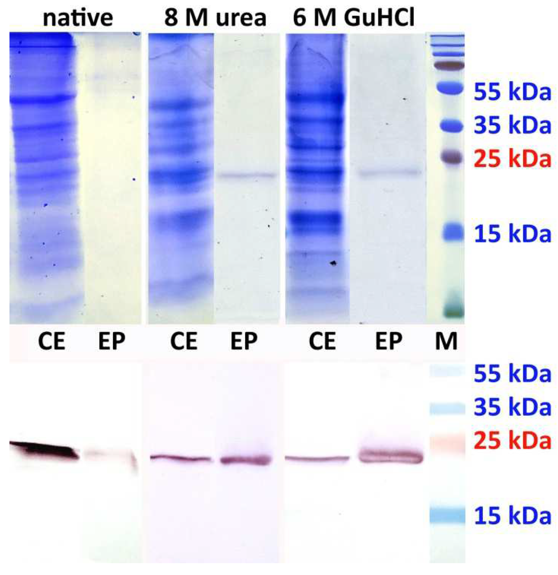
Based on these results, CoN1-his was purified by IMAC under both native and denaturing conditions. Native IMAC resulted in insufficient elution of CoN1-his from the resin. Higher efficiency was observed under denaturing conditions (comparable if 8 M urea or 6 M GuHCl was used), giving a high purity product with a yield of approximately 78 μg/g of fresh leaf tissue (Figure 3). Chaotropic IMAC was also successful for CoN3-his in contrast to full-length CoN2-his which could not be so far purified due to low expression level and proteolytic degradation.

4. Conclusions

Due to the polyprotein expression strategy, all the potyviral proteins are produced in equimolar amounts including the product of any foreign gene cloned in the genome. Not all of them, however, possess the same turnover. While PPV CP concentration is stabilized by forming virions, other gene products may be deposited in inclusion bodies or quickly degraded. Our PPV-based vector can mediate the transient expression of various heterologous polypeptides in planta. The results summarized in Table 3 demonstrated that each expression requires an individual approach concerning stability, solubility, or tissue localization, as these parameters may differ dramatically, depending on the cloned gene. The stability of produced exogenous polypeptides varied from low (N, HA2-2) to fair (plant viral CPs, N-proximal fragment of N, PB1-F2). Some proteins preferentially accumulated in root tissues (sHSP-his, HA2-2, PB1-F2), likely due to their increased susceptibility to leaf proteases. The extraction of the products from root tissues, however, was less efficient. No clear correlation was found between the molecular weight of the target polypeptides and their accumulation rates. Although mainly polypeptides in the range of 10–25 kDa were produced, even the largest protein (N, nearly 46 kDa) reached levels over the detection limit.
The purification efficiency is dependent on the source biological material. Roots contain much less total protein than green tissues, but the manipulation is more challenging—their acquirement, per se, from the soil in a relatively clean state is complicated and the homogenization mostly requires liquid nitrogen. Plant hydroponic culture or root-inducing bacterial systems would be of benefit to increase the yields from root tissues.
Low solubility may be a fair problem with some proteins produced in plants, as demonstrated especially by PB1-F2. Chaotropic agents such as urea or GuHCl may help to release the protein of interest for further maintenance in the liquid phase; however, if the product should serve other than immunological purposes and biological activity is required, renaturation conditions must be carefully optimized.
IMAC proved to be very efficient and fast for the purification of his-tagged polypeptides from N. benthamiana. Although scaling-up optimization is required, preliminary experiments lead to yields of dozens of micrograms per gram of fresh plant tissue, which was comparable to data obtained with other published potyvirus-based vectors [11,12].
Our recent findings demonstrated the susceptibility of different PPV hosts to agroinfection with pAD-agro, including oilseed poppy (Papaver somniferum) and plum (Prunus domestica), thus enabling the potential production of heterologous proteins in edible plant species [53].

Author Contributions

Conceptualization and methodology, Z.Š.; investigation, A.A. and Z.Š.; writing—original draft preparation, Z.Š. and A.A.; writing—review and editing, Z.Š. and M.G.; funding acquisition, M.G. All authors have read and agreed to the published version of the manuscript.

Funding

This work was supported by the grant 2/0003/22 from the Scientific Grant Agency of the Ministry of Education and Slovak Academy of Sciences (VEGA) and by the project APVV-18-0005 (Slovak Research and Development Agency).

Data Availability Statement

The nucleotide sequences of genes reported in this paper have been retrieved from or deposited in the public databases under the accession numbers listed in Table 1.

Acknowledgments

The authors are thankful to H. Drahovská (Faculty of Natural Sciences, Comenius University, Bratislava), I. Košík, E, Varečková, and B. Klempa (Biomedical Research Center, Slovak Academy of Sciences, Bratislava) for kindly providing source templates for gene amplification and specific monoclonal antibodies.

Conflicts of Interest

The authors declare no conflict of interest.

References

  1. Hefferon, K. Plant virus expression vector development: New perspectives. Biomed. Res. Int. 2014, 2014, 785382. [Google Scholar] [CrossRef]
  2. Nagyová, A.; Subr, Z. Infectious full-length clones of plant viruses and their use for construction of viral vectors. Acta Virol. 2007, 51, 223–237. [Google Scholar]
  3. Peyret, H.; Lomonossoff, G.P. When plant virology met Agrobacterium: The rise of the deconstructed clones. Plant Biotechnol. J. 2015, 13, 1121–1135. [Google Scholar] [CrossRef]
  4. Gleba, Y.; Marillonnet, S.; Klimyuk, V. Engineering viral expression vectors for plants: The ‘full virus’ and the ‘deconstructed virus’ strategies. Curr. Opin. Plant Biol. 2004, 7, 182–188. [Google Scholar] [CrossRef]
  5. Kelloniemi, J.; Mäkinen, K.; Valkonen, J.P. A potyvirus-based gene vector allows producing active human S-COMT and animal GFP, but not human sorcin, in vector-infected plants. Biochimie 2006, 88, 505–513. [Google Scholar] [CrossRef]
  6. Dolja, V.V.; Hong, J.; Keller, K.E.; Martin, R.R.; Peremyslov, V.V. Suppression of potyvirus infection by coexpressed closterovirus protein. Virology 1997, 234, 243–252. [Google Scholar] [CrossRef]
  7. Bedoya, L.; Martínez, F.; Rubio, L.; Daròs, J.A. Simultaneous equimolar expression of multiple proteins in plants from a disarmed potyvirus vector. J. Biotechnol. 2010, 150, 268–275. [Google Scholar] [CrossRef]
  8. Majer, E.; Navarro, J.A.; Daròs, J.A. A potyvirus vector efficiently targets recombinant proteins to chloroplasts, mitochondria and nuclei in plant cells when expressed at the amino terminus of the polyprotein. Biotechnol. J. 2015, 10, 1792–1802. [Google Scholar] [CrossRef]
  9. Seo, J.K.; Choi, H.S.; Kim, K.H. Engineering of soybean mosaic virus as a versatile tool for studying protein-protein interactions in soybean. Sci. Rep. 2016, 6, 22436. [Google Scholar] [CrossRef]
  10. Gao, R.; Tian, Y.P.; Wang, J.; Yin, X.; Li, X.D.; Valkonen, J.P. Construction of an infectious cDNA clone and gene expression vector of Tobacco vein banding mosaic virus (genus Potyvirus). Virus Res. 2012, 169, 276–281. [Google Scholar] [CrossRef]
  11. Arazi, T.; Lee Huang, P.; Huang, P.L.; Zhang, L.; Moshe Shiboleth, Y.; Gal-On, A.; Lee-Huang, S. Production of antiviral and antitumor proteins MAP30 and GAP31 in cucurbits using the plant virus vector ZYMV-AGII. Biochem. Biophys. Res. Commun. 2002, 292, 441–448. [Google Scholar] [CrossRef]
  12. Hsu, C.H.; Lin, S.S.; Liu, F.L.; Su, W.C.; Yeh, S.D. Oral administration of a mite allergen expressed by zucchini yellow mosaic virus in cucurbit species downregulates allergen-induced airway inflammation and IgE synthesis. J. Allergy Clin. Immunol. 2004, 113, 1079–1085. [Google Scholar] [CrossRef]
  13. Beauchemin, C.; Bougie, V.; Laliberté, J.F. Simultaneous production of two foreign proteins from a polyvirus-based vector. Virus Res. 2005, 112, 1–8. [Google Scholar] [CrossRef]
  14. German-Retana, S.; Candresse, T.; Alias, E.; Delbos, R.P.; Le Gall, O. Effects of green fluorescent protein or beta-glucuronidase tagging on the accumulation and pathogenicity of a resistance-breaking Lettuce mosaic virus isolate in susceptible and resistant lettuce cultivars. Mol. Plant Microbe Interact. 2000, 13, 316–324. [Google Scholar] [CrossRef]
  15. Choi, I.R.; Stenger, D.C.; Morris, T.J.; French, R. A plant virus vector for systemic expression of foreign genes in cereals. Plant J. 2000, 23, 547–555. [Google Scholar] [CrossRef]
  16. Masuta, C.; Yamana, T.; Tacahashi, Y.; Uyeda, I.; Sato, M.; Ueda, S.; Matsumura, T. Development of clover yellow vein virus as an efficient, stable gene-expression system for legume species. Plant J. 2000, 23, 539–546. [Google Scholar] [CrossRef]
  17. Kelloniemi, J.; Mäkinen, K.; Valkonen, J.P. Three heterologous proteins simultaneously expressed from a chimeric potyvirus: Infectivity, stability and the correlation of genome and virion lengths. Virus Res. 2008, 135, 282–291. [Google Scholar] [CrossRef]
  18. Fernández-Fernández, M.R.; Mouriño, M.; Rivera, J.; Rodríguez, F.; Plana-Durán, J.; García, J.A. Protection of rabbits against rabbit hemorrhagic disease virus by immunization with the VP60 protein expressed in plants with a potyvirus-based vector. Virology 2001, 280, 283–291. [Google Scholar] [CrossRef]
  19. Kamencayová, M.; Šubr, Z. Preparation of vectors based on the genome of plum pox virus PPV-Rec for heterologous gene expression in plants. Acta Fytotechn. Zootech. 2012, 15, 24–26. [Google Scholar]
  20. Atreya, P.L.; Lopez-Moya, J.J.; Chu, M.; Atreya, C.D.; Pirone, T.P. Mutational analysis of the coat protein N-terminal amino acids involved in potyvirus transmission by aphids. J. Gen. Virol. 1995, 76, 265–270. [Google Scholar] [CrossRef]
  21. Predajňa, L.; Nagyová, A.; Subr, Z. Simple and efficient biolistic procedure for the plant transfection with cDNA clones of RNA viruses. Acta Virol. 2010, 54, 303–306. [Google Scholar] [CrossRef]
  22. Šubr, Z.; Nagyová, A.; Glasa, M. Biolistic transfection of plants by infectious cDNA clones of Plum pox virus. In Proceedings of the 21st International Conference on Virus and other Graft Transmissible Diseases of Fruit Crops, Neustadt, Germany, 5–10 July 2009. [Google Scholar]
  23. Pepinsky, R.B. Selective precipitation of proteins from guanidine hydrochloride-containing solutions with ethanol. Anal. Biochem. 1991, 195, 177–181. [Google Scholar] [CrossRef]
  24. Šubr, Z.; Matisová, J. Preparation of diagnostic monoclonal antibodies against two potyviruses. Acta Virol. 1999, 43, 255–257. [Google Scholar]
  25. Varečková, E.; Mucha, V.; Wharton, S.A.; Kostolanský, F. Inhibition of fusion activity of influenza A haemagglutinin mediated by HA2-specific monoclonal antibodies. Arch. Virol. 2003, 148, 469–486. [Google Scholar] [CrossRef]
  26. Krejnusová, I.; Gocníková, H.; Bystrická, M.; Blaskovicová, H.; Poláková, K.; Yewdell, J.; Bennink, J.; Russ, G. Antibodies to PB1-F2 protein are induced in response to influenza A virus infection. Arch. Virol. 2009, 154, 1599–1604. [Google Scholar] [CrossRef]
  27. Kúdela, O.; Gallo, J. Characterization of the alfalfa mosaic virus strain T6. Acta Virol. 1995, 39, 131–135. [Google Scholar]
  28. Glasa, M.; Pittnerová, S. Complete genome sequence of a Slovak isolate of Zucchini Yellow Mosaic Virus (ZYMV) provides further evidence of a close molecular relationship among Central European ZYMV isolates. J. Phytopathol. 2006, 154, 436–440. [Google Scholar]
  29. Samuel, G. The Movement of tobacco mosaic virus within the plant. Ann. Appl. Biol. 1934, 21, 90–111. [Google Scholar]
  30. Bol, J.F. Alfalfa mosaic virus. In Encyclopedia of Virology, 3rd ed.; Mahy, B.W.J., Van Regenmortel, M.H.V., Eds.; Academic Press: Cambridge, MA, USA, 2008; pp. 81–87. [Google Scholar]
  31. Kumar, A.; Reddy, V.S.; Yusibov, V.; Chipman, P.R.; Hata, Y.; Fita, I.; Fukuyama, K.; Rossmann, M.G.; Loesch-Fries, L.S.; Baker, T.S.; et al. The structure of alfalfa mosaic virus capsid protein assembled as a T=1 icosahedral particle at 4.0—A resolution. J. Virol. 1997, 71, 7911–7916. [Google Scholar] [CrossRef]
  32. Gallo, A.; Valli, A.; Calvo, M.; García, J.A. A Functional Link between RNA Replication and Virion Assembly in the Potyvirus Plum Pox Virus. J. Virol. 2018, 92, e02179-17. [Google Scholar] [CrossRef]
  33. Edwards, S.J.; Hayden, M.B.; Hamilton, R.C.; Haynes, J.A.; Nisbet, I.T.; Jagadish, M.N. High level production of potyvirus-like particles in insect cells infected with recombinant baculovirus. Arch. Virol. 1994, 136, 375–380. [Google Scholar] [CrossRef]
  34. Cuesta, R.; Yuste-Calvo, C.; Gil-Cartón, D.; Sánchez, F.; Ponz, F.; Valle, M. Structure of Turnip mosaic virus and its viral-like particles. Sci. Rep. 2019, 9, 15396. [Google Scholar] [CrossRef]
  35. Han, M.J.; Yun, H.; Lee, S.Y. Microbial small heat shock proteins and their use in biotechnology. Biotechnol. Adv. 2008, 26, 591–609. [Google Scholar] [CrossRef]
  36. Gajdošová, J.; Benedikovičová, K.; Kamodyová, N.; Tóthová, L.; Kaclíková, E.; Stuchlík, S.; Turňa, J.; Drahovská, H. Analysis of the DNA region mediating increased thermotolerance at 58 °C in Cronobacter sp. and other enterobacterial strains. Antonie Van Leeuwenhoek 2011, 100, 279–289. [Google Scholar] [CrossRef]
  37. Gičová, A.; Oriešková, M.; Oslanecová, L.; Drahovská, H.; Kaclíková, E. Identification and characterization of Cronobacter strains isolated from powdered infant foods. Lett. Appl. Microbiol. 2014, 58, 242–247. [Google Scholar] [CrossRef]
  38. Wu, N.C.; Wilson, I.A. Structural Biology of Influenza Hemagglutinin: An Amaranthine Adventure. Viruses 2020, 12, 1053. [Google Scholar] [CrossRef]
  39. Fan, X.; Hashem, A.M.; Chen, Z.; Li, C.; Doyle, T.; Zhang, Y.; Yi, Y.; Farnsworth, A.; Xu, K.; Li, Z.; et al. Targeting the HA2 subunit of influenza A virus hemagglutinin via CD40L provides universal protection against diverse subtypes. Mucosal Immunol. 2015, 8, 211–220. [Google Scholar] [CrossRef]
  40. Skehel, J.J.; Wiley, D.C. Receptor binding and membrane fusion in virus entry: The influenza hemagglutinin. Annu. Rev. Biochem. 2000, 69, 531–569. [Google Scholar] [CrossRef]
  41. Vanlandschoot, P.; Beirnaert, E.; Barrère, B.; Calder, L.; Millar, B.; Wharton, S.; Jou, W.M.; Fiers, W. An antibody which binds to the membrane-proximal end of influenza virus haemagglutinin (H3 subtype) inhibits the low-pH-induced conformational change and cell-cell fusion but does not neutralize virus. J. Gen. Virol. 1998, 79, 1781–1791. [Google Scholar] [CrossRef]
  42. Edwards, M.J.; Dimmock, N.J. Hemagglutinin 1-specific immunoglobulin G and Fab molecules mediate postattachment neutralization of influenza A virus by inhibition of an early fusion event. J. Virol. 2001, 75, 10208–10218. [Google Scholar] [CrossRef]
  43. Sautto, G.A.; Kirchenbaum, G.A.; Ross, T.M. Towards a universal influenza vaccine: Different approaches for one goal. Virol. J. 2018, 15, 17. [Google Scholar] [CrossRef]
  44. Staneková, Z.; Mucha, V.; Sládková, T.; Blaškovičová, H.; Kostolanský, F.; Varečková, E. Epitope specificity of anti-HA2 antibodies induced in humans during influenza infection. Influenza Other Respir. Viruses 2012, 6, 389–395. [Google Scholar] [CrossRef]
  45. Schmolke, M.; Manicassamy, B.; Pena, L.; Sutton, T.; Hai, R.; Varga, Z.T.; Hale, B.G.; Steel, J.; Pérez, D.R.; García-Sastre, A. Differential contribution of PB1-F2 to the virulence of highly pathogenic H5N1 influenza A virus in mammalian and avian species. PLoS Pathog. 2011, 7, e1002186. [Google Scholar] [CrossRef]
  46. Chevalier, C.; Al Bazzal, A.; Vidic, J.; Février, V.; Bourdieu, C.; Bouguyon, E.; Le Goffic, R.; Vautherot, J.F.; Bernard, J.; Moudjou, M.; et al. PB1-F2 influenza A virus protein adopts a beta-sheet conformation and forms amyloid fibers in membrane environments. J. Biol. Chem. 2010, 285, 13233–13243. [Google Scholar] [CrossRef]
  47. Kamencayová, M.; Košík, I.; Hunková, J.; Subr, Z.W. Transient expression of the influenza A virus PB1-F2 protein using a plum pox virus-based vector in Nicotiana benthamiana. Acta Virol. 2014, 58, 274–277. [Google Scholar] [CrossRef]
  48. Mamedov, T.; Yuksel, D.; Ilgın, M.; Gürbüzaslan, I.; Gulec, B.; Mammadova, G.; Ozdarendeli, A.; Yetiskin, H.; Kaplan, B.; Islam Pavel, S.T.; et al. Production and Characterization of Nucleocapsid and RBD Cocktail Antigens of SARS-CoV-2 in Nicotiana benthamiana Plant as a Vaccine Candidate against COVID-19. Vaccines 2021, 9, 1337. [Google Scholar] [CrossRef]
  49. Williams, L.; Jurado, S.; Llorente, F.; Romualdo, A.; González, S.; Saconne, A.; Bronchalo, I.; Martínez-Cortes, M.; Pérez-Gómez, B.; Ponz, F.; et al. The C-Terminal Half of SARS-CoV-2 Nucleocapsid Protein, Industrially Produced in Plants, Is Valid as Antigen in COVID-19 Serological Tests. Front. Plant. Sci. 2021, 12, 699665. [Google Scholar] [CrossRef]
  50. Dangi, T.; Class, J.; Palacio, N.; Richner, J.M.; Penaloza MacMaster, P. Combining spike- and nucleocapsid-based vaccines improves distal control of SARS-CoV-2. Cell Rep. 2021, 36, 109664. [Google Scholar] [CrossRef]
  51. Matchett, W.E.; Joag, V.; Stolley, J.M.; Shepherd, F.K.; Quarnstrom, C.F.; Mickelson, C.K.; Wijeyesinghe, S.; Soerens, A.G.; Becker, S.; Thiede, J.M.; et al. Cutting Edge: Nucleocapsid Vaccine Elicits Spike-Independent SARS-CoV-2 Protective Immunity. J. Immunol. 2021, 207, 376–379. [Google Scholar] [CrossRef]
  52. Dobaño, C.; Santano, R.; Jiménez, A.; Vidal, M.; Chi, J.; Rodrigo Melero, N.; Popovic, M.; López-Aladid, R.; Fernández-Barat, L.; Tortajada, M.; et al. Immunogenicity and crossreactivity of antibodies to the nucleocapsid protein of SARS-CoV-2: Utility and limitations in seroprevalence and immunity studies. Transl. Res. 2021, 232, 60–74. [Google Scholar] [CrossRef]
  53. Achs, A.; Glasa, M.; Alaxin, P.; Šubr, Z. Suitability of different plant species for experimental agroinfection with Plum pox virus-based expression vector for potential production of edible vaccines. Acta Virol. 2022, 66, 995–997. [Google Scholar] [CrossRef]
Figure 1. Scheme of the vector pAD. Details of the cloning cassette are shown at the bottom. Restriction sites and amino acid sequences recognized by viral protease are highlighted in bold and underlined. Cleavage positions are specified by vertical arrows. Primer-binding sites for analytical PCR are indicated by horizontal arrows.
Processes 10 01526 g001
Figure 2. Western blot analyses of particular proteins produced in N. benthamiana. AMV CP, ZYMV CP, CoN2-his and CoN3-his—crude leaf extracts, HA2-2 and PB1-F2—crude root extracts, sHSP-his and CoN1-his—IMAC-purified products, M: PageRuler Plus Prestained Protein Ladder (Thermo Scientific, Waltham, MA, USA).
Processes 10 01526 g002
Figure 3. IMAC purification of CoN1-his under various conditions. Coomassie Brilliant Blue-stained gels (upper panel) and Western blot using anti-his antibody (lower panel). CE—crude leaf extracts; EP—eluted products; M—PageRuler Plus Prestained Protein Ladder (Thermo Scientific, Waltham, MA, USA).
Processes 10 01526 g003
Table 1. Amplification details for target genes/gene fragments.
Gene
Designation
Gene OriginForward/Reverse Primer Sequence (5′-3′) *Ann. Temp./Elong. TimeAmplicon
Size **
AMV CPCapsid protein of alfalfa mosaic virus (isolate T6, GenBank accession ID ON706363.1)AAACGGCCGGAAACGTTCTCAGAACTATGCTGCCTTACGCAA/
AAAGGTACCATGACGATCAAGATCGTCAG
58 °C/60 s614 bp
ZYMV CPCapsid protein of zucchini yellow mosaic virus (isolate Kuchyna, GenBank accession ID DQ124239.1)AACGGCCGGTCAGGCACTCAGCCAACTG/
AAGGTACCCTGCATTGTATTCACACC
56 °C/60 s837 bp
sHSP-hisSmall heat-shock protein of Cronobacter sakazakii (strain ATCC 29544, GenBank accession ID FR714908.1)TCGGCCGGCATCATCATCATCATCATATGTCTGCATTGACTCCGTG/
TGGTACCATGATGATGATGATGATGGTTGACTGAGATTTCAATCTG
52 °C/60 s456 bp
HA2-2Hemagglutinin of influenza A virus (isolate A/Aichi/2-1/1968 (H3N2), GenBank accession ID AB847411.1)AACGGCCGGAGGCATCAAAATTCTGAGGGC/
AAGGTACCACCTTTGATCTGAAACCGG
56 °C/30 s453 bp
PB1-F2PB1-F2 protein of influenza A virus (isolate A/Puerto Rico/8/34 (H1N1), GenBank accession ID EF467818.1)AACGGCCGGATGGGACAGGAACAGGATAC/
AAGGTACCCTCGTGTTTGCTGAACAACC
58 °C/30 s261 bp
CoN2-hisNucleoprotein of severe acute respiratory syndrome coronavirus 2 (isolate hCoV-19_Slovakia/SK-BMC5/2020, GISAID.org accession ID EPI_ISL_417879)ATCAGGCCGGCCGGGGTACCCATCATCATCATCATCATATGTCTGATAATGGACCCC/
GTGCACAACAACGTTGGTACCATGATGATGATGATGATGGGCCTGAGTTGAGTCAGC
56 °C/90 s1256 bp
CoN1-hisNucleoprotein of severe acute respiratory syndrome coronavirus 2 (N-terminal fragment)ATCAGGCCGGCCGGGGTACCCATCATCATCATCATCATATGTCTGATAATGGACCCC/
GTGCACAACAACGTTGGTACCATGATGATGATGATGATGACTGTTGCGACTACGTGATG
56 °C/60 s578 bp
CoN3-hisNucleoprotein of severe acute respiratory syndrome coronavirus 2 (C-terminal fragment)ATCAGGCCGGCCGGGGTACCCATCATCATCATCATCATTCATCACGTAGTCGCAACAG/
GTGCACAACAACGTTGGTACCATGATGATGATGATGATGGGCCTGAGTTGAGTCAGC
56 °C/60 s698 bp
* Linker sequences underlined; ** without linkers.
Table 2. Antibodies used for the detection of PPV-CP and expressed polypeptides.
AntigenAntibody Specificity
and Origin
Dilution Used
PPV CPAnti-PPV, polyclonal
[24]
1:500
AMV CPAnti-AMV, polyclonal
(Šubr, unpublished)
1:1000
ZYMV CPAnti-ZYMV, polyclonal
(DSMZ # AS-0234)
1:1000
sHSP-hisAnti-his, monoclonal
(Sigma # H1029)
1:3000
HA2-2Anti-HA, monoclonal, IIF4
[25]
1:200
PB1-F2Anti-PB1-F2, monoclonal, AG55
[26]
1:500
CoN1-his, CoN2-his, CoN3-hisanti-N, polyclonal
(Invitrogen # PA5-114346)
1:1000
Table 3. Summary of expression experiments using pAD and pAD-agro vectors.
Expressed Polypeptide
(Vector)
Aim of
Production
Mr
(His-Tags
Included)
Accumulation Rate
and Stability
LocalizationExtraction Conditions
AMV CP
(pAD)
Pilo
texpression
23 kDaHighNo tissue
specificity
Native
ZYMV CP
(pAD)
Pilo
texpression
31.2 kDaHigh,
partial degradation
No tissue
specificity
Native
sHSP-his
(pAD, pAD-agro)
Research19 kDaFairPreferentially roots6 M GuHCl
HA2-2
(pAD, pAD-agro)
Vaccine17.5 kDaLowExclusively rootsNot successful
PB1-F2
(pAD)
Research10 kDaHighPreferentially roots8 M urea
CoN1-his
(pAD-agro)
Vaccine
Antigen for
serodiagnosis
22.5 kDaHighNo tissue
specificity
Native or chaotropic
CoN2-his
(pAD-agro)
Vaccine
Antigen for
serodiagnosis
43.7 kDaLow,
prone to
degradation
No tissue
specificity
Not successful
CoN3-his
(pAD-agro)
Vaccine
Antigen for
serodiagnosis
27.2 kDaFair,
prone to
degradation
No tissue
specificity
6 M GuHCl
Publisher’s Note: MDPI stays neutral with regard to jurisdictional claims in published maps and institutional affiliations.