Article

Bergenin from Bergenia Species Produces a Protective Response against Myocardial Infarction in Rats †

1
Department of Pharmacy, COMSATS University Islamabad, Abbottabad Campus, University Road, Abbottabad 22060, KPK, Pakistan
2
Department of Pharmacology, College of Pharmacy, University of Sargodha, University Road, Sargodha 40100, Punjab, Pakistan
3
Department of Pharmaceutical Chemistry, College of Pharmacy, Najran University, Najran 61441, Saudi Arabia
4
Medical Department, College of Medicine, Najran University, Najran 61441, Saudi Arabia
5
Pediatric Department, College of Medicine, Najran University, Najran 61441, Saudi Arabia
6
Department of Clinical Laboratory Sciences, Faculty of Applied Medical Sciences, Najran University, P.O. Box 1988, Najran 61441, Saudi Arabia
*
Author to whom correspondence should be addressed.
This paper is an extended version of paper published in the international conference: 1st International e-Conference on Antioxidants in Health and Disease, Basel, Switzerland, 1–15 December 2020.
Processes 2022, 10(7), 1403; https://doi.org/10.3390/pr10071403
Submission received: 28 June 2022 / Revised: 12 July 2022 / Accepted: 15 July 2022 / Published: 19 July 2022
(This article belongs to the Special Issue Bioactive Compounds from Natural Plants)

Abstract

:
Bergenin is a phenolic glycoside that has been reported to occur naturally in several plant species, reported as a cardioprotective. However, bergenin, one of the important phytochemicals in these plants, is still not reported as a cardioprotective. The present study was designed to investigate the cardioprotective effects of bergenin on isoproterenol-induced myocardial infarction in rats. Bergenin and atenolol were administered through intraperitoneal (i.p.) injection to Sprague Dawley (SD) rats in separate experiments for five (5) days. At the end of this period, rats were administered isoproterenol (80 mg/kg s.c.) to induce myocardial injury. After induction, rats were anaesthetized to record lead II ECG, then sacrificed, blood was collected to analyze cardiac marker enzymes, and a histopathological study of the heart tissues was also performed. Pretreatment with bergenin showed a significant decrease in ST-segment elevation, deep Q-wave, infarct size, and also normalized cardiac marker enzymes (cTnI, CPK, CK-MB, LDH, ALT, and AST), particularly at 3 mg/kg, as compared to isoproterenol treated group. Our findings revealed, for the first time, the use of glycoside bergenin as a potential cardioprotective agent against the isoproterenol-induced MI in rats.

Graphical Abstract

1. Introduction

Cardioprotection comprises all mechanism(s) that protect the heart by preventing myocardial damage during myocardial ischemia [1]. Myocardial ischemia is described by discrepancy between oxygen supply to myocardial and demand, lead to cardiac dysfunction, including myocardial infarction (MI) [2]. Multiple options are available to treat MI, such as nitroglycerine; vasodilator, atenolol; β1 receptor antagonist, and verapamil as a Ca2+ channel antagonist. Unfortunately, the mentioned therapeutic options are reported to have some undesirable effects, including cluster headache, bronchoconstriction, sexual dysfunction, hypotension, and reflex tachycardia [3]. Many medicinal plants and their phytochemical constituents have been investigated and reported for cardioprotective activity through different signaling pathways [4,5,6].
Bergenin is a 4-O-methylgallic acid, belong to the type of glycoside called c-glucoside (Figure 1) [7]. It exhibits a wide array of biological activities and also in several cases is responsible for the traditional use of its natural sources [8]. Bergenin has been reported to occur naturally in several plant species, such as Bergenia crassifolia, Bergenia stracheyi, and Bergenia ligulata Wall, which are traditionally used and reported for their antioxidant and cardioprotective effects. Moreover, the Bergenia ligulata Wall extract showed negative inotropic and chronotropic effects [9,10]. Bergenin is reported for a wide range of activities, including antiulcer [11], antiplatelet [12], antioxidant [13], antimicrobial [14], hypolipidemic [15], and anti-inflammatory activity [16,17]. However, no study has been found on the cardioprotective activity of bergenin. Hence, the present study was designed to evaluate the cardioprotective activity of bergenin in isoproterenol-induced myocardial damage in rats. This noninvasive myocardial infarction rat model was used.

2. Methods and Materials

2.1. Chemicals and Reagents

All the drugs and reagents used in this study were of the analytical standard and purchased from industries such as Sigma-Aldrich, St. Louis, MO, USA. The maximum 1% DMSO (dimethyl sulfoxide) was used as solvent.

2.2. Rat Housing Conditions

Adult SD rats (200 g) were used as an experimental model for cardioprotective study. The rats were hosed at the animal house of COMSATS University Islamabad, Abbottabad Campus, at standard conditions. The experimental procedures were approved (EC/PHM/07-2013/CIIT/ATD) by the Ethical Committee of Department of Pharmacy, COMSATS University Islamabad, Abbottabad campus.

2.3. Cardioprotective Study

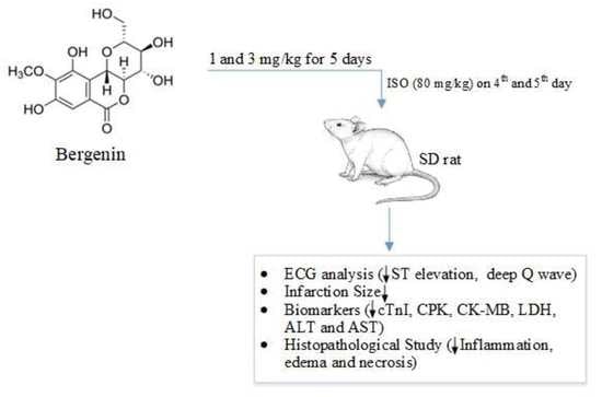
2.3.1. Response of Bergenin against the Isoproterenol-Induced MI in Rats

The myocardial infarction (MI) in rats was induced by administering isoproterenol (ISO) at the dose of 80 mg/kg (s.c.) twice at an interval of 24 h. At 1 and 3 mg/kg doses, bergenin was tested to determine its cardioprotective response against ISO-induced MI in SD rats. The experimental animals were randomly divided into five (5) groups consisting of six (6) rats (n = 6), as shown in Table 1. To control group (Group I), DMSO (1%) was administered (1 mL/kg, i.p.) for five days regularly and normal saline (1 mL/kg, s.c.) on the 4th and 5th days, with a gap of 24 h. Group II rats were given normal saline (1 mL/kg, i.p.) for five consecutive days and ISO (80 mg/kg, s.c.) on the 4th and 5th days, with a gap of 24 h. Group III rats received atenolol (1 mg kg−1) for 5 consecutive days and then ISO (80 mg/kg, s.c.) on the 4th and 5th days, with a gap of 24 h. Moreover, in Groups IV and V, (bergenin + ISO) bergenin in two different doses (1 and 3 mg/kg) were injected (i.p.) for 5 consecutive days and then ISO (80 mg kg−1, s.c.) on the 4th and 5th days, with a gap of 24 h. After 24 h from the second dose of ISO, rats were anesthetized with Pentothal (60 mg/kg). The body weights of all experimental rats were noted. After induction of anesthesia, the ECGs were recorded using a PowerLab supplemented with an Animal BioAmp, analyzed by the software LabChart 7 (AD Instruments, Australia). Through cardiac puncture, the blood samples were collected, and heart weights were measured and then preserved in 10% formalin solution for histopathology and infarct size assessment. The heart to body to weight ratio of all rats was calculated to observe the degree of pathological cardiac changes [18,19,20].

2.3.2. Analysis of Cardiac Biomarkers

During MI, the heart is under severe stress and becomes injured; in response, the cardiac biomarkers show up in the blood. The serums were separated from blood by centrifugation for cardiac biomarkers analysis. The cardiac marker enzymes, such as cardiac troponin I (cTnI), creatine phosphokinase (CPK), creatine kinase-MB (CK-MB), lactate dehydrogenase (LDH), aspartate transaminase (AST), and alanine transaminase (ALT), were assessed using commercially available standard assay kits (Standbio Laboratory, Boerne, TX, USA; JAJ International Inc., San Diego, CA, USA). The serum cardiac markers were evaluated according to procedure followed [21,22].

2.3.3. Histopathological Study

The SD rat heart tissues were sliced up, washed instantly with ice-cold normal saline, and fixed in buffered formalin (10%). Then, tissues were dehydrated in 80–100% alcohol, and xylene was used for cleaning. The fixed heart tissues were fixed in paraffin. Microtome was used to prepared 5 mm sections of tissues and then stained with H and E stains for light microscopy. Then, using a microscope, the stained tissues were studied for changes, such as variations in branches of cardiac muscles fibers, inflammation, edema, and necrosis. After that, advanced research microscopes were used to obtain images of the slides [23,24].

2.4. Statistical Analysis

The results were analyzed by applying one-way ANOVA followed by Tukey’s test through GraphPad Prism Software version 8. A p < 0.05 reflected statistical significance.

3. Results

3.1. Bergenin Response against Myocardial Injury Induced by Isoproterenol in Rats

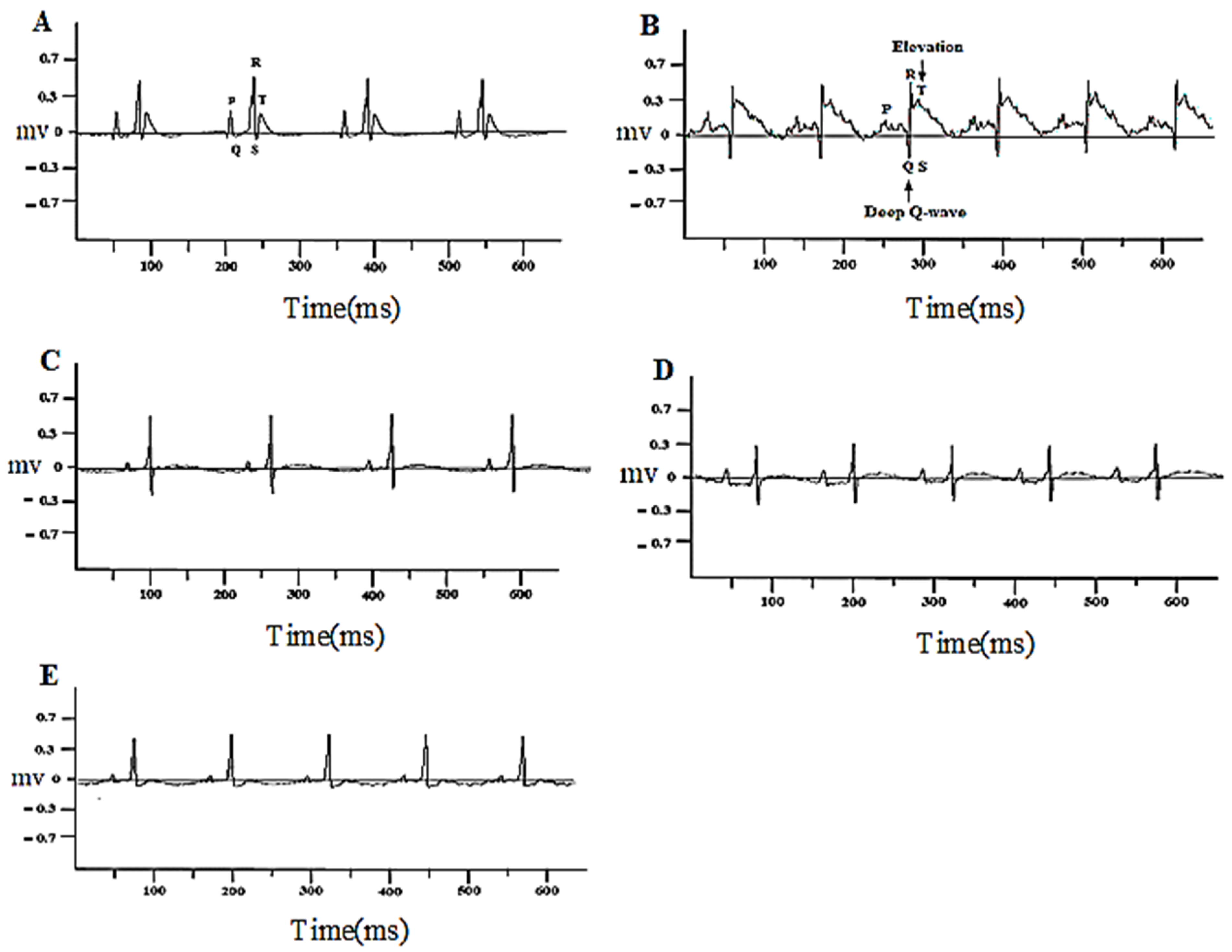
3.1.1. Bergenin Prevented Changes in the ECG Pattern

No significant changes in the ECG pattern were observed in the control group (1% DMSO) (Figure 2A). The isoproterenol-alone-treated rats exhibited marked ST-segment rise and deep Q-wave (Figure 2B). These isoproterenol-induced changes were restored to approximately normal in rats pretreated with atenolol (1 mg/kg) and bergenin (1 mg/kg), and a highly significant effect was observed in 3 mg/kg bergenin-treated rats when compared with the isoproterenol-alone-injected group (Figure 2A–E).

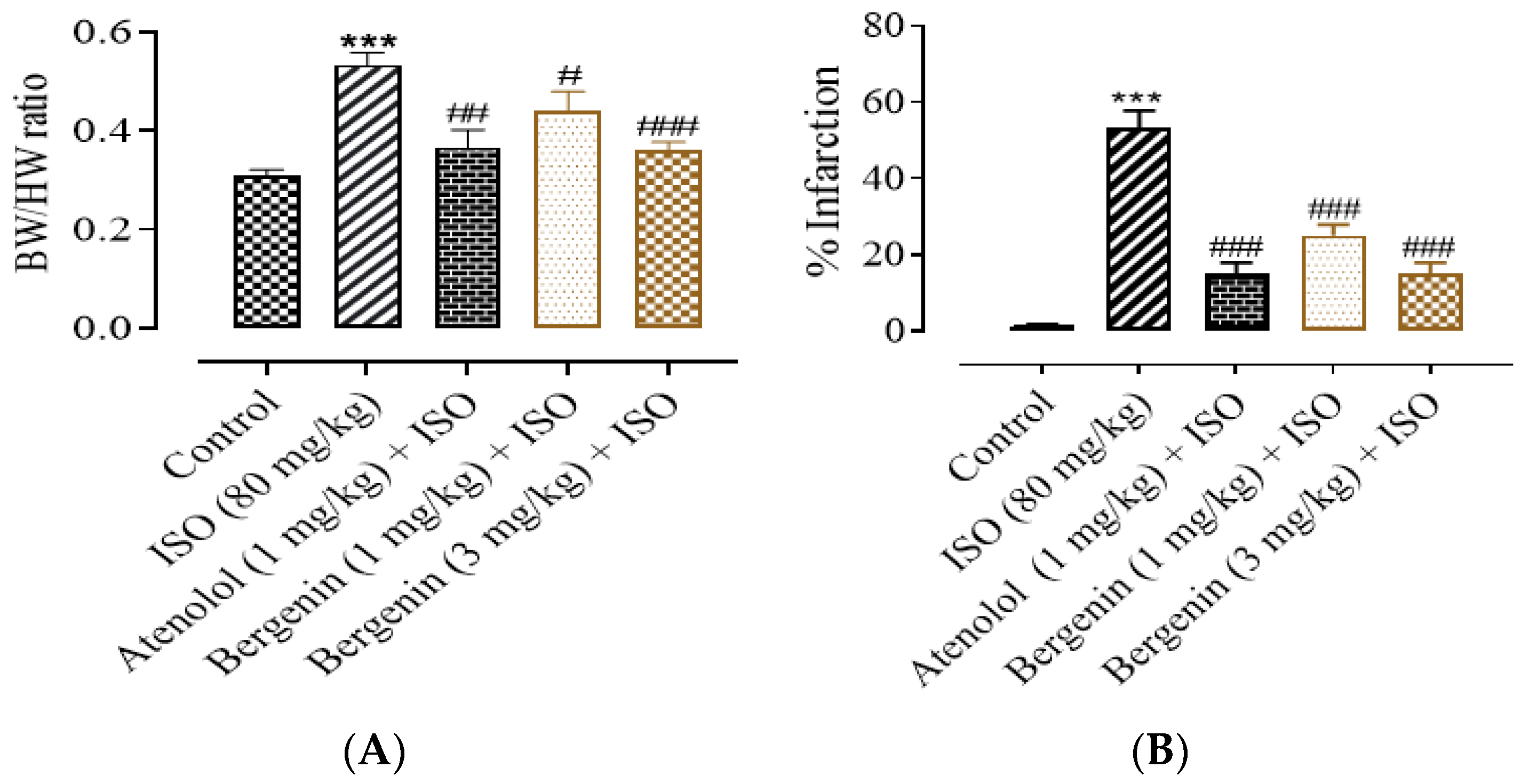
3.1.2. Effect of Bergenin on Heart-To-Body-Weight Ratio, Structural Anatomy, and Infarct Size

In all experimental groups nonsignificant changes were witnessed in the average body weight of rats at the end of experimental procedure (data not shown). In the ISO-alone-treated group, a significant (p < 0.001) increase in the ratio of heart to body weight was observed when compared with the normal control group. The rats which were pre- and co-treated with the atenolol and bergenin exhibited a significant decrease in heart-to-body-weight ratio in comparison to the ISO-alone-treated group (Figure 3A). In comparison to the control group, the heart shape of rats changed from elliptical to spherical when treated with ISO and the heart shape was significantly protected in the atenolol- and bergenin-treated groups. The infarcted area of the myocardium appeared white or pale grey, from which the percent infarct area was identified. The infarction was significantly prevented in the treated groups; atenolol (1 mg/kg) + ISO (15%), bergenin 1 + ISO (25%) mg/kg, and 3 mg/kg + ISO (15%), as compared to the ISO-alone-treated group (53%), as shown in Figure 3B.

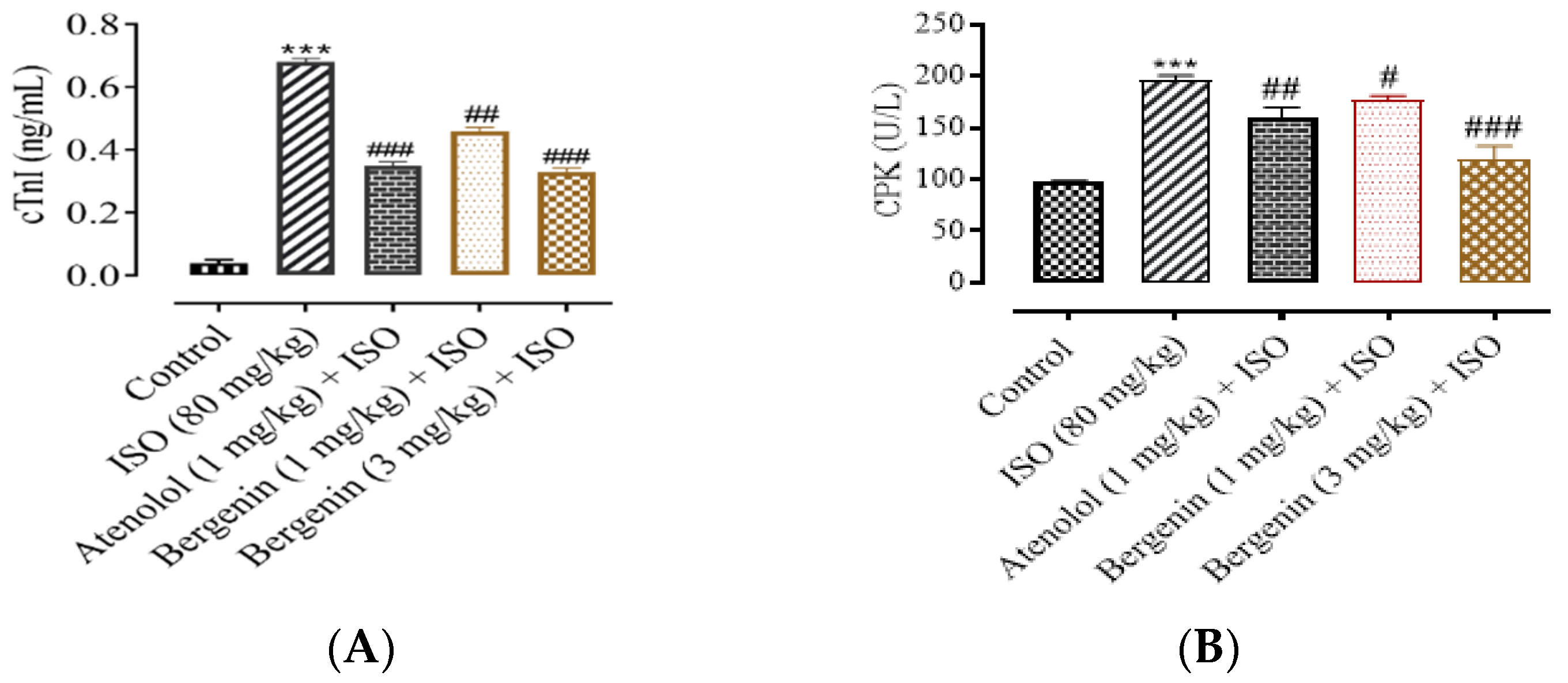
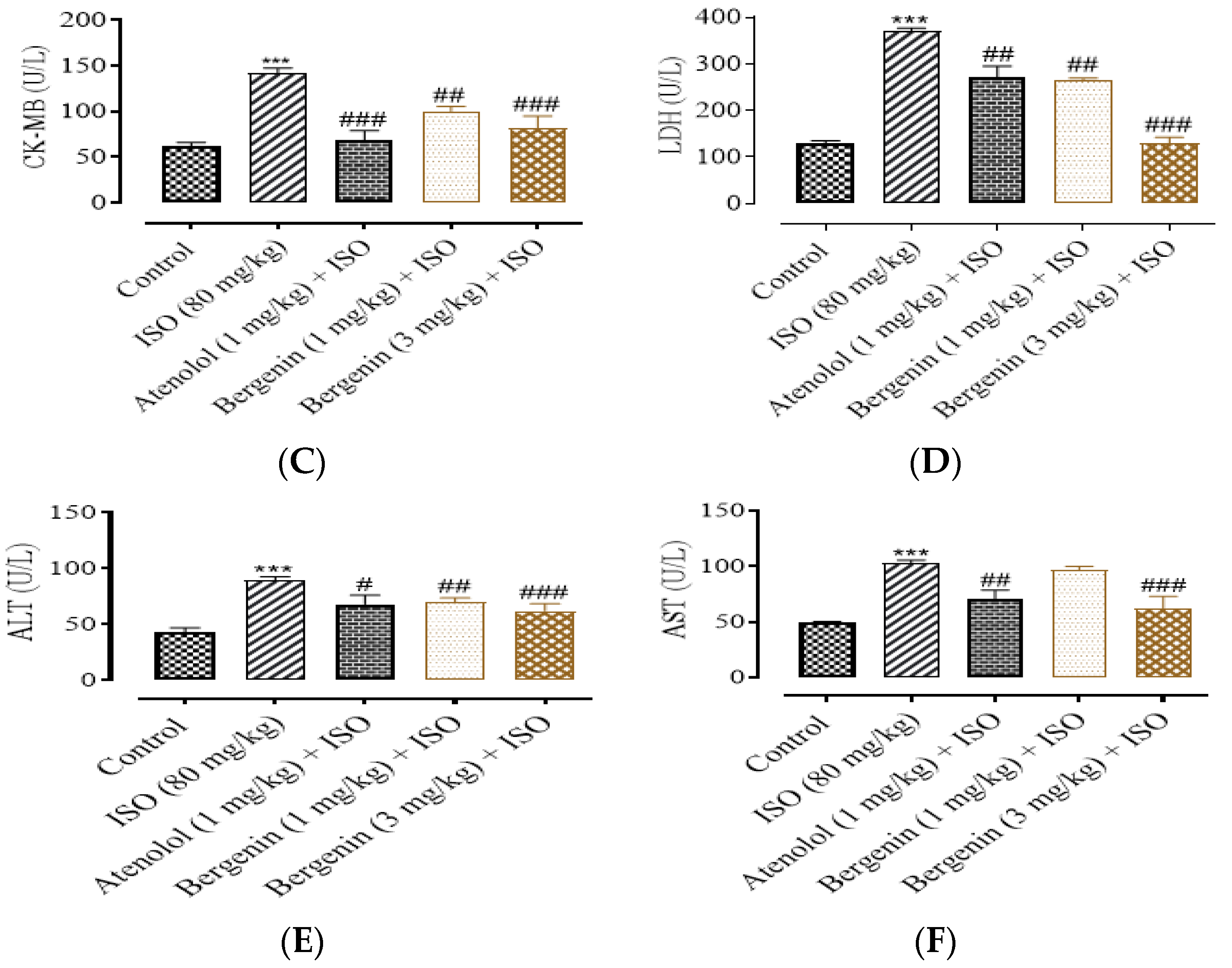
3.1.3. Effect of Bergenin on Cardiac Biomarkers

As shown in Figure 4A–F, a significant increase was observed in the serum levels of cardiac biomarkers (cTnI, CPK, CK-MB, LDH, AST, and ALT) of ISO-alone-treated rats. Compared with the control, ISO-alone administration produced a significant rise in the serum level of biomarkers cTnI (82.3 ± 3.05%), CPK (49.50 ± 4.01%), CK-MB (55.04 ± 3.03%), LDH (61.35% ± 5.01%), ALT (45.01 ± 3.02%), and AST (50.05 ± 1.08%) (Figure 4A–F). Pretreatment with atenolol (1 mg/kg) + ISO significantly restored most of the cardiac biomarkers alterations induced by ISO, such as cTnI (42.20 ± 2.02%), CPK (24.75 ± 1.24%), CK-MB (53.04 ± 3.01%), LDH (25.31 ± 3.02%), ALT (28.13.80 ± 2.02%), and AST (29.34 ± 2.51%). Moreover, bergenin (1 mg/kg) + ISO significantly decreased cTnI (32.35 ± 1.91%), CPK (14.12 ± 1.68%), CK-MB (30.10 ± 2.01%), LDH (24.01 ± 11%), ALT (24.05 ± 3.14%), and AST (9.0 ± 1.05%) serum levels. In addition, bergenin (3 mg/kg) + ISO also significantly restored the cardiac biomarker changes, including cTnI (51.52 ± 3.25%), CPK (36.22 ± 2.01%), CK-MB (43.35 ± 3.01%), LDH (37.03 ± 3.01%), ALT (36.01 ± 3.20%), and AST (36.06 ± 2.31%), as compared to the ISO-alone-injected group (Figure 4A–F).

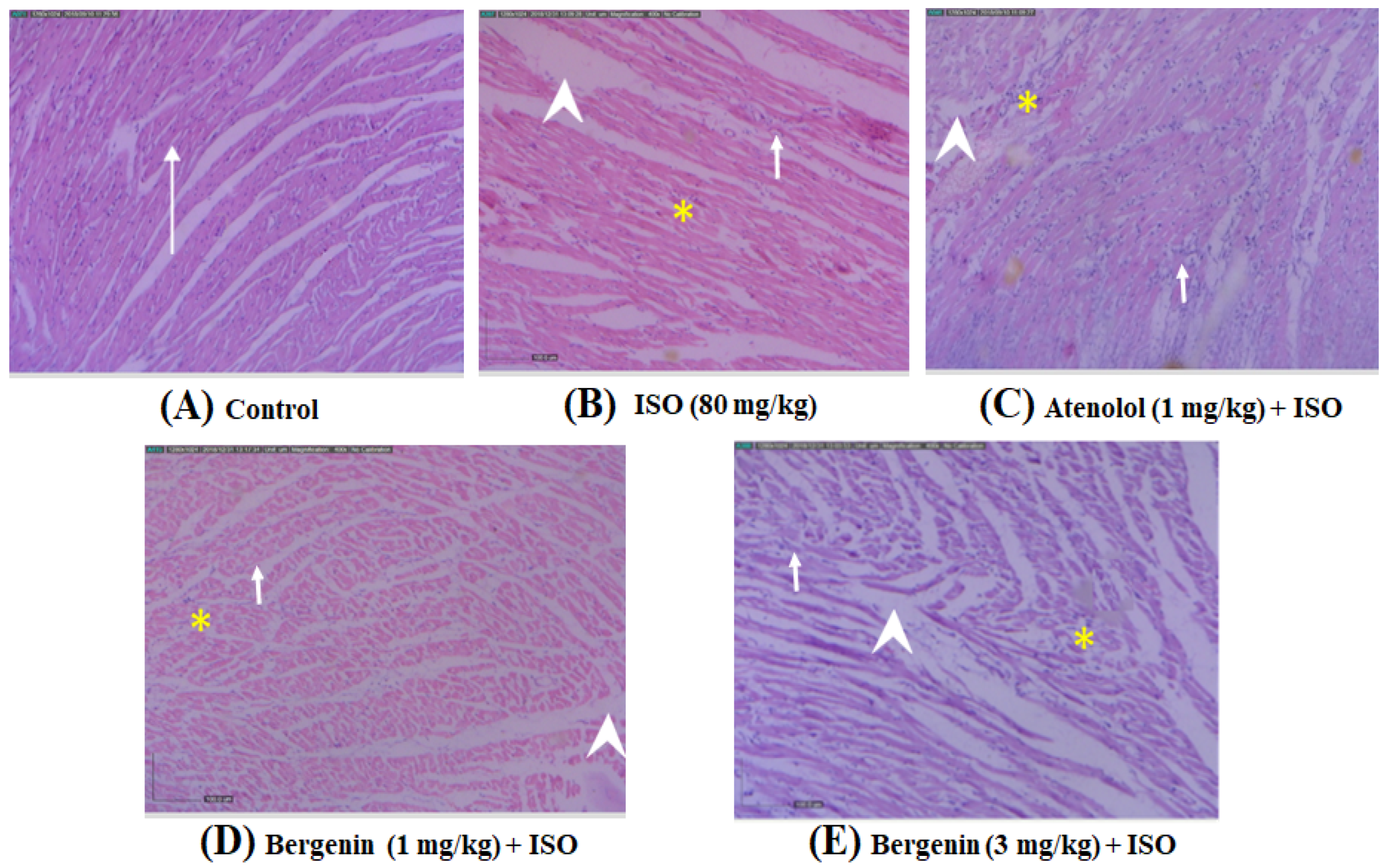
3.1.4. Effect of Bergenin on Histopathological Variations Induced by ISO

The histopathological investigation of the hearts of the control group revealed a normal shape of myocardium with branched look and striations. ISO-administered rats showed apparent inflammation, edema, and necrosis (Figure 3A). The tissue sections from all treated groups, atenolol 1 mg/kg (Figure 5C) and bergenin (1 and 3 mg/kg), showed mild to moderate inflammation, edema, and necrosis, and the shape of cardiac muscle fibers was comparatively well protected with minimum indication of necrosis in comparison to the ISO-alone-treated group (Figure 5A–E) (Table 1).

4. Discussion

In the past, some glycosides have been reported as a cardioprotective against ISO-induced myocardial ischemia in rats [25,26]. The ISO-induced myocardial ischemic rat model was used to study the effect bergenin as a cardioprotective. ISO is a nonselective β-adrenergic receptor agonist. Activation of β-adrenergic receptor (β-AR) is the primary mechanism to stimulate cardiac contractility. However, chronic β-AR stimulation (at sub-maximal doses of ISO) results in dysregulation of the β-adrenergic signaling pathway, leading to activation of protein kinase A (PKA)-dependent phosphorylation of L-type Ca2+ channels, which in response lead to increase of the intracellular Ca2+ level and oxidative stress, and ultimately lead to MI [27,28].
Administration of ISO at high doses leads to increase oxygen consumption in heart muscles and ultimately accelerates myocardium necrosis, elevated ST-segment in the ECG, and structural changes in the heart very similar to those that occur in patients with myocardial infarction [23,29]. Although some differences exist, essential resemblances between rat and human ECGs cannot be ignored [30].
The ECG correct interpretation is considered one of most important initial clinical markers for diagnosing myocardial ischemia and infarction. ECG abnormalities such as ST-segment elevation and deep Q-wave might be due to abnormal electrical signaling pathway and rupture of cell membrane in the injured myocardium [31]. In transmural myocardial ischemia, myocardial injury or necrosis occur; this event is called ST-elevation myocardial infarction (STEMI) [32].
In the present study, a significant ST-segment elevation in isoproterenol-treated rats was observed; however, pretreatment with bergenin and atenolol markedly restrained isoproterenol-induced ST-segment elevation, Q-wave, and ECG pattern, suggestive of its cell-membrane-protecting effects. The cardioprotective action of bergenin was further confirmed.
Injury in myocardium leads to release of an abundant amount of biomarker enzymes for MI in to the blood stream [33]. Hence, the serum levels of these marker enzymes reflect the alterations in membrane integrity and/or permeability [34].
The ISO administration to the rats also caused the leakage of cardiac marker enzymes, such as cTnI, CPK, CK-MB, LDH, ALT, and AST, of heart tissue to the circulation, rupture of cell membrane, hypoxia, and cardiac hypertrophy [35].
Cardiac troponin is considered a gold standard to confirm MI [36]. cTnI elevation in the blood is an important indicator of heart damage for detecting MI [37]. cTnI stays in the bloodstream for enough time after all other cardiac markers return to normal levels (Rajadurai and Prince 2007). The appearance of CPK in the blood has been generally considered to be an indirect marker of muscle damage. CPK acts as a catalyst to phosphorylate the creatine. Creatine facilitates recycling of adenosine triphosphate (ATP) [38]. CK-MB (subunit of CPK) is more sensitive for heart damage due to MI. However, troponin has largely replaced CK-MB [39]. The other cardiac marker recommended initially by WHO was LDH. LDH is an enzyme and is present in almost every major organ of the body [35]. The LDH turns sugar into energy [40]. The levels remain elevated for 8 to 14 days, making it a late marker for MI. LDH is a nonspecific marker for MI [41]. The cardiac cells have transaminases (AST and ALT) to synthesize and break down amino acids and to convert energy storage molecules. The concentrations of AST and ALT in the serum at normal physiological condition are normally low. However, if the myocardial muscles are damaged, the cardiac cell membrane becomes more permeable and AST and ALT leak out into the blood circulation. After myocardial infarction, AST and ALT are released into the circulation. Measurement of ALT and AST has been substituted by better biomarkers that are more specific for cardiac damage, such as CK-MB [42].
Pretreatment with bergenin (1 and 3 mg/kg, respectively) and atenolol (1 mg/kg) followed by ISO administration significantly decreased not only the serum levels of cTnI, CPK, CK-MB, LDH, AST, and ALT but also decreased the heart-to-body-weight ratio and infarct size when compared to ISO-only-treated rats. The bergenin showed highly significant results at the 3 mg/kg dose. It revealed that bergenin will possibly maintain cardiac membrane permeability, function, and structural integrity, thus restricting the leakage of these enzymes and cTnI from the myocardium. These effects are evident from the markedly blunted levels of these enzymes and cTnI in bergenin + ISO. Administration of high doses of isoproterenol leads to oxidative stress [43]. Oxidative stress is a reason for impairment of cellular functions in the cardiac cells of rats. However, some endogenous antioxidants, such as glutathione peroxidase (GPX), superoxide dismutase (SOD), and catalase (CAT), provide a first-line defense against the free radicals [44,45]. Previous studies reported that bergenin and the plant sources Bergenia ciliate, Bergenia ligulata Wall, and Ficus racemose L. significantly restore the SOD, GPX, and CAT enzymes’ activities [46,47,48]. It is also reported that bergenin exhibits a significant activity against free radicals [13]. The structure of bergenin (Figure 1) possesses OH groups in the benzene ring, which might be the reason for its free radicals’ scavenging ability [49].
To further confirm the response of bergenin, histopathological study was performed. The ISO-alone-injected group exhibited irregular cardiac muscle fibers and large infarcted zone with inflammation, edema, and necrosis. In SD rats, ISO is reported to increase oxidative stress, exhaust ATP levels, and cause Ca2+ overload [50]. However, bergenin (1 and 3 mg/kg) and atenolol (1 mg/kg) pretreatment followed by ISO significantly reduced the pathological changes such as inflammation, edema, and necrosis when compared to ISO-alone-injected SD rats. The most significant effects were observed at higher dose, 3 mg/kg, of bergenin. Bergenin is also reported to downregulate inflammatory factors such as TNF-α, IL-6, and IL-1β [51], which is again confirmed by a significant anti-inflammatory response. In addition, it was also previously reported that bergenin reduces the cell necrosis [51]. In summary, these findings of the present investigation may trigger a renewed interest in the usage of bergenin as a cardioprotective.

5. Conclusions

In conclusion, the results of our present study suggest that prior administration of bergenin from Bergenia species proved to be effective as a cardioprotective therapeutic agent in preventing the extent of myocardial damage (induced by ISO) by fortifying the myocardial cell membrane and heart tissue architecture, and also by normalizing cardiac marker enzymes. Overall, the results of this study offer scientific evidence of the importance of bergenin as a cardioprotection agent. Further studies, such as electrophysiology and its effect on protein expression of Caspase-3, etc., would provide more insight into the molecular aspects of these mechanisms.

Author Contributions

T.A. and A.J.S. designed the study; T.A. performed the experiment; A.J.S., I.U.H. and T.K. supervised the study; data were analyzed and reviewed by A.J.S. and T.A.; M.H.M. and M.Y.A. collected study data and performed statistical analysis and visualization; M.A.A., S.A.A. and H.A.S. contributed to the writing and proofreading of the manuscript. All authors have read and agreed to the published version of the manuscript.

Funding

This work is funded by Deanship of Scientific Research, Najran University, Kingdom of Saudi Arabia, under the research collaborations funding program grant code number NU/RC/MRC/11/2.

Institutional Review Board Statement

The experiments performed comply with the rulings of the Institute of Laboratory Animal Re-sources, Commission on Life Sciences, National Research Council (NRC, 1996) and were approved by the Ethical Committee of Department of Pharmacy, COMSATS University Islamabad, Abbottabad Campus, in its meeting held on 17-06-2013, video notification EC/PHM/07-2013/CIIT/ATD.

Informed Consent Statement

Not applicable.

Data Availability Statement

The datasets used and analyzed during the current study are available from the corresponding author upon reasonable request.

Acknowledgments

The authors acknowledge the support from the Deanship of Scientific Research, Najran University, Kingdom of Saudi Arabia, for funding this work under the research collaborations funding program grant code number NU/RC/MRC/11/2.

Conflicts of Interest

The authors declare no conflict of interest.

References

  1. Kubler, W.; Haass, M. Cardioprotection: Definition, classification, and fundamental principles. Heart 1996, 75, 330–333. [Google Scholar] [CrossRef] [Green Version]
  2. Shimokawa, H.; Yasuda, S. Myocardial ischemia: Current concepts and future perspectives. J. Cardiol. 2008, 52, 67–78. [Google Scholar] [CrossRef] [Green Version]
  3. Egred, M.; Shaw, S.; Mohammad, B.; Waitt, P.; Rodrigues, E. Under-use of beta-blockers in patients with ischaemic heart disease and concomitant chronic obstructive pulmonary disease. QJM Int. J. Med. 2005, 98, 493–497. [Google Scholar] [CrossRef] [PubMed] [Green Version]
  4. Khan, M.; Gilani, A.-H. Studies on Blood Pressure Lowering, Vasodilator and Cardiac Suppressant Activities of Vitex Negundo: Involvement of K+ Channel Activation and Ca++ Channel Blockade. Available online: https://scialert.net/abstract/?doi=ijp.2015.137.142 (accessed on 1 April 2021).
  5. Shah, S.M.A.; Akram, M.; Riaz, M.; Munir, N.; Rasool, G. Cardioprotective Potential of Plant-Derived Molecules: A Scientific and Medicinal Approach. Dose-Response 2019, 17, 1559325819852243. [Google Scholar] [CrossRef] [Green Version]
  6. Epure, A.; Pârvu, A.E.; Vlase, L.; Benedec, D.; Hanganu, D.; Gheldiu, A.-M.; Toma, V.A.; Oniga, I. Phytochemical Profile, Antioxidant, Cardioprotective and Nephroprotective Activity of Romanian Chicory Extract. Plants 2020, 10, 64. [Google Scholar] [CrossRef] [PubMed]
  7. Ahmed, F.; Siddesha, J.M.; Urooj, A.; Vishwanath, B.S. Radical scavenging and angiotensin converting enzyme inhibitory activities of standardized extracts of Ficus racemosa stem bark. Phytother. Res. 2010, 24, 1839–1843. [Google Scholar] [CrossRef]
  8. Rastogi, S.; Rawat, A.K.S. A Comprehensive Review on Bergenin, a Potential Hepatoprotective and Antioxidative Phytocon-stituent. Herba Pol. 2008, 54, 66–79. [Google Scholar]
  9. Shikov, A.N.; Pozharitskaya, O.N.; Makarova, M.N.; Makarov, V.G.; Wagner, H. Bergenia crassifolia (L.) Fritsch—Pharmacology and phytochemistry. Phytomedicine 2014, 21, 1534–1542. [Google Scholar] [CrossRef]
  10. Singh, M.; Pandey, N.; Agnihotri, V.; Singh, K.K.; Pandey, A. Antioxidant, antimicrobial activity and bioactive compounds of Bergenia ciliata Sternb.: A valuable medicinal herb of Sikkim Himalaya. J. Tradit. Complement. Med. 2017, 7, 152–157. [Google Scholar] [CrossRef] [Green Version]
  11. Abe, K.; Sakai, K.; Uchida, M. Effects of bergenin on experimental ulcers—Prevention of stress induced ulcers in rats. Gen. Pharmacol. Vasc. Syst. 1980, 11, 361–368. [Google Scholar] [CrossRef]
  12. Lee, Y.Y.; Jang, D.S.; Jin, J.L.; Yun-Choi, H.S. Anti-Platelet Aggregating and Anti-Oxidative Activities of 11-O-(4′-O-Methylgalloyl)-bergenin, a New Compound Isolated from Crassulacv. ‘Himaturi’. Planta Med. 2005, 71, 776–777. [Google Scholar] [CrossRef] [Green Version]
  13. Singh, U.; Barik, A.; Priyadarsini, K.I. Reactions of hydroxyl radical with bergenin, a natural poly phenol studied by pulse radiolysis. Bioorganic Med. Chem. 2009, 17, 6008–6014. [Google Scholar] [CrossRef]
  14. Da Silva, S.L.; De Oliveira, V.G.; Yano, T.; Nunomura, R.D.C.S. Antimicrobial activity of bergenin from Endopleura uchi (Huber) Cuatrec. Acta Amaz. 2009, 39, 187–191. [Google Scholar] [CrossRef] [Green Version]
  15. Kumar, R.; Patel, D.K.; Prasad, S.K.; Laloo, D.; Krishnamurthy, S.; Hemalatha, S. Type 2 antidiabetic activity of bergenin from the roots of Caesalpinia digyna Rottler. Fitoterapia 2012, 83, 395–401. [Google Scholar] [CrossRef] [PubMed]
  16. Gao, X.-J.; Guo, M.-Y.; Zhang, Z.-C.; Wang, T.-C.; Cao, Y.-G.; Zhang, N.-S. Bergenin Plays an Anti-Inflammatory Role via the Modulation of MAPK and NF-κB Signaling Pathways in a Mouse Model of LPS-Induced Mastitis. Inflammation 2014, 38, 1142–1150. [Google Scholar] [CrossRef] [PubMed]
  17. Oliveira, G.A.; Araujo, A.K.; Pacheco, G.; Oliveira, A.P.; Carvalho, J.L.; Chaves, L.S.; Sousa, G.C.; Lopes, A.L.; Silva, P.C.; Leódido, A.C.M. Anti-Inflammatory Properties of Bergenin in Mice. J. Appl. Pharm. Sci. 2019, 9, 069–077. [Google Scholar]
  18. Li, H.; Xie, Y.-H.; Yang, Q.; Wang, S.-W.; Zhang, B.-L.; Wang, J.-B.; Cao, W.; Bi, L.-L.; Sun, J.-Y.; Miao, S.; et al. Cardioprotective Effect of Paeonol and Danshensu Combination on Isoproterenol-Induced Myocardial Injury in Rats. PLoS ONE 2012, 7, e48872. [Google Scholar] [CrossRef] [Green Version]
  19. Sahu, B.D.; Anubolu, H.; Koneru, M.; Kumar, J.M.; Kuncha, M.; Rachamalla, S.S.; Sistla, R. Cardioprotective effect of embelin on isoproterenol-induced myocardial injury in rats: Possible involvement of mitochondrial dysfunction and apoptosis. Life Sci. 2014, 107, 59–67. [Google Scholar] [CrossRef]
  20. Kim, M.J.; Lim, J.E.; Oh, A.B. Validation of Non-invasive Method for Electrocardiogram Recording in Mouse using Lead II. Biomed. Sci. Lett. 2015, 21, 135–143. [Google Scholar] [CrossRef] [Green Version]
  21. Kim, K.; Chini, N.; Fairchild, D.G.; Engle, S.K.; Reagan, W.J.; Summers, S.D.; Mirsalis, J.C. Evaluation of Cardiac Toxicity Biomarkers in Rats from Different Laboratories. Toxicol. Pathol. 2016, 44, 1072–1083. [Google Scholar] [CrossRef] [Green Version]
  22. Ighodaro, O.M.; Akinloye, O.A. First line defence antioxidants-superoxide dismutase (SOD), catalase (CAT) and glutathione peroxidase (GPX): Their fundamental role in the entire antioxidant defence grid. Alex. J. Med. 2018, 54, 287–293. [Google Scholar] [CrossRef] [Green Version]
  23. Wang, S.-B.; Tian, S.; Yang, F.; Yang, H.-G.; Yang, X.-Y.; Du, G.-H. Cardioprotective effect of salvianolic acid A on isoproterenol-induced myocardial infarction in rats. Eur. J. Pharmacol. 2009, 615, 125–132. [Google Scholar] [CrossRef] [PubMed]
  24. Ahmed, S.; Gul, S.; Ze Jaafar, H.; Moga, M.; Zia-Ul-Haq, M.; Dima, L. Anti-Platelet Effects of Nimesulide in Isopro-terenol-Induced Myocardial Ischaemia and Infarction in Rabbits. Acta Cardiol. 2015, 70, 401–408. [Google Scholar] [CrossRef] [PubMed]
  25. Long, J.; Gao, M.; Kong, Y.; Shen, X.; Du, X.; Son, Y.-O.; Shi, X.; Liu, J.; Mo, X. Cardioprotective effect of total paeony glycosides against isoprenaline-induced myocardial ischemia in rats. Phytomedicine 2012, 19, 672–676. [Google Scholar] [CrossRef] [PubMed]
  26. Yu, Q.; Li, X.; Cao, X. Cardioprotective Effects of Phenylethanoid Glycoside-rich Extract from Cistanche deserticola in Ischemia-Reperfusion–Induced Myocardial Infarction in Rats. Ann. Vasc. Surg. 2016, 34, 234–242. [Google Scholar] [CrossRef]
  27. Ho, D.; Yan, L.; Iwatsubo, K.; Vatner, D.E.; Vatner, S.F. Modulation of β-adrenergic receptor signaling in heart failure and longevity: Targeting adenylyl cyclase type 5. Heart Fail. Rev. 2010, 15, 495–512. [Google Scholar] [CrossRef] [Green Version]
  28. De Lucia, C.; Eguchi, A.; Koch, W.J. New Insights in Cardiac β-Adrenergic Signaling During Heart Failure and Aging. Front. Pharmacol. 2018, 9, 904. [Google Scholar] [CrossRef] [Green Version]
  29. Wexler, B.C. Myocardial infarction in young vs old male rats: Pathophysiologic changes. Am. Heart J. 1978, 96, 70–80. [Google Scholar] [CrossRef]
  30. Sambhi, M.P.; White, F.N. The Electrocardiogram of the Normal and Hypertensive Rat. Circ. Res. 1960, 8, 129–134. [Google Scholar] [CrossRef] [Green Version]
  31. Rajadurai, M.; Prince, P.S.M. Preventive effect of naringin on cardiac mitochondrial enzymes during isoproterenol-induced myocardial infarction in rats: A transmission electron microscopic study. J. Biochem. Mol. Toxicol. 2007, 21, 354–361. [Google Scholar] [CrossRef]
  32. Antman, E.M.; Bassand, J.-P.; Klein, W.; Ohman, M.; Sendon, J.L.L.; Rydén, L.; Simoons, M.L.; Tendera, M. Myocardial infarction redefined—A consensus document of the joint european society of cardiology/american college of cardiology committee for the redefinition of myocardial infarction: The Joint European Society of Cardiology/ American College of Cardiology Committee. J. Am. Coll. Cardiol. 2000, 36, 959–969. [Google Scholar] [CrossRef] [Green Version]
  33. Suchalatha, S.; Devi, C.S.S. Protective effect of Terminalia chebula against experimental myocardial injury induced by isoproterenol. Indian J. Exp. Biol. 2004, 42, 174–178. [Google Scholar] [PubMed]
  34. Thygesen, K.; Alpert, J.S.; Jaffe, A.S.; Chaitman, B.R.; Bax, J.J.; Morrow, D.A.; White, H.D.; Executive Group on behalf of the Joint European Society of Cardiology (ESC)/American College of Cardiology (ACC)/American Heart Association (AHA)/World Heart Federation (WHF) Task Force for the Universal Definition of Myocardial Infarction. Fourth Universal Definition of Myocardial Infarction (2018). Circulation 2018, 138, e618–e651. [Google Scholar] [CrossRef] [PubMed]
  35. Roy, S.J.; Prince, P.S.M. Protective effects of sinapic acid on cardiac hypertrophy, dyslipidaemia and altered electrocardiogram in isoproterenol-induced myocardial infarcted rats. Eur. J. Pharmacol. 2013, 699, 213–218. [Google Scholar] [CrossRef] [PubMed]
  36. Body, R.; McDowell, G.; Carley, S.; Wibberley, C.; Ferguson, J.; Mackway-Jones, K. A FABP-ulous ‘rule out’ strategy? Heart fatty acid binding protein and troponin for rapid exclusion of acute myocardial infarction. Resuscitation 2011, 82, 1041–1046. [Google Scholar] [CrossRef]
  37. Roos, A.; Hellgren, A.; Rafatnia, F.; Hammarsten, O.; Ljung, R.; Carlsson, A.C.; Holzmann, M.J. Investigations, findings, and follow-up in patients with chest pain and elevated high-sensitivity cardiac troponin T levels but no myocardial infarction. Int. J. Cardiol. 2017, 232, 111–116. [Google Scholar] [CrossRef]
  38. Barcelos, R.; Stefanello, S.; Mauriz, J.; Gonzalez-Gallego, J.; Soares, F. Creatine and the Liver: Metabolism and Possible Interactions. Mini. Rev. Med. Chem. 2016, 16, 12–18. [Google Scholar] [CrossRef]
  39. Baird, M.F.; Graham, S.M.; Baker, J.S.; Bickerstaff, G.F. Creatine-Kinase- and Exercise-Related Muscle Damage Implications for Muscle Performance and Recovery. Available online: https://www.hindawi.com/journals/jnme/2012/960363/ (accessed on 5 May 2020).
  40. Akila, P.; Vennila, L. Chlorogenic acid a dietary polyphenol attenuates isoproterenol induced myocardial oxidative stress in rat myocardium: An in vivo study. Biomed. Pharmacother. 2016, 84, 208–214. [Google Scholar] [CrossRef]
  41. Mythili, S.; Malathi, N. Diagnostic markers of acute myocardial infarction. Biomed. Rep. 2015, 3, 743–748. [Google Scholar] [CrossRef] [Green Version]
  42. Shen, J.; Zhang, J.; Wen, J.; Ming, Q.; Zhang, J.; Xu, Y. Correlation of Serum Alanine Aminotransferase and Aspartate Ami-notransferase with Coronary Heart Disease. Int J. Clin. Exp. Med. 2015, 8, 4399–4404. [Google Scholar]
  43. Rathore, N.; John, S.; Kale, M.; Bhatnagar, D. Lipid Peroxidation and Antioxidant Enzymes in Isoproterenol In-duced Oxidative Stress in Rat Tissues. Pharmacol. Res. 1998, 38, 297–303. [Google Scholar] [CrossRef] [PubMed]
  44. Ji, L.L.; Stratman, F.W.; Lardy, H.A. Antioxidant enzyme systems in rat liver and skeletal muscle: Influences of Selenium Deficiency, Chronic Training, and Acute Exercise. Arch. Biochem. Biophys. 1988, 263, 150–160. [Google Scholar] [CrossRef]
  45. Wu, J.; Hecker, J.G.; Chiamvimonvat, N. Antioxidant enzyme gene transfer for ischemic diseases. Adv. Drug Deliv. Rev. 2009, 61, 351–363. [Google Scholar] [CrossRef] [PubMed] [Green Version]
  46. Jani, S.; Shukla, V.J.; Harisha, C.R. Comparative pharmacognostical and phytochemical study on Bergenia ligulata Wall. and Ammania buccifera Linn. Ayu 2013, 34, 406–410. [Google Scholar] [CrossRef] [Green Version]
  47. Saha, S.; Shrivastav, P.S.; Verma, R.J. Antioxidative mechanism involved in the preventive efficacy of Bergenia ciliatarhizomes against experimental nephrolithiasis in rats. Pharm. Biol. 2014, 52, 712–722. [Google Scholar] [CrossRef] [PubMed]
  48. Aggarwal, D.; Kaushal, R.; Kaur, T.; Bijarnia, R.K.; Puri, S.; Singla, S.K. The most potent antilithiatic agent ameliorating renal dysfunction and oxidative stress from Bergenia ligulata rhizome. J. Ethnopharmacol. 2014, 158, 85–93. [Google Scholar] [CrossRef]
  49. Bendary, E.; Francis, R.R.; Ali, H.M.G.; Sarwat, M.I.; El Hady, S. Antioxidant and structure–activity relationships (SARs) of some phenolic and anilines compounds. Ann. Agric. Sci. 2013, 58, 173–181. [Google Scholar] [CrossRef] [Green Version]
  50. Padmanabhan, M.; Prince, P.S.M. Preventive effect of S-allylcysteine on lipid peroxides and antioxidants in normal and isoproterenol-induced cardiotoxicity in rats: A histopathological study. Toxicology 2006, 224, 128–137. [Google Scholar] [CrossRef]
  51. Xiang, S.; Chen, K.; Xu, L.; Wang, T.; Guo, C. Bergenin Exerts Hepatoprotective Effects by Inhibiting the Release of Inflammatory Factors, Apoptosis and Autophagy via the PPAR-γ Pathway. Drug Des. Dev. Ther. 2020, 14, 129–143. [Google Scholar] [CrossRef] [Green Version]
Figure 1. Structure of bergenin.
Processes 10 01403 g001
Figure 2. Representative lead II electrocardiogram (ECG) tracings of (A) control, (B) ISO-alone, (C) atenolol (1 mg/kg) + ISO, (D) bergenin, 1 mg + ISO, and (E) 3 mg/kg + ISO treated rats, respectively (recording speed 50 ms/div). Millisecond (ms), division (div), millivolt (mv).
Processes 10 01403 g002
Figure 3. (A) Shows the effect of bergenin + ISO, atenolol + ISO and ISO on body weight/heart weight (BW/HW) ratio in comparison to control. The graph (B) shows the percent myocardial infarct size in the control and treated groups. All values represent the mean ± SEM of six determinations, where #, ##, ### indicates < 0.01 and 0.001 p values vs. ISO, respectively, and *** describes p value < 0.001 vs. Control.
Processes 10 01403 g003
Figure 4. Bar graph (A) shows the response of bergenin and atenolol on serum level of cTnI (cardiac troponin I), (B) CPK (creatine phosphokinase), (C) CK-MB (creatine kinase-MB), (D) LDH (lactate dehydrogenase), (E) ALT (alanine transaminase), and (F) AST (aspartate transaminase) in isoproterenol-induced infarcted rats. Data are presented as mean ± SEM (n = 6), where #, ##, ### indicates < 0.01 and 0.001 p values vs. ISO, respectively, and *** describes p value < 0.001 vs. Control.
Processes 10 01403 g004aProcesses 10 01403 g004b
Figure 5. H and E (200×) staining of cardiac heart tissues of (A) control group, demonstrating normal cardiomyocytes (arrow). (B) Isoproterenol (ISO)-alone-administered indicates marked inflammation (arrow), edema (arrow head), and confluent necrosis (*, esteric). (C) Pretreatment with 1 mg/kg atenolol + ISO produced significant decrease in inflammation (arrow), edema (arrow head), and necrosis (*, esteric). (D) 1 mg/kg bergenin + ISO produced minimal decrease in the edema (head), inflammation (arrow), and necrosis (*, esteric); (E) 3 mg/kg bergenin + ISO produced maximal decrease in the inflammation (arrow), edema (arrow head), and necrosis (*, esteric), respectively.
Processes 10 01403 g005
Table 1. This table shows the whole scheme of isoproterenol-induced myocardial ischemia.
S.NO.GroupsExperiment Procedure
1Group I (Control group)
  • 1% DMSO (1 mL/kg, i.p.) for 5 consecutive days.
  • Normal saline (1 mL/kg, s.c.) on 4th and 5th day.
2Group II (ISO group)
  • 1% DMSO (1 mL/kg, i.p.) for 5 consecutive days.
  • ISO (80 mg/kg, s.c.) on 4th and 5th day, at an interval of 24 h.
3Group III
(Atenolol + ISO)
  • Group III received atenolol (1 mg/kg i.p.) for 5 consecutive days.
  • ISO (80 mg/kg, s.c.) on 4th and 5th day, at an interval of 24 h.
4Group IV, V
(Bergenin + ISO)
  • Bergenin in two different doses (1 and 3 mg/kg i.p.) for 5 consecutive days.
  • ISO (80 mg/kg, s.c.) on 4th and 5th day, at an interval of 24 h.
Publisher’s Note: MDPI stays neutral with regard to jurisdictional claims in published maps and institutional affiliations.

