Abstract
Freeze-drying, also known as lyophilization, significantly improves the storage, stability, shelf life, and clinical translation of biopharmaceuticals. On the downside, this process faces complex challenges, i.e., the presence of freezing and drying stresses for the active compounds, the uniformity and consistency of the final products, and the efficiency and safety of the reconstituted lyophilized formulations. All these requirements can be addressed by adding specific excipients that can protect and stabilize the active ingredient during lyophilization, assisting in the formation of solid structures without interfering with the biological and/or pharmaceutical action of the reconstituted products. However, these excipients, generally considered safe and inert, could play an active role in the formulation interacting with the biological cellular machinery and promoting toxicity. Any side effects should be carefully identified and characterized to better tune any treatments in terms of concentrations and administration times. In this work, various concentrations in the range of 1 to 100 mg/mL of cellobiose, lactose, sucrose, trehalose, isoleucine, glycine, methionine, dextran, mannitol, and (2-hydroxypropyl)-β-cyclodextrin were evaluated in terms of their ability to create uniform and solid lyophilized structures. The freeze-dried products were then reconstituted in the appropriate cell culture media to assess their in vitro cytotoxicity on both a healthy cell line (B-lymphocytes) and their tumoral lymphoid counterpart (Daudi). Results showed that at 10 mg/mL, all the excipients demonstrated suitable lyophilized solid structures and high tolerability by both cell lines, while dextran was the only excipient well-tolerated also up to 100 mg/mL. An interesting result was shown for methionine, which even at 10 mg/mL, selectively affected the viability of the cancerous cell line only, opening future perspectives for antitumoral applications.
1. Introduction
Since the 1980s, biopharmaceuticals, produced through biotechnological processes using molecular biology methods, have revolutionized the treatment of a broad range of diseases [1]. They include enzymes, vaccines, monoclonal antibodies, cytokines, hormones, recombinant blood products, hematopoietic growth factors, and nucleic acid- gene- or cell-based therapeutics presenting higher specificity and targeting ability with respect to conventional small molecule drugs [2]. However, biopharmaceuticals have high molecular weight and complex structures, which confer certain physical and chemical instability, negatively affecting their therapeutic action and patients’ health [3]. Furthermore, exposure to organic solvents and environmental stresses, such as pH and temperature variations, light, agitation, and shear stress, can be detrimental to maintaining the biopharmaceuticals’ properties [4]. For these reasons, pharmaceuticals are commonly produced as solid dosage forms using freeze-drying to enhance their pharmaceutical stability, increase their shelf life and preserve their in vivo efficacy [5].
Lyophilization or freeze-drying is a three-step preservation method based on water freezing, followed by its removal, initially by sublimation (primary drying) and then by desorption (secondary drying). It is the most suitable method to preserve thermo-liable materials such as vaccines, viruses, proteins, peptides, and colloidal carriers. Freeze-dried materials can often be stored at room temperature (RT) and quickly reconstituted in water or physiological solutions [6,7]. However, stresses may arise during this process, impair product stability and lead to loss of biological activity. For instance, damage or denaturation of biomolecules can be promoted by low temperatures, cryoconcentration, interaction with the ice–water interface, and phase separation [8,9]. In addition, freeze-drying is a highly time-consuming and energy-intensive process, and the pharmaceutical industry increasingly requires optimizing lyophilization cycles by shortening the drying steps, adjusting the process parameters, and customizing the formulations [3].
The addition of specific excipients contributes to the stability of biopharmaceuticals and indirectly determines the most suitable parameters for developing a cost-effective lyophilization process to obtain freeze-dried products with desirable appearance and properties. According to the International Pharmaceutical Excipient Council (IPEC), excipients are defined as “any substance other than active drug that is included in the manufacturing process or is contained in finished pharmaceutical dosage form”, and their dosage is significantly higher than the active pharmaceutical ingredient (API) [10].
In biopharmaceutical formulations, different excipients are usually added to the preparation before the freeze-drying process with the aims of stabilizing the biological product, preventing aggregation and surface adsorption, conferring the physiological osmolality and tonicity, pH control, minimizing adverse reactions upon injection, favoring the targeted drug delivery, and acting as antimicrobial agent. Furthermore, excipients for sterile formulation must withstand the final sterilization and not be impaired by the aseptic process [10,11]. The ideal characteristics of an excipient are its chemical stability and low sensitivity to the process. Furthermore, it must be inert and non-toxic to the human body, efficient for the intended use, with accepted organoleptic properties, and possibly, inexpensive [12]. Despite their widespread use, relatively little is known about the biological effects of excipients because the importance of investigating the possible adverse effects has been underestimated, and their safety is often taken for granted [13]. Like most chemicals, excipients are potentially toxic, able to cause renal failure, osmotic diarrhea, hypersensitivity and allergic reactions, cardiotoxicity, and even death if not strictly regulated [14]. For these reasons, different studies investigated the safety of various excipients, increasing the awareness of systematic testing of possible adverse side effects. For instance, Kiss et al. proved the toxicity of Cremophor EL and RH40 on endothelial and epithelial cells, even at clinical doses [15], while Dai et al. associated with Cremophor EL the metabolic dysregulation and unfolded protein response caused by Taxol [16]. Bajaj et al. studied the P-glycoprotein inhibition mediated by different excipients since its modulation played a critical role in oral drug bioavailability and safety [17]. Many researchers focused on the effects of excipients on fragile people, such as pediatric patients [18,19,20,21]. Some studies investigated the in vitro cytotoxicity of different excipients against different biological targets [22,23,24], while others used machine learning algorithms to estimate the toxicity risk of small molecular entities [25,26].
In this view, this study aims to combine thermal characterizations as differential scanning calorimetry (DSC) and freeze-drying microscopy (FDM) with an in vitro cytotoxicity test to evaluate the suitability of using a wide range of sugars, polyols, and amino acids for freeze-drying, rehydration, and cell culture applications. In detail, sugars (i.e., cellobiose, lactose, sucrose, and trehalose), (2-hydroxypropyl)-β-cyclodextrin (hp-β-cyclodextrin), bulking agents such as mannitol, and amino acids (i.e., isoleucine, glycine, and methionine), and a collapse temperature modifier (dextran) were investigated [3].
Stabilizers, such as sucrose, trehalose, polyethylene glycol, and cyclodextrins, are the most widely used and studied excipients since they protect the API against chemical and physical instability during both the freezing and drying steps. Their stabilizing activity is mainly driven by different physicochemical phenomena such as preferential exclusion, water replacement and entrapment, and vitrification [27]. Excipients stabilize API by establishing repulsive forces between the proteins and the carbohydrates molecules or acting as a water substitute [28]. In other cases, it has been shown that excipients form a cage around API, entrapping and slowing down water molecules, or otherwise, the API can be immobilized inside the stabilizer’s glassy matrix, which prevents translational molecular movements and thus its degradation [29,30,31,32].
Bulking agents, such as mannitol, glycine and amino acids, provide a proper porous structure to the freeze-dried product [3,33], while adding a collapse temperature modifier, such as dextran, can affect the lyophilization process by increasing the maximum allowable product temperature, significantly reducing the primary drying time [34].
Most of the excipients considered in this study are widely used for the formulation of intravenous and intramuscular injectable, oral, topical administrable or inhalable drugs [35]. Excipients in pharmaceutical formulations are used at a broad range of concentrations, from 0.001 to up to 20% w/v. According to the most recurring concentrations of the inactive ingredients in biologics, we investigated the 0.1, 1, and 10% w/v concentrations in our study [36,37]. After 24 and 48 h of treatments, we evaluated their effects on both a healthy cell line (B-lymphocytes) and on their tumoral lymphoid counterpart (Daudi).
2. Materials and Methods
2.1. Preparation of Formulations to Be Lyophilized
The effect of the freeze-drying process on solutions containing different excipients at three different concentrations, i.e., a very high one, 100 mg/mL corresponding to 10% w/v, a medium one, 10 mg/mL as 1% w/v and 1 mg/mL as 0.1% w/v. For this purpose, different freeze-drying excipients were used to evaluate their impact on cell viability after thermal characterizations: four disaccharides, i.e., cellobiose (Sigma, Darmstadt, Germany), lactose (Fluka, Charlotte, NC, USA), sucrose (Fisher Chemical, Waltham, MA, USA), and trehalose (Sigma), three amino acids, i.e., isoleucine (Sigma), glycine (Sigma), methionine (Sigma), a polymer, i.e., dextran (40 kDa, PanReac AppliChem, Darmstadt, Germany), a polyol, i.e., mannitol (Chem-Lab, Zedelgem, Belgium), and (2-hydroxypropyl)-β-cyclodextrin (Sigma).
For each excipient, except for isoleucine and methionine, three solutions were prepared in double-distilled (DD) water. The more concentrated one was prepared by dissolving the excipient in DD water until the concentration of 100 mg/mL; the other two solutions at 10 and 1 mg/mL were obtained by subsequent dilutions of the more concentrated one. Isoleucine and methionine had a lower solubility than the other excipients (34 and 57 mg/mL, respectively); thus, only the solutions at 10 and 1 mg/mL were prepared following the previously described protocol. All the tested solutions are summarized in Table 1.

Table 1.
List of the tested solutions.
2.2. Thermal Characterization
The thermal characterization of the solutions was carried out with two complementary techniques: differential scanning calorimetry (DSC) and freeze-drying microscopy (FDM). The DSC is a consolidated technique for thermal analysis that detects the variations of the specific heat capacity (Cp), measured as changes in the heat flow, comparing the sample with a reference. It measures the temperature at which thermal events such as the glass transition (Tg′) for amorphous materials and the eutectic point (Teu) for crystalline ones occur [38]. The FDM mimics the different steps of the freeze-drying process in miniature by freezing and drying small volumes of formulation under the microscope. It allows for the measurement of the collapse (Tc), for amorphous excipients, and melting (Tm), for crystalline ones, temperatures identified as the onset of visible collapse [39].
Differential scanning calorimetry (DSC Q200, TA Instruments, New Castle, DE, USA) was used to evaluate the thermal behavior of samples constituted by DD water and different concentrations of the excipients listed above. An aluminum pan was filled with ~20 μL of the solution and sealed; the reference was an identical empty pan. The dynamic temperature program included a freezing step until −60 °C at 1 °C/min and a heating one up to 25 °C at 5 °C/min. Results were then evaluated using Universal Analysis software (TA Instruments).
Freeze-drying microscopy (BX51, Olympus Europa, Hamburg, Germany; temperature controller: PE95-T95, Linkam, Scientific Instruments, Tadworth, Surrey, UK) was employed to verify the freeze-drying behavior of the excipient solutions and to identify the critical temperatures that lead to collapse events. First, the freezing ramp was set at 5 °C/min to −60 °C; then, lyophilization was performed at 10 Pascal, progressively increasing the product temperature. At this point, the sublimation front was accurately tracked. The temperature at which the sublimation of ice into water vapor from the sample was observed, resulting in the movement of the sublimation front, was recorded as Tc [40].
2.3. Freeze-Drying Process
Samples were freeze-dried using a laboratory-scale freeze-dryer (Revo™ series, Millrock Technology, Kingston, NY, USA). The chamber pressure was monitored through a thermal conductive gauge (Pirani, PSG-101-S model, Inficon, Bad Ragaz, Svizzera) and a capacitive sensor (Baratron, 626A model, MKS Instruments, Andover, MA, USA). The primary drying endpoint was determined based on the Pirani-to-Baratron pressure ratio signal [41].
Then, 300 μL of each excipient solution, at each concentration, was filled into 2R vials (Nuova Ompi glass division, Stevanato Group, Piombino Dese, Italy), partially sealed with silicon igloo stoppers (West Pharmaceutical Services, Milan, Italy), and loaded into the freeze-dryer in direct contact with the temperature-controlled shelf.
During freezing, shelf temperature was decreased down to −45 °C at 0.5 °C/min and held for 1 h; after that, primary drying was carried out at 100 μBar and −25 °C. When all ice sublimed, secondary drying was performed at the same pressure, increasing shelf temperature until 20 °C in 5 h, and held at those conditions for 12 h. At the end of the cycle, the pressure was released to the atmospheric value by bleeding nitrogen into the drying chamber. The lyophilization cycle is represented in Figure 1 in terms of temperature and pressure evolution.
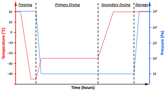
Figure 1.
The graph represents the freeze-drying cycle. The red line represents the temperature variations and the blue one the pressure changes.
2.4. Freeze-Dried Samples Evaluation
The freeze-dried products were characterized in terms of cake appearance and residual moisture. Ideally, the cake should preserve the same size and shape as the frozen product, with uniform color and texture [42]. The residual moisture of the lyophilized samples was investigated by the Karl-Fischer titration (Karl Fischer Moisture Meter CA-31, Mitsubishi, Japan). The Hydranal titration solvent (Sigma-Aldrich, Milano, Italy) reconstituted most of the formulations to be analyzed, whereas formamide (Honeywell, Fluka) was added to those samples containing dextran, glycine, and mannitol for their low solubility in methanol.
2.5. Cell Cultures
The cell lines used were maintained according to standard mammalian cell culture protocols and sterile technique at 37 °C under 5% CO2 atmosphere in 75 cm2 not treated cell culture flasks (Corning, Glendale, AZ, USA).
The cell culture medium of each cell line was supplemented with heat-inactivated fetal bovine serum (FBS) obtained by heating at 56 °C for 30 min to destroy the action of serum complement, without affecting the growth properties of FBS, and 1% of penicillin/streptomycin (P/S, 10,000 units penicillin and 10 mg streptomycin/mL, Sigma).
Lymphocytes (IST-EBV-TW6B) were purchased from the cell bank IRCCS AOU San Martino IST (Genova, Italy) and maintained in advanced RPMI 1640 cell culture medium (Gibco) supplemented with 20% of FBS (Gibco, Thermo Fisher Scientific, Waltham, MA, USA) and 1% L-glutamine 200 mM (Q, Lonza), with a cell density between 9 × 10⁴ and 9 × 10⁵ cells/mL. After 20 days of use, the cell culture medium was supplemented with 1% Q and 1% of non-essential amino acid solution (Sigma).
Daudi cell line (ATCC® CCL-213TM), derived from the peripheral blood of a Burkitt’s lymphoma patient, was cultured in RPMI 1640 culture medium (ATCC), with a cell density between 3 × 10⁵ and 3 × 10⁶ cells/mL.
2.6. Cytotoxicity Assay
The cytotoxicity of the different excipients was tested at different concentrations, i.e., 100, 10, and 1 mg/mL, and treatment times, i.e., 24 and 48 h, through the WST-1 cell proliferation assay. This technique was based on the conversion operated by cellular mitochondrial dehydrogenases of the tetrazolium salt WST-1 in formazan. The quantity of formazan dye converted depended on the number of viable cells.
The freeze-dried excipient was reconstituted with lymphocytes or Daudi’s culture media to obtain a final concentration of 100 mg/mL. For optimal reconstitution, after the addition of the media, samples were left for 30 min at 4 °C and then further 30 min at 37 °C with 180 rpm shaking. After the reconstitution, the treatment solutions at 10 and 1 mg/mL concentrations were prepared by diluting the 100 mg/mL solution.
Then, 2 × 10⁵ cells for each mL of treatment were centrifuged, resuspended in the treatment solution and 100 μL plated in each well of a 96-well flat-bottom plastic culture plate (Greiner Bio-one, Kremsmünster, AT, Austria). After 20 and 44 h of incubation at 37 °C and 5% CO2, 10 μL of WST-1 reagent (CELLPRO-RO, Roche, Basel, CH) was added to each well, and after further 4 h of incubation, in the same conditions, the formazan absorbance was detected at 450 nm through a microplate spectrophotometer (Multiskan Go microplate spectrophotometer, Thermo Fisher Scientific) using a 620 nm reference. Independent experiments were carried out in triplicate three times for each cell line, time, concentration, and excipient, and results were normalized to the untreated sample.
2.7. Statistical Analysis
Data were plotted as mean ± standard error (SE). For cytotoxicity results, the three-way analysis of variance (ANOVA) tools of the SIGMA Plot software’s data analysis package was used to compare the two cell lines and the two treatment times. Regarding the different excipients and concentrations, a different approach was used and evaluated using the two-way ANOVA.
Since isoleucine and methionine are significantly less water-soluble than all the other selected excipients, only the concentrations of 10 and 1 mg/mL were tested. Thus, two data pools were evaluated separately to refine the statistical analysis. The former, represented with black asterisks on the graphs, excluded isoleucine and methionine from the comparison made between all the other excipients at the three concentrations. The latter, represented with red asterisks, compared all the excipients only at the concentrations of 10 and 1 mg/mL.
* p ≤ 0.05 and ** p ≤ 0.001 were considered significant.
3. Results
3.1. Thermal Characterization
Understanding the thermal behavior of lyophilized formulations is critical for developing an efficient freeze-drying cycle. Thus, DSC and FDM analyses were performed to evaluate the maximum allowable temperature of the formulation, i.e., the glass transition temperature or the eutectic melting temperature, and to estimate the collapse or melting temperatures, respectively [43]. The Tm of the FDM experiments corresponded to a visible loss of structure of the formulation, which referred to a viscous flow in an amorphous material (i.e., collapse) or to a eutectic melt in a crystalline one [44].
Table 2 shows the Tg′ or Teu and Tm results of the DSC and FDM analyses for the different excipients at 100 and 10 mg/mL. The results from the tests with 1 mg/mL were not shown since all the solutions were too diluted to appreciate a significant thermal event in the DSC graph or to resolve the formation of a solid structure in the FDM.

Table 2.
Results of DSC and FDM measurements. ND represents not detectable samples.
DSC and FDM results were not significantly affected by the decreasing excipient’s concentration; the only exception was the Tm of glycine and mannitol, which noticeably decreased by decreasing the concentration. However, the solution was too diluted to maintain the structure and collapsed in this case.
Disaccharides, i.e., cellobiose, lactose, sucrose, and trehalose, showed a Tg′ around −30 °C, and the FDM results were slightly higher than the DSC ones. The amino acids isoleucine and methionine did not display relevant thermal events before the fusion peak, while glycine demonstrated glass transition temperatures at lower values. Dextran and hp-β-cyclodextrin had similar Tg′ and Tm between the two concentrations. On the contrary, mannitol displayed two thermal events due to its polymorphism [45,46].
3.2. Freeze-Dried Samples Evaluation
Freeze-drying was generally applied to different thermolabile compounds to prevent deleterious processes such as aggregation, deamidation, oxidation, and other degradation pathways that can occur in solution.
An efficient freeze-drying process provides products with a residual moisture content (RM) below 2% [47]; otherwise, it can act as a plasticizer, lowering the Tg′ of the lyophilized product. Small variations in the RM value might cause relevant changes in the physical and chemical stability in the shelf life of the dried product.
The analyzed formulations at 100 and 10 mg/mL displayed an RM below 2%, which was acceptable for a good-quality freeze-dried product.
The evaluation of the cake appearance of the freeze-dried products showed a homogenous and structured cake for all the excipients at the concentrations of 100 and 10 mg/mL. In contrast, the ones at 1 mg/mL were generally less compact, probably for the low concentration of solutes that caused a collapse of the structures, probably due to gravity. A non-ideal cake appearance was considered a visual indication of a poor formulation, due to the process not being totally under control or due to a poor drug product presentation, or even to both cases. However, sometimes a non-ideal appearance can result from the physics of freeze-drying, and it has no impact on the product quality [42].
3.3. Cytotoxicity Assay Results
The cytotoxicity tests were conducted on an in vitro model that included two different cell lines, a healthy one (lymphocytes) and its tumoral counterpart (Daudi). The three-way ANOVA comparisons showed that when cells were treated with different concentrations of excipients, there were no significant differences between the two cell lines (lymphocytes and Daudi) and the treatment times (24 and 48 h).
Figure 2 shows the results of the viability of lymphocytes after 24 h of treatment. Comparing the concentrations among the different excipients, there was a statistically significant difference between 1 and 100 mg/mL (p ≤ 0.001) and between 10 and 100 mg/mL (p = 0.010 for glycine, and p ≤ 0.001 for the others) for all the excipients analyzed apart from dextran, which did not demonstrate any significant difference between the three tested concentrations. Lastly, the viability assessment of lymphocytes at 1 and 10 mg/mL was comparable for all the tested excipients.
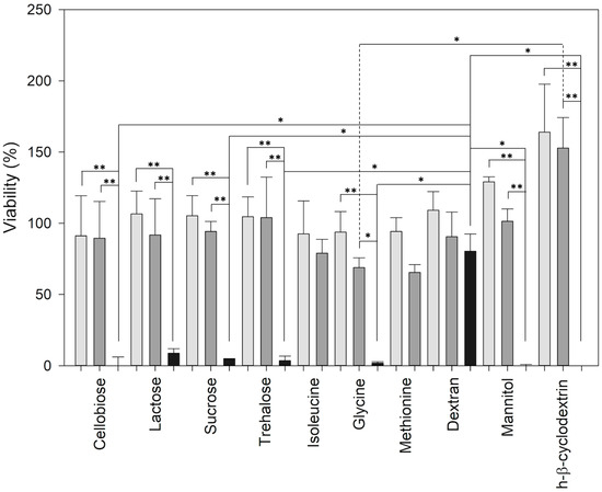
Figure 2.
The graph represents the viability of lymphocytes after 24 h of treatment with the different excipients. Light grey is 1 mg/mL, dark grey is 10 mg/mL, and black is 100 mg/mL. Data are displayed as mean ± SE. ** for p ≤ 0.001 and * for p ≤ 0.05. Black asterisks represent the comparison of excipients at the three concentrations, without isoleucine and methionine.
Comparing the different excipients at the same concentrations, the viability of cells treated with dextran at 100 mg/mL was considerably higher than the ones treated with hp-β-cyclodextrin (p = 0.006), cellobiose (p = 0.018), mannitol (p = 0.024), glycine (p = 0.031), trehalose (p = 0.037), and sucrose (p = 0.041). Differently, at 10 mg/mL, cells were less affected by the treatment with hp-β-cyclodextrin if compared with glycine (p = 0.017).
Considering the second pool of data in Figure 2, which compared all the excipients at 1 and 10 mg/mL, lymphocytes treated with hp-β-cyclodextrin were more viable than the ones with methionine (p = 0.005), glycine (p = 0.007), isoleucine (p = 0.013), and cellobiose (p = 0.027).
Figure 3 shows the viability of Daudi after 24 h of treatment. Comparing the concentrations among the different excipients, there was a statistically significant difference between 1 and 100 mg/mL (p = 0.035 for dextran and p ≤ 0.001 for the others), 10 and 100 mg/mL (p = 0.008 for dextran and p ≤ 0.001 for the others), and 1 and 10 mg/mL only in the treatments with lactose, glycine, isoleucine, and methionine (p = 0.018, p ≤ 0.001, p = 0.002, and p ≤ 0.001 respectively).
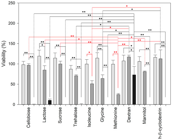
Figure 3.
The graph represents the viability of Daudi after 24 h of treatment with the different excipients. Light grey is 1 mg/mL, dark grey is 10 mg/mL, and black is 100 mg/mL. Data are displayed as mean ± SE. ** for p ≤ 0.001 and * for p ≤ 0.05. Black asterisks represent the comparison of excipients at the three concentrations, without isoleucine and methionine; red asterisks represent excipients at 10 and 1 mg/mL.
Comparing the different excipients at the same concentrations, the viability of cells treated with dextran at 100 mg/mL was considerably higher than all the other treatments (p ≤ 0.001 for all the comparisons). At 10 mg/mL, glycine strongly affected the viability of Daudi when compared with dextran, hp-β-cyclodextrin, and trehalose (p = 0.011, p = 0.029 and p = 0.043, respectively).
Considering the second pool of data, which compared all the excipients at 1 and 10 mg/mL in Figure 3, Daudi treated with methionine was significantly impaired by the treatment, if compared with hp-β-cyclodextrin (p ≤ 0.001), dextran (p ≤ 0.001), sucrose (p = 0.004), and lactose (p = 0.017), and they were also affected by the treatments with isoleucine than with hp-β-cyclodextrin (p = 0.021) and dextran (p = 0.037). Comparing the viability of cells at 10 mg/mL, it was impaired more by methionine than by dextran, hp-β-cyclodextrin, sucrose, cellobiose (p ≤ 0.001), lactose (p = 0.007), mannitol (p = 0.016), by isoleucine than by dextran (p = 0.002) and hp-β-cyclodextrin (p = 0.006), and by glycine than by dextran (p = 0.030).
Figure 4 shows the viability of lymphocytes after 48 h of treatment. Comparing the concentrations among the different excipients, there was a statistically significant difference between 1 and 100 mg/mL for all the excipients with the exception of lactose (p = 0.001 for cellobiose, p = 0.012 for sucrose, p = 0.005 for trehalose, p = 0.003 for glycine, p = 0.005 for dextran, p = 0.004 for mannitol, p ≤ 0.001 for hp-β-cyclodextrin); between 10 and 100 mg/mL (p = 0.012 for cellobiose, p = 0.017 for sucrose, p = 0.007 for trehalose, p = 0.009 for mannitol, and p ≤ 0.001 for hp-β-cyclodextrin); and between 1 and 10 mg/mL only in the treatment with methionine (p = 0.041).
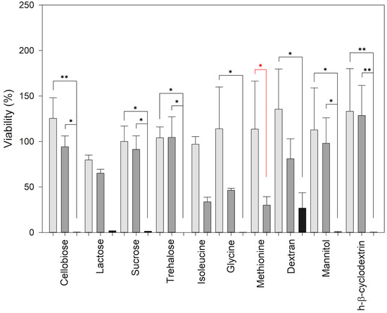
Figure 4.
The graph represents the viability of lymphocytes after 48 h of treatment with the different excipients. Light grey is 1 mg/mL, dark grey is 10 mg/mL, and black is 100 mg/mL. Data are displayed as mean ± SE. ** for p ≤ 0.001 and * for p ≤ 0.05. Black asterisks represent the comparison of excipients at the three concentrations, without isoleucine and methionine, red asterisks, for all excipients at 10 and 1 mg/mL.
There were no significant differences between the viability of cells treated with the same concentration of excipients.
Figure 5 shows the viability of Daudi after 48 h of treatment. Considering the different excipients, cells were less affected by the treatment with dextran if compared with mannitol, glycine, and trehalose (p ≤ 0.001), sucrose (p = 0.004), cellobiose (p = 0.007), lactose (p = 0.041), and by the ones with hp-β-cyclodextrin in comparison with mannitol and glycine (p = 0.012 and p = 0.042, respectively).
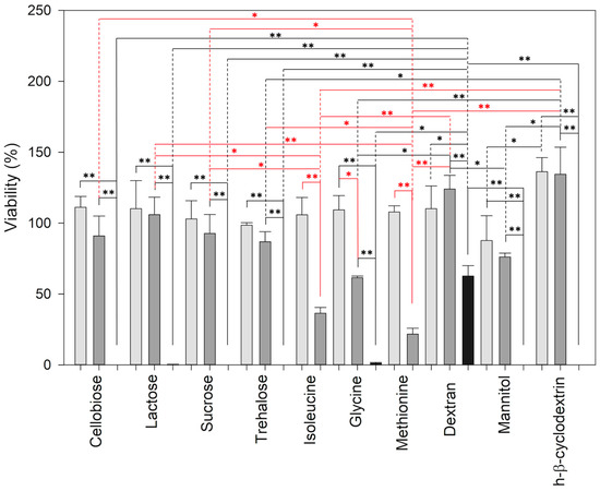
Figure 5.
The graph represents the viability of Daudi after 48 h of treatment with the different excipients. Light grey is 1 mg/mL, dark grey is 10 mg/mL, and black is 100 mg/mL. Data are displayed as mean ± SE. ** for p ≤ 0.001 and * for p ≤ 0.05. Black asterisks represent the comparison of excipients at the three concentrations, without isoleucine and methionine; red asterisks represent all excipients at 10 and 1 mg/mL.
Comparing the concentrations among the different excipients, there was a statistically significant difference between 1 and 100 mg/mL (p ≤ 0.001, p = 0.004 for dextran), between 10 and 100 mg/mL (p ≤ 0.001), and between 1 and 10 mg/mL only in the treatments with glycine (p = 0.002), isoleucine, and methionine (p ≤ 0.001 for both).
Comparing the different excipients at the same concentrations, dextran affected the viability of cells less than other treatments both at 100 mg/mL (p ≤ 0.001 for hp-β-cyclodextrin, trehalose, cellobiose, mannitol, sucrose, and lactose, p = 0.002 for glycine) and at 10 mg/mL, (p = 0.002 for glycine and p = 0.041 for mannitol). Hp-β-cyclodextrin affected the viability of Daudi less if compared with glycine (p ≤ 0.001), mannitol (p = 0.005), and trehalose (p = 0.043) at 10 mg/mL, and with mannitol (p = 0.041) at 1 mg/mL.
Considering the second pool of data in Figure 5, which compared all the excipients at 1 and 10 mg/mL, Daudi treated with hp-β-cyclodextrin had higher viability than the ones treated with methionine, isoleucine, and mannitol (p ≤ 0.001), glycine (p = 0.004), and trehalose (p = 0.023); in addition, cells with dextran had higher viability than the ones with methionine (p = 0.002) and isoleucine (p = 0.010). Lactose also impaired less Daudi than methionine (p = 0.020). The viability of cells at 10 mg/mL was impaired more by methionine than by hp-β-cyclodextrin, dextran, lactose (p ≤ 0.001), sucrose (p = 0.003), cellobiose (p = 0.004), trehalose (p = 0.009). Similarly, the cell viability was affected more by isoleucine than by hp-β-cyclodextrin, dextran (p ≤ 0.001), lactose (p = 0.004), and sucrose (p = 0.040), by glycine than by hp-β-cyclodextrin (p = 0.002) and dextran (p = 0.014), and by mannitol than by hp-β-cyclodextrin (p = 0.029).
4. Discussion
The development of a new medical product requires the use of animal models for some crucial preclinical stages, but for ethical reasons, many efforts have been made to find alternatives. After the introduction of the 3R rules (reduce, refine, and replace) to regulate the use of animals in in vivo experiments, international organizations such as the FDA and EMA enforce the use of in vitro and in silico methods for drug testing [48] and quantitative risk assessment [49]. Although in vitro concentration–response curves data cannot be directly used for clinical practice [50], modeling approaches, such as physiologically based kinetic (PBK), can integrate data from in vitro experiments to obtain reverse dosimetry or quantitative in vitro–in vivo extrapolation (QIVIVE). This new approach values the importance of the in vitro data as a reliable background to determine safe and effective treatment doses for in vivo applications [51].
To accomplish this increasing request for reliable in vitro models, we used a human cellular model constituted by an ad hoc designed physio (B lymphocytes) pathological (tumoral lymphoid cells, Daudi) system that allowed us to selectively assess the effect on cells viability of some of the most used excipients for lyophilized formulations.
Cytotoxicity results showed that for all excipients except dextran, the viability of both lymphocytes and Daudi cells was strongly impaired by the 100 mg/mL treatments. It has been shown that the use of excipients in high concentrations induces an osmotic unbalancing, causing intracellular water loss with consequent cell shrinkage. As a result, this phenomenon affects many homeostatic processes, halting cell proliferation and inducing cell death [52,53,54]. Lower concentrations of excipients, i.e., 1 and 10 mg/mL, caused hyperosmotic stress that induced no cell apoptosis but, in some cases, their proliferation. In the case of osmotic stresses, mammalian cells exploit the only known osmosensitive transcription factor, i.e., the nuclear factor of activated T cells 5/tonicity enhancer binding protein (NFAT5), to control the transcription of genes, with the aim of increasing the intracellular osmolytes’ concentration. Considering the immune system, lymphoid tissues are hyperosmotic relative to blood; thus, NFAT5 is highly expressed in the thymus, suggesting that osmotic stress may be relevant to lymphocyte physiological function in vivo. Following this observation, many researchers demonstrated that cells’ proliferation under conditions of hypertonic stress strongly depends on the NFAT5-mediated response [55,56,57,58]. In addition, cells under hyperosmotic stress, caused by the addition in the culture medium of some non-penetrating excipients, demonstrated changes in the plasma membrane, such as the glycosylation and translocation of the EGFR, which caused an upregulated proliferation of cells [59].
A different behavior from all other excipients is demonstrated by dextran, which seemed to be highly biocompatible even at the highest tested concentration. Dextran 40 is already employed in pharmaceutical products alone at 10% w/v for intravenous infusion as a plasma volume expander for the enhancement of the blood flow in solutions, or as an excipient in some formulations such as Kymriah®, used for leukemia and lymphoma at 11 mg/mL, or ocular drops at 0.1% w/v. Our results confirm the high biocompatibility of Dextran 40, at the doses already used for human applications, due both to its high molecular weight, which minimizes the osmotic shock to the treated cells [60,61], and to its antioxidant and immunomodulatory activity [62].
Considering the other excipients, mannitol is one of the most used as a tonicity agent since it is included in commercial drug products in the range of 0.04 to 10% w/v [36,63]. For instance, in the oncological field, Yervoy® for melanoma treatment contains 10 mg/mL of mannitol, Opdivo® used for a wide range of tumoral pathologies, among which are lymphoma and gastrointestinal oncology, contains 30 mg/mL, and Gemzar®, for breast and ovarian cancer, contains 40 mg/mL. Another bulking agent is glycine, used between 0.1 and 23 mg/mL, which is present at 1 mg/mL in Erbitux® and at 10 mg/mL in Cyramza®. Sucrose is one of the most used stabilizers, usually employed in a wide concentration range between 4 and 500 mg/mL [36]. High concentrations of these excipients are contained in oncological drugs such as Keytruda® (70 mg/mL) and Tecentriq® (~40 mg/mL). Lactose is used as excipients especially for oral administrable formulations of many oncological drugs for breast cancer, such as Afinitor®, Tamoxifen®, Alecensa®, and Temodal®. In addition, trehalose is commonly used in oncological formulations, in Mvasi® at ~6 mg/mL, in Herceptin® at ~20 mg/mL, and in Trodelvy® at ~7 mg/mL [64].
Our results pointed out that 10 mg/mL methionine 24 h treatments selectively impaired the viability of Daudi cancer cells if compared to the healthy ones, lymphocytes (p = 0.003). From the results of our preliminary study, the evident effect that the addition of methionine, recently introduced in oncological formulations, such as Phesgo® at 1.5 mg/mL for the treatment of breast cancer [65], has on the reduction of the viability of Daudi cancer cells opens the way to a series of possible applications of this molecule for the development of new and more effective oncological drugs.
5. Conclusions
To conclude, biopharmaceutical discovery and development represent the driving force for in vitro drug tests and preclinical and clinical trials studies. Freeze-drying is one of the most widely spread methods to remove water from sensitive, thermo-liable samples to stabilize, store and transport them, increasing their shelf life and reducing handling steps. In addition, a stabilized dry product can significantly limit phenomena such as reductive oxide, deamidation, and aggregation commonly known as occurring in an aqueous solution and negatively affecting the effectiveness and/or the safety of biopharmaceuticals.
Since APIs could be strongly affected and/or damaged by freezing and drying stresses during lyophilization, the addition of specific excipients acting as cryo- and lyoprotectants is mandatory. Excipients should give consistency and stability to the formulation, improving the API’s bioavailability and avoiding side effects during administration after reconstitution [66].
Our studies, in addition to confirming what has already been published by several colleagues on the cytotoxic effects of the use of high concentrations of excipients both in in vitro and in vivo experiments [23,24,25,67,68], have shed light on the fact that higher concentrations of specific excipients are well tolerated as in the case of the use of dextran. What emerged in the behavior of molecules such as methionine is unprecedented, since it seems that, already at 10 mg/mL, it could have exceptional potential in the preparation of freeze-dried products to be used in oncology. Our preliminary results showed that it could selectively increase the effectiveness of lyophilized formulations, making them more cytotoxic to tumor cells than to healthy ones. Further in vitro and in vivo studies could confirm this behavior at different concentrations using different tumor models.
Author Contributions
Conceptualization, R.P. and T.L.; methodology, R.P., T.L. and F.S.; validation, R.P., T.L. and F.S.; formal analysis R.P., T.L. and F.S.; investigation, F.S. and M.M.; resources, V.C. and R.P.; data curation, T.L. and F.S.; writing—original draft preparation, F.S. and T.L.; writing—review and editing, R.P., T.L., V.C. and F.S.; supervision, R.P. and T.L.; project administration, R.P.; funding acquisition, V.C. and R.P. All authors have read and agreed to the published version of the manuscript.
Funding
This research received no external funding.
Conflicts of Interest
The authors declare no conflict of interest.
References
- Kesik-Brodacka, M. Progress in biopharmaceutical development. Biotechnol. Appl. Biochem. 2018, 65, 306–322. [Google Scholar] [CrossRef] [PubMed]
- Zeb, A.; Rana, I.; Choi, H.I.; Lee, C.H.; Baek, S.W.; Lim, C.W.; Khan, N.; Arif, S.T.; Sahar, N.U.; Alvi, A.M.; et al. Potential and Applications of Nanocarriers for Efficient Delivery of Biopharmaceuticals. Pharmaceutics 2020, 12, 1184. [Google Scholar] [CrossRef] [PubMed]
- Bjelošević, M.; Zvonar Pobirk, A.; Planinšek, O.; Ahlin Grabnar, P. Excipients in freeze-dried biopharmaceuticals: Contributions toward formulation stability and lyophilisation cycle optimisation. Int. J. Pharm. 2020, 576, 119029. [Google Scholar] [CrossRef] [PubMed]
- Emami, F.; Vatanara, A.; Park, E.J.; Na, D.H. Drying Technologies for the Stability and Bioavailability of Biopharmaceuticals. Pharmaceutics 2018, 10, 131. [Google Scholar] [CrossRef]
- Arsiccio, A.; Paladini, A.; Pattarino, F.; Pisano, R. Designing the Optimal Formulation for Biopharmaceuticals: A New Approach Combining Molecular Dynamics and Experiments. J. Pharm. Sci. 2019, 108, 431–438. [Google Scholar] [CrossRef]
- Khairnar, S.; Kini, R.; Harwalkar, M.; Salunkhe, K.; Chaudhari, S. A Review on Freeze Drying Process of Pharmaceuticals. Int. J. Res. Pharm. Sci. 2012, 2013, 76–94. [Google Scholar]
- Bahr, M.M.; Amer, M.S.; Abo-El-Sooud, K.; Abdallah, A.N.; El-Tookhy, O.S. Preservation techniques of stem cells extracellular vesicles: A gate for manufacturing of clinical grade therapeutic extracellular vesicles and long-term clinical trials. Int. J. Vet. Sci. Med. 2020, 8, 1–8. [Google Scholar] [CrossRef]
- Chang, L.; Shepherd, D.; Sun, J.; Ouellette, D.; Grant, K.L.; Tang, X.C.; Pikal, M.J. Mechanism of protein stabilization by sugars during freeze-drying and storage: Native structure preservation, specific interaction, and/or immobilization in a glassy matrix? J. Pharm. Sci. 2005, 94, 1427–1444. [Google Scholar] [CrossRef]
- Tang, X.; Pikal, M.J. Design of freeze-drying processes for pharmaceuticals: Practical advice. Pharm. Res. 2004, 21, 191–200. [Google Scholar] [CrossRef]
- Rayaprolu, B.M.; Strawser, J.J.; Anyarambhatla, G. Excipients in parenteral formulations: Selection considerations and effective utilization with small molecules and biologics. Drug Dev. Ind. Pharm. 2018, 44, 1565–1571. [Google Scholar] [CrossRef]
- Medi, M.B.; Chintala, R.; Bhambhani, A. Excipient selection in biologics and vaccines formulation development. Eur. Pharm. Rev. 2014, 19, 16–20. [Google Scholar]
- Chaudhari, S.P.; Patil, P.S. Pharmaceutical excipients: A review. Int. J. Adv. Pharm. Biol. Chem. 2012, 1, 21–34. [Google Scholar]
- Pifferi, G.; Restani, P. The safety of pharmaceutical excipients. Il Farm. 2003, 58, 541–550. [Google Scholar] [CrossRef] [PubMed]
- Osterberg, R.E.; See, N.A. Toxicity of excipients—A Food and Drug Administration perspective. Int. J. Toxicol. 2003, 22, 377–380. [Google Scholar] [CrossRef] [PubMed]
- Kiss, L.; Walter, F.R.; Bocsik, A.; Veszelka, S.; Ozsvári, B.; Puskás, L.G.; Szabó-Révész, P.; Deli, M.A. Kinetic analysis of the toxicity of pharmaceutical excipients Cremophor EL and RH40 on endothelial and epithelial cells. J. Pharm. Sci. 2013, 102, 1173–1181. [Google Scholar] [CrossRef]
- Dai, Q.; Liu, X.; He, T.; Yang, C.; Jiang, J.; Fang, Y.; Fu, Z.; Yuan, Y.; Bai, S.; Qiu, T.; et al. Excipient of paclitaxel induces metabolic dysregulation and unfolded protein response. iScience 2021, 24, 103170. [Google Scholar] [CrossRef] [PubMed]
- Bajaj, R.; Chong, L.B.; Zou, L.; Tsakalozou, E.; Ni, Z.; Giacomini, K.M.; Kroetz, D.L. Interaction of Commonly Used Oral Molecular Excipients with P-glycoprotein. AAPS J. 2021, 23, 106. [Google Scholar] [CrossRef]
- Belayneh, A.; Tadese, E.; Molla, F. Safety and Biopharmaceutical Challenges of Excipients in Off-Label Pediatric Formulations. Int. J. Gen. Med. 2020, 13, 1051–1066. [Google Scholar] [CrossRef]
- Rouaz, K.; Chiclana-Rodríguez, B.; Nardi-Ricart, A.; Suñé-Pou, M.; Mercadé-Frutos, D.; Suñé-Negre, J.M.; Pérez-Lozano, P.; García-Montoya, E. Excipients in the Paediatric Population: A Review. Pharmaceutics 2021, 13, 387. [Google Scholar] [CrossRef]
- Schmitt, G. Safety of Excipients in Pediatric Formulations—A Call for Toxicity Studies in Juvenile Animals? Children 2015, 2, 191–197. [Google Scholar] [CrossRef]
- Valeur, K.S.; Holst, H.; Allegaert, K. Excipients in Neonatal Medicinal Products: Never Prescribed, Commonly Administered. Pharmaceut. Med. 2018, 32, 251–258. [Google Scholar] [CrossRef] [PubMed]
- Pottel, J.; Armstrong, D.; Zou, L.; Fekete, A.; Huang, X.P.; Torosyan, H.; Bednarczyk, D.; Whitebread, S.; Bhhatarai, B.; Liang, G.; et al. The activities of drug inactive ingredients on biological targets. Science 2020, 369, 403–413. [Google Scholar] [CrossRef] [PubMed]
- Horváth, T.; Bartos, C.; Bocsik, A.; Kiss, L.; Veszelka, S.; Deli, M.A.; Újhelyi, G.; Szabó-Révész, P.; Ambrus, R. Cytotoxicity of Different Excipients on RPMI 2650 Human Nasal Epithelial Cells. Molecules 2016, 21, 658. [Google Scholar] [CrossRef] [PubMed]
- Nemes, D.; Kovács, R.; Nagy, F.; Mező, M.; Poczok, N.; Ujhelyi, Z.; Pető, Á.; Fehér, P.; Fenyvesi, F.; Váradi, J.; et al. Interaction between Different Pharmaceutical Excipients in Liquid Dosage Forms—Assessment of Cytotoxicity and Antimicrobial Activity. Molecules 2018, 23, 1827. [Google Scholar] [CrossRef] [PubMed]
- Bieberich, A.A.; Rajwa, B.; Irvine, A.; Fatig, R.O., 3rd; Fekete, A.; Jin, H.; Kutlina, E.; Urban, L. Acute cell stress screen with supervised machine learning predicts cytotoxicity of excipients. J. Pharmacol. Toxicol. Methods 2021, 111, 107088. [Google Scholar] [CrossRef]
- Mi, X.; Shukla, D. Predicting the Activities of Drug Excipients on Biological Targets using One-Shot Learning. J. Phys. Chem. B 2022, 126, 1492–1503. [Google Scholar] [CrossRef]
- Arsiccio, A.; Pisano, R. Clarifying the role of cryo- and lyo-protectants in the biopreservation of proteins. Phys. Chem. Chem. Phys. 2018, 20, 8267–8277. [Google Scholar] [CrossRef]
- Ohtake, S.; Kita, Y.; Arakawa, T. Interactions of formulation excipients with proteins in solution and in the dried state. Adv. Drug Deliv. Rev. 2011, 63, 1053–1073. [Google Scholar] [CrossRef]
- Grasmeijer, N.; Stankovic, M.; de Waard, H.; Frijlink, H.W.; Hinrichs, W.L. Unraveling protein stabilization mechanisms: Vitrification and water replacement in a glass transition temperature controlled system. Biochim. Biophys Acta 2013, 1834, 763–769. [Google Scholar] [CrossRef]
- Toniolo, S.P.; Afkhami, S.; Mahmood, A.; Fradin, C.; Lichty, B.D.; Miller, M.S.; Xing, Z.; Cranston, E.D.; Thompson, M.R. Excipient selection for thermally stable enveloped and non-enveloped viral vaccine platforms in dry powders. Int. J. Pharm. 2019, 561, 66–73. [Google Scholar] [CrossRef]
- Susa, F.; Bucca, G.; Limongi, T.; Cauda, V.; Pisano, R. Enhancing the preservation of liposomes: The role of cryoprotectants, lipid formulations and freezing approaches. Cryobiology 2021, 98, 46–56. [Google Scholar] [CrossRef] [PubMed]
- Arsiccio, A.; Pisano, R. Water entrapment and structure ordering as protection mechanisms for protein structural preservation. J. Chem. Phys. 2018, 148, 055102. [Google Scholar] [CrossRef] [PubMed]
- Horn, J.; Tolardo, E.; Fissore, D.; Friess, W. Crystallizing amino acids as bulking agents in freeze-drying. Eur. J. Pharm. Biopharm. 2018, 132, 70–82. [Google Scholar] [CrossRef] [PubMed]
- Larsen, B.S.; Skytte, J.; Svagan, A.J.; Meng-Lund, H.; Grohganz, H.; Löbmann, K. Using dextran of different molecular weights to achieve faster freeze-drying and improved storage stability of lactate dehydrogenase. Pharm. Dev. Technol. 2019, 24, 323–328. [Google Scholar] [CrossRef] [PubMed]
- U.S. Food & Drug Administration: Inactive Ingredient Search for Approved Drug Products U.S. Federal Government Regulatory Agency. Available online: https://www.accessdata.fda.gov/scripts/cder/iig/ (accessed on 7 July 2022).
- Ionova, Y.; Wilson, L. Biologic excipients: Importance of clinical awareness of inactive ingredients. PLoS ONE 2020, 15, e0235076. [Google Scholar] [CrossRef] [PubMed]
- Rao, V.A.; Kim, J.J.; Patel, D.S.; Rains, K.; Estoll, C.R. A Comprehensive Scientific Survey of Excipients Used in Currently Marketed, Therapeutic Biological Drug Products. Pharm. Res. 2020, 37, 200. [Google Scholar] [CrossRef]
- Rycerz, L. Practical remarks concerning phase diagrams determination on the basis of differential scanning calorimetry measurements. J. Therm. Anal. Calorim. 2013, 113, 231–238. [Google Scholar] [CrossRef]
- Horn, J.; Friess, W. Detection of Collapse and Crystallization of Saccharide, Protein, and Mannitol Formulations by Optical Fibers in Lyophilization. Front. Chem. 2018, 6, 4. [Google Scholar] [CrossRef]
- Ray, P.; Rielly, C.D.; Stapley, A.G.F. A freeze-drying microscopy study of the kinetics of sublimation in a model lactose system. Chem. Eng. Sci. 2017, 172, 731–743. [Google Scholar] [CrossRef]
- Pisano, R. Automatic control of a freeze-drying process: Detection of the end point of primary drying. Dry. Technol. 2022, 40, 140–157. [Google Scholar] [CrossRef]
- Patel, S.M.; Nail, S.L.; Pikal, M.J.; Geidobler, R.; Winter, G.; Hawe, A.; Davagnino, J.; Rambhatla Gupta, S. Lyophilized Drug Product Cake Appearance: What Is Acceptable? J. Pharm. Sci. 2017, 106, 1706–1721. [Google Scholar] [CrossRef] [PubMed]
- Bjelošević, M.; Seljak, K.B.; Trstenjak, U.; Logar, M.; Brus, B.; Ahlin Grabnar, P. Aggressive conditions during primary drying as a contemporary approach to optimise freeze-drying cycles of biopharmaceuticals. Eur. J. Pharm. Sci. 2018, 122, 292–302. [Google Scholar] [CrossRef]
- Ward, K.R.; Matejtschuk, P. Characterization of Formulations for Freeze-Drying. In Lyophilization of Pharmaceuticals and Biologicals: New Technologies and Approaches; Ward, K.R., Matejtschuk, P., Eds.; Springer: New York, NY, USA, 2019; pp. 1–32. [Google Scholar]
- Kim, A.I.; Akers, M.J.; Nail, S.L. The Physical State of Mannitol after Freeze-Drying: Effects of Mannitol Concentration, Freezing Rate, and a Noncrystallizing Cosolute. J. Pharm. Sci. 1998, 87, 931–935. [Google Scholar] [CrossRef] [PubMed]
- Gil, A.; Barreneche, C.; Moreno, P.; Solé, C.; Inés Fernández, A.; Cabeza, L.F. Thermal behaviour of d-mannitol when used as PCM: Comparison of results obtained by DSC and in a thermal energy storage unit at pilot plant scale. Appl. Energy 2013, 111, 1107–1113. [Google Scholar] [CrossRef]
- De Beer, T.R.; Wiggenhorn, M.; Hawe, A.; Kasper, J.C.; Almeida, A.; Quinten, T.; Friess, W.; Winter, G.; Vervaet, C.; Remon, J.P. Optimization of a pharmaceutical freeze-dried product and its process using an experimental design approach and innovative process analyzers. Talanta 2011, 83, 1623–1633. [Google Scholar] [CrossRef]
- Jaroch, K.; Jaroch, A.; Bojko, B. Cell cultures in drug discovery and development: The need of reliable in vitro-in vivo extrapolation for pharmacodynamics and pharmacokinetics assessment. J. Pharm. Biomed. Anal. 2018, 147, 297–312. [Google Scholar] [CrossRef]
- Hamon, J.; Renner, M.; Jamei, M.; Lukas, A.; Kopp-Schneider, A.; Bois, F.Y. Quantitative in vitro to in vivo extrapolation of tissues toxicity. Toxicol. Vitr. 2015, 30, 203–216. [Google Scholar] [CrossRef]
- Louisse, J.; Beekmann, K.; Rietjens, I.M. Use of physiologically based kinetic modeling-based reverse dosimetry to predict in vivo toxicity from in vitro data. Chem. Res. Toxicol. 2017, 30, 114–125. [Google Scholar] [CrossRef]
- Algharably, E.A.H.; Kreutz, R.; Gundert-Remy, U. Importance of in vitro conditions for modeling the in vivo dose in humans by in vitro–in vivo extrapolation (IVIVE). Arch. Toxicol. 2019, 93, 615–621. [Google Scholar] [CrossRef]
- Uchida, T.; Furukawa, M.; Kikawada, T.; Yamazaki, K.; Gohara, K. Trehalose uptake and dehydration effects on the cryoprotection of CHO–K1 cells expressing TRET1. Cryobiology 2019, 90, 30–40. [Google Scholar] [CrossRef]
- Brocker, C.; Thompson, D.C.; Vasiliou, V. The role of hyperosmotic stress in inflammation and disease. BioMol. Concepts 2012, 3, 345–364. [Google Scholar] [CrossRef]
- Cvetkovic, L.; Perisic, S.; Titze, J.; Jäck, H.-M.; Schuh, W. The impact of hyperosmolality on activation and differentiation of B lymphoid cells. Front. Immunol. 2019, 10, 828. [Google Scholar] [CrossRef]
- Go, W.Y.; Liu, X.; Roti, M.A.; Liu, F.; Ho, S.N. NFAT5/TonEBP mutant mice define osmotic stress as a critical feature of the lymphoid microenvironment. Proc. Natl. Acad. Sci. USA 2004, 101, 10673–10678. [Google Scholar] [CrossRef] [PubMed]
- Chen, B.L.; Li, Y.; Xu, S.; Nie, Y.; Zhang, J. NFAT5 Regulated by STUB1, Facilitates Malignant Cell Survival and p38 MAPK Activation by Upregulating AQP5 in Chronic Lymphocytic Leukemia. Biochem. Genet. 2021, 59, 870–883. [Google Scholar] [CrossRef] [PubMed]
- Sana, I.; Mantione, M.E.; Angelillo, P.; Muzio, M. Role of NFAT in Chronic Lymphocytic Leukemia and Other B-Cell Malignancies. Front. Oncol. 2021, 11, 651057. [Google Scholar] [CrossRef] [PubMed]
- Drews-Elger, K.; Ortells, M.C.; Rao, A.; López-Rodriguez, C.; Aramburu, J. The Transcription Factor NFAT5 Is Required for Cyclin Expression and Cell Cycle Progression in Cells Exposed to Hypertonic Stress. PLoS ONE 2009, 4, e5245. [Google Scholar] [CrossRef]
- Yoshimoto, S.; Morita, H.; Matsuda, M.; Katakura, Y.; Hirata, M.; Hashimoto, S. NFAT5 promotes oral squamous cell carcinoma progression in a hyperosmotic environment. Lab. Investig. 2021, 101, 38–50. [Google Scholar] [CrossRef] [PubMed]
- Hunger, J.; Bernecker, A.; Bakker, H.J.; Bonn, M.; Richter, R.P. Hydration dynamics of hyaluronan and dextran. Biophys. J. 2012, 103, L10–L12. [Google Scholar] [CrossRef]
- Reich-Slotky, R.; Bachegowda, L.S.; Ancharski, M.; Mendeleyeva, L.; Rubinstein, P.; Rennert, H.; Shore, T.; van Besien, K.; Cushing, M. How we handled the dextran shortage: An alternative washing or dilution solution for cord blood infusions. Transfusion 2015, 55, 1147–1153. [Google Scholar] [CrossRef]
- Soeiro, V.C.; Melo, K.R.T.; Alves, M.G.C.F.; Medeiros, M.J.C.; Grilo, M.L.P.M.; Almeida-Lima, J.; Pontes, D.L.; Costa, L.S.; Rocha, H.A.O. Dextran: Influence of Molecular Weight in Antioxidant Properties and Immunomodulatory Potential. Int. J. Mol. Sci. 2016, 17, 1340. [Google Scholar] [CrossRef]
- Thakral, S.; Sonje, J.; Munjal, B.; Bhatnagar, B.; Suryanarayanan, R. Mannitol as an Excipient for Lyophilized Injectable Formulations. J. Pharm. Sci. 2022, in press. [Google Scholar] [CrossRef] [PubMed]
- National Cancer Institute: Drugs Approved for Dfferent Types of Cancer. Available online: https://www.cancer.gov/about-cancer/treatment/drugs/cancer-type (accessed on 7 July 2022).
- Perkey, C. Pertuzumab/Trastuzumab/Hyaluronidase-Zzxf (Phesgo™). Oncol. Times 2021, 43, 6–17. [Google Scholar] [CrossRef]
- Abrantes, C.G.; Duarte, D.; Reis, C.P. An Overview of Pharmaceutical Excipients: Safe or Not Safe? J. Pharm. Sci. 2016, 105, 2019–2026. [Google Scholar] [CrossRef] [PubMed]
- Dirain, C.O.; Karnani, D.N.; Antonelli, P.J. Cytotoxicity of Ear Drop Excipients in Human and Mouse Tympanic Membrane Fibroblasts. Otolaryngol. Head Neck Surg. 2019, 162, 204–210. [Google Scholar] [CrossRef]
- Gurjar, R.; Chan, C.Y.S.; Curley, P.; Sharp, J.; Chiong, J.; Rannard, S.; Siccardi, M.; Owen, A. Inhibitory Effects of Commonly Used Excipients on P-Glycoprotein In Vitro. Mol. Pharm. 2018, 15, 4835–4842. [Google Scholar] [CrossRef] [PubMed]
Publisher’s Note: MDPI stays neutral with regard to jurisdictional claims in published maps and institutional affiliations. |
© 2022 by the authors. Licensee MDPI, Basel, Switzerland. This article is an open access article distributed under the terms and conditions of the Creative Commons Attribution (CC BY) license (https://creativecommons.org/licenses/by/4.0/).