Article

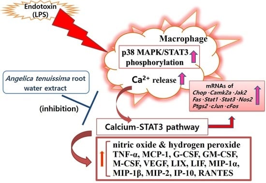
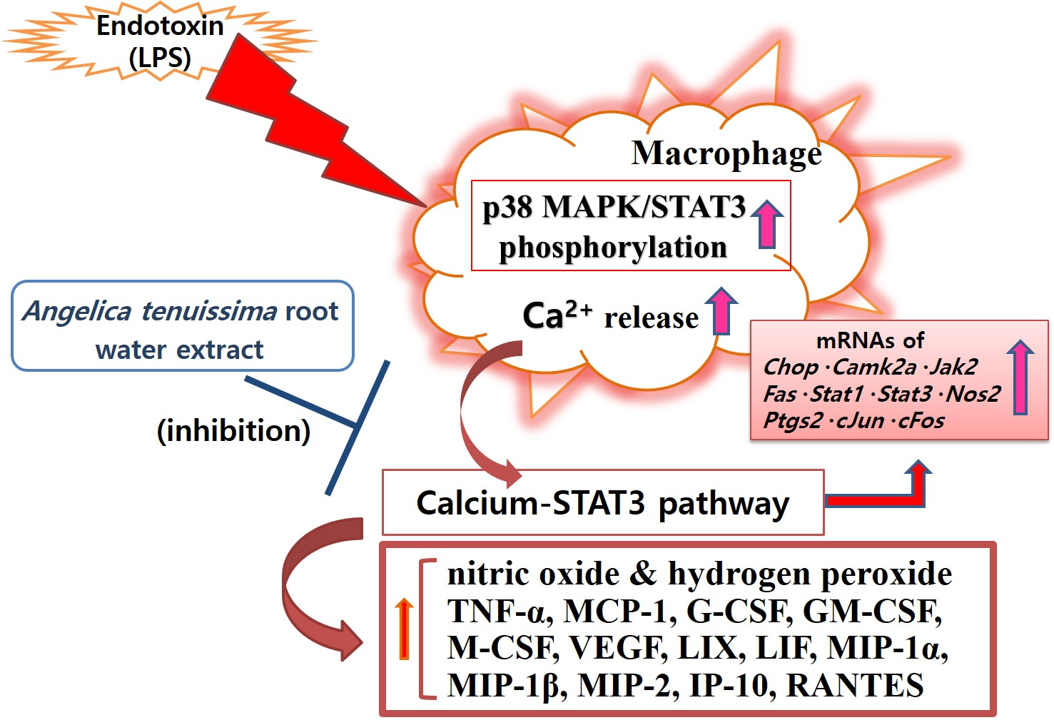
Conioselinum tenuissimum Root Extract Modulates Macrophage Activation via the Calcium–STAT3 Pathway

1
Korean Medicine Clinical Trial Center, Korean Medicine Hospital, Kyung Hee University, Seoul 02447, Korea
2
Department of Medical Classics and History, College of Korean Medicine, Gachon University, Seongnam 13120, Korea
3
Department of Pathology, College of Korean Medicine, Gachon University, Seongnam 13120, Korea
*
Author to whom correspondence should be addressed.
Processes 2022, 10(11), 2238; https://doi.org/10.3390/pr10112238
Submission received: 1 September 2022 / Revised: 13 October 2022 / Accepted: 25 October 2022 / Published: 1 November 2022
(This article belongs to the Special Issue Immune Regulatory Properties of Natural Products)

Abstract

:
Despite the development of many antibiotics, excessive inflammation caused by endotoxins is still a subject of interest to biomedical researchers. The hyper-inflammatory response of macrophages activated by endotoxins is an important topic in the development of natural product-based anti-inflammatory drugs. Conioselinum tenuissimum, a perennial herb of the family Apiaceae, contains levistolide A, demethylsuberosin, and fraxetin. One of the synonyms of Conioselinum tenuissimum is Angelica tenuissima. The objective of this study was to determine the effects of Conioselinum tenuissimum root water extract (AT) on the hyper-inflammatory responses of macrophages activated by endotoxin (lipopolysaccharide; LPS) and the mechanisms involved in such effects. Levels of cytokines, nitric oxide (NO), hydrogen peroxide, and cytosolic calcium in LPS-activated RAW 264.7 murine macrophages were evaluated by the multiplex cytokine assay (MCA), Griess reagent assay (GRA), dihydrorhodamine 123 assay (DHR), and Fluo-4 calcium assay (FCA), respectively. Additionally, real-time PCR and the flow cytometry assay (FLA) was performed to determine the effects of AT on LPS-activated RAW 264.7. Data from MCA, GRA, DHR, and FCA revealed that AT lowered levels of IL-6, MCP-1, TNF-α, G-CSF, GM-CSF, VEGF, M-CSF, LIF, LIX, MIP-1α, MIP-1β, MIP-2, RANTES, IP-10, NO, hydrogen peroxide, and calcium in LPS-activated RAW 264.7. Real-time PCR results revealed that AT significantly lowered mRNA expression levels of inflammatory genes such as Chop, Nos2, c-Jun, Stat1, Stat3, c-Fos, Camk2a, Ptgs2, Fas, and Jak2. FLA showed that AT significantly reduced phosphorylation levels of P38 MAPK and STAT3 in LPS-activated RAW 264.7. These results indicate that AT can exert anti-inflammatory effects in LPS-activated macrophages via the calcium–STAT3 pathway.

Graphical Abstract

1. Introduction

Despite the development of many treatments and medical technologies, including antibiotics, microbial infections are still threatening human life [1,2]. Lipopolysaccharide (LPS) is one of the substances that make up the outer leaflet of Gram-negative bacteria’s outer membrane [3,4,5,6]. Gram-negative bacteria that have intruded into the body can be destroyed by immune cells in the innate immune system. They can release LPS, which is an endotoxin. Endotoxins are resistant to heat. Unlike exotoxins released from Gram-positive bacteria, endotoxins have low immunogenicity, making it difficult to develop antibodies and vaccines. When endotoxins stimulate immune cells (such as neutrophils and macrophages), interleukins (ILs), tumor necrosis factors (TNFs), prostaglandins, and colony stimulation factors are produced [7,8]. When endotoxins are found in the blood, it is called endotoxemia. High levels of endotoxemia can lead to sepsis shock [9,10]. Sepsis is still a major cause of death.
Macrophage activation induced by endotoxins and massive productions of inflammatory mediators has important pathophysiological implications for septic shock, although NO and cytokines can help remove invading pathogens and strengthen the immune system against infection. Among inflammatory factors produced by macrophages, NO is one of the reactive nitrogen species (RNS) that can directly destroy the source of infection. On the other hand, it can worsen septic shock by lowering blood pressure and increasing blood vessel permeability [11]. Cytokine is an essential factor in response to an infection. Cytokines include IL, TNFs, and interferons (IFNs). They can induce immune cell migration and amplify immune responses. However, during the inflammation process, TNF could degrade endothelial glycocalyx of vascular endothelium and cause vascular permeation, leukocyte adhesion, and platelet aggregation, resulting in disseminated vascular coagulation and multiple organ failure [12]. Therefore, decreasing the excessive production of inflammatory factors caused by endotoxin is required to alleviate endotoxemia and septic shock. In this respect, natural products that can suppress macrophage activation and excessive production of inflammatory factors in macrophages caused by endotoxin have attracted attention. Many studies have been conducted on natural anti-inflammatory materials using in vitro experimental models and endotoxin-stimulated macrophages [13,14,15]. For example, Scutellariae Radix was reported to decrease levels of NO, IL-6, and interferon-inducible protein (IP)-10 (C-X-C motif chemokine ligand 10; CXCL10), and so on in mouse macrophages activated by endotoxins [16]. Mass production of reactive oxygen species (ROS), which occur during an oxidative burst in activated macrophages, is crucial for macrophages to attack and remove infectious pathogens. However, they can damage surrounding tissues around infection sites and cause endoplasmic reticulum (ER) stress by altering redox signaling in cells [17,18,19,20]. In sepsis, ROS and RNS can cause oxidative stress and oxidative damage by modifying intracellular proteins, DNA, and lipids, resulting in organ dysfunction [21,22]. Signal transducer and activator of transcription (STAT) is a well-known transcription factor (TF) related to inflammatory responses caused by infections. Inflammatory mediators such as cytokines, chemokines, and growth factors can activate STAT protein, one of the important TFs in the inflammatory cascade launched by Gram-negative bacterial infection and viral infection [23]. So far, seven members of the mammalian STAT protein family group have been reported. STAT is activated by Janus kinase (Jak) [24]. For this reason, the term Jak–STAT is used to describe the mechanism of the immuno-inflammatory response of immune cells, including macrophages and lymphocytes [25]. Jak–STAT is an important signaling pathway for the expression of IL-6 and chemokines in ER stress-related inflammation [26].
Conioselinum tenuissimum, a perennial herb of the family Apiaceae, contains levistolide A, demethylsuberosin, fraxetin, marmesinin, and isopraeroside IV [27]. One of the synonyms of Conioselinum tenuissimum is Angelica tenuissima. In traditional medicine, Angelica tenuissima is used to treat pain (such as joint pain and abdominal pain) and gynecological diseases [28,29]. So far, there have been no reports concerning the effect of Conioselinum tenuissimum root (Angelicae tenuissimae Radix) on inflammatory responses of macrophages stimulated by endotoxins. Thus, we checked the effects of Angelica tenuissima root (AT) water extract on the hyper-inflammatory responses of macrophages activated by endotoxin (lipopolysaccharide) and the mechanisms involved in such effects. Data revealed that the water extract of AT could inhibit the excessive production of inflammatory mediators in murine macrophages triggered by lipopolysaccharide (LPS) and that the calcium–STAT3 pathway was involved in its mechanism.

2. Materials and Methods

The experimental methods and substances used in this study have been referenced from previous studies [30,31]. More details are described in the supplementary file.

2.1. Materials

Baicalein (Cat No. 465119), indomethacin (Cat No. 17378), and Dulbecco’s modified Eagle medium (DMEM, Cat No. D5796) were purchased from Millipore (Billerica, MA, USA).

2.2. Preparation of AT

Commercial Angelicae tenuissimae Radix materials were obtained from Omniherb (Daegu, Korea) in August 2021. Angelicae tenuissimae Radix (voucher specimen No. 2021-025) was authenticated by referring to the website of the Korea Food and Drug Administration accessed on 25 August 2021 (https://www.nifds.go.kr/nhmi/analscase/snststMnl/view.do?selectedSnststMnlNo=29&pageIndex=1&pageSize=10&searchText=%EA%B3%A0%EB%B3%B8&searchTarget=all&sortField=snststMnlNo&direction=DESC) and stored at Gachon University College of Korean Medicine. Angelicae tenuissimae Radix materials were extracted with hot water (yield: 33.98%) [30,31].

2.3. Diethylene Glycol Colorimetric Assay

The total flavonoid content (TFC) of AT was determined using the diethylene glycol colorimetric assay [30].

2.4. MTT Assay

Murine macrophage RAW 264.7 cell line (passage number 2) was obtained from the Korea Cell Line Bank (Seoul, Korea). Cell viability was accessed with a tetrazolium-based colorimetric assay (MTT assay), as described previously [30]. After 24 h incubation with AT (concentrations of 25~200 mg/mL), absorbance values were measured with a microplate reader (Bio-Rad, Hercules, CA, USA) to calculate the viability of RAW 264.7. Cell viability data were compared with the group treated with 25 µg/mL of AT, the group treated with 50 µg/mL, the group treated with 100 µg/mL, the group treated with 200 µg/mL, and the group treated with cell culture media alone.

2.5. Griess Reagent Assay

NO production in RAW 264.7 (1 × 10⁴ cells/well) after 24 h treatment with AT was measured with Griess reagent (Thermo Fisher Scientific, Waltham, MA, USA), as described previously [30].

2.6. Fluo-4 Calcium Assay

Cytosolic calcium release in RAW 264.7 (1 × 10⁵ cells/well) after 24 h treatment with AT was measured with a Fluo-4 calcium assay kit (Thermo Fisher Scientific) [30].

2.7. Dihydrorhodamine 123 Assay

The levels of hydrogen peroxide in RAW 264.7 (1 × 10⁴ cells/well) were measured with a dihydrorhodamine 123 assay [30].

2.8. Multiplex Cytokine Assay

Multiplex Cytokine Assay kits of Millipore were used to evaluate concentrations of cytokines in RAW 264.7 (1 × 10⁴ cells/well) [30,31].

2.9. Real-Time PCR

The mRNA expressions of Chop, Camk2a, Stat1, Stat3, Jak2, Fas, c-Jun, c-Fos, Nos2, Ptgs2, and β-Actin were quantified with real-time PCR. GenBank accession numbers used for designing primers are listed in Table 1.

2.10. Flow Cytometry Assay

Flow cytometry was used to check phospho-STAT3 and phospho-P38 MAPK with Attune NxT flow cytometer (Thermo Fisher Scientific) [30]. After 18 h treatment, RAW 264.7 were stained with phospho-P38 MAPK and phospho-P38 MAPK antibody. The level of the antibody was analyzed with Attune NxT software. Details of flow cytometry are presented in the supplementary file.

2.11. Statistical Analyses

Values are expressed in means ± standard deviation. ANOVA was used to check the statistical significance.

3. Results

3.1. Total Flavonoid Content of AT
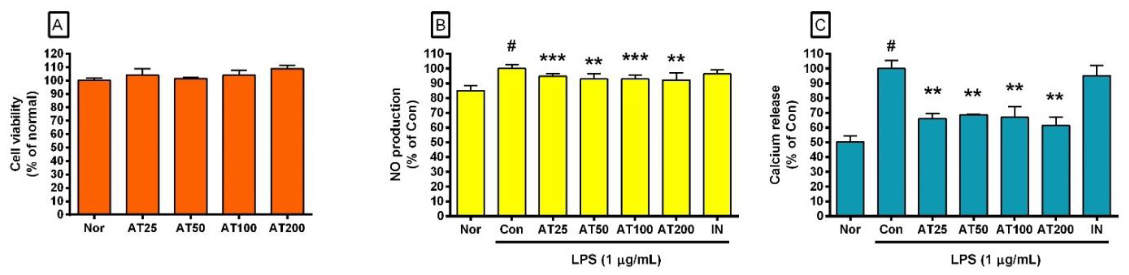
The TFC of AT was 1.62 mg RE/g extract.

3.2. Cell Viability

AT did not show any cytotoxicity to RAW 264.7. After 24 h of treatment, cell viability was measured with a tetrazolium-based colorimetric assay. Cell viability data were compared with the group treated with 25 µg/mL of AT (AT25), the group treated with 50 µg/mL (AT50), the group treated with 100 µg/mL (AT100), the group treated with 200 µg/mL (AT200), and the group treated with cell culture media alone (Nor). Experimental results represented that the cell viability of AT25 was 104.19 ± 4.63% compared to Nor, 101.41 ± 0.94% of AT50, 104.00 ± 3.65% of AT100, and 108.89 ± 2.37% of AT200, respectively (Figure 1A). Along with these experimental results, up to 200 µg/mL of AT concentration were used in this study.

3.3. NO Production

Although macrophages tend to produce RNS such as NO, as shown in Figure 1, LPS significantly induced NO production of RAW 264.7 (Figure 1B), whereas AT significantly inhibited NO production of RAW 264.7 against the stimulation of LPS (Figure 1B). NO production data were compared with AT25, AT50, AT100, and AT200 and the group treated with lipopolysaccharide (LPS, 1 µg/mL) alone (Con). Experimental results represented that NO production of AT25 was 94.71 ± 1.76% compared to Con, 92.88 ± 3.43% of AT50, 92.8 ± 2.82% of AT100, and 92.15 ± 4.96% of AT200, respectively. These experimental results mean that AT can regulate NO-caused harmful inflammatory processes by controlling the amount of NO produced from activated macrophages.

3.4. Calcium Release

Calcium release data were compared with AT25, AT50, AT100, and AT200 and the group treated with LPS (1 µg/mL) alone (Con). Experimental results represented that calcium release of AT25 was 66.07 ± 3.39% compared to Con, 68.53 ± 0.43% of AT50, 66.96 ± 7.37% of AT100, and 61.38 ± 5.83% of AT200, respectively (Figure 1C). These results of AT treatment on LPS-activated macrophages can be interpreted as having the effect of controlling harmful inflammatory processes in LPS-activated macrophages by modulating the calcium release signaling in activated macrophages.

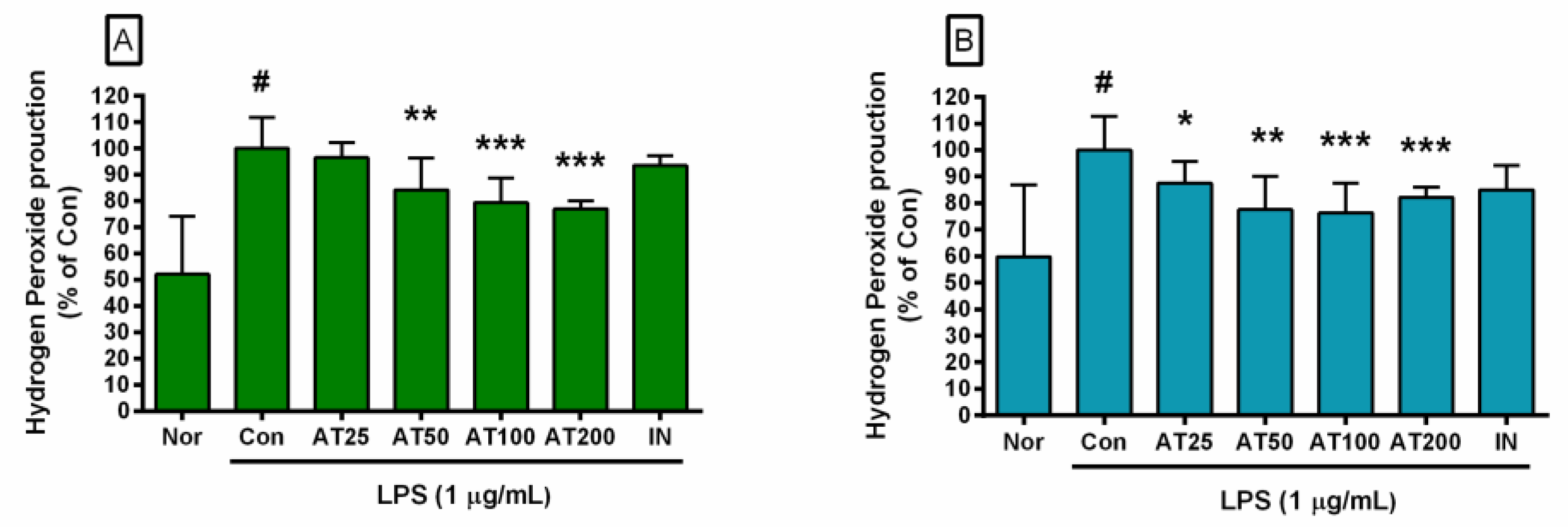
3.5. Hydrogen Peroxide Production

Figure 2 represents that AT dose-dependently decreased hydrogen peroxide production from RAW 264.7 activated by LPS. Hydrogen peroxide data were compared with AT25, AT50, AT100, and AT200 and the group treated with LPS (1 µg/mL) alone (Con). Experimental results for 24 h treatment represented that hydrogen peroxide production of AT25 was 96.57 ± 5.65% compared to Con, 84.29 ± 12.01% of AT50, 79.44 ± 9.43% of AT100, and 77.05 ± 3.1% of AT200, respectively (Figure 2A). Experimental results for 48 h treatment represented that hydrogen peroxide production of AT25 was 87.51 ± 8.15% compared to Con, 77.56 ± 12.56% of AT50, 76.3 ± 11.25% of AT100, and 82.24± 3.67% of AT200, respectively (Figure 2B). These results of AT treatment on LPS-activated macrophages can be interpreted as having the effect of relieving oxidative stress phenomena during LPS-induced macrophage oxidative burst by modulating hydrogen peroxide production in activated macrophages, resulting in the inhibition of oxidative stress-linked inflammatory processes.

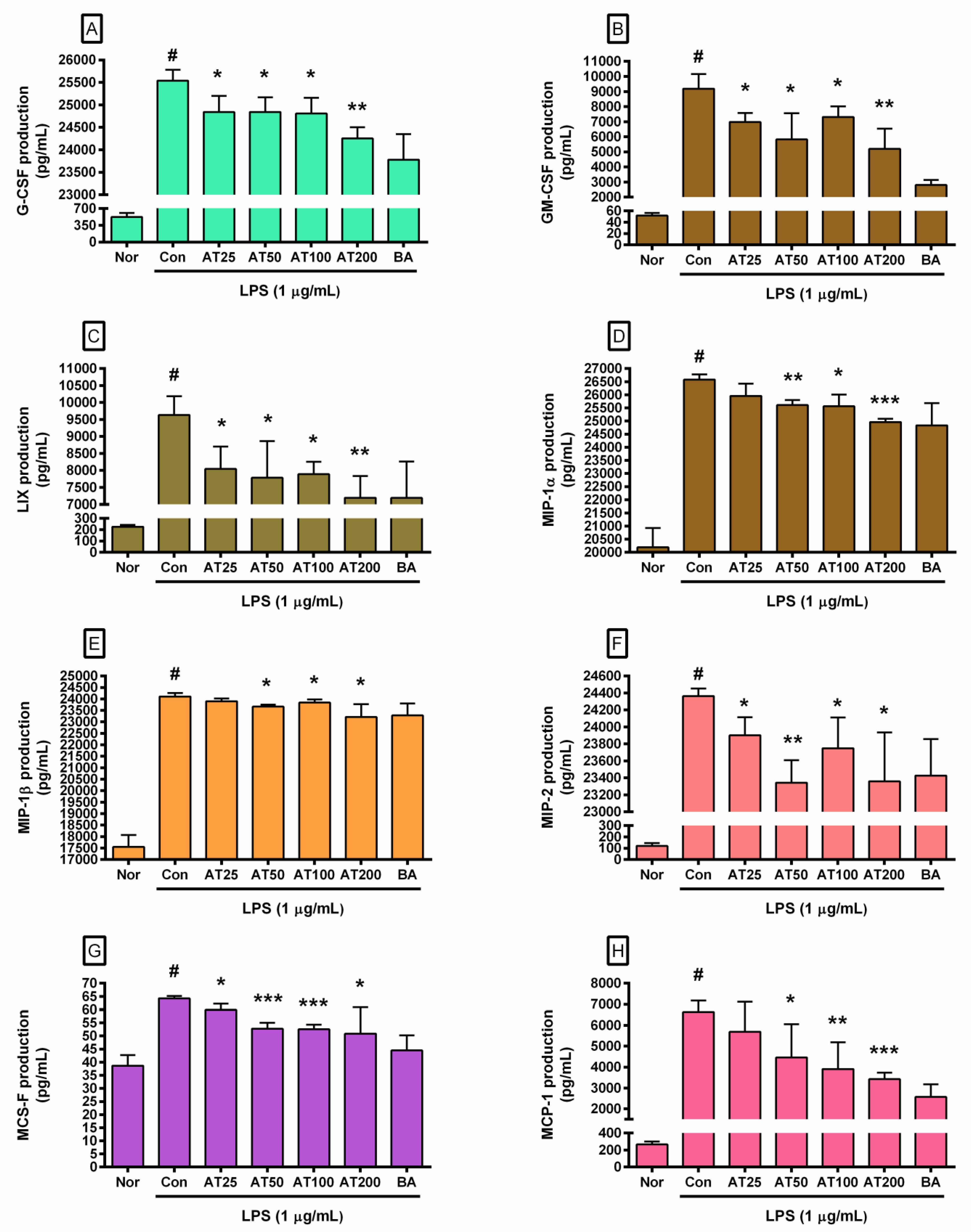
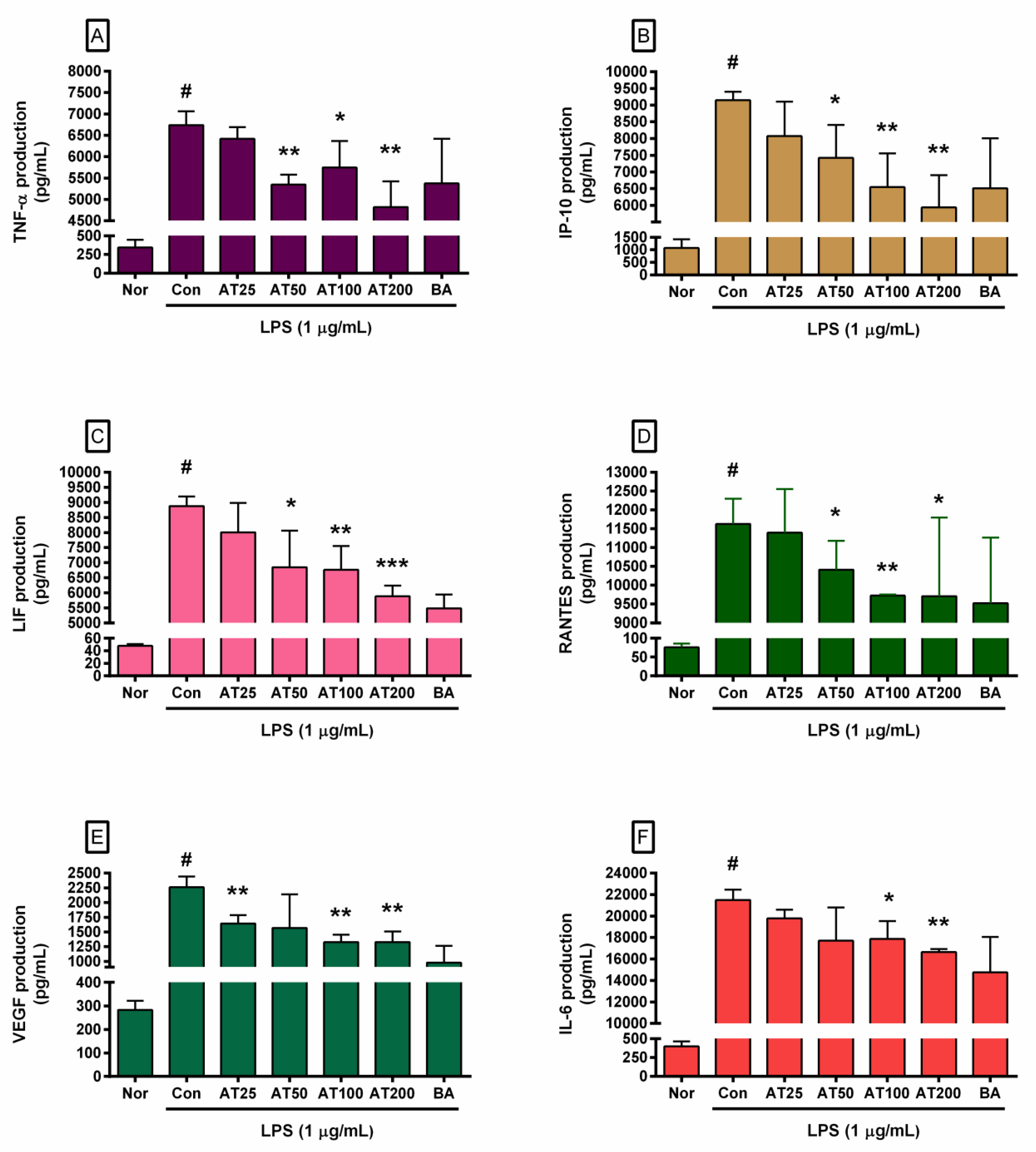
3.6. Cytokine Production

Since one of the goals of this study regards the activity of AT on increased productions of various cytokines and growth factors related to the Jak–STAT signaling pathway in LPS-stimulated macrophages, the level of cytokines and growth factors in cell culture supernatants was checked after 24 h of incubation with LPS (1 µg/mL) and AT. In detail, concentrations of B cell stimulatory factor 2 (BSF2; IL-6), chemokine ligand 2 (CCL2; MCP-1), tumor necrosis factor-alpha (TNF-α), colony-stimulating factor 1 (CSF1; M-CSF), colony-stimulating factor 2 (CSF2; GM-CSF), colony-stimulating factor 3 (CSF3; G-CSF), vascular permeability factor (VPF; VEGF), cholinergic differentiation factor (CDF; LIF), C-X-C motif chemokine 5 (CXCL5; LIX), chemokine (C-C motif) ligand 3 (CCL3; MIP-1α), chemokine (C-C motif) ligands 4 (CCL4; MIP-1β), chemokine (C-X-C motif) ligand 2 (CXCL2; MIP-2), chemokine (C-C motif) ligand 5 (CCL5; RANTES), and CXCL10 (IP-10) were evaluated using the Bio-plex 200 system for the multiplex cytokine assay. Experimental results represented that AT at concentrations of 25, 50, 100, and 200 µg/mL significantly inhibited the LPS-induced production of CSF1, CSF2, CSF3, CXCL5, and CXCL2 in RAW 264.7 (Figure 3, Table 2). In addition, AT at concentrations of 50, 100, and 200 µg/mL significantly inhibited the LPS-induced production of CCL3, CCL4, CCL2, TNF-α, CXCL10, CDF, and CCL5 in RAW 264.7 (Figure 3 and Figure 4, Table 2). The level of VPF was significantly decreased by AT at concentrations of 25, 100, and 200 µg/mL in RAW 264.7 activated by LPS. BSF2 was significantly decreased by AT at concentrations of 100 and 200 µg/mL (Figure 4 and Table 2).
However, concentrations of CCL4 (MIP-1β), CXCL2 (MIP-2), and TNF-α did not decrease by AT dose-dependently (Figure 3 and Figure 4). After treatment with AT at 25 µg/mL, levels of CCL3 (MIP-1α), CCL4 (MIP-1β), CCL2 (MCP-1), TNF-α, CXCL10 (IP-10), CDF (LIF), and CCL5 (RANTES) were decreased, although such decreases were not statistically significant (Figure 3 and Figure 4). AT at a concentration of 50 µg/mL decreased VPF (VEGF) production (Figure 4), and such decrease did not reach significance either. In the case of BSF2 (IL-6), AT at concentrations of 25 and 50 µg/mL did not significantly inhibit its production (Figure 4).
Our data show that AT regulates hyper-inflammatory responses in activated macrophages by decreasing excessive levels of various cytokines and growth factors, thus modulating the hyper-inflammatory response associated with endotoxemia.

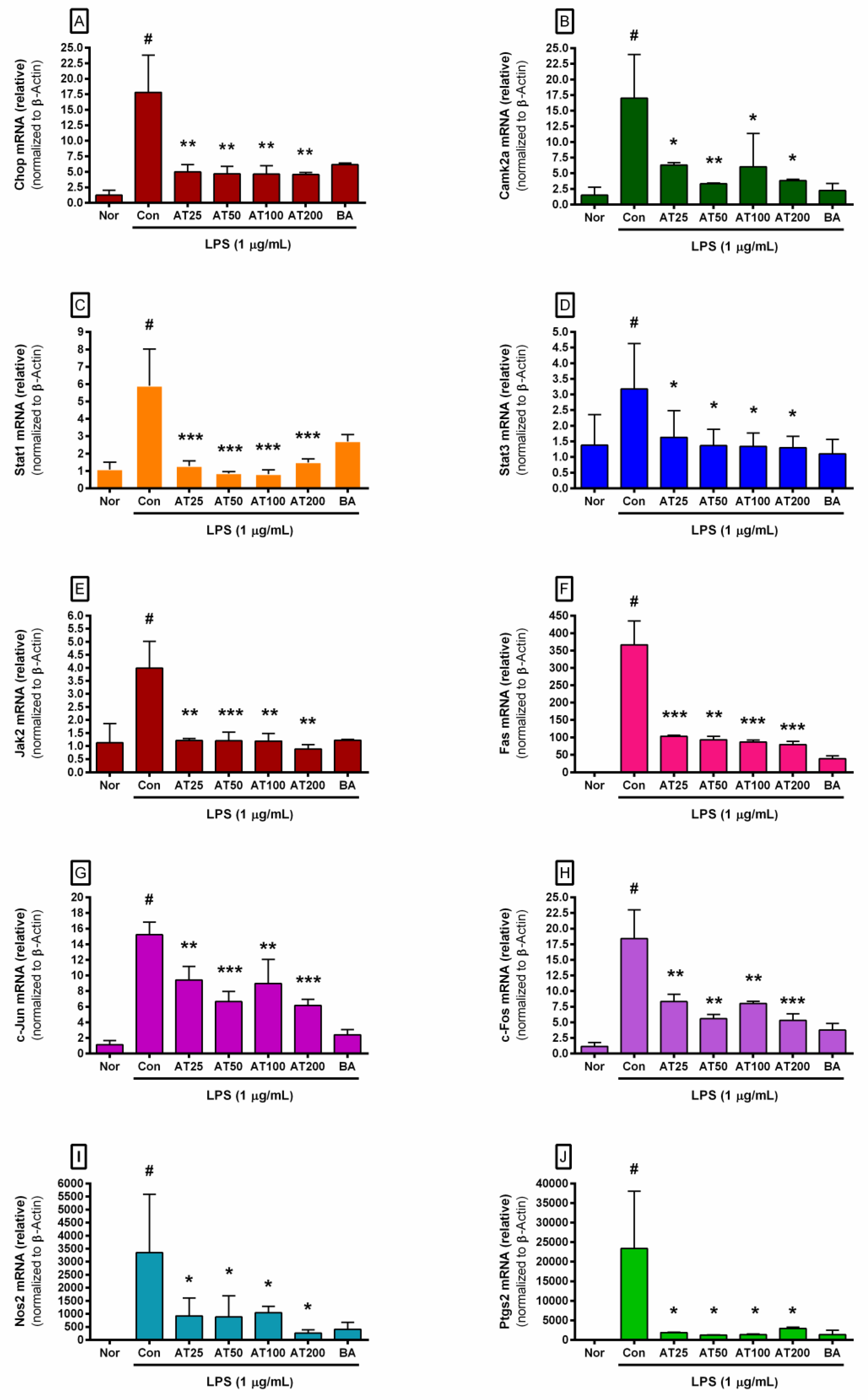
3.7. The Level of Inflammatory Gene Expression

To verify the effects of AT on expressions of inflammatory genes in LPS-stimulated macrophages, mRNA expression levels of inflammatory genes related to ER stress and the Jak–STAT signaling pathway were measured with real-time PCR. CHOP, CAMK2a, and FAS might play a key role in the ER stress cascade in activated macrophages [18,19,20]. Jak–STAT signaling is associated with increased expression levels of STAT, Jak, c-Jun, c-Fos, Nos2, and Ptgs2. Our data showed that treatment with AT at concentrations of 25, 50, 100, and 200 µg/mL for 24 h significantly reduced mRNA expression levels of Stat1, Stat3, Nos2, Ptgs2, Jak2, Fas, c-Jun, c-Fos, Chop, and Camk2a genes in LPS-stimulated RAW 264.7 (Figure 5, Table 2). Our data show that the anti-inflammatory effect of AT on macrophages activated by LPS is achieved through the CHOP-related pathway.

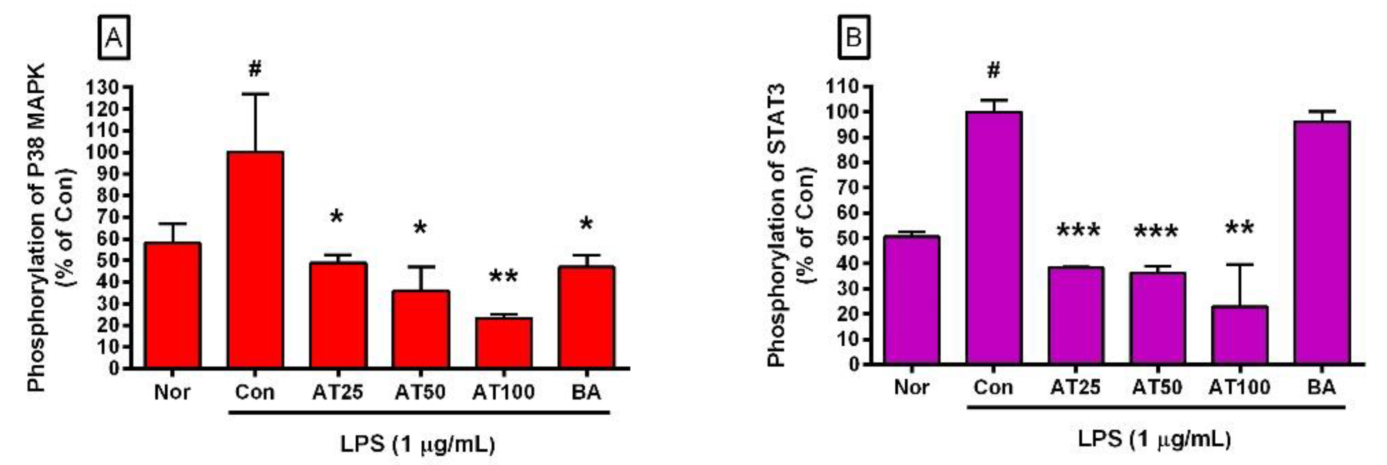
3.8. The Level of Phospho-STAT3 and Phospho-P38 MAPK

To determine the pathway for the inhibitory effect of AT on RAW 264.7 activated by LPS, the effects of AT on the phosphorylation of P38 MAPK and STAT3 in RAW 264.7 were investigated using the flow cytometry assay. AT significantly downregulated the phosphorylation of P38 MAPK and STAT3 in RAW 264.7 activated by LPS (Figure 6). In detail, phosphorylation levels of P38 MAPK in RAW 264.7 macrophages treated with AT at 25, 50, and 100 µg/mL were decreased to 48.76 ± 3.79%, 35.97 ± 11.12%, and 23.37 ± 1.74% of that treated with LPS alone, respectively. Phosphorylation levels of STAT3 were decreased to 38.23 ± 0.49%, 36.29 ± 2.55%, and 22.87 ± 16.9% of that treated with LPS alone, respectively. These data show that AT inhibits hyper-inflammatory responses in RAW 264.7 activated by LPS via P38 MAPK and STAT3 signaling.

4. Discussion

Conioselinum tenuissimum is traditionally used to treat pain (such as headaches, joint pain, limb pain, toothache, and abdominal pain) and gynecological diseases in East Asia [28,29]. Lee et al. reported the efficacy of Conioselinum tenuissimum in treating headaches [32]. They found that Angelicae tenuissimae Radix could decrease the level of NO and Nos2 gene transcripts in LPS-stimulated BV-2 (C57/BL6 murine Microglia) activated by endotoxins as well as the level of prostaglandin E and Ptgs2 gene transcripts, suggesting that Angelicae tenuissimae Radix might lessen brain inflammation and headache. Kim et al. reported that the combination of Angelica tenuissima, Angelica dahurica, Scutellaria baicalensis, and acetaminophen can synergistically decrease LPS-induced inflammation in a microglia cell line [33]. These results support the activity of Angelicae tenuissimae Radix in relieving brain inflammation and lessening headaches.
Angelicae tenuissimae Radix and Aspergillus oryzae-fermented Angelicae tenuissimae Radix (FAT) inhibit melanin production in melanocytes insulted by α-melanocyte-stimulating hormone as well as tyrosinase activity [28]. In 2018, Park et al. reported that FAT could protect keratinocytes against ultraviolet light B exposure [29]. This means that FAT has an anti-photoaging effect [29]. FAT can increase procollagen Type-1 and hemeoxygease-1 while suppressing the expression of MMP-1, elastase, and Ptgs2 [29]. Thus, FAT could be a candidate with anti-aging and anti-wrinkle effects. Despite these studies, there are not many detailed studies on the inhibitory activity of Conioselinum tenuissimum root on the production of cytokines and growth factors in endotoxin-activated macrophages yet.
Current data revealed that AT did not show any toxicity to RAW 264.7. Even if natural products have biomedical effects, if they are toxic to immune cells, they can rather reduce the host’s immune function in response to infection. In this regard, when developing anti-inflammatory drugs using natural products, it is important to confirm whether the natural product is toxic to macrophages. Since our data represent AT modulate hyper-inflammatory responses in endotoxins-activated macrophages without cytotoxicity, AT might be a safe candidate material for treating inflammatory diseases concerned with excessive production of cytokines and growth factors.
First, AT inhibited the level of NO, cytosolic calcium, and hydrogen peroxide in RAW 264.7 activated by LPS. These inhibitory effects of AT on activated macrophages can be interpreted as having the effect of relieving oxidative stress phenomena during endotoxins-induced macrophage oxidative burst by regulating the production of ROS such as hydrogen peroxide as well as RNS such as NO, resulting in the modulation of oxidative stress-linked inflammatory processes via calcium signaling. Of course, oxidative stress causes various effects on human pathophysiological phenomena. Not only ROS but also RNS play different roles in immune cells (i.e., macrophages) and brain tissue cells such as neuroglia. In other words, in the innate immune system, RNS or ROS belong to an effective method of responding to infection. Therefore, proper control may be more beneficial for disease control than the complete removal of RNS and ROS which are produced during oxidative stress. In this respect, the activity of AT that regulates the production of RNS and ROS can be effective in solving inflammatory responses caused by bacterial infection. Furthermore, considering that calcium signaling in ER stress is involved in increasing Chop transcripts, it means that AT’s activity to control calcium release and suppress the expression of the Chop gene is involved in relieving ER stress in endotoxin-induced macrophages.
STAT proteins are transcription factors that can mediate various cellular functions such as immunity, proliferation, and even apoptosis. They are activated by Jak [34,35]. In detail, ligands such as cytokines and growth factors can activate STAT proteins, which are phosphorylated by receptor-associated Jak. Among STAT proteins, STAT3 is activated through the phosphorylation of tyrosine 705 by Janus kinases. In addition, Tkach et al. reported that STAT3 is phosphorylated at Ser727 residue via MAPK signaling in progestin-induced murine C4HD cells (breast cancer cells) [36]. Activated STAT3 can translocate to the nucleus and subsequently stimulate the expression of various genes associated with inflammation, oncogenesis, and even tumor suppression. In addition, STAT3 can interact with various proteins such as EP300, JUN, NR3C1, PML, Stathmin, MYOD1, mTOR, TRIP10, RELA, and so on for cell signaling. Interestingly, Yang and Stark reported that unphosphorylated STAT can strengthen NFκB signaling to promote the expression of RANTES in response to secreted IL-6 [37]. Jak–STAT signaling is an important pathway for LPS-induced inflammatory processes in macrophages [35]. Simon et al. reported that ROS, such as hydrogen peroxide in mammalian cells, might activate the Jak–STAT pathway [38]. The Jak–STAT3 pathway is also important for signaling the expression of IL-6 and chemokines in ER stress-related inflammation [39]. Increases in ROS and RNS can cause oxidative stress to cells, break cellular redox homeostasis, and increase unfolded or misfolded proteins in ER lumen, resulting in ER stress [40]. That is, the increase in ROS and RNS might provoke ER stress and unfolded protein reactions. In 2013, Ahyi et al. reported that ER stress could activate STAT3 signaling in infection [41]. Our data suggest that the inhibitory effects of AT on oxidative stress in endotoxins-activated RAW 264.7 could relieve ER stress through the P38 MAPK–STAT3 pathway (Figure 7).
As opposed to oxidative stress-induced redox imbalance (the impaired redox homeostasis) causing ER stress, cellular ROS could be increased by ER stress as well as CHOP expression [42,43]. ROS occur in both ER and mitochondria. This study could not reveal where hydrogen peroxide occurred (mitochondria or ER) in RAW 264.7. However, the level of ROS changed in response to changes in NO production and calcium release. The regulatory effect of AT on the production of cytokines such as CCL3 and mRNA expressions of inflammatory genes such as Chop and Camk2a could be related to the modulation of ER stress in activated RAW 264.7. Phosphorylated STAT proteins and P38 MAPK are important in ER stress and inflammatory cascade in macrophages activated by endotoxins via the CHOP–caspase-11 pathway, which can finally induce the expression of Fas and cytokines [44,45,46]. Liu et al. reported that LPS-stimulated RAW 264.7 can generate ROS and cytokines via P38 MAPK activation [45]. In addition, activating protein-1 (AP-1), a well-known transcription factor for apoptosis, is associated with CHOP overexpression in mouse aortic smooth muscle cells [47]. Components of AP-1 include c-Fos and c-Jun. Klymenko et al. reported that AP-1 can not only induce Chop expression in type-II alveolar epithelial cells under ER stress conditions but also is essential for the induction of CHOP [48]. CHOP-amplified calcium released from ER calcium stores can activate Camk2a, which can increase ROS generation, Fas induction, and/or Stat1 activation in the processes of macrophage apoptosis [43,46]. During inflammatory processes, LPS can increase Chop expression and calcium, which might be released from ER calcium stores [49,50].
Regarding the actual usage of Angelica tenuissima root, Yi et al. evaluated the effect of AT on cytochrome P450 activities after healthy volunteers were administered daily AT (183 mg/kg) for 11 days [51]. As a reference, mice were administered daily i.p. injections of BA (10 mg/kg) to determine whether or not BA could inhibit β-amyloid production [52]. Additional research is required to determine the therapeutic dosage of AT. Meanwhile, it was not possible to reveal which bioactive ingredients of AT could suppress the production of RNS, ROS, cytokines, and growth factors and the phosphorylation of P38 MAPK and STAT3. Since the anti-inflammatory effects of decursin [53] and Z-ligustilide [54] have already been reported, decursin and Z-ligustilide may have been involved in the anti-inflammatory effect of AT. More research is required to investigate the clinical efficacy of AT for inflammatory diseases.
The current study showed that AT (50~ 200 µg/mL) significantly inhibited levels of NO, hydrogen peroxide, cytosolic Ca2+, cytokines, and growth factors (CSF1, CSF2, CSF3, CCL3, CCL4, CCL5, CXCL2, etc.) in RAW 264.7 activated by endotoxins. AT also decreased transcriptional levels of ER stress-related genes such as Chop and Camk2a. In addition, it inhibited the phosphorylation levels of P38 MAPK and STAT3. When referring to prior studies, the results of this study indicated that AT might inhibit endotoxin-induced inflammatory responses in RAW 264.7 through calcium-STAT3 signaling.

5. Conclusions

AT can significantly inhibit levels of NO, Ca2+, hydrogen peroxide, CSF1, CSF2, CSF3, CXCL10, CDF, CXCL5, TNF-α, CCL2, CCL3, CCL4, CCL5, and CXCL2 in RAW 264.7 activated by endotoxins through calcium-STAT3 signaling.

Supplementary Materials

The following supporting information can be downloaded at: https://www.mdpi.com/article/10.3390/pr10112238/s1, Method S1-Details for Method.

Author Contributions

Conceptualization, W.P. and T.-H.K.; Methodology, W.P. and D.-H.K.; Validation, W.P. and D.-H.K.; Formal analysis, W.P.; Investigation, W.P. and D.-H.K.; Resources, W.P.; Data curation, W.P.; Writing—original draft preparation, W.P. and T.-H.K.; Writing—review and editing, W.P. and D.-H.K.; Visualization, W.P.; Supervision, W.P. and T.-H.K.; Funding acquisition, W.P. All authors have read and agreed to the published version of the manuscript.

Funding

This study was funded by a grant (2017R1A2B4004933) from the Basic Science Research Program through the National Research Foundation funded by the Ministry of Science, ICT, and Future Planning, Republic of Korea. The funder had no role in the design of the study, data collection, data analyses, or data interpretation.

Acknowledgments

The author thanks Ji-Young Lee, Young-Jin Kim, and Hyun-Joo Kim (College of Korean Medicine, Gachon University) for their technical assistance.

Conflicts of Interest

The authors have no conflict of interest relevant to this study to disclose. The funders had no role in the design of the study, data collection, data analyses, data interpretation, writing of the manuscript, or the decision to publish the results.

References

  1. Jansen, K.U.; Anderson, A.S. The Role of Vaccines in Fighting Antimicrobial Resistance (AMR). Hum. Vaccines Immunother. 2018, 14, 2142–2149. [Google Scholar] [CrossRef] [PubMed] [Green Version]
  2. Perez de la Lastra, J.M.; Anand, U.; Gonzalez-Acosta, S.; Lopez, M.R.; Dey, A.; Bontempi, E.; Morales delaNuez, A. Antimicrobial Resistance in the COVID-19 Landscape: Is there an Opportunity for Anti-Infective Antibodies and Antimicrobial Peptides? Front. Immunol. 2022, 13, 921483. [Google Scholar] [CrossRef] [PubMed]
  3. Gupta, R.S. Protein Phylogenies and Signature Sequences: A Reappraisal of Evolutionary Relationships among Archaebacteria, Eubacteria, and Eukaryotes. Microbiol. Mol. Biol. Rev. 1998, 62, 1435–1491. [Google Scholar] [CrossRef] [Green Version]
  4. Koonin, E.V.; Makarova, K.S.; Wolf, Y.I. Evolutionary Genomics of Defense Systems in Archaea and Bacteria. Annu. Rev. Microbiol. 2017, 71, 233–261. [Google Scholar] [CrossRef] [PubMed]
  5. Giordano, N.P.; Cian, M.B.; Dalebroux, Z.D. Outer Membrane Lipid Secretion and the Innate Immune Response to Gram-Negative Bacteria. Infect. Immun. 2020, 88, e00920-19. [Google Scholar] [CrossRef] [PubMed]
  6. Bertani, B.; Ruiz, N. Function and Biogenesis of Lipopolysaccharides. EcoSal Plus 2018, 8, 1. [Google Scholar] [CrossRef]
  7. Prins, J.M.; Lauw, F.N.; Derkx, B.H.; Speelman, P.; Kuijper, E.J.; Dankert, J.; van Deventer, S.J. Endotoxin Release and Cytokine Production in Acute and Chronic Meningococcaemia. Clin. Exp. Immunol. 1998, 114, 215–219. [Google Scholar] [CrossRef]
  8. Mathias, B.; Mira, J.C.; Rehfuss, J.P.; Rincon, J.C.; Ungaro, R.; Nacionales, D.C.; Lopez, M.C.; Baker, H.V.; Moldawer, L.L.; Larson, S.D. LPS Stimulation of Cord Blood Reveals a Newborn-Specific Neutrophil Transcriptomic Response and Cytokine Production. Shock 2017, 47, 606–614. [Google Scholar] [CrossRef] [Green Version]
  9. Lu, J.Y.; Sadri, N.; Schneider, R.J. Endotoxic Shock in AUF1 Knockout Mice Mediated by Failure to Degrade Proinflammatory Cytokine mRNAs. Genes Dev. 2006, 20, 3174–3184. [Google Scholar] [CrossRef] [Green Version]
  10. Hagar, J.A.; Powell, D.A.; Aachoui, Y.; Ernst, R.K.; Miao, E.A. Cytoplasmic LPS Activates Caspase-11: Implications in TLR4-Independent Endotoxic Shock. Science 2013, 341, 1250–1253. [Google Scholar] [CrossRef]
  11. Barth, E.; Radermacher, P.; Thiemermann, C.; Weber, S.; Georgieff, M.; Albuszies, G. Role of Inducible Nitric Oxide Synthase in the Reduced Responsiveness of the Myocardium to Catecholamines in a Hyperdynamic, Murine Model of Septic Shock. Crit. Care Med. 2006, 34, 307–313. [Google Scholar] [CrossRef] [PubMed]
  12. Hu, Z.; Cano, I.; D’Amore, P.A. Update on the Role of the Endothelial Glycocalyx in Angiogenesis and Vascular Inflammation. Front. Cell. Dev. Biol. 2021, 9, 734276. [Google Scholar] [CrossRef]
  13. Muniandy, K.; Gothai, S.; Badran, K.M.H.; Suresh Kumar, S.; Esa, N.M.; Arulselvan, P. Suppression of Proinflammatory Cytokines and Mediators in LPS-Induced RAW 264.7 Macrophages by Stem Extract of Alternanthera Sessilis Via the Inhibition of the NF-kappaB Pathway. J. Immunol. Res. 2018, 2018, 3430684. [Google Scholar] [CrossRef] [PubMed] [Green Version]
  14. Chen, Y.; Luo, G.; Yuan, J.; Wang, Y.; Yang, X.; Wang, X.; Li, G.; Liu, Z.; Zhong, N. Vitamin C Mitigates Oxidative Stress and Tumor Necrosis Factor-Alpha in Severe Community-Acquired Pneumonia and LPS-Induced Macrophages. Mediat. Inflamm. 2014, 2014, 426740. [Google Scholar] [CrossRef] [PubMed] [Green Version]
  15. Kim, S.Y.; Choi, M.Y.; Do, E.j.; Kim, M.R. Comparative evaluation on biological activities of Ziingiber officinale Roscoe extracted from different solvents. Korea J. Herbol. 2021, 36, 19–29. [Google Scholar]
  16. Yoon, S.B.; Lee, Y.J.; Park, S.K.; Kim, H.C.; Bae, H.; Kim, H.M.; Ko, S.G.; Choi, H.Y.; Oh, M.S.; Park, W. Anti-Inflammatory Effects of Scutellaria Baicalensis Water Extract on LPS-Activated RAW 264.7 Macrophages. J. Ethnopharmacol. 2009, 125, 286–290. [Google Scholar] [CrossRef]
  17. Forman, H.J.; Torres, M. Redox Signaling in Macrophages. Mol. Asp. Med. 2001, 22, 189–216. [Google Scholar] [CrossRef]
  18. Lendeckel, U.; Venz, S.; Wolke, C. Macrophages: Shapes and Functions. ChemTexts 2022, 8, 12. [Google Scholar] [CrossRef]
  19. Forman, H.J.; Torres, M. Reactive Oxygen Species and Cell Signaling: Respiratory Burst in Macrophage Signaling. Am. J. Respir. Crit. Care Med. 2002, 166, S4–S8. [Google Scholar] [CrossRef]
  20. Chong, W.C.; Shastri, M.D.; Eri, R. Endoplasmic Reticulum Stress and Oxidative Stress: A Vicious Nexus Implicated in Bowel Disease Pathophysiology. Int. J. Mol. Sci. 2017, 18, 771. [Google Scholar] [CrossRef]
  21. Sygitowicz, G.; Sitkiewicz, D. Molecular Mechanisms of Organ Damage in Sepsis: An Overview. Braz. J. Infect. Dis. 2020, 24, 552–560. [Google Scholar] [CrossRef] [PubMed]
  22. Victor, V.M.; Rocha, M.; Esplugues, J.V.; De la Fuente, M. Role of Free Radicals in Sepsis: Antioxidant Therapy. Curr. Pharm. Des. 2005, 11, 3141–3158. [Google Scholar] [CrossRef] [PubMed]
  23. Recio, C.; Guerra, B.; Guerra-Rodriguez, M.; Aranda-Tavio, H.; Martin-Rodriguez, P.; de Mirecki-Garrido, M.; Brito-Casillas, Y.; Garcia-Castellano, J.M.; Estevez-Braun, A.; Fernandez-Perez, L. Signal Transducer and Activator of Transcription (STAT)-5: An Opportunity for Drug Development in Oncohematology. Oncogene 2019, 38, 4657–4668. [Google Scholar] [CrossRef] [PubMed]
  24. Zhao, J.; Qi, Y.F.; Yu, Y.R. STAT3: A Key Regulator in Liver Fibrosis. Ann. Hepatol. 2021, 21, 100224. [Google Scholar] [CrossRef] [PubMed]
  25. Bao, L.; Zhang, H.; Chan, L.S. The Involvement of the JAK-STAT Signaling Pathway in Chronic Inflammatory Skin Disease Atopic Dermatitis. JAKSTAT 2013, 2, e24137. [Google Scholar] [CrossRef] [Green Version]
  26. Montero, P.; Milara, J.; Roger, I.; Cortijo, J. Role of JAK/STAT in Interstitial Lung Diseases; Molecular and Cellular Mechanisms. Int. J. Mol. Sci. 2021, 22, 6211. [Google Scholar] [CrossRef]
  27. Choi, H.G.; Je, I.G.; Kim, G.J.; Nam, J.W.; Shim, S.H.; Kim, S.H.; Choi, H. Chemical Constituents of the Root of Angelica tenuissima and their Anti-Allergic Inflammatory Activity. Nat. Prod. Commun. 2017, 12, 779–780. [Google Scholar] [CrossRef] [Green Version]
  28. Park, Y.; Kim, D.; Yang, I.; Choi, B.; Lee, J.W.; Namkoong, S.; Koo, H.J.; Lee, S.R.; Park, M.R.; Lim, H.; et al. Decursin and Z-Ligustilide in Angelica tenuissima Root Extract Fermented by Aspergillus Oryzae Display Anti-Pigment Activity in Melanoma Cells. J. Microbiol. Biotechnol. 2018, 28, 1061–1067. [Google Scholar] [CrossRef]
  29. Park, Y.A.; Lee, S.R.; Lee, J.W.; Koo, H.J.; Jang, S.A.; Yun, S.W.; Kim, H.J.; Woo, J.S.; Park, M.R.; Kang, S.C.; et al. Suppressive Effect of Fermented Angelica tenuissima Root Extract Against Photoaging: Possible Involvement of Hemeoxygenase-1. J. Microbiol. Biotechnol. 2018, 28, 1391–1400. [Google Scholar] [CrossRef] [Green Version]
  30. Kim, D.H.; Lee, J.Y.; Kim, Y.J.; Kim, H.J.; Park, W. Rubi Fructus Water Extract Alleviates LPS-Stimulated Macrophage Activation Via an ER Stress-Induced Calcium/CHOP Signaling Pathway. Nutrients 2020, 12, 3577. [Google Scholar] [CrossRef]
  31. Kim, Y.J.; Lee, J.Y.; Kim, H.J.; Kim, D.H.; Lee, T.H.; Kang, M.S.; Park, W. Anti-Inflammatory Effects of Angelica Sinensis (Oliv.) Diels Water Extract on RAW 264.7 Induced with Lipopolysaccharide. Nutrients 2018, 10, 647. [Google Scholar] [CrossRef] [PubMed]
  32. Lee, S.H.; Choi, H.; Kim, H.; Lee, H.; Sung, Y.H.; Kim, S.E.; Chang, H.K.; Shin, M.C.; Shin, M.S.; Kim, C.J. Inhibitory Effect of Angelicae Tenuissimae Radix on Expressions of Cyclooxygenase-2 and Inducible Nitric Oxide Synthase in Mouse BV2 Microglial Cells. Neurol. Res. 2010, 32 (Suppl. S1), 58–63. [Google Scholar] [CrossRef] [PubMed]
  33. Kim, H.; Bae, S.; Kwon, K.Y.; Hwang, Y.I.; Kang, J.S.; Lee, W.J. A Combinational Effect of Acetaminophen and Oriental Herbs on the Regulation of Inflammatory Mediators in Microglia Cell Line, BV2. Anat. Cell Biol. 2015, 48, 244–250. [Google Scholar] [CrossRef] [PubMed] [Green Version]
  34. Villarino, A.V.; Kanno, Y.; Ferdinand, J.R.; O’Shea, J.J. Mechanisms of Jak/STAT Signaling in Immunity and Disease. J. Immunol. 2015, 194, 21–27. [Google Scholar] [CrossRef] [PubMed] [Green Version]
  35. Ma, Y.; Tang, T.; Sheng, L.; Wang, Z.; Tao, H.; Zhang, Q.; Zhang, Y.; Qi, Z. Aloin Suppresses Lipopolysaccharideinduced Inflammation by Inhibiting JAK1STAT1/3 Activation and ROS Production in RAW264.7 Cells. Int. J. Mol. Med. 2018, 42, 1925–1934. [Google Scholar]
  36. Tkach, M.; Rosemblit, C.; Rivas, M.A.; Proietti, C.J.; Diaz Flaque, M.C.; Mercogliano, M.F.; Beguelin, W.; Maronna, E.; Guzman, P.; Gercovich, F.G.; et al. P42/p44 MAPK-Mediated Stat3Ser727 Phosphorylation is Required for Progestin-Induced Full Activation of Stat3 and Breast Cancer Growth. Endocr. Relat. Cancer 2013, 20, 197–212. [Google Scholar] [CrossRef] [Green Version]
  37. Yang, J.; Stark, G.R. Roles of Unphosphorylated STATs in Signaling. Cell Res. 2008, 18, 443–451. [Google Scholar] [CrossRef]
  38. Simon, A.R.; Rai, U.; Fanburg, B.L.; Cochran, B.H. Activation of the JAK-STAT Pathway by Reactive Oxygen Species. Am. J. Physiol. 1998, 275, C1640–C1652. [Google Scholar] [CrossRef]
  39. Meares, G.P.; Liu, Y.; Rajbhandari, R.; Qin, H.; Nozell, S.E.; Mobley, J.A.; Corbett, J.A.; Benveniste, E.N. PERK-Dependent Activation of JAK1 and STAT3 Contributes to Endoplasmic Reticulum Stress-Induced Inflammation. Mol. Cell. Biol. 2014, 34, 3911–3925. [Google Scholar] [CrossRef] [Green Version]
  40. Bao, Y.; Liang, W.; Ye, Y.; Yi, B. PERK-Dependent Activation of the JAK2/STAT3 Pathway Contributes to High Glucose-Induced Extracellular Matrix Deposition in Renal Tubular Epithelial Cells. Int. J. Endocrinol. 2021, 2021, 8475868. [Google Scholar] [CrossRef]
  41. Ahyi, A.N.; Quinton, L.J.; Jones, M.R.; Ferrari, J.D.; Pepper-Cunningham, Z.A.; Mella, J.R.; Remick, D.G.; Mizgerd, J.P. Roles of STAT3 in Protein Secretion Pathways during the Acute-Phase Response. Infect. Immun. 2013, 81, 1644–1653. [Google Scholar] [CrossRef] [PubMed] [Green Version]
  42. Cao, S.S.; Kaufman, R.J. Endoplasmic Reticulum Stress and Oxidative Stress in Cell Fate Decision and Human Disease. Antioxid. Redox Signal. 2014, 21, 396–413. [Google Scholar] [CrossRef] [PubMed] [Green Version]
  43. Tabas, I.; Seimon, T.; Timmins, J.; Li, G.; Lim, W. Macrophage Apoptosis in Advanced Atherosclerosis. Ann. N. Y. Acad. Sci. 2009, 1173 (Suppl. S1), E40–E45. [Google Scholar] [CrossRef] [PubMed] [Green Version]
  44. Wang, X.Z.; Ron, D. Stress-Induced Phosphorylation and Activation of the Transcription Factor CHOP (GADD153) by p38 MAP Kinase. Science 1996, 272, 1347–1349. [Google Scholar] [CrossRef] [Green Version]
  45. Liu, H.; Xu, R.; Feng, L.; Guo, W.; Cao, N.; Qian, C.; Teng, P.; Wang, L.; Wu, X.; Sun, Y.; et al. A Novel Chromone Derivative with Anti-Inflammatory Property Via Inhibition of ROS-Dependent Activation of TRAF6-ASK1-p38 Pathway. PLoS ONE 2012, 7, e37168. [Google Scholar] [CrossRef] [Green Version]
  46. Timmins, J.M.; Ozcan, L.; Seimon, T.A.; Li, G.; Malagelada, C.; Backs, J.; Backs, T.; Bassel-Duby, R.; Olson, E.N.; Anderson, M.E.; et al. Calcium/calmodulin-Dependent Protein Kinase II Links ER Stress with Fas and Mitochondrial Apoptosis Pathways. J. Clin. Invest. 2009, 119, 2925–2941. [Google Scholar] [CrossRef] [Green Version]
  47. Sun, D.; Zhang, M.; Li, Y.; Mei, S.; Qin, J.; Yan, J. CJun/Ap1 is Upregulated in an Ang IIinduced Abdominal Aortic Aneurysm Formation Model and Mediates Chop Expression in Mouse Aortic Smooth Muscle Cells. Mol. Med. Rep. 2019, 19, 3459–3468. [Google Scholar]
  48. Klymenko, O.; Huehn, M.; Wilhelm, J.; Wasnick, R.; Shalashova, I.; Ruppert, C.; Henneke, I.; Hezel, S.; Guenther, K.; Mahavadi, P.; et al. Regulation and Role of the ER Stress Transcription Factor CHOP in Alveolar Epithelial Type-II Cells. J. Mol. Med. 2019, 97, 973–990. [Google Scholar] [CrossRef] [Green Version]
  49. Endo, M.; Mori, M.; Akira, S.; Gotoh, T. C/EBP Homologous Protein (CHOP) is Crucial for the Induction of Caspase-11 and the Pathogenesis of Lipopolysaccharide-Induced Inflammation. J. Immunol. 2006, 176, 6245–6253. [Google Scholar] [CrossRef] [Green Version]
  50. Stout, B.A.; Melendez, K.; Seagrave, J.; Holtzman, M.J.; Wilson, B.; Xiang, J.; Tesfaigzi, Y. STAT1 Activation Causes Translocation of Bax to the Endoplasmic Reticulum during the Resolution of Airway Mucous Cell Hyperplasia by IFN-Gamma. J. Immunol. 2007, 178, 8107–8116. [Google Scholar] [CrossRef] [Green Version]
  51. Yi, S.; Cho, J.Y.; Lim, K.S.; Kim, K.P.; Kim, J.; Kim, B.H.; Hong, J.H.; Jang, I.J.; Shin, S.G.; Yu, K.S. Effects of Angelicae Tenuissima Radix, Angelicae Dahuricae Radix and Scutellariae Radix Extracts on Cytochrome P450 Activities in Healthy Volunteers. Basic Clin. Pharmacol. Toxicol. 2009, 105, 249–256. [Google Scholar] [CrossRef] [PubMed]
  52. Zhang, S.Q.; Obregon, D.; Ehrhart, J.; Deng, J.; Tian, J.; Hou, H.; Giunta, B.; Sawmiller, D.; Tan, J. Baicalein Reduces Beta-Amyloid and Promotes Nonamyloidogenic Amyloid Precursor Protein Processing in an Alzheimer’s Disease Transgenic Mouse Model. J. Neurosci. Res. 2013, 91, 1239–1246. [Google Scholar] [CrossRef] [PubMed] [Green Version]
  53. Cho, J.H.; Kwon, J.E.; Cho, Y.; Kim, I.; Kang, S.C. Anti-Inflammatory Effect of Angelica Gigas Via Heme Oxygenase (HO)-1 Expression. Nutrients 2015, 7, 4862–4874. [Google Scholar] [CrossRef] [PubMed]
  54. Wang, J.; Du, J.R.; Wang, Y.; Kuang, X.; Wang, C.Y. Z-Ligustilide Attenuates Lipopolysaccharide-Induced Proinflammatory Response Via Inhibiting NF-kappaB Pathway in Primary Rat Microglia. Acta Pharmacol. Sin. 2010, 31, 791–797. [Google Scholar] [CrossRef] [PubMed]
Figure 1. Effects of Angelicae tenuissimae Radix (AT) water extract on cell viability (A), Nitric Oxide (NO) production (B), and Calcium release (C) in RAW 264.7. Values are presented as mean ± SD. Nor, the group treated with cell culture media alone; Con, the group treated with lipopolysaccharide (LPS, 1 µg/mL) alone. AT25 means 25 µg/mL of AT, AT50 means 50 µg/mL of AT, AT100 means 100 µg/mL of AT, and AT200 means 200 µg/mL of AT. IN, indomethacin at 0.5 µM. #, p < 0.05 vs. Nor; **, p < 0.01 vs. Con; ***, p < 0.001 vs. Con.
Processes 10 02238 g001
Figure 2. Effects of AT on hydrogen peroxide production after incubation with RAW 264.7 macrophages for 24 h (A) and 48 h (B). Values are presented as mean ± SD. Nor, the group treated with cell culture media alone; Con, the group treated with lipopolysaccharide (LPS, 1 µg/mL) alone. AT25 means 25 µg/mL of AT, AT50 means 50 µg/mL of AT, AT100 means 100 µg/mL of AT, and AT200 means 200 µg/mL of AT. IN, indomethacin at 0.5 µM. #, p < 0.05 vs. Nor; *, p < 0.05 vs. Con; **, p < 0.01 vs. Con; ***, p < 0.001 vs. Con.
Processes 10 02238 g002
Figure 3. Production of G-CSF (A), GM-CSF (B), LIX (C), MIP-1α (D), MIP-1β (E), MIP-2 (F), M-CSF (G), and MCP-1 (H) in RAW 264.7 activated by LPS. Values are presented as mean ± SD. Nor, the group treated with cell culture media alone; Con, the group treated with lipopolysaccharide (LPS, 1 µg/mL) alone. AT25 means 25 µg/mL of AT, AT50 means 50 µg/mL of AT, AT100 means 100 µg/mL of AT, and AT200 means 200 µg/mL of AT. BA, baicalein (25 µM). #, p < 0.05 vs. Nor; *, p < 0.05 vs. Con; **, p < 0.01 vs. Con; ***, p < 0.001 vs. Con.
Processes 10 02238 g003
Figure 4. Production of TNF-α (A), IP-10 (B), LIF (C), RANTES (D), VEGF (E), and IL-6 (F) in RAW 264.7 activated by LPS. Values are presented as mean ± SD. Nor, the group treated with cell culture media alone; Con, the group treated with lipopolysaccharide (LPS, 1 µg/mL) alone. AT25 means 25 µg/mL of AT, AT50 means 50 µg/mL of AT, AT100 means 100 µg/mL of AT, and AT200 means 200 µg/mL of AT. BA, baicalein (25 µM). #, p < 0.05 vs. Nor; *, p < 0.05 vs. Con; **, p < 0.01 vs. Con; ***, p < 0.001 vs. Con.
Processes 10 02238 g004
Figure 5. Effects of AT on mRNA expression levels of Chop (A), Camk2a (B), Stat-1 (C), Stat-3 (D), c-Jun (E), c-Fos (F), Jak2 (G), Fas (H), Nos2 (I), and Ptgs2 (J) in RAW 264.7 activated by LPS. Values are presented as mean ± SD. Nor, the group treated with cell culture media alone; Con, the group treated with lipopolysaccharide (LPS, 1 µg/mL) alone. AT25 means 25 µg/mL of AT, AT50 means 50 µg/mL of AT, AT100 means 100 µg/mL of AT, and AT200 means 200 µg/mL of AT. BA, baicalein (25 µM). #, p < 0.05 vs. Nor; *, p < 0.05 vs. Con; **, p < 0.01 vs. Con; ***, p < 0.001 vs. Con.
Processes 10 02238 g005
Figure 6. P38 MAPK phosphorylation (A) and STAT3 phosphorylation (B) in RAW 264.7 activated by LPS. Values are presented as mean ± SD. Nor, the group treated with cell culture media alone; Con, the group treated with lipopolysaccharide (LPS, 1 µg/mL) alone. AT25 means 25 µg/mL of AT, AT50 means 50 µg/mL of AT, and AT100 means 100 µg/mL of AT. BA, baicalein (25 µM). #, p < 0.05 vs. Nor; *, p < 0.05 vs. Con; **, p < 0.01 vs. Con; ***, p < 0.001 vs. Con.
Processes 10 02238 g006
Figure 7. A schematic diagram of the inhibitory effect of Angelica tenuissima root (AT) on endotoxins-activated RAW 264.7 via the calcium–STAT3 pathway.
Processes 10 02238 g007
Table 1. List of GenBank accession numbers used for PCR primers.
Gene NameGenBank Accession Number
ChopNM_007837
Camk2aNM_012920
Stat1NM_009283.4
Stat3NM_011486.5
Jak2NM_001048177.3
FasNM_007987
c-JunNM_010591
c-FosNM_010234
Nos2NM_010927.3
Ptgs2NM_011198
β-ActinNM_007393.3
Table 2. Effects of Angelica tenuissima root extract (AT) on hyper-inflammatory responses in RAW 264.7 activated by endotoxin (1 µg/mL of lipopolysaccharide).
Inflammatory FactorNormal
(Media Only)
Control
(LPS Alone)
Concentration (µg/mL) of AT with Lipopolysaccharide (LPS)
2550100200
IL-6 (pg/mL)397.67 ±68.05 21,494.67 ±973.61 19,787.50 ±797.22 17,699.33 ±3085.14 17,879.83 ±1655.03*16,650.00 ±272.53**
TNF-α (pg/mL)342.50 ±104.26 6740.63 ±324.74 6420.00 ±268.06 5346.00 ±230.36**5745.50 ±621.53*4817.83 ±606.39**
G-CSF (pg/mL)521.00 ±83.72 25,543.00 ±238.10 24,842.50 ±357.68*24,842.83 ±323.51*24,810.50 ±347.73*24,253.50 ±248.47**
GM-CSF (pg/mL)51.67 ±5.03 9191.88 ±961.95 6978.33 ±597.92*5821.33 ±1745.5*7315.83 ±712.23*5202.25 ±1347**
M-CSF (pg/mL)38.67 ±4.04 64.25 ±0.87 59.88 ±2.39*52.75 ±2.22***52.50 ±1.78***50.88 ±10.02*
VEGF (pg/mL)282.67 ±39.03 2258.50 ±181.85 1641.00 ±146.21**1567.00 ±575.31 1324.67 ±127.45**1329.33 ±176.38**
IP-10 (pg/mL)925.67 ±235.72 9146.33 ±257.94 8075.17 ±1031.92 7064.33 ±816.24*6314.50 ±1103.26**5472.33 ±270.81**
LIF (pg/mL)48.00 ±3.04 8874.63 ±321.15 8004.38 ±979.19 6841.63 ±1219.48*6759.38 ±790.78**5881.25 ±354.84***
LIX (pg/mL)227.00 ±14.93 9632.00 ±556.22 8041.67 ±656.98*7785.50 ±1082.63*7891.83 ±365.63*7194.67 ±642.9**
MCP-1 (pg/mL)265.33 ±36.83 6632.75 ±542.50 5690.33 ±1434.00 4457.67 ±1597.81*3903.75 ±1278.68**3420.13 ±313.43***
MIP-1α (pg/mL)20,198.33 ±733.50 26,577.30 ±201.23 25,957.67 ±465.5*25,611.50 ±189.76**25,562.83 ±443.83*24,959.83 ±121.64***
MIP-1β (pg/mL)17,552.50 ±520.21 24,111.80 ±145.97 23,903.67 ±122.45 23,671.67 ±75.18*23,840.00 ±139.53*23,215.33 ±553.15*
MIP-2 (pg/mL)120.67 ±24.17 24,363.00 ±89.64 23,902.00 ±212.08*23,343.50 ±265.26**23,749.50 ±360.7*23,358.83 ±576.98*
RANTES (pg/mL)76.00 ±10.39 11,624.71 ±674.17 11,397.50 ±1156.28 10,413.83 ±763.51*9724.33 ±30.09**9703.50 ±2096.06*
Chop mRNA (ratio) 1.24 ±0.80 17.83 ±6.00 4.98 ±1.18**4.69 ±1.19**4.63 ±1.36**4.57 ±0.34**
Camk2a mRNA (ratio) 1.51 ±1.28 17.03 ±6.96 6.32 ±0.38*3.32 ±0.12**6.06 ±5.32*3.83 ±0.2*
Stat-1 mRNA (ratio)1.08 ±0.43 5.90 ±2.12 1.28 ±0.31***0.86 ±0.1***0.81 ±0.25***1.49 ±0.21***
Stat-3 mRNA (ratio) 1.38 ±0.98 3.18 ±1.45 1.63 ±0.85*1.37 ±0.52*1.34 ±0.43*1.30 ±0.36*
Jak2 mRNA (ratio) 1.13 ±0.73 3.99 ±1.03 1.21 ±0.08**1.20 ±0.33***1.19 ±0.29**0.89 ±0.17**
Fas mRNA (ratio) 1.09 ±0.59 366.74 ±68.43 103.31 ±2.7***93.07 ±9.9**86.25 ±5.84***79.15 ±9.41***
c-Jun mRNA (ratio) 1.14 ±0.53 15.22 ±1.60 9.42 ±1.75*6.65 ±1.33*8.97 ±3.09*6.18 ±0.77*
c-Fos mRNA (ratio) 1.16 ±0.61 18.40 ±4.62 8.33 ±1.17*5.59 ±0.67*8.02 ±0.36*5.32 ±1.07*
Nos2 mRNA (ratio) 1.38 ±1.78 3354.89 ±2239.32 920.95 ±687.25*884.32 ±806.5*1046.78 ±244.75*262.17 ±123.65*
Ptgs2 mRNA (ratio) 2.05 ±2.56 23,427.87 ±14,650.27 1886.16 ±49.8*1260.22 ±33.95*1361.26 ±202.44*2966.61 ±268.69*
Values are the mean ± SD (n = 4); *, p < 0.05 vs. Con; **, p < 0.01 vs. Con; ***, p < 0.001 vs. Con.
Publisher’s Note: MDPI stays neutral with regard to jurisdictional claims in published maps and institutional affiliations.

