Next Article in Journal
Adverse-Mode FFF: Multi-Force Ideal Retention Theory
Next Article in Special Issue
Determination of Sesquiterpenes in Wines by HS-SPME Coupled with GC-MS
Previous Article in Journal / Special Issue
Recent Developments and Applications of Solid Phase Microextraction (SPME) in Food and Environmental Analysis—A Review
 
 
Please note that, as of 1 January 2016, Chromatography has been renamed to Separations and is now published here.
Article

Automated Analysis of Oxytocin by On-Line in-Tube Solid-Phase Microextraction Coupled with Liquid Chromatography-Tandem Mass Spectrometry

School of Pharmacy, Shujitsu University, 1-6-1, Nishigawara, Okayama 703-8516, Japan
*
Author to whom correspondence should be addressed.
Chromatography 2015, 2(3), 382-391; https://doi.org/10.3390/chromatography2030382
Submission received: 12 May 2015 / Revised: 21 June 2015 / Accepted: 26 June 2015 / Published: 30 June 2015
(This article belongs to the Special Issue Solid Phase Micro-Extraction)

Abstract

:
A simple and sensitive method for the analysis of oxytocin was developed using automated on-line in-tube solid-phase microextraction (SPME) coupled with liquid chromatography-tandem mass spectrometry (LC–MS/MS). Oxytocin was separated within 3 min on a Zorbax Eclipse XDB-C8 column, with water/methanol (10/90, v/v) as the mobile phase at a flow rate of 0.2 mL min−1. Electrospray ionization conditions in the positive ion mode were optimized for MS/MS detection by multiple reaction monitoring. The optimum in-tube SPME conditions were 20 draw/eject cycles of 40 µL sample at a flow rate of 250 µL min−1 using a Supel-Q PLOT capillary column as an extraction device. The extracted oxytocin was easily desorbed from the capillary by passage of the mobile phase, and no carryover was observed. The calibration curves for oxytocin were linear (r = 0.9981) in the range of 0−5.0 ng mL−1, and the relative standard deviations at each point were below 14.7% (n = 3). The limit of detection of this method was 4.0 pg mL−1, and its sensitivity was 58-fold higher than that of the direct injection method. This method was applied successfully to the analysis of oxytocin in saliva samples without any other interference peaks.

1. Introduction

Oxytocin (Figure 1) is a mammalian neurohypophysial nonapeptide hormone secreted by the posterior pituitary gland and shown to play important roles in various regulatory functions. For example, oxytocin behaves as a neuromodulator, and has been shown to be involved in stress, anxiety, trust, empathy, social recognition, orgasm, parturition, lactation, maternal behaviors, and mother-child and pair bonding [1,2,3,4,5,6,7], with these functions altered by variations in both oxytocin and oxytocin receptor concentrations [3,5,8,9]. Oxytocin may also have broader pro-social effects on behavior and cognition, suggesting that this hormone has potential as a neuroprotective agent, improving social cognition and functioning in psychiatric disorders, including schizophrenia, depression, post-traumatic stress disorder and autism [7,10,11,12,13,14,15,16], and intranasal administration of oxytocin in animals and humans has shown many positive effects [16,17,18,19]. In addition to understanding the role of endogenous oxytocin and its related neuropeptide systems in influencing behaviors, it is necessary to evaluate the expected benefits and risks of exogenous oxytocin administration. Methods are therefore needed to accurately and sensitively measure oxytocin levels in biological fluids.
Figure 1. Structure of oxytocin.
Chromatography 02 00382 g001
Oxytocin in biological fluids has been measured by radioimmunoassay [20,21], enzyme immunoassay [21,22,23,24], high performance liquid chromatography (HPLC) [24,25], and LC plus tandem mass spectrometry (MS/MS) [26,27]. Although immunological methods are sensitive, they are relatively non-specific due to the cross-reactivity of the antibodies with structurally related peptides. In addition, HPLC methods with UV detection have low sensitivity in biological samples. LC−MS/MS methods are highly selective and sensitive, and are becoming increasingly popular. In addition, most of above methods require time-consuming sample preparation procedures, such as protein precipitation [24], solvent extraction [22], evaporation to dryness [24], immunoaffinity purification [20,25], HPLC and gel chromatography [21,23], microdialysis [27], and solid-phase extraction (SPE) [21,24,26], to concentrate oxytocin in plasma [20,21,22,26], saliva [23], milk [24], and culture media supernatant [25], and to remove proteins and other substances present in these samples prior to analysis. Measuring oxytocin concentrations in plasma requires blood sampling, an invasive procedure that may itself induce stress. Although saliva can be easily collected in a non-invasive method without medical supervision, and salivary oxytocin may be a good biomarker, the lower concentrations of oxytocin in saliva than in plasma make it necessary to develop a simple, sensitive, and accurate method for determination of salivary oxytocin concentrations.
In-tube solid-phase microextraction (SPME), using an open tubular fused-silica capillary with an inner surface coating as the SPME device, is a simple method that can be easily coupled online with LC. In-tube SPME allows convenient automation of the extraction process, not only reducing the analysis time, but providing greater precision and sensitivity than manual off-line techniques. We have already developed in-tube SPME methods for the determination by LC−MS of various compounds in saliva samples [28,29]; the details of these in-tube SPME techniques and applications have been summarized in several reviews [30,31,32,33]. This study describes a simple and sensitive on-line in-tube SPME LC–MS/MS method for analysis of oxytocin in saliva samples. Using this method, we analyzed the effects of intranasal oxytocin administration.

2. Materials and Methods

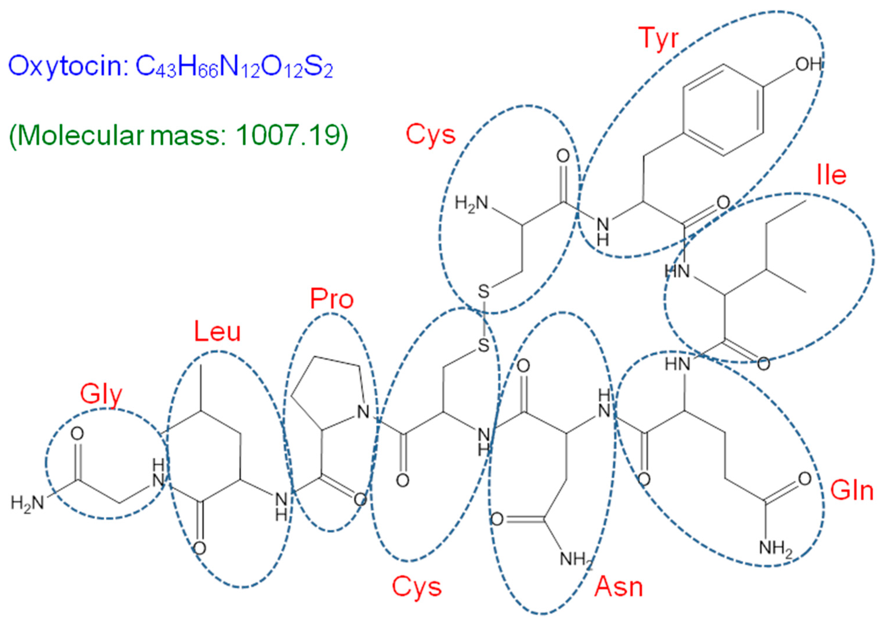
2.1. Reagents and Materials

Oxytocin was purchased from Peptide Institution Inc. (Osaka, Japan) and was dissolved in water to a concentration of 0.1 mg mL−1. The stock solution was stored at 4 °C and diluted to the required concentrations with water prior to use. LC−MS grade methanol and water used as mobile phases were purchased from Kanto Chemical (Tokyo, Japan). All other chemicals were of analytical grade.

2.2. Instruments and Analytical Conditions

The LC system was a Model 1100 series (Agilent Technologies, Boeblingen Germany), consisting of a binary pump, an online-degasser, an autosampler, a column compartment, a diode array detector, and an HP ChemStation. A Zorbax Eclipse XDB-C8 column (50 mm × 2.1 mm, particle size 3.5 µm; Agilent Technologies) was used for LC separation at a column temperature of 30 °C, with a mobile phase consisting of water/methanol (10/90, v/v), at a flow rate of 0.2 mL min−1.
Electrospray MS/MS for oxytocin was performed on an API 4000 triple quadruple mass spectrometer (Applied Biosystems, Foster City, CA, USA), equipped with a turbo ion spray interface operated in positive ion mode at 4500 V and 500 °C. Nitrogen as a nebulizing and drying gas was generated from compressed air using a Kaken N2 generator (System Instruments Co., Ltd., Tokyo, Japan). The ion sources gases 1 (GS1) and 2 (GS2) were set at flow rates of 50 and 80 L min−1, respectively. The curtain gas (CUR) and collision gas (CAD) flows were set at 10 L min−1 and 12 L min−1, respectively. Other set-up parameters included a declustering potential (DP) of 100 V; an entrance potential (EP) of 10 V; a collision energy (CE) of 42 eV; and a collision cell exit potential (CXP) of 22 V. Quantification was performed by multiple reaction monitoring (MRM) of the protonated precursor molecular ions [M+H]+ and the related product ions. Quadrupole Q1 mass (precursor ion) and Q3 mass (product ion) were set at unit resolution. MRM in the positive ionization mode was performed using a dwell time of 500 msec per transition to detect ion pairs m/z 1008.0→723.8 for quantitation and m/z 1008.0→991.0 for confirmation. LC−MS/MS data were processed using Analyst Software 1.3.1 (Applied Biosystems).

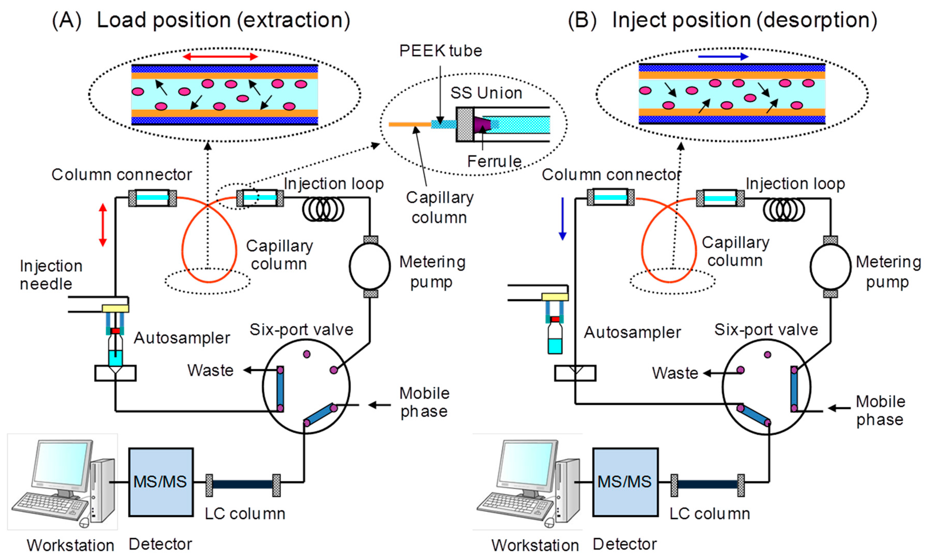
2.3. In-tube Solid-Phase Microextraction

The in-tube SPME device, a GC capillary column (60 cm × 0.32 mm i.d.), was placed between the injection loop and injection needle of the autosampler. The injection loop was retained in the system to avoid fouling of the metering pump. A 2.5-cm sleeve of 1/16-in. polyetheretherketone (PEEK) tubing, with an internal diameter of 330 µm, was placed at each end of the capillary. Standard 1/16-in. stainless steel nuts, ferrules, and connectors were used to complete the connections. The extraction efficiencies of non-coated fused silica (including active surface), CP-Sil 5CB (100% polydimethylsiloxane, film thickness 5 µm), CP-Sil 19CB (14% cyanopropyl phenyl methylsilicone, film thickness 1.2 µm), CP-Wax 52CB (polyethylene glycol, film thickness 1.2 µm) (Varian Inc., Lake Forest, CA, USA), Carboxen 1006 PLOT (Carboxen molecularsives, film thickness 15 µm), and Supel-Q PLOT (divinylbenzene polymer, film thickness 17 µm) (Supelco, Bellefonte, PA, USA) were compared. The autosampler software was programmed to control in-tube SPME extraction, desorption, and injection. For sampling and extraction, 2-mL screw-cap autosampler vials equipped with silicon/PTFE septa were filled with 1 mL of sample for extraction and set onto the sample tray in the autosampler, which had been programmed to control the SPME extraction, desorption and injection steps. In addition, 2-mL autosampler vials with septa, one containing 1.5 mL of methanol and the other containing 1.5 mL of water, were set into the autosampler. The capillary column was washed and conditioned by two repeated draw/eject cycles (40 µL each) of these solvents, and a 50-μL air plug was drawn prior to the extraction step. This air gap was necessary not only to avoid sample mixing but to desorb the analyte from the capillary coating by the mobile phase during the eject step. Oxytocin was extracted onto the capillary coating by 20 repeated draw/eject cycles of 40 µL sample at a flow rate of 250 µL min−1 with the six-port valve in the LOAD position. After extraction, the tip of the injection needle was washed by one draw/eject cycle of 2 µL methanol. The extracted compounds were desorbed from the capillary coating with the mobile phase, transported to the LC column by switching the six-port valve to the INJECT position, and detected with the MS/MS system. During analysis, the SPME capillary was washed and conditioned with the mobile phase for the next extraction. The program for the in-tube SPME process is shown in Figure 2.

2.4. Preparation of Saliva Samples

The protocol of this study was approved by the Ethics Committee of Shujitsu University and the aim of the experiment was explained to each subject, who provided informed consent after confirming that they fully understood the experiment. To assess the salivary secretion of oxytocin, 2 mg mL−1 oxytocin solution was administered by four sprays (containing ca. 1.47 mg of oxytocin) into the nasal cavities of 59 male volunteers. Saliva was collected by rinsing the mouth of each subject with water, followed by the collection of saliva samples in Salisoft tubes containing a polypropylene-polyethylene sponge (Assist, Tokyo, Japan). The tubes were centrifuged at 2500 rpm for 5 min, and the supernatants were centrifuged in tubes containing Amicon Ultra 0.5 mL 10K regenerated cellulose 10,000 molecular weight cut-off centrifugal filter devices (Millipore, Tullagreen, Ireland) at 15,000 × g for 15 min at 20 °C. A 0.2 mL aliquot of each saliva ultrafiltrate was pipetted into a MonoTip C18 (GL Sciences, Tokyo, Japan) and centrifuged at 5000 × g for 2 min at 20 °C. After washing with 0.4 mL distilled water, 0.2 mL methanol was added to this adsorbent, and the adsorbed oxytocin adsorbent was desorbed into a 2.0-mL autosampler vial containing a septum. The methanol was evaporated, the residue was dissolved in 0.5 mL distilled water, and the vial was placed in the autosampler for in-tube SPME LC−MS/MS analysis.
Figure 2. Schematic diagrams of the automated on-line in-tube solid-phase microextraction (SPME)/ liquid chromatography-tandem mass spectrometry (LC–MS/MS) system.
Chromatography 02 00382 g002

3. Results and Discussion

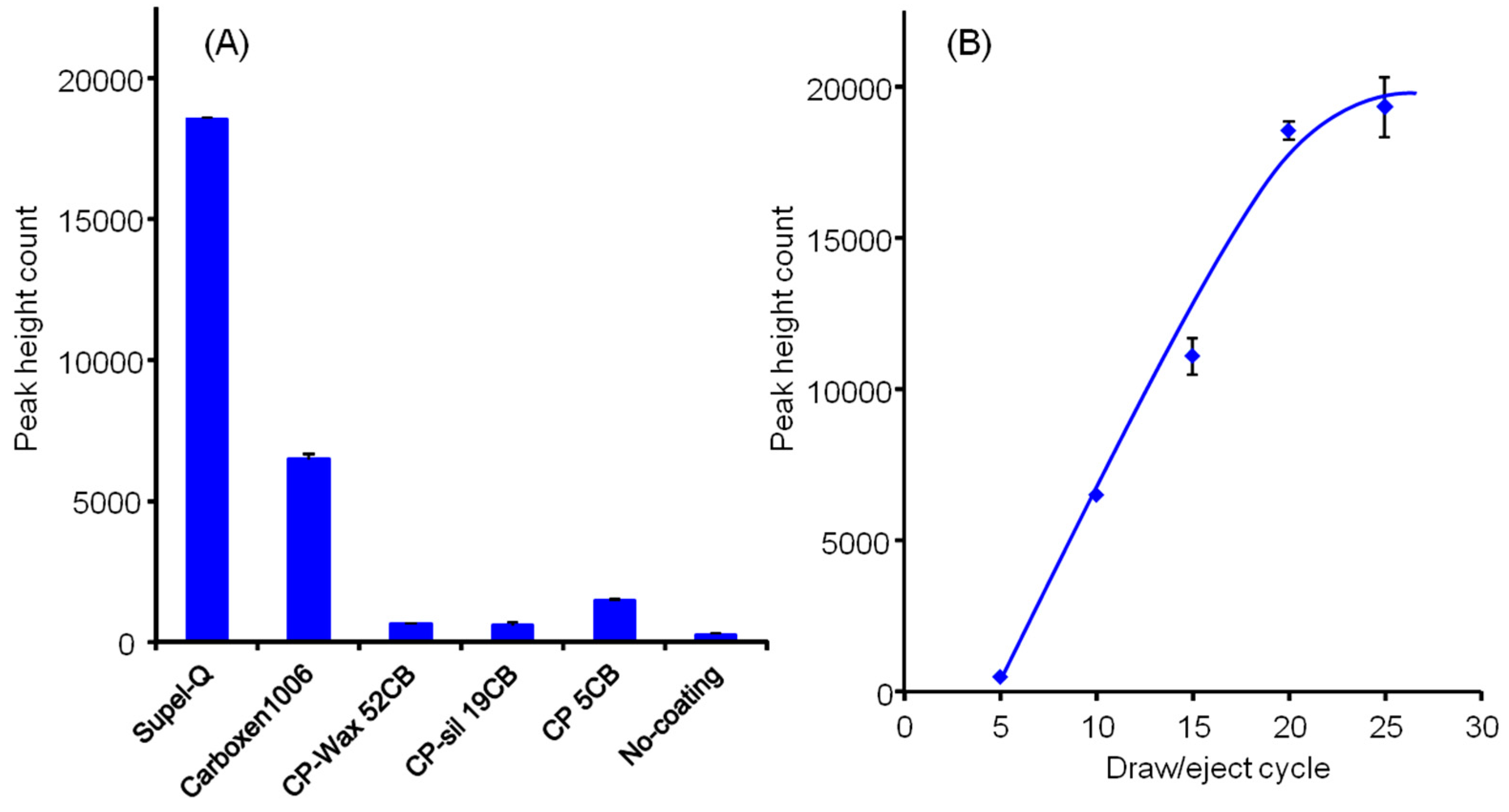
3.1. Optimization of In-tube Solid-phase Microextraction and Desorption

To optimize the extraction of oxytocin by in-tube SPME, several parameters were investigated, including the number and volume of draw/eject cycles and the stationary phase coated onto the inner surface of the in-tube SPME capillary columns. We found that the extraction efficiency of the Supel-Q PLOT column was better than that of the five other columns tested, Carboxen 1006 PLOT, CP-Wax 52CB, CP-Sil 19CB, CP-Sil 5CB, and non-coated fused silica (Figure 3A). As PLOT columns such as Supel-Q and Carboxen 1006 have a larger adsorption surface area and thicker film layer, the amounts extracted were greater for these than for other liquid-phase columns. The large surface area of Supel-Q PLOT (divinylbenzene polymeric material) especially enhanced the adsorption of oxytocin by hydrophobic interaction. Using in-tube SPME, the extraction time and flow rate were proportional to the amounts of compounds extracted. Varying the number of draw/eject cycles from 1 to 25 showed that the extraction equilibrium of these compounds was reached after almost 20 draw/eject cycles of 40 µL of sample (Figure 3B), whereas varying draw/eject rates from 100 to 250 µL min−1 had little effect on extraction efficiency. We selected 250 µL min−1 as the optimal draw/eject flow rate, since extraction took an inconveniently long time below this rate and extraction efficiency tended to decrease above this rate.
Figure 3. Effects of (A) capillary coating and (B) numbers of draw/eject cycles on the in-tube SPME of oxytocin.
Chromatography 02 00382 g003
The mobile phase was found suitable for desorption of oxytocin extracted into the stationary phase of the capillary column. Dynamic desorption of these compounds from the capillary was readily achieved by switching the six-port valve. The desorbed oxytocin was transported to the LC column by flow of the mobile phase. No carryover was observed because the capillary column was washed and conditioned by draw/eject cycles of methanol and mobile-phase prior to extraction. The extraction and desorption of oxytocin by the in-tube SPME LC−MS/MS method were accomplished automatically within 20 min, making possible the automated analysis of about 72 samples per day with overnight operation.

3.2. Linearity, Reproducibility and Detection Limit

To test the linearity of the calibration curve, we analyzed various concentrations of oxytocin (0, 0.2, 0.5, 1.0, 2.0 and 5.0 ng mL−1). Calibration curves constructed by peak height count showed a linear relationship for six-point calibration, with correlation coefficients 0.9981 (n = 18). The RSDs at each point were below 14.7% (n = 3). Oxytocin provided excellent responses in MRM mode detection. Under our LC−MS/MS conditions, the detection limit (S/N = 3) of oxytocin was 4.0 pg mL−1. The sensitivity of the in-tube SPME method was over 58-fold higher than the sensitivity of the direct injection method (10 µL injection), because oxytocin was concentrated in the capillary column during draw/eject cycles.

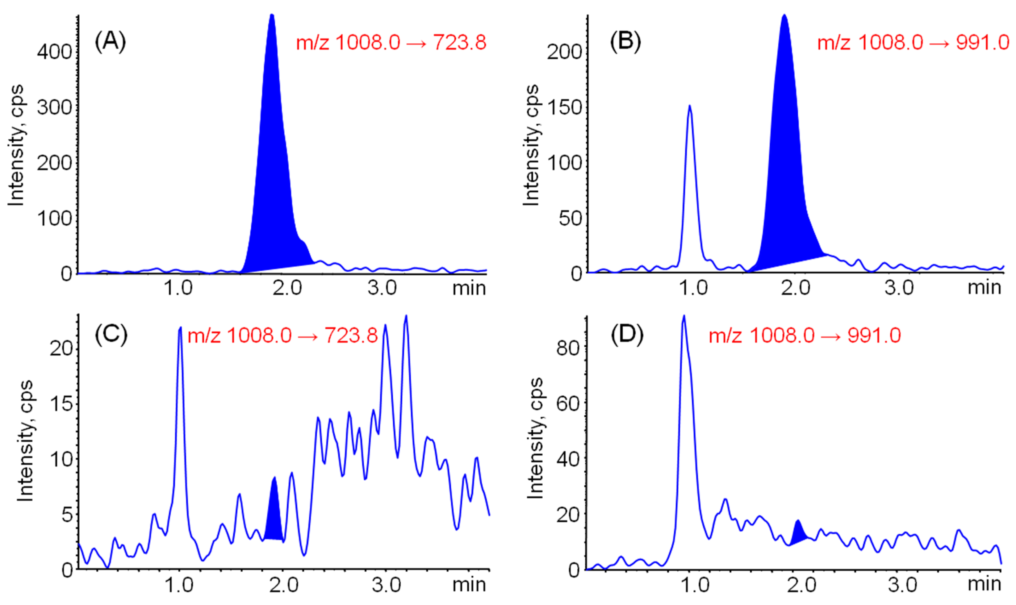
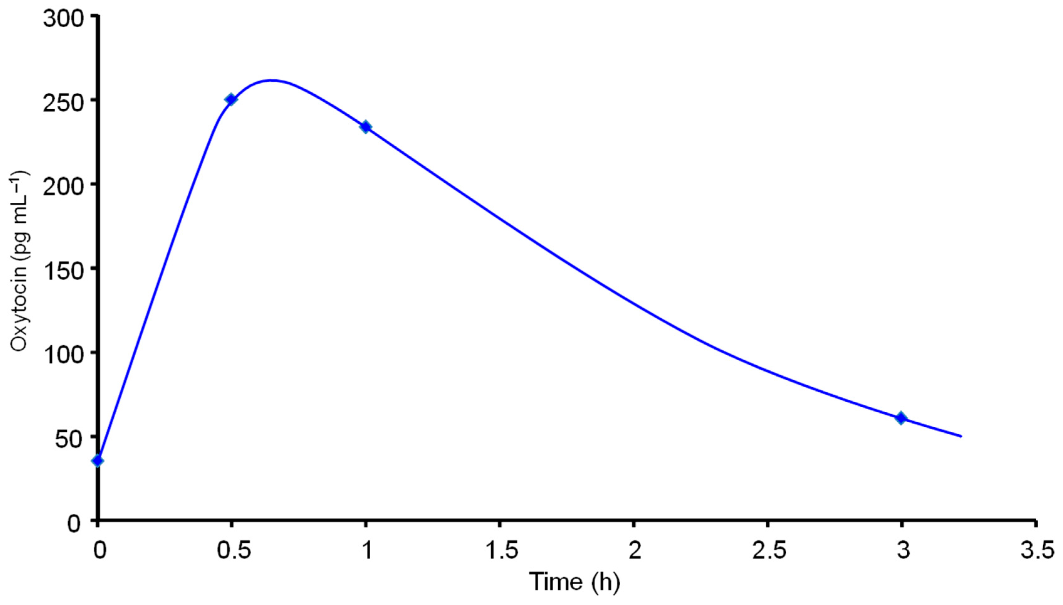
3.3. Application to the Analysis of Saliva Samples

Saliva samples were collected easily into Salisoft tubes containing polypropylene-polyethylene sponges, followed by ultracentrifugation with Amicon Ultra to remove the proteins. To remove salivary interfering substances such as mucin, the filtrate was extracted with MonoTip C18, a monolithic silica adsorbent packed into a micro-tip. The saliva samples were successfully analyzed without interference peaks using the established in-tube SPME LC−MS/MS method with MRM mode detection, and the oxytocin peak was confirmed by detection of another ion pair (Figure 4). The quantification limit (S/N = 10) of oxytocin in a 0.2 mL saliva sample was about 30 pg mL−1. Although the quantification limit of oxytocin in human plasma is reported to be 1 pg mL−1 [26], large plasma samples (1.4 mL) are needed. In contrast, our method could measure trace levels of oxytocin in saliva samples of only 0.2 mL, and it was easier and more rapid. We utilized this method to analyze changes in salivary oxytocin concentrations following intranasal oxytocin administration. We found that oxytocin concentration was maximum at 30 min and was returned to baseline at 3 h (Figure 5). These changes indicate that oxytocin is slightly absorbed by the nasal mucosa.
Figure 4. Typical chromatograms obtained from (A, B) standard oxytocin (0.5 ng m L−1) and (C, D) saliva samples (0.2 mL) by on-line in-tube SPME/LC–MS/MS.
Chromatography 02 00382 g004
Figure 5. Salivary excretion of oxytocin after intranasal oxytocin administration.
Chromatography 02 00382 g005

4. Conclusions

The on-line in-tube SPME LC−MS/MS method described in the present study can be used for the continuous extraction and concentration of oxytocin in saliva samples, allowing their analyses by LC−MS/MS. This method is automated, simple, rapid, selective, and sensitive and can be applied easily to the analyses of small volumes of saliva samples. This method should therefore be very useful for analysis of oxytocin in saliva samples and for objective evaluations of the physiological effects of oxytocin.

Acknowledgments

This work was supported by a Grant-in-Aid for Basic Scientific Research (C, No. 26460053).

Author Contributions

Hiroyuki Kataoka designed the experiments and wrote the paper. Eri Moriyama performed the experiments and analyzed the data under Dr. Kataoka’s supervision.

Conflicts of Interest

The authors have declared no conflict of interest.

References

  1. Ross, H.E.; Young, L.J. Oxytocin and the neural mechanisms regulating social cognition and affiliative behavior. Front. Neuroendocrinol. 2009, 30, 534–547. [Google Scholar] [CrossRef] [PubMed]
  2. Lee, H.J.; Macbeth, A.H.; Pagani, J.H.; Young, W.S., 3rd. Oxytocin: The great facilitator of life. Prog. Neurobiol. 2009, 88, 127–151. [Google Scholar] [CrossRef] [PubMed]
  3. Viero, C.; Shibuya, I.; Kitamura, N.; Verkhratsky, A.; Fujihara, H.; Katoh, A.; Ueta, Y.; Zingg, H.H.; Chvatal, A.; Sykova, E.; et al. Oxytocin: Crossing the bridge between basic science and pharmacotherapy. CNS Neurosci. Ther. 2010, 16, e138–e156. [Google Scholar] [CrossRef] [PubMed]
  4. Galbally, M.; Lewis, A.J.; van Ijzendoorn, M.; Permezel, M. The role of oxytocin in mother-infant relations: A systematic review of human studies. Harv. Rev. Psychiatry 2011, 19, 1–14. [Google Scholar] [CrossRef] [PubMed]
  5. Gordon, I.; Martin, C.; Feldman, R.; Leckman, J.F. Oxytocin and social motivation. Dev. Cogn. Neurosci. 2011, 1, 471–493. [Google Scholar] [CrossRef] [PubMed]
  6. Bisagno, V.; Cadet, J.L. Stress, sex, and addiction: potential roles of corticotropin-releasing factor, oxytocin, and arginine-vasopressin. Behav. Pharmacol. 2014, 25, 445–457. [Google Scholar] [CrossRef] [PubMed]
  7. Vargas-Martínez, F.; Uvnäs-Moberg, K.; Petersson, M.; Olausson, H.A.; Jiménez-Estrada, I. Neuropeptides as neuroprotective agents: Oxytocin a forefront developmental player in the mammalian brain. Prog. Neurobiol. 2014, 123C, 37–78. [Google Scholar] [CrossRef] [PubMed]
  8. Kumsta, R.; Heinrichs, M. Oxytocin, stress and social behavior: neurogenetics of the human oxytocin system. Curr. Opin. Neurobiol. 2013, 23, 11–16. [Google Scholar] [CrossRef] [PubMed]
  9. Arrowsmith, S.; Wray, S. Oxytocin: Its mechanism of action and receptor signalling in the myometrium. J. Neuroendocrinol. 2014, 26, 356–369. [Google Scholar] [CrossRef] [PubMed]
  10. Bartz, J.A.; Hollander, E. Oxytocin and experimental therapeutics in autism spectrum disorders. Prog. Brain Res. 2008, 170, 451–462. [Google Scholar] [PubMed]
  11. Matsuzaki, M.; Matsushita, H.; Tomizawa, K.; Matsui, H. Oxytocin: A therapeutic target for mental disorders. J. Physiol. Sci. 2012, 62, 441–444. [Google Scholar] [CrossRef] [PubMed]
  12. Olff, M.; Frijling, J.L.; Kubzansky, L.D.; Bradley, B.; Ellenbogen, M.A.; Cardoso, C.; Bartz, J.A.; Yee, J.R.; van Zuiden, M. The role of oxytocin in social bonding, stress regulation and mental health: An update on the moderating effects of context and interindividual differences. Psychoneuroendocrinology 2013, 38, 1883–1894. [Google Scholar] [CrossRef] [PubMed]
  13. Cochran, D.M.; Fallon, D.; Hill, M.; Frazier, J.A. The role of oxytocin in psychiatric disorders: a review of biological and therapeutic research findings. Harv. Rev. Psychiatry 2013, 21, 219–247. [Google Scholar] [CrossRef] [PubMed]
  14. Preti, A.; Melis, M.; Siddi, S.; Vellante, M.; Doneddu, G.; Fadda, R. Oxytocin and autism: A systematic review of randomized controlled trials. J. Child Adolesc. Psychopharmacol. 2014, 24, 54–68. [Google Scholar] [CrossRef] [PubMed]
  15. McQuaid, R.J.; McInnis, O.A.; Abizaid, A.; Anisman, H. Making room for oxytocin in understanding depression. Neurosci. Biobehav. Rev. 2014, 45, 305–322. [Google Scholar] [CrossRef] [PubMed]
  16. Koch, S.B.; van Zuiden, M.; Nawijn, L.; Frijling, J.L.; Veltman, D.J.; Olff, M. Intranasal oxytocin as strategy for medication-enhanced psychotherapy of PTSD: Salience processing and fear inhibition processes. Psychoneuroendocrinology 2014, 40, 242–256. [Google Scholar] [CrossRef] [PubMed]
  17. Guastella, A.J.; MacLeod, C. A critical review of the influence of oxytocin nasal spray on social cognition in humans: evidence and future directions. Horm. Behav. 2012, 61, 410–418. [Google Scholar] [CrossRef] [PubMed]
  18. Guastella, A.J.; Hickie, I.B.; McGuinness, M.M.; Otis, M.; Woods, E.A.; Disinger, H.M.; Chan, H.K.; Chen, T.F.; Banati, R.B. Recommendations for the standardisation of oxytocin nasal administration and guidelines for its reporting in human research. Psychoneuroendocrinology 2013, 38, 612–625. [Google Scholar] [CrossRef] [PubMed]
  19. De Berardis, D.; Marini, S.; Iasevoli, F.; Tomasetti, C.; de Bartolomeis, A.; Mazza, M.; Valchera, A.; Fornaro, M.; Cavuto, M.; Srinivasan, V.; et al. The role of intranasal oxytocin in the treatment of patients with schizophrenia: A systematic review. CNS Neurol. Disord. Drug Targets 2013, 12, 252–264. [Google Scholar] [CrossRef] [PubMed]
  20. Vecsernyés, M.; Török, A.; Jójárt, I.; Laczi, F.; Penke, B.; Julesz, J. Specific radioimmunoassay of oxytocin in rat plasma. Endocr. Regul. 1994, 28, 145–150. [Google Scholar] [PubMed]
  21. Szeto, A.; McCabe, P.M.; Nation, D.A.; Tabak, B.A.; Rossetti, M.A.; McCullough, M.E.; Schneiderman, N.; Mendez, A.J. Evaluation of enzyme immunoassay and radioimmunoassay methods for the measurement of plasma oxytocin. Psychosom. Med. 2011, 73, 393–400. [Google Scholar] [CrossRef] [PubMed]
  22. Prakash, B.S.; Metten, M.; Schams, D.; Wuttke, W. Development of a sensitive enzymeimmunoassay for oxytocin determination in bovine plasma. Anim. Reprod. Sci. 1998, 51, 185–194. [Google Scholar] [CrossRef]
  23. Carter, C.S.; Pournajafi-Nazarloo, H.; Kramer, K.M.; Ziegler, T.E.; White-Traut, R.; Bello, D.; Schwertz, D. Oxytocin: Behavioral associations and potential as a salivary biomarker. Ann. NY Acad. Sci. 2007, 1098, 312–322. [Google Scholar] [CrossRef] [PubMed]
  24. Mishra, M.; Ali, S.; Das, M. Analysis of oxytocin in milk samples and intake pattern in different age groups of Indian population. Toxicol. Mech. Methods 2014, 24, 342–346. [Google Scholar] [CrossRef] [PubMed]
  25. Kukucka, M.A.; Misra, H.P. Determination of oxytocin in biological samples by isocratic high-performance liquid chromatography with coulometric detection using C18 solid-phase extraction and polyclonal antibody-based immunoaffinity column purification. J. Chromatogr. B 1994, 653, 139–145. [Google Scholar] [CrossRef]
  26. Zhang, G.; Zhang, Y.; Fast, D.M.; Lin, Z.; Steenwyk, R. Ultra sensitive quantitation of endogenous oxytocin in rat and human plasma using a two-dimensional liquid chromatography-tandem mass spectrometry assay. Anal. Biochem. 2011, 416, 45–52. [Google Scholar] [CrossRef] [PubMed]
  27. Mabrouk, O.S.; Kennedy, R.T. Simultaneous oxytocin and arg-vasopressin measurements in microdialysates using capillary liquid chromatography-mass spectrometry. J. Neurosci. Methods 2012, 209, 127–133. [Google Scholar] [CrossRef] [PubMed]
  28. Kataoka, H.; Inoue, R.; Yagi, K.; Saito, K. Determination of nicotine, cotinine, and related alkaloids in human urine and saliva by automated in-tube solid-phase microextraction coupled with liquid chromatography-mass spectrometry. J. Pharm. Biomed. Anal. 2009, 49, 108–114. [Google Scholar] [CrossRef] [PubMed]
  29. Kataoka, H.; Ehara, K.; Yasuhara, R.; Saito, K. Simultaneous determination of testosterone, cortisol and dehydroepiandrosterone in saliva by stable isotope dilution on-line in-tube solid-phase microextraction coupled with liquid chromatography−tandem mass spectrometry. Anal. Bioanal. Chem. 2013, 405, 331–340. [Google Scholar] [CrossRef] [PubMed]
  30. Kataoka, H. Automated sample preparation using in-tube solid-phase microextraction and its application—A review. Anal. Bioanal. Chem. 2002, 373, 31–45. [Google Scholar] [CrossRef] [PubMed]
  31. Kataoka, H.; Ishizaki, A.; Nonaka, Y.; Saito, K. Developments and applications of capillary microextraction techniques: A review. Anal. Chim. Acta 2009, 655, 8–29. [Google Scholar] [CrossRef] [PubMed]
  32. Kataoka, H. Recent developments and applications of microextraction techniques in drug analysis. Anal. Bioanal. Chem. 2010, 396, 339–364. [Google Scholar] [CrossRef] [PubMed]
  33. Kataoka, H.; Saito, K. Recent advances in SPME techniques in biomedical analysis. J. Pharm. Biomed. Anal. 2011, 54, 926–950. [Google Scholar] [CrossRef] [PubMed]

