Article

Does a Graf Type-I Hip Justify the Discontinuation of Pavlik Harness Treatment in Patients with Developmental Dislocation of the Hip?

1
Department of Pediatric Orthopaedics, Guangzhou Women and Children’s Medical Center, Guangzhou Medical University, Guangzhou 510623, China
2
Department of Pediatric Surgery and Orthopedics, Jeanne de Flandre Hospital, Lille University Center, 59800 Lille, France
*
Author to whom correspondence should be addressed.
Children 2022, 9(5), 752; https://doi.org/10.3390/children9050752
Submission received: 30 March 2022 / Revised: 18 May 2022 / Accepted: 19 May 2022 / Published: 20 May 2022
(This article belongs to the Special Issue Bone Development and Disease in Infants (Volume II))

Abstract

:
Background: To analyze the clinical data of patients aged < 6 months with developmental dislocation of the hip (DDH) treated with Pavlik Harness (PH) in order to identify the best time to terminate PH treatment. Method: Fifty-four patients (47 females, 7 males; 63 hips) met the inclusion criteria and were included in the study; there were 33 (61.1%) left, 12 (22.2%) right and 9 (16.7%) bilateral DDH. The mean age at diagnosis was 11.8 ± 5.9 weeks (range, 1.4–25.5). All patients underwent fulltime PH treatment for about three months. At completion of PH treatment, patients were then divided into Group A, including patients with clinically stable hip joint and Graf type-I hip on ultrasound (US), and Group B, including patients with clinically stable hip joint and well-reduced hip on anterior-posterior (AP) radiographs without acetabular dysplasia. Six months after completion of PH treatment, the presence/absence of residual acetabular dysplasia (RAD) was evaluated on AP pelvis radiographs. The t-test and chi-square test were used to compare the differences in age, gender, side, Graf classification and RAD rate between the two groups of patients. Results: At completion of PH treatment, 45 hips were in Group A and 18 in Group B. There were no significant differences in age, gender, side, preoperative alpha angle and Graf classification between the two groups. Six months after discontinuation of PH, the AI in Group A (27.1° ± 6.8°) was significantly higher than that in Group B (21.9° ± 3.5°; p = 0.001); moreover 23 hips (51.1%) in Group A developed RAD compared to one hip in Group B (5.6%; p = 0.001). Among Group A patients, those with RAD were significantly older (13.7 ± 4.9 weeks) than those with normal hips (7.6 ± 3.8 weeks; p < 0.001); the incidence of RAD was significantly lower in patients with Graf type-II D hips (22.2%) than in patients with Graf type-III (70%) and type-IV hips (71.4%; p = 0.006). However, logistic regression analysis identified age as the only risk factor for RAD. All 24 hips with RAD (24/63, 38.1%) were treated with abduction braces. At final follow-up, AI in Group A (20.5° ± 3.3°) was not significantly different from that in Group B (21.9° ± 3.3°; p = 0.132). At the last follow-up visit, five hips (11.1%) in Group A still had RAD, compared to none in Group B (p = 0.31). Conclusions: In patients with DDH treated by PH, Graf type-I on US is not an absolute timing to terminate PH treatment. In addition, patients ≥ 13 weeks had a high risk of RAD despite PH treatment as 51.1% of infants developed RAD during follow up. Follow-up radiographs should be requested in all patients achieving Graf type-I hips at completion of PH treatment.

1. Introduction

Developmental dislocation of the hip (DDH) is a fairly common condition in children, and successful treatment is based on early, gentle and stable hip reduction to promote the development of the acetabulum and femoral epiphysis and to avoid the onset of avascular necrosis (AVN) [1,2].
Pavlik Harness (PH) treatment is indicated in children < 6 months of age with confirmed diagnosis of DDH; the device maintains the hips at no more than 75° of abduction and 90° of flexion [2]. Generally, PH treatment is recommended for patients with Graf type IIb to Graf type IV hips, and it has very variable success rates depending on the severity of DDH, as assessed by ultrasound (US) [2,3]. In particular, the reported overall short-, mid- and long-term success rates of PH treatment range from 45% to 100% [4,5,6]. However, in patients with dislocated hips, the success rate decreases significantly, especially in Graf type IV hips [7]. Complications such as failure of reduction, femoral nerve palsy, AVN and residual acetabular dysplasia (RAD) have been reported by several authors [3,4,8].
Currently, most caregivers consider PH treatment should be discontinued when hips return to Graf type-I on US. In general, the duration of PH treatment is three months [9,10]. In clinical practice, however, we encountered some patients with Graf-I hip at the end of PH treatment and acetabular dysplasia three to six months after the end of treatment. In particular, previous studies have also reported that some patients develop RAD following successful PH treatment [11,12].
In this study, we retrospectively analyzed the clinical data of patients aged < 6 months of age with DDH treated with PH in order to identify the best time to terminate PH treatment. Specifically, we wanted to answer the following question: is a Graf-I hip an absolute indication for immediate discontinuation of PH treatment?

2. Materials and Methods

The medical records of children with DDH who were aged < 6 months of age at the beginning of PH treatment during the period 2014–2019 were collected and retrospectively reviewed.
The following inclusion criteria were applied: (1) diagnosis of DDH (radiographic evidence of dislocation, or US suggestive of Graf type-D, III, or IV [13]); (2) patients < 6 months of age at the beginning of PH treatment; (3) hip meeting the US or radiographic criteria for normalization upon completion of PH treatment; (4) complete clinical and radiographic data.
The exclusion criteria were: (1) failure of treatment by PH; (2) follow-up time < 12 months and/or insufficient radiographic data; (3) abnormal hip at the end of PH treatment as per US or radiographic criteria; (4) presence of underlying pathology such as cerebral palsy, spinal cord thrombosis, myelomeningocele, joint contracture and other neuromuscular diseases.
Fifty-four patients (47 females, 7 males; 63 hips) met the inclusion criteria and were included in the study; there were 33 (61.1%) left, 12 (22.2%) right and 9 (16.7%) bilateral DDH.
The mean age at diagnosis was 11.8 ± 5.9 weeks (range, 1.4–25.5). All patients underwent fulltime PH treatment for about 3 months (12 weeks); treatment started the same day of the diagnosis. The patients were evaluated once a week in the first 3 to 4 weeks to check if the hip reduced and the wearing of the PH.

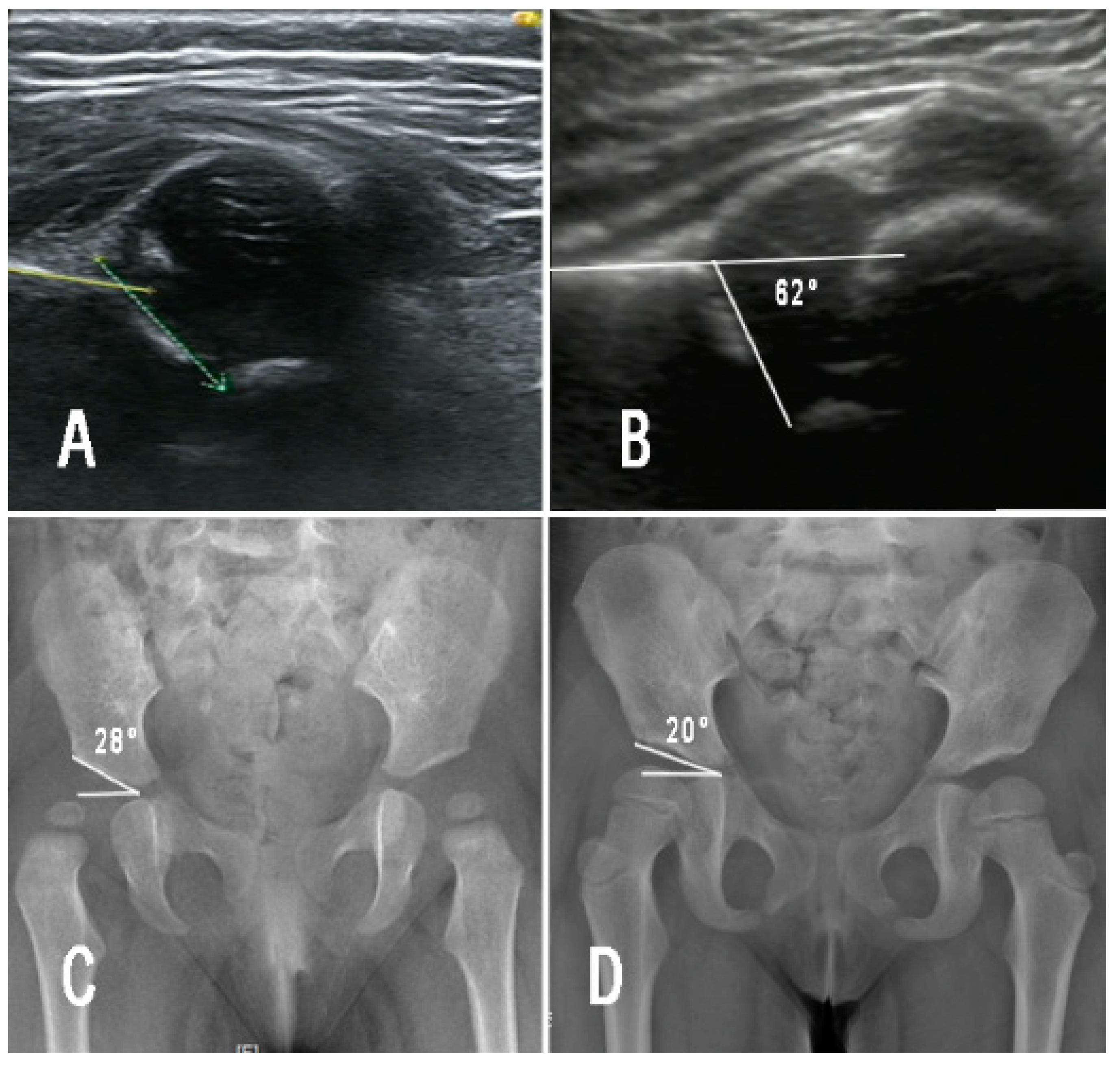
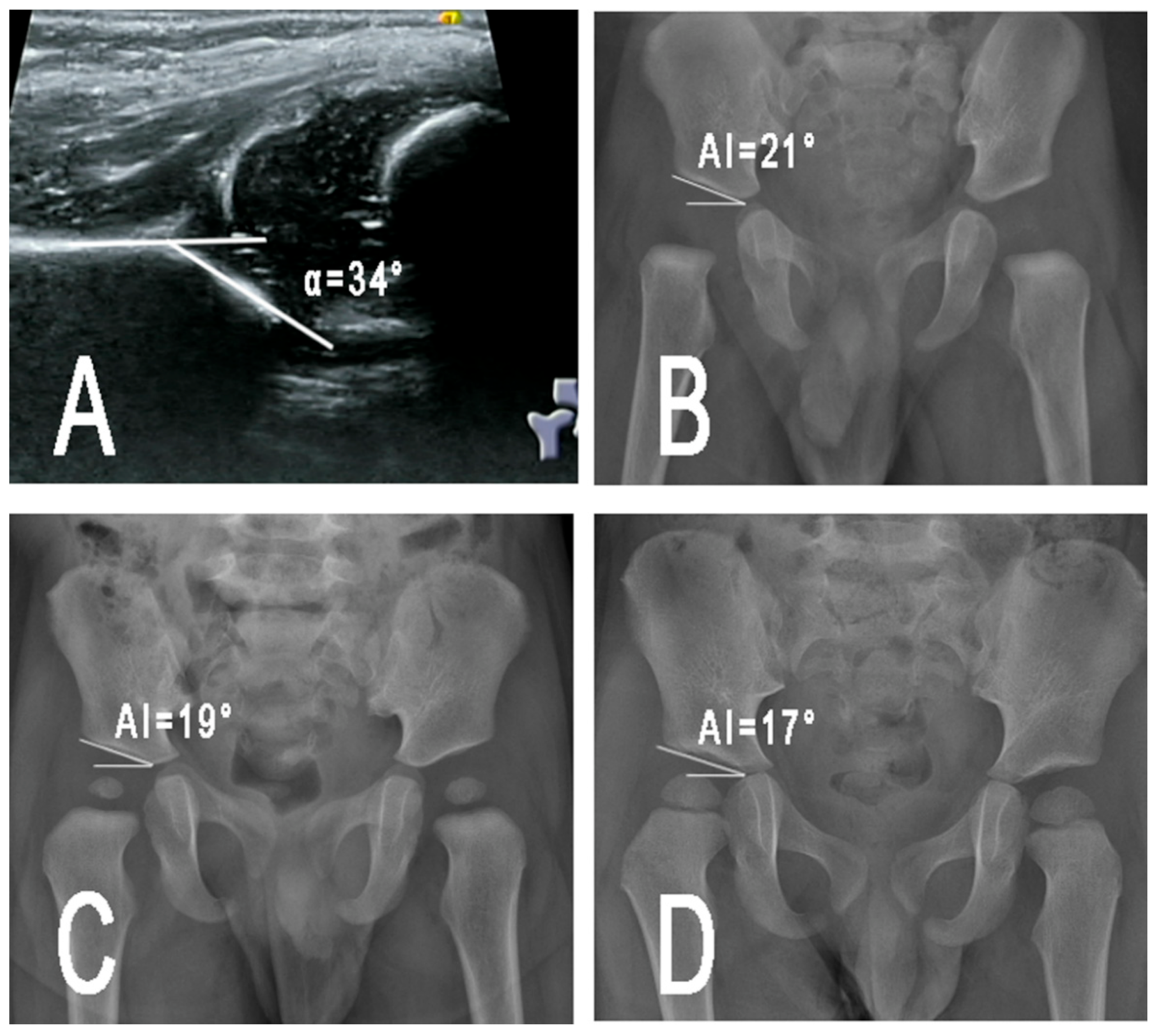
2.1. Ultrasonography and Radiographic Assessment

Hip US was performed according to the standard method described by Graf [14]; all hip US examinations and classification of the hip were performed by pediatric radiologists with at least 5 years of experience. They all received standard training of hip US-course.
Hip US examination was performed before PH treatment, and during PH treatment at 3 to 4 week intervals, and at completion of PH treatment. The alpha angle of the hip was recorded for each hip US examination. In patients with successful hip reduction and normal hip anatomy, PH treatment was discontinued after 3 months, while in those with reduced hip and RAD, treatment was switched to an abduction brace treatment. Patients with failed PH treatment requiring closed reduction and spica cast immobilization and those with RAD at the end of PH treatment were excluded from the analysis.
Hip US or pelvis AP pelvis radiographs (supine position; non-weight bearing) were performed at the end of PH treatment in order to determine whether the hip achieved normal morphology. Forty-five hips (71.4%) underwent US examination to evaluate hip morphology; for those not able to attend US or those > 6 months of age at completion of PH treatment (n = 18; 28.6%), AP pelvis radiographs were performed.
Six months after completion of PH treatment, all patients underwent regular anterior-posterior (AP) pelvis radiographs to evaluate Acetabular Index (AI) during follow up; in addition, center edge angle of Wiberg (CEA) was measured on AP radiographs of the pelvis in patients older than 4 years of age [15]. AP pelvis radiographs were also used to evaluate the presence/absence of RAD. RAD was diagnosed if: (1) AI in patients younger than 4 years was greater than one standard deviation than the mean value for normal age matched children [16], or there was evidence of subluxation; (2) patients older than 4 years with Severin grade III or IV hips [17].
At final follow-up, AVN was assessed and graded according to the Kalamchi and MacEwen method on AP pelvis radiographs [18]. Since type I AVN is a temporary ischemic change that can be completely recovered, we considered type I AVN as normal [19].
Patients were then divided into two groups, according to the type of examination (hip US or AP radiograph of the pelvis), and hip morphology: Group A included patients with clinically stable hip joint and Graf type-I hip on US while Group B included those with clinically stable hip joint and well-reduced hip on plain radiographs with AI no more than one standard deviation above the mean value for normal age matched children [16].
Two pediatric orthopedic surgeons (LYQ and LYH) separately reviewed all AP pelvis radiographs on the Picture Archiving and Communication System of our Institution and the mean value of each measurement was used for statistical analysis.

2.2. Statistical Analysis

Statistical analysis was performed using SPSS 22.0 (SPSS, Chicago, IL, USA). Data included continuous numerical variables, frequencies and percentages. The t-test and chi-square test were used to compare the differences between the indicators of groups A and B. Logistic regression analysis (LRA) was performed to identify potential risk factors for RAD six months after completion of PH treatment. p < 0.05 was considered statistically significant.

3. Results

Of the 63 hips (54 patients), 21 (33.3%) were type-II D, 26 (41.3%) were type-III and 10 (15.9%) were type-IV according to Graf classification; the remaining 6 hips (9.5%) did not undergo pretreatment US examination, and AP pelvis radiographs revealed fully dislocated hips.
Mean age at diagnosis was 11.8 ± 5.9 weeks (range, 1.4–25.5), the mean duration of PH treatment was 3.2 ± 0.7 months (range, 2–6) and mean follow-up time was 24.4 ± 14.1 months (range, 12–61.5).
At completion of PH treatment, 45 hips (71.4%) were included in Group A and 18 hips (28.6%) in Group B.
In Group A patients, mean age at diagnosis was 10.7 ± 5.4 weeks, while it was 13.7 ± 7.0 weeks in Group B (p = 0.078). There were no significant differences in sex, laterality, preoperative alpha angle, Graf type or follow-up time between the two groups of patients (Table 1).
Six months after completion of PH treatment, AI of Group A patients (27.1° ± 6.8°) was significantly higher than that of Group B (21.9° ± 3.5°; p < 0.001). Twenty-three hips (51.1%) of Group A patients had RAD at six months after discontinuation of PH, which was significantly higher than that of patients in Group B (n = 1, 5.6%; p = 0.001) (Table 1). All 24 hips with RAD (24/63, 38.1%) were treated with abduction braces.
Among Group A patients, those with RAD were significantly older (13.7 ± 4.9 weeks) than those with normal hips (7.6 ± 3.8 weeks; p < 0.001); the incidence of RAD was significantly lower in patients with Graf type-II D (23.5%) than in those with type-III (70%) and type-IV hips (71.4%; p = 0.006) (Table 2). However, LRA found age as the only risk factor for RAD (Table 3).
SE, standard error; RR, relative risk; CI, confidence interval.
At last follow-up visit, the AI in Group A patients (20.5° ± 3.3°) was comparable to that of Group B patients (21.9° ± 3.3°; p = 0.132). However, at last follow-up visit five hips (11.1%) in Group A still had RAD, while no case of RAD was identified among Group B patients; although no statistically significant difference between the two groups could be identified (p = 0.31) (Table 1).

4. Discussion

Currently, there is no consensus on the duration of PH treatment in patients with DDH [7]. Weinstein et al. [15] suggested that the duration of PH treatment should be twice the patient’s age in months. Westacott et al. [7] studied the experience of 111 members of the British Society for Children’s Orthopaedic Surgery (BSCOS) in treating DDH in patients < 6 months. They found the majority of BSCOS members used PH for the treatment of DDH and the duration of treatment ranged between 6 and 16 weeks (mostly 12 weeks). Another survey by Kelley et al. [6] confirmed most pediatric orthopedic surgeons believe that PH treatment should last three months. However, this is only the personal opinion of the individual surgeon and there is no reliable evidence to show whether a PH treatment time of three months is sufficient to normalize the hip joint. The results of the present study showed that for patients with hip dislocation, an average of three months of PH treatment proved to be insufficient, as 51.1% of patients in Group A developed RAD even though all of them had achieved normal hip US and needed further abduction braces treatment. Dornacher et al. [12] examined 90 patients with DDH treated with PH, although US normalization of the pathological hip was achieved prior to discontinuation of PH treatment, 32.9% of hips developed RAD on radiographs taken once ambulation was acquired. Harris et al. [17] retrospectively examined 720 dislocated or subluxated hips in 550 patients treated with PH, and found that 9% of these had acetabular dysplasia at the end of PH treatment. Thus, in more severe forms of DDH, particularly Graf types III and IV, three months of PH treatment is not sufficient and longer treatment or a switch to an abduction brace is necessary to restore a normal hip morphology.
This study also showed that achieving a Graf-I hip is not an absolute indication to stop PH treatment (Figure 1). Currently, most pediatric orthopedic surgeons usually use US examination to determine the timing of cessation of PH treatment. In the survey by Westacott et al. [7] described above, 74% of surgeons used only US examination to assess the timing of discontinuation of PH treatment. Kelley et al. [6] surveyed pediatric orthopedic surgeons from several countries and found similar results. In our study, 45 hips with DDH achieved a Graf-I hip at the end of PH treatment, but 23 hips (51.1%) still developed acetabular dysplasia during subsequent follow-up. Similarly, Sarkissian et al. [11] retrospectively analyzed 115 patients with DDH who were treated with PH and whose hips reverted to Graf-I. Of these, 33% developed significant acetabular dysplasia on radiographs taken at one year of age. The studies by Bradley et al. [18] and Dornacher et al. [12] also showed that 16.7% to 29.4% of patients still had acetabular dysplasia at one year of age, even though US found normalization of the hip at the time of discontinuation of PH treatment.
Our data suggest that a Graf type-I hip on US does not guarantee that the hip will remain normal on plain radiographs. The reason for this may be the relatively low reproducibility of US. Despite the fact that the hip US technique developed by Graf involves rigorous examination procedures, including probe selection, patient positioning, US section selection, angle measurement and precise classification [14,19], there are still large intra- and inter-examiner discrepancies. The results of the systematic review and meta-analysis by Quader et al. [20], (n = 28 studies) suggested high variability and low agreement in all reported dysplasia metrics. Roovers et al. [21], asked five sonographers to classify 200 US images according to Graf’s method and found that the inter-observer concordance for accurate classification was k = 0.47, whereas the concordance for judging whether it was a Graf type-I was not high (k = 0.65). In contrast, radiographic examination of the hip joint has high reliability, and most studies have shown good concordance (ICC and k > 0.8) for AI and CEA measurements on AP radiographs of the pelvis [15,22]. Consequently, once treatment with PH has ended, radiographic examination should be performed to assess whether the hip joint is indeed normal. If this is not the case, it is necessary to continue treatment with PH or to switch to an abduction brace.
This study also showed that the incidence of RAD after achieving US normalization of the hip (Graf-I) was significantly increased in patients ≥13 weeks at the time of treatment. Even though LRA failed to identify Graf type as a risk factor of RAD, among patients with Graf-III or IV hips, 70% to 71.4% still had signs of RAD 6 months after PH removal in Group A. Previous studies have also shown that age and Graf classification are important factors influencing the outcome of PH treatment. Novais et al. [23] studied 84 patients (134 hips) with DDH treated with PH and reported that 11.8% of these patients still had signs of RAD on AP pelvis radiographs taken at one year of age; they also noted Graf-IV hips had significantly higher incidence of RAD. Ömeroğlu et al. [24] retrospectively analyzed 130 patients (181 hips) with DDH treated with PH, and the results showed that 93% of patients < 3 months of age at the beginning of treatment evolved towards a Graf-I hip compared to only 37% of patients older than 5 months of age. Therefore, for patients ≥13 weeks at diagnosis, extra care should be taken because they are at high risk for RAD despite treatment with PH. Further studies should be performed to identify the correlation between RAD and Graf classification.
In our opinion, the variability in US results should not only be attributed to the skills of the examiners but also to the imaging examination itself: radiographs are easier to be reliably performed than US examination. Thus, patients with DDH treated by PH should undergo hip radiographic examination at the completion of PH treatment. PH should be terminated when radiographs are normal (Figure 2). In case ossific nucleus of the femoral head is not present, hip radiographs should be performed after four months of age, as suggested by the clinical guidelines of the American Academy of Orthopaedic Surgeons (AAOS) for management of pediatric developmental dysplasia of the hip in infants [25]. It is possible that when both ultrasound and radiograph are normal, PH can be safely discontinued, so as to decrease the occurrence of RAD.

5. Conclusions

In conclusion, in patients with DDH treated by PH, Graf type-I on US is not an absolute timing to terminate PH treatment. In addition, patients ≥13 weeks have a high risk of RAD despite PH treatment as 51.1% of infants developed RAD during follow up. If the US-results cannot be safely relied upon, follow-up radiographs should be requested at the completion of PH treatment.

Author Contributions

Conceptualization, methodology, supervision and writing (review and editing): Y.L. (Yiqiang Li), F.C., H.X. Original draft: Y.L. (Yiqiang Li). Data collection and editing the manuscript: Y.L.(Yanhan Liu), J.W., J.L., Z.Y., Q.Z., Y.L. (Yuanzhong Liu), W.C. Formal Data Analysis: Y.L. (Yiqiang Li), F.C. All authors have read and agreed to the published version of the manuscript.

Funding

This work was supported by funding from Guangzhou Women and Children’s Medical Center/Guangzhou Institute of Pediatrics (Grant No. GWCMC2020-6-005).

Institutional Review Board Statement

This study was approved by the Ethics Committee of Guangzhou Women and Children’s Medical Center (No: 2021-129A01).

Informed Consent Statement

Patient consent was waived because this is a retrospective observation cohort study without identified information of any patient.

Data Availability Statement

Data are available from the corresponding author.

Conflicts of Interest

The authors declare that they have no conflict interest. No benefits in any form have been received or will be received from a commercial party related directly or indirectly to the subject of this article.

References

  1. Kotlarsky, P.; Haber, R.; Bialik, V.; Eidelman, M. Developmental dysplasia of the hip: What has changed in the last 20 years? World J. Orthop. 2015, 6, 886–901. [Google Scholar] [CrossRef]
  2. Vaquero-Picado, A.; González-Morán, G.; Garay, E.G.; Moraleda, L. Developmental dysplasia of the hip: Update of management. EFORT Open Rev. 2019, 4, 548–556. [Google Scholar] [CrossRef] [PubMed]
  3. Swarup, I.; Penny, C.L.; Dodwell, E.R. Developmental dysplasia of the hip: An update on diagnosis and management from birth to 6 months. Curr. Opin. Pediatr. 2018, 30, 84–92. [Google Scholar] [CrossRef] [PubMed]
  4. Ömeroglu, H. Treatment of developmental dysplasia of the hip with the Pavlik harness in children under six months of age: Indications, results and failures. J. Child. Orthop. 2018, 12, 308–316. [Google Scholar] [CrossRef] [PubMed]
  5. Hines, A.C.; Neal, D.C.; Beckwith, T.; Jo, C.; Kim, H.K.W. A Comparison of Pavlik Harness Treatment Regimens for Dislocated But Reducible (Ortolani+) Hips in Infantile Developmental Dysplasia of the Hip. J. Pediatr. Orthop. 2019, 39, 505–509. [Google Scholar] [CrossRef]
  6. Imerci, A.; Rogers, K.J.; Bhattacharjee, A.; Bowen, J.R.; Thacker, M.M. Risk Factors for Failure of Pavlik Harness Treatment in Infants With Dislocated Hips That Are Evaluated by Dynamic Sonography. J. Pediatr. Orthop. 2021, 41, e386–e391. [Google Scholar] [CrossRef]
  7. Novais, E.N.; Sanders, J.; Kestel, L.A.; Carry, P.M.; Meyers, M.L. Graf Type-IV Hips Have a Higher Risk of Residual Acetabular Dysplasia at 1 Year of Age Following Successful Pavlik Harness Treatment for Developmental Hip Dysplasia. J. Pediatr. Orthop. 2018, 38, 498–502. [Google Scholar] [CrossRef]
  8. Shaw, K.A.; Moreland, C.M.; Olszewski, D.; Schrader, T. Late acetabular dysplasia after successful treatment for developmental dysplasia of the hip using the Pavlik method: A systematic literature review. J. Orthop. 2019, 16, 5–10. [Google Scholar] [CrossRef]
  9. Kelley, S.P.; Feeney, M.M.; Maddock, C.L.; Murnaghan, M.L.; Bradley, C.S. Expert-Based Consensus on the Principles of Pavlik Harness Management of Developmental Dysplasia of the Hip. JBJS Open Access 2019, 4, e0054. [Google Scholar] [CrossRef]
  10. Westacott, D.J.; Perry, D.C. The treatment of neonatal hip dysplasia with splints in the United Kingdom: Time for consensus? J. Child. Orthop. 2020, 14, 112–117. [Google Scholar] [CrossRef]
  11. Sarkissian, E.J.; Sankar, W.N.; Zhu, X.; Wu, C.H.; Flynn, J.M. Radiographic Follow-up of DDH in Infants: Are X-rays Necessary After a Normalized Ultrasound? J. Pediatr. Orthop. 2015, 35, 551–555. [Google Scholar] [CrossRef]
  12. Dornacher, D.; Cakir, B.; Reichel, H.; Nelitz, M. Early radiological outcome of ultrasound monitoring in infants with developmental dysplasia of the hips. J. Pediatr. Orthop. B 2010, 19, 27–31. [Google Scholar] [CrossRef]
  13. Graf, R. Advantages and disadvantages of various access routes in sonographic diagnosis of dysplasia and luxation in the infant hip. J. Pediatr. Orthop. B 1997, 6, 248–252. [Google Scholar] [CrossRef]
  14. Graf, R. Hip sonography: Background; technique and common mistakes; results; debate and politics; challenges. Hip Int. 2017, 27, 215–219. [Google Scholar] [CrossRef]
  15. Li, Y.; Guo, Y.; Li, M.; Zhou, Q.; Liu, Y.; Chen, W.; Li, J.; Canavese, F.; Xu, H. Acetabular index is the best predictor of late residual acetabular dysplasia after closed reduction in developmental dysplasia of the hip. Int. Orthop. 2018, 42, 631–640. [Google Scholar] [CrossRef]
  16. Shi, Y.; Liu, T.; Zhao, Q.; Zhang, L.; Ji, S. The measurements of normal acetabular index and Sharp acetabular angle in Chinese hips. Chin. J. Orthop. 2010, 30, 748–753. [Google Scholar] [CrossRef]
  17. Harris, I.E.; Dickens, R.; Menelaus, M.B. Use of the Pavlik harness for hip displacements. When to abandon treatment. Clin. Orthop. Relat. Res. 1992, 281, 29–33. [Google Scholar] [CrossRef]
  18. Bradley, J.; Wetherill, M.; Benson, M.K. Splintage for congenital dislocation of the hip. Is it safe and reliable? J. Bone Jt. Surg. Br. 1987, 69, 257–263. [Google Scholar] [CrossRef] [Green Version]
  19. Kolb, A.; Benca, E.; Willegger, M.; Puchner, S.E.; Windhager, R.; Chiari, C. Measurement considerations on examiner-dependent factors in the ultrasound assessment of developmental dysplasia of the hip. Int. Orthop. 2017, 41, 1245–1250. [Google Scholar] [CrossRef]
  20. Quader, N.; Schaeffer, E.K.; Hodgson, A.J.; Abugharbieh, R.; Mulpuri, K. A Systematic Review and Meta-analysis on the Reproducibility of Ultrasound-based Metrics for Assessing Developmental Dysplasia of the Hip. J. Pediatr. Orthop. 2018, 38, e305–e311. [Google Scholar] [CrossRef]
  21. Roovers, E.A.; Boere-Boonekamp, M.M.; Geertsma, T.S.; Zielhuis, G.A.; Kerkhoff, A.H. Ultrasonographic screening for developmental dysplasia of the hip in infants. Reproducibility of assessments made by radiographers. J. Bone Jt. Surg. Br. 2003, 85, 726–730. [Google Scholar] [CrossRef] [Green Version]
  22. Shi, Y.Y.; Liu, T.J.; Zhao, Q.; Zhang, L.J.; Ji, S.J.; Wang, E.B. The normal centre-edge angle of Wiberg in the Chinese population: A population-based cross-sectional study. J. Bone Jt. Surg. Br. 2010, 92, 1144–1147. [Google Scholar] [CrossRef] [Green Version]
  23. Novais, E.N.; Kestel, L.A.; Carry, P.M.; Meyers, M.L. Higher Pavlik Harness Treatment Failure Is Seen in Graf Type IV Ortolani-positive Hips in Males. Clin. Orthop. Relat. Res. 2016, 474, 1847–1854. [Google Scholar] [CrossRef] [Green Version]
  24. Ömeroğlu, H.; Köse, N.; Akceylan, A. Success of Pavlik Harness Treatment Decreases in Patients ≥ 4 Months and in Ultrasonographically Dislocated Hips in Developmental Dysplasia of the Hip. Clin. Orthop. Relat. Res. 2016, 474, 1146–1152. [Google Scholar] [CrossRef] [Green Version]
  25. Mulpuri, K.; Song, K.M.; Gross, R.H.; Tebor, G.B.; Otsuka, N.Y.; Lubicky, J.P.; Szalay, E.A.; Harcke, H.T.; Zehr, B.; Spooner, A.; et al. The American Academy of Orthopaedic Surgeons Evidence-Based Guideline on Detection and Nonoperative Management of Pediatric Developmental Dysplasia of the Hip in Infants up to Six Months of Age. J. Bone Jt. Surg. Am. 2015, 97, 1717–1718. [Google Scholar] [CrossRef]
Figure 1. Female patient aged 3 months with right DDH (Graf type IV) (A), treated by PH for three months and achieved Graf type I (B), then the PH treatment was terminated. However, radiographic examination showed significant subluxation on the right hip 6 months later (C), and then the patient was treated by a night time abduction brace. At final follow-up (4 years old), the hip recovered to normal (D).
Children 09 00752 g001
Figure 2. A patient aged 4 months with right DDH (Graf type III) (A), treated by PH for 4 months and recovered to normal radiograph (B). Six months later, radiographic examination showed good development of the hip (C). At final follow-up (2 years old), the outcome is satisfactory (D).
Children 09 00752 g002
Table 1. Comparison of clinical data between patients achieving normal hips on hip US (Group A) and pelvis radiographs (Group B) at the end of PH treatment.
Group AGroup Bχ2/tp
Hipsn45 (71.4%)18 (28.6%)--
Age (weeks) 10.7 ± 5.413.7 ± 7.01.7940.078
GenderFemale37 (69.8%)16 (30.2%)-0.710
Male8 (80%)2 (20%)
SideLeft25 (75.8%)8 (24.2%)3.3590.239
Right6 (50%)6 (50%)
Bilateral14 (77.8%)4 (22.2%)
Preoperative α-angle 42.4° ± 4.5°40.8° ± 6.4°1.0230.311
Graf classificationII-D18 (85.7%)3 (14.3)1.2660.583
III20 (76.9%)6 (23.1%)
IV7 (70%)3 (30%)
Duration of PH treatment (months) 3.0 ± 0.33.2 ± 0.41.2130.236
Follow-up time (months) 26.0 ± 16.020.5 ± 6.31.9310.058
RAD following removal of PHNo22 (56.4%)17 (43.6%)11.3140.001
Yes23 (95.8)1 (4.2%)
AI (6 months after completion of PH treatment) 27.1° ± 6.8°21.9° ± 3.5°4.0450.000
Final AI 20.5° ± 3.3°21.9° ± 3.3°1.5280.132
Final CEA 17.4° ± 6.1°19.3° ± 4.5°1.1530.255
RAD at final follow-upNo40 (69%)18 (31%)-0.31
Yes5 (100%)0 (0%)
Table 2. Comparison of clinical data between patients with and without RAD six months after the completion of PH treatment.
RADχ2/tp
NoYes
Hipsn22 (48.9%)23 (51.1%)--
Age (weeks) 7.6 ± 3.813.7 ± 4.94.6660.000
GenderFemale16 (43.2%)21 (56.8%)-0.135
Male6 (75%)2 (25%)
SideLeft11 (44%)14 (56%)-0.239
Right5 (83.3%)1 (16.7%)
Bilateral6 (42.9%)8 (57.1%)
Preoperative α-angle 43.6° ± 4.7°41.1° ± 4.0°1.9010.064
Graf classificationII-D14 (77.8%)4 (22.2%)-0.006
III6 (30%)14 (70%)
IV2 (28.6%)5 (71.4%)
Duration of PH treatment (months) 3.1 ± 0.33.1 ± 0.40.0750.94
Table 3. Logistic regression analysis to evaluate risk factors for RAD.
CoefficientSEWaldpRR95% of CI for RR
Age0.1790.0647.9270.0051.1961.056, 1.355
Graf type0.5640.4471.5930.2071.7580.732, 4.222
Publisher’s Note: MDPI stays neutral with regard to jurisdictional claims in published maps and institutional affiliations.

