

Prevention of Tracheo-Innominate Artery Fistula Formation as a Complication of Tracheostomy: Two Case Reports


Abstract
:1. Introduction
2. Case Description
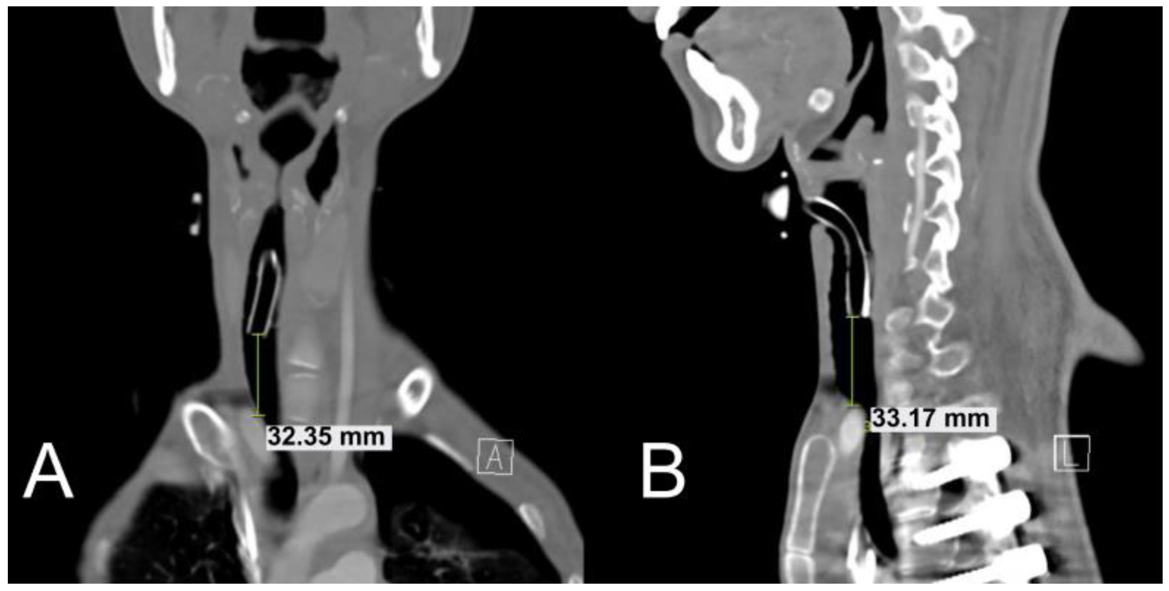
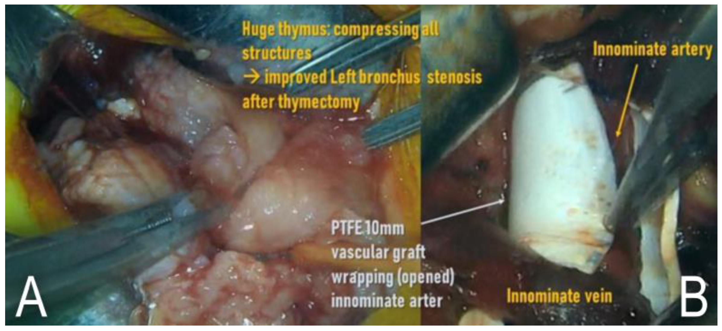
2.1. Case 1
2.2. Case 2
3. Discussion
4. Conclusions
Author Contributions
Funding
Institutional Review Board Statement
Informed Consent Statement
Data Availability Statement
Conflicts of Interest
References
- Hasegawa, T.; Oshima, Y.; Hisamatsu, C.; Matsuhisa, H.; Maruo, A.; Yokoi, A.; Bitoh, Y.; Nishijima, E.; Okita, Y. Innominate artery compression of the trachea in patients with neurological or neuromuscular disorders. Eur. J. Cardio-Thoracic Surg. 2014, 45, 305–311. [Google Scholar] [CrossRef] [PubMed] [Green Version]
- Menen, R.S.; Pak, J.J.; Dowell, M.A.; Patel, A.R.; Ashiku, S.K.; Velotta, J.B. Treatment of Tracheoinnominate Fistula with Ligation of the Innominate Artery: A Case Report. Perm. J. 2016, 20, 15–166. [Google Scholar] [CrossRef] [PubMed] [Green Version]
- Lee, D.J.; Yang, W.; Propst, E.J.; Rosenblatt, S.D.; Hseu, A.; Wolter, N.E. Tracheo-innominate fistula in children: A systematic review of literature. Laryngoscope 2020, 130, 217–224. [Google Scholar] [CrossRef]
- Hamaguchi, S.; Nakajima, Y. Two cases of tracheoinnominate artery fistula following tracheostomy treated successfully by endovascular embolization of the innominate artery. J. Vasc. Surg. 2012, 55, 545–547. [Google Scholar] [CrossRef] [PubMed]
- Komatsu, T.; Sowa, T.; Fujinaga, T.; Handa, N.; Watanabe, H. Tracheo-innominate artery fistula: Two case reports and a clinical review. Ann. Thorac. Cardiovasc. Surg. 2013, 19, 60–62. [Google Scholar] [CrossRef] [PubMed] [Green Version]
- Machado, A.T.; Barroso, M. Endovascular management of tracheo-innominate artery fistula: A case report and literature review. J. Vasc. Bras. 2018, 17, 348–352. [Google Scholar] [CrossRef] [PubMed] [Green Version]
- Obatake, M.; Tokunaga, T.; Hashizume, K.; Mochizuki, K.; Nagayasu, T. Prophylactic ligation of the innominate artery and creation of tracheostomy in a neurologically impaired girl: A case report. Case Rep. Med. 2011, 2011, 790746. [Google Scholar] [CrossRef] [Green Version]
- Ogawa, K.; Nitta, N.; Sonoda, A.; Takahashi, M.; Suzuki, T.; Kitamura, S.; Hanaoka, J.; Tezuka, N.; Murata, K. Tracheo-brachiocephalic artery fistula after tracheostomy associated with thoracic deformity: A case report. J. Med. Case Rep. 2011, 5, 595. [Google Scholar] [CrossRef] [Green Version]
- Gasparri, M.G.; Nicolosi, A.C.; Almassi, G.H. A novel approach to the management of tracheoinnominate artery fistula. Ann. Thorac. Surg. 2004, 77, 1424–1426. [Google Scholar] [CrossRef]
- Grant, C.A.; Dempsey, G.; Harrison, J.; Jones, T. Tracheo-innominate artery fistula after percutaneous tracheostomy: Three case reports and a clinical review. Br. J. Anaesth. 2006, 96, 127–131. [Google Scholar] [CrossRef]
- Khanafer, A.; Hellstern, V.; Meißner, H.; Harmening, C.; Schneider, K.; Henkes, H. Tracheoinnominate fistula: Acute bleeding and hypovolemic shock due to a trachea-innominate artery fistula after long-term tracheostomy, treated with a stent-graft. CVIR Endovasc. 2021, 4, 30. [Google Scholar] [CrossRef] [PubMed]
- Kapural, L.; Sprung, J.; Gluncic, I.; Kapural, M.; Andelinovic, S.; Primorac, D.; Schoenwald, P.K. Tracheo-innominate artery fistula after tracheostomy. Anesth. Analg. 1999, 88, 777–780. [Google Scholar] [CrossRef] [PubMed] [Green Version]
- Deguchi, J.; Furuya, T.; Tanaka, N.; Nobori, M.; Seki, Y.; Nomura, Y.; Umehara, I.; Saito, H.; Miyata, T. Successful management of tracheo-innominate artery fistula with endovascular stent graft repair. J. Vasc. Surg. 2001, 33, 1280–1282. [Google Scholar] [CrossRef] [PubMed] [Green Version]
- Nakai, M.; Sato, H.; Sato, M.; Ikoma, A.; Sanda, H.; Nakata, K.; Minamiguchi, H.; Kawai, N.; Sonomura, T.; Nishimura, Y.; et al. Tracheo-innominate artery fistula successfully treated by endovascular stent-graft repair. Jpn. J. Radiol. 2013, 31, 65–70. [Google Scholar] [CrossRef]
- Sato, H.; Kawase, H.; Furuta, S.; Shima, H.; Wakisaka, M.; Kitagawa, H. Tracheoinnominate artery fistula after laryngotracheal separation: Prevention and management. J. Pediatr. Surg. 2012, 47, 341–346. [Google Scholar] [CrossRef]
- Fujimoto, Y.; Hirose, K.; Ota, N.; Murata, M.; Ide, Y.; Tosaka, Y.; Tachi, M.; Sakamoto, K. Suprasternal approach for impending tracheo-innominate artery fistula. Gen. Thorac. Cardiovasc. Surg. 2010, 58, 480–484. [Google Scholar] [CrossRef]
- Meyers, T.A.; Townsend, D. Cardiac Pathophysiology and the Future of Cardiac Therapies in Duchenne Muscular Dystrophy. Int. J. Mol. Sci. 2019, 20, 4098. [Google Scholar] [CrossRef] [Green Version]
- Mukoyama, M.; Kondo, K.; Hizawa, K.; Nishitani, H. Life spans of Duchenne muscular dystrophy patients in the hospital care program in Japan. J. Neurol. Sci. 1987, 81, 155–158. [Google Scholar] [CrossRef]
- Sawamura, Y.; Takase, K.; Higuchi, N.; Kikuchi, S.; Ito, T.; Tabayashi, K. Surgical repair for tracheo-innominate artery fistula with a muscle flap. Jpn. J. Thorac. Cardiovasc. Surg. 2003, 51, 630–633. [Google Scholar] [CrossRef]
- Tomoyasu, M.; Tanita, T.; Nakajima, T.; Deguchi, H.; Koizumi, J.; Horie, K.; Nagumo, T.; Sasaki, H.; Mizuno, M.; Kawazoe, K. Successful repair using innominate vein flap, pericardial flap and thymus pedicle flap for tracheo-innominate artery fistula. Ann. Thorac. Cardiovasc. Surg. 2007, 13, 143. [Google Scholar]
- Hazarika, P.; Kamath, S.G.; Balakrishnan, R.; Girish, R.; Harish, K. Tracheo-innominate artery fistula: A rare complication in a laryngectomized patient. J. Laryngol. Otol. 2002, 116, 562–564. [Google Scholar] [CrossRef] [PubMed]
- Silva, R.C.; Chi, D.H. Successful management of a tracheo-innominate fistula in a 7-year-old child. Int. J. Pediatr. Otorhinolaryngol. 2010, 74, 946–948. [Google Scholar] [CrossRef] [PubMed]
- Wang, X.-L.; Xu, Z.-G.; Tang, P.-Z.; Yu, Y. Tracheo-innominate artery fistula: Diagnosis and surgical management. Head Neck J. Sci. Spec. 2013, 35, 1713–1718. [Google Scholar] [CrossRef] [PubMed]
- Iodice, F.G.; Brancaccio, G.; Lauri, A.; Di Donato, R. Preventive ligation of the innominate artery in patients with neuromuscular disorders. Eur. J. Cardio-Thoracic Surg. 2007, 31, 747–749. [Google Scholar] [CrossRef] [PubMed] [Green Version]
- Kurose, M.; Takano, K.; Mitsuzawa, H.; Himi, T. Tracheo-innominate artery fistula with severe motor and intellectual disability: Incidence and therapeutic management. Int. J. Pediatr. Otorhinolaryngol. 2014, 78, 1348–1351. [Google Scholar] [CrossRef] [PubMed]
- Suzuki, K.; Fujishiro, J.; Ichijo, C.; Watanabe, E.; Tomonaga, K.; Sunouchi, T.; Watanabe, Y. Prophylactic innominate artery transection to prevent tracheoinnominate artery fistula: A retrospective review of single institution experiences. Pediatr. Surg. Int. 2021, 37, 267–273. [Google Scholar] [CrossRef]
- Furukawa, K.; Kamohara, K.; Itoh, M.; Morokuma, H.; Morita, S. Operative technique for tracheo-innominate artery fistula repair. J. Vasc. Surg. 2014, 59, 1163–1167. [Google Scholar] [CrossRef]


Publisher’s Note: MDPI stays neutral with regard to jurisdictional claims in published maps and institutional affiliations. |
© 2022 by the authors. Licensee MDPI, Basel, Switzerland. This article is an open access article distributed under the terms and conditions of the Creative Commons Attribution (CC BY) license (https://creativecommons.org/licenses/by/4.0/).
Share and Cite
Yoo, B.; Lee, B.; Park, J.D.; Kwon, S.K.; Kwak, J.G. Prevention of Tracheo-Innominate Artery Fistula Formation as a Complication of Tracheostomy: Two Case Reports. Children 2022, 9, 1603. https://doi.org/10.3390/children9111603
Yoo B, Lee B, Park JD, Kwon SK, Kwak JG. Prevention of Tracheo-Innominate Artery Fistula Formation as a Complication of Tracheostomy: Two Case Reports. Children. 2022; 9(11):1603. https://doi.org/10.3390/children9111603
Chicago/Turabian StyleYoo, Byungsun, Bongjin Lee, June Dong Park, Seong Keun Kwon, and Jae Gun Kwak. 2022. "Prevention of Tracheo-Innominate Artery Fistula Formation as a Complication of Tracheostomy: Two Case Reports" Children 9, no. 11: 1603. https://doi.org/10.3390/children9111603
APA StyleYoo, B., Lee, B., Park, J. D., Kwon, S. K., & Kwak, J. G. (2022). Prevention of Tracheo-Innominate Artery Fistula Formation as a Complication of Tracheostomy: Two Case Reports. Children, 9(11), 1603. https://doi.org/10.3390/children9111603