Article

The Significance of Elevated sST2 in Children with Kawasaki Disease

Pedietric Department of Shengjing Hospital, China Medical University, Shenyang 110004, China
*
Author to whom correspondence should be addressed.
These authors contributed equally to this work.
Children 2025, 12(7), 868; https://doi.org/10.3390/children12070868
Submission received: 18 May 2025 / Revised: 21 June 2025 / Accepted: 25 June 2025 / Published: 30 June 2025
(This article belongs to the Special Issue Kawasaki Disease in Children: Advance and Challenges)

Abstract

Objectives: Kawasaki Disease (KD) is an acute vasculitis associated with systemic inflammation. This study aimed to investigate the level and clinical significance of soluble ST2 (sST2) in children with KD. Methods: A retrospective analysis was conducted on 287 pediatric KD patients treated at the Pediatric Cardiology Department of Shengjing Hospital, China Medical University, from November 2021 to December 2022. Patients were stratified into subgroups based on the presence of myocardial damage (MD), coronary artery lesions (CAL), multi-organ involvement (MOD; ≥3 organs) and/or intravenous immunoglobulin-resistant KD (IVIG-R KD). In each group, we analyzed the correlation between sST2 levels and various laboratory parameters, including white blood cell count (WBC), hemoglobin (HB), platelet count (PLT), C-reactive protein (CRP), interleukin-6 (IL-6), erythrocyte sedimentation rate (ESR), N-terminal pro-brain natriuretic peptide (NT-pro BNP), D-dimer, and albumin (ALB). Results: Patients in the CAL group were significantly younger and predominantly male (p < 0.05). In the MD, CAL, MOD, and IVIG-R KD groups, levels of sST2, CRP, NT-pro BNP, and D-dimer were significantly higher than in their respective comparison groups (p < 0.05). sST2 showed weak positive correlations with WBC, CRP, IL-6, NT-pro BNP, and D-dimer, and weak negative correlations with HB and ALB (p < 0.05). sST2, HB, and IL-6 were identified as independent risk factors for MOD (p < 0.05). sST2 and HB were independent risk factors for IVIG-R KD (p < 0.05). Among acute-phase patients, four cases had sST2 levels > 200 ng/mL—all were classified as IVIG-R KD and MOD; three of these also developed coronary artery aneurysms (CAA). Conclusions: Elevated sST2 levels in the acute phase of KD may serve as a clinical indicator of IVIG-R KD, CAA, MOD, and MD.

1. Introduction

Exploring the relationship between elevated soluble growth stimulation expressed gene 2 protein (sST2) levels during the acute phase of Kawasaki Disease (KD) and the occurrence of coronary artery lesions (CAL), intravenous immunoglobulin-resistant KD (IVIG-R KD), and multi-organ damage (MOD) may contribute to timely diagnosis, targeted intervention, and the prevention of serious complications in children with KD. KD is an acute autoimmune vasculitis that primarily affects medium-sized arteries, with a predisposition towards coronary arteries. Among its complications, CALs are the most serious, with an incidence of 15–20% in untreated cases (defined as coronary artery Z score ≥ 2.5) [1]. KD has now become the leading cause of acquired heart disease in children worldwide. The use of IVIG has significantly reduced the incidence of CAL; however, IVIG resistance remains a critical challenge, as it is associated with a higher risk of coronary artery aneurysms (CAA) [2].
KD is characterized not only by coronary artery involvement, but also by systemic inflammation affecting multiple organs during the acute febrile phase. Clinical manifestations may include cardiac (myocarditis, pericarditis, valvulitis), gastrointestinal (abdominal pain, vomiting, diarrhea, gallbladder hydrops), hepatic (hepatitis), pulmonary (interstitial pneumonia), neurological (aseptic meningitis), renal (sterile pyuria) and pancreatic (pancreatitis). In rare but severe cases, KD Shock Syndrome (KDSS) or macrophage activation syndrome (MAS) may occur, posing life-threatening risks.
ST2, a member of the interleukin-1 receptor family, exists in two isoforms: the soluble form (sST2) and the transmembrane form (ST2L). First identified by Shin-ichi Tominaga in 1989, its cardiac relevance was later described by Richard Lee in 2002. In 2005, IL-33 was identified as the functional ligand for ST2 [3].
ST2 is produced by cardiomyocytes in response to mechanical stress, and plays a critical role in cardiovascular inflammation and remodeling. Under normal physiological conditions, ST2L binds IL-33 and initiates protective immune signaling pathways that regulate cardiomyocyte and fibroblast responses [4,5]. However, under pathological conditions, elevated sST2 acts as a decoy receptor, competitively binding IL-33 and blocking its interaction with ST2L. This inhibits cardioprotective signaling, thereby exacerbating myocardial injury, cell death, and fibrosis, and promoting disease progression. Since sST2 lacks the transmembrane and cytoplasmic domains of ST2L, it is secreted into the circulation and can be readily measured in blood samples. To date, most clinical research on sST2 has focused on heart failure and cardiovascular disease [6]. However, emerging evidence suggests that sST2 is also elevated during the acute phase of KD, which is characterized by necrotizing vasculitis, predominantly infiltrated by neutrophils and macrophages, along with elevated systemic inflammatory markers [7].
Given KD’s potential to cause long-term cardiovascular complications, understanding the role of sST2 as a biomarker may provide important insights into disease severity, the risk of IVIG resistance, and systemic involvement such as MOD.

2. Methods

2.1. Patients

A total of 287 children diagnosed with KD were included in this study. All patients were diagnosed and treated at the Department of Pediatric Cardiology, Shengjing Hospital, affiliated with the China Medical University, between November 2021 and December 2022.

2.2. sST2 Measurement

Serum sST2 levels were quantitatively measured using a double-antibody sandwich ELISA on a 96-well plate. A 2 mL venous blood sample was collected from each child prior to treatment with IVIG or glucocorticoids, and within 10 days of fever onset. The ELISA kit was supplied by Shanghai Ruidi Biotechnology Co., Ltd., and testing was performed using the Freedom Evolyzer-2100 automated immunoassay system (TECAN, Switzerland, Zurich).

2.3. Clinical and Laboratory Data Collection

In addition to sST2 testing, the following biochemical parameters were collected: complete blood count, including white blood cell count (WBC), hemoglobin (HB), and platelet count (PLT); inflammatory markers such as C-reactive protein (CRP), erythrocyte sedimentation rate (ESR) and interleukin-6 (IL-6); and cardiac biomarkers such as high-sensitivity troponin I, high-sensitivity troponin T and N-terminal pro-B-type natriuretic peptide (NT-proBNP). Other indicators include albumin (ALB), D-dimer, serum electrolytes, and alanine aminotransferase (ALT).

2.4. Imaging Data

Relevant imaging studies were undertaken, including electrocardiography (ECG), echocardiography (ECHO), electroencephalography (EEG), and chest computed tomography (CT). It has been reported that ST2 > 71 ng/mL in adults is regarded as the critical value of cardiac insufficiency. The higher the ST2, the more severe the cardiovascular damage. Four patients with sST2 levels > 200 ng/mL were identified and followed up through April 2025 to monitor clinical outcomes.

3. Definitions and Diagnostic Criteria

CAL: Diagnosed based on the 6th revised Japanese diagnostic guidelines using the Z-score system (http://raise.umin.jp/zsp/CALculator, accessed on 1 October 2020), with Z ≥ 2.5 considered diagnostic for CAL.
MOD: Defined as the involvement of three or more organ systems, including but not limited to cardiovascular (e.g., CAL, MD), hematologic (e.g., granulocytopenia, anemia, thrombocytopenia), electrolyte disturbances (e.g., hyponatremia, hypokalemia), neurological (e.g., sterile meningitis, facial nerve palsy), respiratory (e.g., interstitial pneumonia), hepatic (e.g., elevated ALT), genitourinary (e.g., urethritis), musculoskeletal (e.g., arthritis), and macrophage activation syndrome (MAS).
IVIG-R KD: Defined as a persistent or recurrent fever (≥38 °C) occurring 36–48 h after the completion of IVIG treatment at a dose of 2 g/kg, in the absence of any other identifiable cause [5].

3.1. Inclusion Criteria

Children diagnosed with KD were enrolled based on the 2017 American Heart Association (AHA) guidelines [8] and the sixth revised diagnostic criteria issued in Japan [9]. Only patients who received treatment and were available for follow-up were included.

3.2. Exclusion Criteria

Patients were excluded if they met any of the following conditions:
  • Duration of fever exceeded 10 days at the time of hospital admission;
  • sST2 testing was not performed, as measurements were only available on working days;
  • Administration of IVIG or corticosteroids within one month prior to sST2 testing.

3.3. Diagnostic Criteria for Myocardial Damage (MD)

MD was diagnosed based on the presence of elevated troponin I and/or troponin T levels above the normal upper limit; ECG abnormalities, including arrhythmias, prolonged P-R interval, non-specific ST-T changes, or low QRS voltage; ECHO findings, including left ventricular dilation, weakened ventricular wall motion, valve regurgitation, or reduced ejection fraction.

3.4. Patient Groups

A total of 287 patients with KD were categorized into the following groups based on laboratory, ECG, and ECHO results:
  • Myocardial Damage (MD)
Group A—MD present (n = 17)
Group B—MD absent (n = 270);
2.
Coronary Artery Lesions (CAL)
Group C—CAL present (n = 48)
Group D—CAL absent (n = 239);
3.
Multi-Organ Damage (MOD)
Group E—≥3 organs involved (n = 58)
Group F—<3 organs involved (n = 229);
4.
IVIG-Resistant KD (IVIG-R KD)
Group G—IVIG-R KD present (n = 24)
Group H—IVIG-R KD absent (n = 263).

4. Statistical Analysis

All statistical analyses were conducted using SPSS version 27.0. Continuous variables conforming to a normal distribution were expressed as mean ± standard deviation (x ± s). Independent samples t-tests were used for comparisons with equal variances. Welch’s t-tests were used for unequal variances. Data with a skewed distribution were expressed as median (interquartile range, IQR), and the Mann–Whitney U test was applied. Categorical variables were presented as frequency and percentage, and compared using the Chi-square test. Binary logistic regression analysis was used to identify independent risk factors for MOD and IVIG-R KD. The receiver operating characteristic (ROC) curve was employed to evaluate the predictive value of sST2. Spearman correlation analysis was used to assess the relationships between sST2 levels and laboratory indicators. A p-value < 0.05 was considered statistically significant.

Ethical Approval

This study was reviewed and approved by the Ethics Committee of Shengjing Hospital, China Medical University (Approval No. 2023PS152J).

5. Results

5.1. The Age and Gender Distribution of Children with KD

Among the 287 children with KD, 213 (72.20%) were under 3 years old, and the male-to-female ratio was 1.58:1. The percentage of males and the age were both lower in the KD with CAL group (p < 0.05) (Table 1).

5.2. Comparison of sST2 Levels Among Different Groups

The sST2 levels in groups A, C, E and G were significantly higher than those in groups B, D, F, and H (p < 0.05) (Table 2).

5.3. Comparison of Other Indicators Among Different Groups

The levels of CRP, NT-pro BNP and D-dimer in group A, C, E, and G were higher than those in groups B, D, F, and H, respectively (p < 0.05) (Table 3).

5.4. Correlation Analysis Between sST2 and Other Indicators

The correlation coefficient r was calculated using Spearman correlation analysis. A correlation was considered weak when 0.3 ≤ |r| < 0.5. sST2 had a weak positive correlation with WBC, CRP, IL-6, NT-pro BNP, and D-dimer, and a weak negative correlation with HB and ALB. There was no correlation between sST2 and ESR or PLT (Table 4).

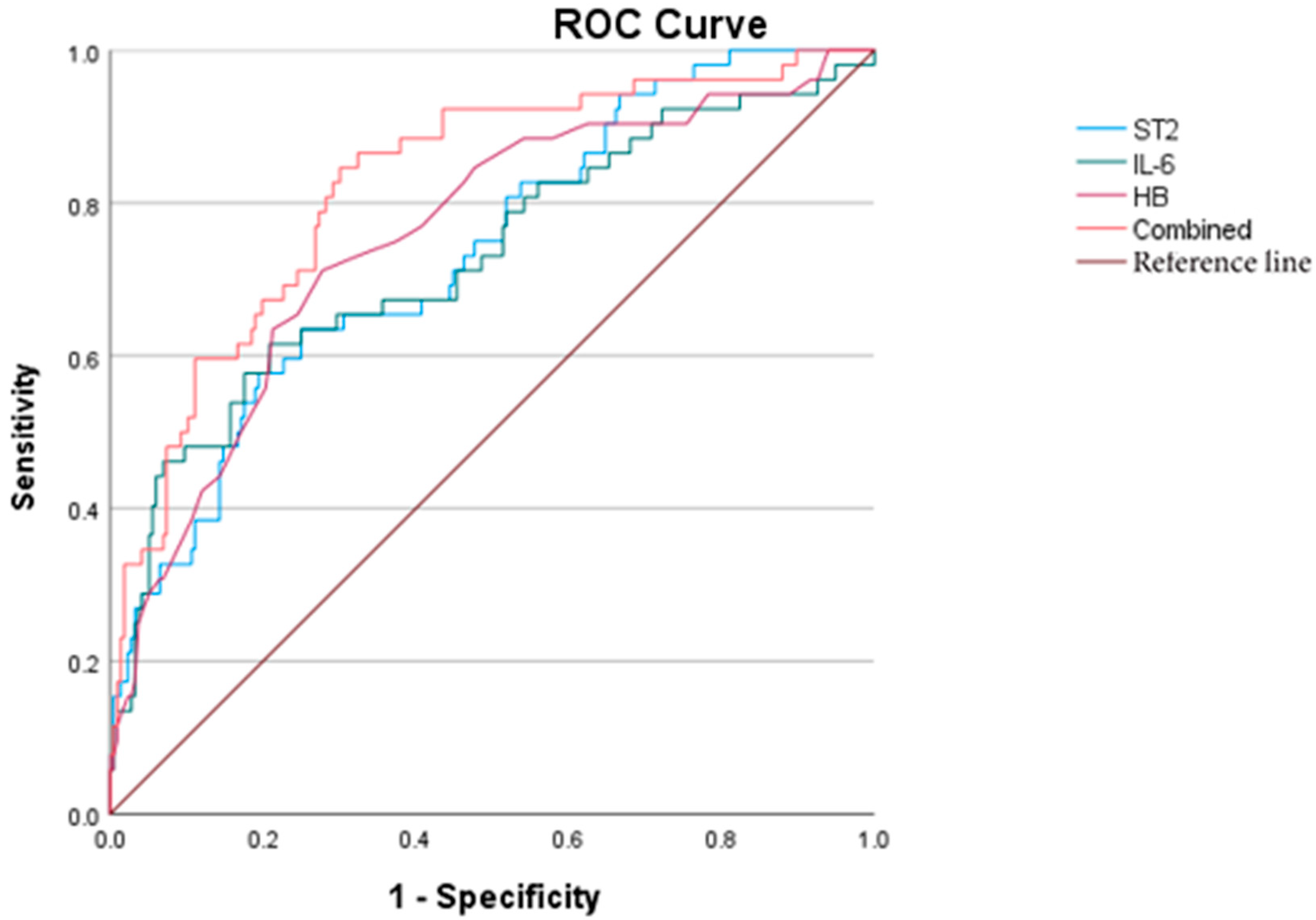
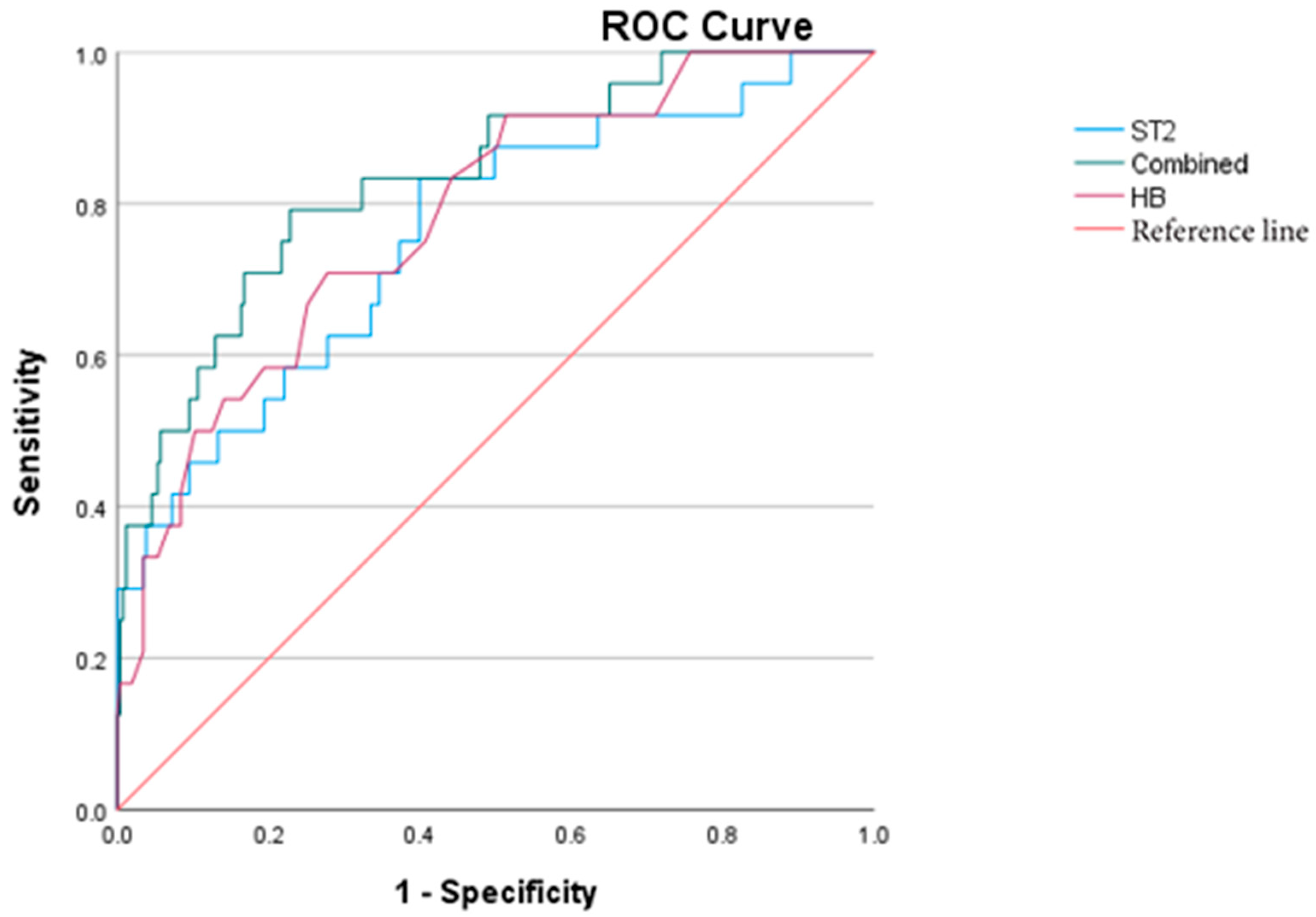
5.5. KD Combined with MD

Based on the differences observed between group A (KD with MD) and group B (KD without MD), as shown in Table 3, the following variables were included as independent predictors in a univariate binary logistic regression analysis: sST2, WBC, PLT, CRP, IL-6, D-dimer, and NT-pro BNP. The analysis reveals that elevated levels of sST2, WBC, and CRP were significant factors promoting MD in patients with KD (p < 0.05). However, when these variables were further analyzed using multivariate logistic regression, none of them remained statistically significant, suggesting that no single factor independently predicted myocardial damage after adjusting for covariates (Table 5).

5.6. KD Combined with CAL

Based on the differences observed between group C (with CAL, CAL) and group D (without CAL) in Table 3, the following variables were included as independent predictors in a univariate binary logistic regression analysis: sST2, HB, PLT, CRP, NT-pro BNP, D-dimer, ALB, age, and gender (coded as male = 1). The models for sST2, HB, PLT, CRP, ALB, age, and gender demonstrated good fit, indicating reliable performance.
The univariate analysis shows that elevated sST2, PLT, and CRP, as well as decreased HB and ALB, younger age, and male gender, were all significantly associated with an increased risk of coronary artery lesions (p < 0.05). However, in the subsequent multivariate logistic regression analysis, only male gender remained a statistically significant independent risk factor for coronary artery damage (p < 0.05) (Table 6).

5.7. KD Combined with MOD

Based on the differences between group E (three or more organs involved, MOD) and group F (less than three organs involved) shown in Table 3, the following variables were included as independent predictors in a univariate binary logistic regression analysis: sST2, WBC, HB, PLT, CRP, IL-6, NT-pro BNP, D-dimer, and ALB.
The models constructed with sST2, WBC, HB, PLT, IL-6, and D-dimer demonstrated good fit and were considered valid. These variables were subsequently included in a multivariate binary logistic regression analysis, which revealed that elevated sST2 and IL-6, along with reduced HB, were independent risk factors for multi-organ involvement in children with KD (p < 0.05) (Table 7).
To further evaluate the predictive values of key variables for MOD in KD, a receiver operating characteristic (ROC) curve analysis was performed. The area under the curve (AUC) values for sST2, IL-6, and HB were 0.735, 0.728, and 0.756, respectively. When these three markers were combined, the AUC increased to 0.823, suggesting improved predictive performance. The optimal cut-off value for sST2 in predicting MOD was determined to be 51.264 ng/mL (Figure 1).

5.8. IVIG-R KD

Binary logistic regression analysis demonstrated that models incorporating sST2, HB, CRP, IL-6, ALB, and IVIG-R KD exhibited good fit. Among these, sST2 and HB were identified as independent risk factors for IVIG-R KD (p < 0.05) (Table 8).
To further assess the predictive value of sST2 and HB for IVIG-R KD, a ROC curve analysis was performed. The AUCs for sST2 and HB were 0.760 and 0.783, respectively. When combined, the AUC increased to 0.835, demonstrating improved predictive performance. Notably, elevation in sST2 occurred earlier in the disease course than the decline in HB. The optimal cut-off value of sST2 for predicting IVIG-R KD was determined to be 43.412 ng/mL (Figure 2).

5.9. Clinical Data of Four Cases with ST2 > 200 ng/mL

Among acute-phase patients, four cases had sST2 levels > 200 ng/mL—all were classified as IVIG-R KD and MOD; three of these also developed CAA (Table 9).

6. Discussion

Soluble ST2 (sST2), a member of the interleukin-1 (IL-1) receptor family, has emerged as a critical biomarker in cardiovascular and inflammatory diseases. Its specific ligand, interleukin-33 (IL-33), also part of the IL-1 family, forms the IL-33/ST2 axis, a signaling pathway increasingly recognized for its role in immune regulation and cardiovascular pathology. Since its initial identification by Shin-ichi Tominaga in 1989 and its association with cardiac disease described by Richard Lee’s group in 2002, sST2 has been extensively studied for its predictive value in heart failure, myocardial fibrosis, and cardiac remodeling [10,11,12].
There are two forms of ST2—transmembrane ST2L, which binds IL-33 and activates protective signaling pathways, and soluble ST2 (sST2), which acts as a decoy receptor. Under physiological conditions, IL-33/ST2L signaling inhibits cardiac hypertrophy and fibrosis, contributing to myocardial protection [11]. However, elevated sST2 levels competitively inhibit IL-33/ST2L binding, disrupting these protective mechanisms and allowing disease progression. Unlike NT-pro BNP, sST2 levels are not influenced by renal function, making this a potentially superior biomarker in settings of volume depletion or pre-renal insufficiency common during the acute phase of KD [12,13].
Our study demonstrates that sST2 is significantly elevated in patients with CAA, consistent with prior research [4]. Notably, three patients with CAA exhibited elevated sST2 despite normal NT-pro BNP levels, highlighting sST2′s sensitivity and the early rise in acute inflammation. This may be attributed to necrosis of the mid-coronary artery, which triggers the release of sST2.
Furthermore, patients with MOD or IVIG-R KD also exhibited elevated sST2, though to a lesser degree than those with CAA. In our cohort, four patients had sST2 levels exceeding 200 ng/mL, and all developed both IVIG-R KD and MOD. Their clinical courses illustrate the severity of illness and the extent of systemic involvement.
Case 1: Eight-organ involvement, cardiogenic shock, heart failure (recovery in 10 days), aseptic meningitis (recovered in 3 months).
Case 2: Six-organ involvement, leukemia-like reaction (recovered in 3 weeks), middle CAA (resolved within 2 years).
Case 3: Persistent fever (>3 weeks), six-organ involvement, giant CAA (resolved in 2 years).
Case 4: Fever for 22 days, seven-organ involvement, persistent large CAA (ongoing follow-up through April 2025).
These cases suggest that a marked elevation in sST2 is associated with severe inflammation and poor prognosis, consistent with findings from sepsis studies, where higher sST2 levels were linked to shock, multi-organ failure, and mortality [3].
Logistic regression analysis in our study identified elevated sST2 and IL-6 and low HB as independent risk factors for MOD. ROC curve analysis showed that HB reduction had the highest predictive value, followed by sST2. However, sST2 elevation occurred earlier in the disease course than HB reduction, suggesting that sST2 may serve as an early warning marker for severe KD. The combination of sST2, IL-6, and HB yielded the best predictive accuracy for MOD.
For IVIG-R KD, multivariate analysis also showed that sST2 and HB were independent predictors, with sST2 having superior predictive value. These findings emphasize the clinical utility of sST2 not only in identifying coronary complications, but also in forecasting treatment resistance and systemic involvement.
Additionally, our study corroborated previous findings that CAL is more common in younger, male patients, and that MD is associated with elevated sST2. However, in multivariate analysis, sST2 was not a significant independent predictor of CAL or MD, potentially due to sample size limitations and the short study duration, indicating the need for further investigation.
The relevance of the IL-33/ST2 axis extends beyond KD. Emerging literature has drawn parallels between KD and Multisystem Inflammatory Syndrome in Children (MIS-C) associated with COVID-19. MIS-C shares features with KD, including fever, rash, conjunctivitis, mucosal changes, and multi-organ involvement [14,15,16]. IL-33 signaling plays a pivotal role in immune activation in MIS-C, involving MAPK, NF-κB, and MyD88 pathways, contributing to a cytokine storm and MOD [17]. The elevation of sST2 in MIS-C and KD likely reflects this shared pathophysiological basis.
Importantly, persistent CAA in KD patients poses long-term risks. Although the precise prognostic significance is not fully understood, autopsy findings in patients with long-standing CAA have shown extensive myocardial fibrosis, likely due to chronic coronary inflammation and ischemia. Thus, the long-term monitoring of sST2 may provide prognostic insight into cardiac remodeling and guide post-acute care.

7. Conclusions

In summary, elevated sST2 levels during the acute phase of KD are strongly associated with IVIG resistance, coronary artery involvement, multi-organ damage, and myocardial injury. sST2 may serve as an early biomarker for identifying severe or atypical KD cases, guiding timely and aggressive intervention. Its independence from renal function and early rise enhance its clinical utility over traditional markers like NT-pro BNP.
What is particularly promising is the growing evidence that the IL-33/sST2 axis may represent a therapeutic target in KD vasculitis [4]. However, due to the limited sample size and a lack of non-KD febrile controls, further studies with larger cohorts and comparative groups are needed. The long-term follow-up of KD patients with persistent CAA and elevated sST2 levels may provide valuable insight into disease progression and outcomes.

Author Contributions

Z.Y.: Data collection, data analysis, and Chinese editing of manuscript. Y.X.: Co-first author—the diagnosis, treatment and follow-up of some patients, analysis of some data, image production, and English editing of manuscript. Y.C.: The diagnosis, treatment and follow-up of some patients. J.L.: The diagnosis, treatment and follow-up of some patients. H.W.: The design of the project, ethical application, diagnosis, treatment and follow-up of the vast majority of patients, editing of the preface, conclusions and discussions of the paper, as well as English revision. All authors have read and agreed to the published version of the manuscript.

Funding

This research received no external funding.

Institutional Review Board Statement

This study was approved by Shengjing hospital, affiliated with the China medical university ethics committee, ethical number: 2023PS152J, approved on 27 April 2023.

Informed Consent Statement

Informed consent was obtained from all subjects involved in the study. Written informed consent has been obtained from the patient(s) to publish this paper.

Data Availability Statement

Because it involves patient privacy, especially for those with CAA, these clinical data cannot be made public.

Acknowledgments

Thanks to Xianyi Yu for the diagnosis and follow-up of some patients, and to Rui Chen and Ce Wang for the follow-up of some patients.

Conflicts of Interest

The authors declare no conflicts of interest.

References

  1. Jone, P.-N.; Tremoulet, A.; Choueiter, N.; Dominguez, S.R.; Harahsheh, A.S.; Mitani, Y.; Zimmerman, M.; Lin, M.-T.; Friedman, K.G. Update on diagnosis and management of Kawasaki disease: A scientific statement from the American Heart Association. Circulation 2024, 150, e481–e500. [Google Scholar] [CrossRef] [PubMed]
  2. Fabi, M.; Andreozzi, L.; Frabboni, I.; Dormi, A.; Corinaldesi, E.; Lami, F.; Cicero, C.; Tchana, B.; Francavilla, R.; Sprocati, M.; et al. Non-coronary cardiac events, younger age, and IVIG unresponsiveness increase the risk for coronary aneurysms in Italian children with Kawasaki disease. Clin. Rheumatol. 2021, 40, 1507–1514. [Google Scholar] [CrossRef] [PubMed]
  3. Homsak, E.; Gruson, D. Soluble ST2: A complex and diverse role in several diseases. Clin. Chim. Acta 2020, 507, 75–87. [Google Scholar] [CrossRef] [PubMed]
  4. Okada, S.; Yasudo, H.; Ohnishi, Y.; Matsuguma, C.; Fukano, R.; Motonaga, T.; Waniishi, T.; Hasegawa, S. Interleukin-33/ST2 Axis as Potential Biomarker and Therapeutic Target in Kawasaki Disease. Inflammation 2023, 46, 480–490. [Google Scholar] [CrossRef] [PubMed]
  5. Kotsiou, O.S.; Gourgoulianis, K.I.; Zarogiannis, S.G. IL-33/ST2 Axis in Organ Fibrosis. Front. Immunol. 2018, 9, 2432. [Google Scholar] [CrossRef] [PubMed] [PubMed Central]
  6. Pascual-Figal, D.A.; Januzzi, J.L. The biology of ST2: The International ST2 Consensus Panel. Am. J. Cardiol. 2015, 115 (Suppl. S7), 3B–7B. [Google Scholar] [CrossRef] [PubMed]
  7. Xu, H.; Turnquist, H.R.; Hoffman, R.; Billiar, T.R. Role of the IL-33-ST2 axis in sepsis. Mil. Med. Res. 2017, 4, 3. [Google Scholar] [CrossRef] [PubMed]
  8. McCrindle, B.W.; Rowley, A.H.; Newburger, J.W.; Burns, J.C.; Bolger, A.F.; Gewitz, M.; Baker, A.L.; Jackson, M.A.; Takahashi, M.; Shah, P.B.; et al. American Heart Association Rheumatic Fever, Endocarditis, and Kawasaki Disease Committee of the Council on Cardiovascular Disease in the Young; Council on Cardiovascular and Stroke Nursing; Council on Cardiovascular Surgery and Anesthesia; and Council on Epidemiology and Prevention. Diagnosis, Treatment, and Long-Term Management of Kawasaki Disease: A Scientific Statement for Health Professionals from the American Heart Association. Circulation 2017, 135, e927–e999, Erratum in Circulation 2019, 140, e181–e184. [Google Scholar] [CrossRef] [PubMed]
  9. Kobayashi, T.; Ayusawa, M.; Suzuki, H.; Abe, J.; Ito, S.; Kato, T.; Kamada, M.; Shiono, J.; Suda, K.; Tsuchiya, K.; et al. Revision of diagnostic guidelines for Kawasaki disease (6th revised edition). Pediatr. Int. 2020, 62, 1135–1138. [Google Scholar] [CrossRef] [PubMed]
  10. Schmitz, J.; Owyang, A.; Oldham, E.; Song, Y.; Murphy, E.; McClanahan, T.K.; Zurawski, G.; Moshrefi, M.; Qin, J.; Li, X.; et al. IL-33, an interleukin-1-like cytokine that signals via the IL-1 receptor-related protein sST2 and induces T helper type 2-associated cytokines. Immunity 2005, 23, 479–490. [Google Scholar] [CrossRef] [PubMed]
  11. Savarimuthu, S.; Goel, P.; Harky, A. Soluble ST2: A valuable prognostic marker in heart failure. Heart Fail. Rev. 2022, 27, 2155–2164. [Google Scholar] [CrossRef] [PubMed]
  12. Yu, M. Chinese expert consensus on biomarkers for heart failure. Chin. J. Lab. Med. 2020, 43, 130–141. [Google Scholar] [CrossRef]
  13. Okada, S.; Sakai, A.; Ohnishi, Y.; Yasudo, H.; Motonaga, T.; Fukano, R.; Waniishi, T.; Sugiyama, M.; Hasegawa, S. Necrotic Change of Tunica Media Plays a Key Role in the Development of Coronary Artery Lesions in Kawasaki Disease. Circ. J. 2024, 88, 1709–1714. [Google Scholar] [CrossRef] [PubMed]
  14. Algarni, A.S.; Alamri, N.M.; Khayat, N.Z.; Alabdali, R.A.; Alsubhi, R.S.; Alghamdi, S.H. Clinical practice guidelines in multisystem inflammatory syndrome (MIS-C) related to COVID-19: A critical review and recommendations. World J. Pediatr. 2022, 18, 83–90. [Google Scholar] [CrossRef] [PubMed] [PubMed Central]
  15. Kabeerdoss, J.; Pilania, R.K.; Karkhele, R.; Kumar, T.S.; Danda, D.; Singh, S. Severe COVID-19, multisystem inflammatory syndrome in children, and Kawasaki disease: Immunological mechanisms, clinical manifestations and management. Rheumatol. Int. 2021, 41, 19–32. [Google Scholar] [CrossRef] [PubMed] [PubMed Central]
  16. Kakkar, R.; Lee, R.T. The IL-33/ST2 pathway: Therapeutic target and novel biomarker. Nat. Rev. Drug Discov. 2008, 7, 827–840. [Google Scholar] [CrossRef] [PubMed] [PubMed Central]
  17. Hoshino, S.; Jain, S.; Shimizu, C.; Roberts, S.; He, F.; Daniels, L.B.; Kahn, A.M.; Tremoulet, A.H.; Gordon, J.B.; Burns, J.C. Biomarkers of inflammation and fibrosis in young adults with history of Kawasaki disease. Int. J. Cardiol. Heart Vasc. 2021, 36, 100863. [Google Scholar] [CrossRef] [PubMed] [PubMed Central]
Figure 1. ROC curves of sST2, IL-6, HB, and the combined diagnosis for KD with MOD.
Children 12 00868 g001
Figure 2. ROC curves of sST2, HB, and the combined prediction of IVIG-R KD.
Children 12 00868 g002
Table 1. General information.
GroupsAge (year)Male (%)PyearPgender
MDA (17)2.0 (0.6~3.0)9 (52.94)0.0610.464
B (270)2.5 (1.5~4.0)167 (61.85)
CALC (48)1.9 (0.8~2.7)40 (83.33)0.003<0.001
D (239)2.5 (1.5~4.0)136 (56.90)
MODE (58)2.7 (1.0~4.0)36 (62.07)0.8490.896
F (229)2.4 (1.4~4.0)140 (61.14)
IVIG-R KDG (24)2.8 (1.8~5.0)17 (70.83)0.1090.318
H (263)2.4 (1.4~4.0)159 (60.46)
MD, myocardial damage; CAL, coronary artery lesion; MOD, multi-organ damage; IVIG-R KD, IVIG-resistant KD.
Table 2. Comparison of sST2 levels among different groups.
GroupssST2 (ng/mL)Zp
MDA (17)55.53 (41.97~120.58)−3.1500.002
B (270)38.28 (27.25~57.60)
CALC (48)42.82 (32.24~71.78)−2.0860.037
D (239)38.35 (27.14~57.46)
MODE (58)59.58 (37.47~96.14)−5.380<0.001
F (229)37.49 (26.33~51.83)
IVIG-R KDG (24)65.67 (43.96~183.66)−4.214<0.001
H (263)37.73 (27.29~55.62)
MD, myocardial damage; CAL, coronary artery lesion; MOD, multi-organ damage; IVIG-R KD, IVIG-resistant KD.
Table 3. Comparison of other indicators among different groups.
PWBCPHBPPLTPCRPPIL-6PESRPpro-BNPPD-dimerPALB
A vs. B0.0010.1340.0460.0180.0020.348<0.0010.0030.072
C vs. D0.370.0010.0110.0390.0760.9330.0370.0050.032
E vs. F0.002<0.0010.002<0.001<0.0010.772<0.001<0.001<0.001
G vs. H0.032<0.0010.05<0.0010.0010.2880.0090.002<0.001
WBC, white blood cell; HB, hemoglobin; PLT, platelet; CRP, C-reactive protein; ESR, erythrocyte sedimentation rate; IL-6, interleukin 6; pro-BNP, pro-B-type natriuretic peptide; ALB, serum albumin.
Table 4. Correlation analysis between sST2 and other indexes.
Indexes rSig.95% Confidence Interval (CI)
Lower LimitUpper Limit
sST2WBC0.301<0.0010.1880.405
HB−0.333<0.001−0.434−0.222
PLT0.196<0.0010.0790.308
CRP0.412<0.0010.3080.506
IL-60.456<0.0010.3520.548
ESR0.1050.08−0.0160.223
NT-pro BNP0.419<0.0010.3150.514
D-dimer0.367<0.0010.2580.467
ALB−0.403<0.001−0.499−0.299
WBC, white blood cell; HB, hemoglobin; PLT, platelet; CRP, C-reactive protein; ESR, erythrocyte sedimentation rate; IL-6, interleukin 6; pro BNP, pro-B-type natriuretic peptide; ALB, serum albumin.
Table 5. Logistic regression analysis of KD combined with MD.
FactorInfluence FactorBSEWaldpOR95%CI
Lower LimitUpper Limit
singlesST20.0110.0047.0430.0081.0111.0031.020
WBC0.0990.0357.7880.0051.1041.0301.183
CRP0.0120.00410.0340.0021.0121.0041.019
multisST20.0040.0060.5440.4611.0040.9931.015
WBC0.0510.0441.3240.2501.0520.9651.147
CRP0.0070.0051.9090.1671.0070.9971.017
sST2, soluble ST2; WBC, white blood cell; CRP, C-reactive protein.
Table 6. Logistic regression analysis of KD combined with CAL.
FactorInfluence FactorBSEWaldpOR95%CI
Lower LimitUpper Limit
singlesST20.0100.0047.6800.0061.0101.0031.017
HB−0.0490.01511.031<0.0010.9520.9240.980
PLT0.0030.00110.5120.0011.0031.0011.005
CRP0.0090.00311.073<0.0011.0091.0041.014
ALB−0.1200.0466.8040.0090.8870.8110.971
age−0.2530.1036.0240.0140.7760.6340.950
gender1.3320.40910.6120.0013.7871.7008.437
multisST20.0030.0050.2840.5941.0030.9931.012
HB−0.0130.0190.5050.4770.9870.9511.024
PLT0.0020.0012.8890.0891.0021.0001.004
CRP0.0050.0041.4110.2351.0050.9971.012
ALB−0.0650.0591.2040.2720.9370.8341.053
age−0.1740.1102.5030.1140.8400.6771.043
gender1.4300.43510.8040.0014.1791.7819.803
sST2, soluble ST2; HB, hemoglobin; PLT, platelet; CRP, C-reactive protein; ALB, serum albumin.
Table 7. Logistic regression analysis of KD combined with MOD.
FactorInfluence FactorBSEWaldpOR95%CI
Lower LimitUpper Limit
singlesST20.0250.00524.92<0.0011.0251.0151.035
WBC0.0780.0268.910.0031.0811.0271.137
HB−0.0850.01628.99<0.0010.9180.8900.947
PLT0.0020.0017.970.0051.0021.0011.004
IL-60.0050.00122.39<0.0011.0051.0031.008
D-dimer0.0010.00016.44<0.0011.0011.0011.002
multisST20.0130.0056.010.0141.0131.0031.024
HB−0.0670.02110.650.0010.9350.8980.974
IL-60.0030.0015.790.0161.0031.0011.006
WBC−0.0210.0400.280.6000.9790.9051.059
PLT0.0010.0010.510.4771.0010.9981.003
sST2, soluble ST2; HB, hemoglobin; IL-6, interleukin 6; WBC, white blood cell; PLT, platelet.
Table 8. Logistic regression analysis of IVIG-R KD.
FactorInfluence FactorBSEWaldpOR95%CI
Lower LimitUpper Limit
singlesST20.0260.00524.142<0.0011.0251.0161.037
HB−0.1070.02223.786<0.0010.8990.8610.938
CRP0.0170.00324.584<0.0011.0171.0101.024
IL-60.0030.0016.2390.0131.0031.0011.005
ALB−0.2430.06912.369<0.0010.7850.6850.898
multisST20.0170.0067.9870.0051.0171.0051.029
HB−0.0620.0275.3540.0210.9400.8920.991
CRP0.0060.0051.1430.2851.0060.9951.016
IL-60.0000.0010.4160.5191.0000.9991.002
ALB0.0590.0860.4770.4901.0610.8971.256
sST2, soluble ST2; HB, hemoglobin; CRP, C-reactive protein; IL-6, interleukin 6; ALB, albumin.
Table 9. Clinical data of four cases with sST2 > 200.
CaseGenderAgesST2
(ng/mL)
FeverTreatmentMOD
1#
19 kg
107 cm
F3.5 y>200Admission 7 d
Regressive10 d
IVIG 4 g/kg
Dex5 mg*2 d
Methylmethicone:
2 mg/kg*7 d
1.5 mg/kg*7 d
1 mg/kg*1 d
Prednisone Po 14 d
ALB IV 40 g
Cardiogenic shock
Acute heart failure
Hypoproteinemia (27.1 g/L)
Hypokalemia, hyponatremia,
Pneumonia
Aseptic encephalitis (EEG 2–3 Hz)
Localized peritonitis
Thrombocytopenia
2#
13.5 kg
102 cm
M3 y>200Admission 9 d
Regressive20 d
IVIG 2 g/kg
Methylmethicone:
20 mg/kg*3 d
2 mg/kg*3 d
1 mg/kg*1 d
Prednisone Po 7 d
ALB IV 10 g
CAA: LM4.7 mm, Z = 6.07, 3 m recovered
Liver damage (ALT 95 U/L)
Hypoproteinemia (24 g/L)
Leukemoid reaction
Aseptic encephalitis (EEG 5–7 Hz)
Pneumonia,
3#
9.3 kg
82 cm
M23 m285.4Admission 5 d
Regressive27 d
IVIG 4 g/kg
Methylmethicone
20 mg/kg*3 d
10 mg/kg*3 d
2 mg/kg*4 d
1 mg/kg*10 d
Prednisone Po 10 d
TNF inhibitor 5 mg/kg
ALBI V 70 g
CAA: LM5.6 mm (Z = 11.1)
RCA6.5 mm (Z = 12)
Liver damage (ALT 434 U/L)
Hypoproteinemia (24.2 g/L)
Aseptic encephalitis
(CSF:WBC66, Pro 0.56)
Pleural effusion
Moderate anemia (HGB = 76 g/L)
4#
29 kg
130 cm
F9 y287.2Admission 6 d
Regressive 22 d
IVIG 3 g/kg
Methylmethicone:
2 mg/kg*6 d
1 mg/kg*7 d
0.7 md/kg*3 d
Prednison Po 10 d
ALBIV 60 g
CTX 2 mg/kg IV
CAA: LAD 6.9 mm (Z = 7.63) persist
RCA7.7 mm (Z = 10.63) persist
Hypoproteinemia (20.6 g/L)
Aseptic encephalitis (EEG 4–7 Hz)
Knee joint effusion
Granulocytopenia
Hyponatremia,
Moderate anemia (HGB = 86 g/L)
MOD, multi-organ damage; Dex, dexamethasone; ALB, albumin; CAA, coronary artery aneurysm; LM, left main coronary artery; RCA, right coronary artery; CSF, Cerebrospinal fluid; WBC, white blood cell.
Disclaimer/Publisher’s Note: The statements, opinions and data contained in all publications are solely those of the individual author(s) and contributor(s) and not of MDPI and/or the editor(s). MDPI and/or the editor(s) disclaim responsibility for any injury to people or property resulting from any ideas, methods, instructions or products referred to in the content.

