Article

Exploring the Clinical Characteristics and Outcomes of Rhinovirus Infection in Hospitalized Children Compared with Other Respiratory Viruses

1
Carol Davila University of Medicine and Pharmacy, 050474 Bucharest, Romania
2
National Institute for Mother and Child Health, Alessandrescu-Rusescu, 020395 Bucharest, Romania
*
Author to whom correspondence should be addressed.
Children 2024, 11(11), 1303; https://doi.org/10.3390/children11111303
Submission received: 2 October 2024 / Revised: 20 October 2024 / Accepted: 23 October 2024 / Published: 28 October 2024

Abstract

:
Background: Acute viral respiratory tract infections constitute a significant challenge in pediatric healthcare globally, with rhinovirus representing one of the primary etiological agents. In this context, we conducted a study with the objective of identifying the clinical characteristics and outcomes of rhinovirus infection in comparison with other respiratory viruses in children hospitalized in one of the largest pediatric hospitals in the capital of Romania. Methods: We conducted a retrospective study among children hospitalized for influenza-like illness symptoms and who were tested by multiplex RT-PCR with a nasopharyngeal swab between May 2020 and December 2021. Results: A total of 496 children were eligible for inclusion in the study, and the positivity rate for at least one virus was 58.5%. The rhinovirus was identified in 138 patients (median age 12.5 months), representing 27.8% of all children tested and 49.3% of all positive samples. Although the clinical features of children with rhinovirus were dominated by cough (63.7%) and dyspnea (51.6%), no symptoms were identified that were strongly associated with rhinovirus infection in comparison to other respiratory viruses. The probability of receiving an antibiotic prescription was 1.92 times lower (p = 0.011) in children who tested positive for rhinovirus compared to children with negative RT-PCR results. The incidence of acute bronchiolitis or acute bronchitis, acute respiratory failure, and acute otitis media was higher among rhinovirus-positive children than among those who tested negative via RT-PCR. However, the incidence of these conditions was similar among children who tested positive for other respiratory viruses. Conclusions: Rhinovirus was the most prevalent virus identified in children hospitalized with influenza-like illness symptoms. The utilization of multiplex RT-PCR molecular tests is instrumental in elucidating etiology with precision and implementation of these advanced diagnostic methods, which can bring significant benefits in practice. A positive result for rhinovirus helps to reduce the unnecessary administration of antibiotics and optimizes patient management, thus decreasing the risk of severe complications such as acute respiratory failure and acute otitis media.

1. Introduction

Acute respiratory tract infections caused by viruses represent a substantial challenge to global health systems, ranking among the most common reasons for seeking medical care among both adults and children [1,2]. In the past, rhinovirus infections were classified as mild and self-limiting. However, advances in molecular diagnostic techniques have revolutionized our understanding of these infections. It has been found that rhinoviruses not only affect the upper respiratory tract but can also cause severe lower respiratory tract complications such as acute bronchitis, bronchiolitis, or pneumonia [3]. In light of these considerations, a reassessment of the role of rhinoviruses in respiratory diseases is imperative for the advancement of more efficacious prevention and management strategies. This is particularly crucial given the potential of these viruses to contribute to increased morbidity, particularly among vulnerable groups such as children, the elderly, and individuals with pre-existing comorbidities [4,5].
Rhinovirus, a member of the Picornaviridae family, is distinguished by its exceptionally small size, approximately 30 nm, which enables rapid and efficient transmission. The virus has a single-stranded RNA genome and exhibits numerous similarities with other enteroviruses [6,7]. It is highly contagious, transmitted by aerosols and direct contact, and exhibits resistance to dry environments and low temperatures. This latter characteristic facilitates rapid and straightforward replication within the upper respiratory tract [8]. Currently, three distinct rhinovirus species, designated as A, B, and C, have been identified, collectively comprising approximately 160 distinct serotypes. These serotypes are distinguished from one another on the basis of variations in surface glycoprotein and diversity in viral RNA structures [9]. A significant feature of rhinovirus biology is the absence of error-correcting mechanisms in RNA genome replication, which results in a high frequency of mutations. This high mutation rate contributes to the extensive antigenic variability of the virus, which presents a significant challenge in the development of an effective vaccine [9]. Due to the genetic diversity of rhinoviruses, the host immune response is evaded, resulting in recurrent infections and posing a significant challenge to the control strategies employed for viral respiratory infections [5,10]. Rhinovirus exhibits a marked affinity for the epithelial cells of the respiratory tract, initiating infection in the nasal or conjunctival mucosa and subsequently spreading to the posterior nasopharynx and tracheobronchial epithelium [6]. The interaction between rhinovirus and the host is influenced by a number of factors, including the individual’s immune status, age, the presence of comorbidities, and the genetic variability of the virus. These variables can result in a wide range of clinical manifestations, from self-limiting or even asymptomatic infections to severe complications [11].
In this context, we conducted a comprehensive study aimed at identifying and analyzing the distinctive clinical characteristics and outcomes of rhinovirus infection in comparison with other respiratory viruses among children hospitalized in one of the largest pediatric hospitals in the Romanian capital. This study brings new insights by focusing on the specific clinical patterns and complications associated with rhinovirus and by comparing its impact with that of other respiratory pathogens, providing a clearer understanding of its role in pediatric morbidity.

2. Materials and Methods

A retrospective study was conducted among children hospitalized for influenza-like illness (ILI) symptoms and who were tested by multiplex RT-PCR via nasopharyngeal swabs between May 2020 and December 2021. All cases included in the analysis were admitted to the National Institute for Mother and Child Health (NIMCH) “Alessandrescu-Rusescu” in Bucharest, a tertiary care facility with a specialized focus on pediatrics and obstetrics–gynecology. The NIMCH is one of Romania’s major medical institutions, with over 70,000 pediatric patients hospitalized or undergoing emergency evaluation annually [12]. During the period under review, the NIMCH concentrated its efforts on the care of non-COVID-19 patients, with a particular focus on those with respiratory conditions. This was made possible by the availability of a multiplex RT-PCR testing machine, which was introduced in May 2021.
The study population comprised all patients younger than 18 years of age who were hospitalized for at least 24 h due to an acute illness with onset within the last seven days and who exhibited at the time of admission at least one symptom from the ILI definition [13], and who were tested by multiplex RT-PCR via a nasopharyngeal swab on the first day of hospitalization. Patients with a symptom onset of more than seven days, a positive RT-PCR result for at least one of the bacteria, or in whom other laboratory investigations proved bacterial etiology were excluded. In addition, patients with incomplete or uncertain data in their medical records and those tested more than 24 h after hospitalization were excluded.
All patients included in the study underwent testing via nasopharyngeal swab using the same QIAstat-Dx® Respiratory SARS-CoV-2 Panel multiplex RT-PCR kit (Qiagen, Hilden, Germany). Consequently, a total of 21 respiratory pathogens were tested (viruses: influenza A with subtypes H1 and H3, influenza B, coronaviruses 229E, HKU1, NL63, OC43, SARS-CoV-2, SARS-CoV-2, parainfluenza viruses 1-4, respiratory syncytial virus A/B (RSV), human metapneumovirus A/B, adenovirus, and rhinovirus/enterovirus; and bacteria: Mycoplasma pneumoniae, Chlamydophila pneumoniae and Bordetella pertussis).
Patients were identified by analyzing laboratory registers that noted all patients tested via multiplex RT-PCR. All positive and negative patients were recorded in our database. Eligible cases were determined after analyzing electronic patient records and applying inclusion and exclusion criteria. For each patient, we collected demographic data (age, sex), symptoms at admission, blood test results, treatment received, complications and outcome, and duration of hospitalization. All blood tests were performed in the local NIMCH laboratory; their interpretation was made in relation to the age and sex of the patients, according to the normal ranges indicated by the manufacturer. Any deviation from the normal range was noted as decreased or increased, as appropriate.
To facilitate a more comprehensive examination of the data, the patients in the study were classified into four distinct groups based on their RT-PCR results. The first group consisted of children who tested negative for any of the targeted viruses. The second group comprised children who were positive for rhinovirus but did not exhibit any other respiratory virus infections. The third group included children who were positive for other respiratory viruses besides rhinovirus. The fourth and final group consisted of children who were co-infected with rhinovirus and other respiratory viruses. All comparisons were made in relation to the group of children who were positive for rhinovirus only.
Statistical analysis was performed with IBM SPSS Statistics for Windows, version 25 (IBM Corp., Armonk, NY, USA). The level of statistical significance was set at p < 0.05. For continuous variables with normal distribution, we presented mean values, standard deviation, and paired t-test results. For the continuous variables with non-Gaussian distribution, we presented the interquartile range (IQR, defined as 25th percentile and 75th percentile) and the results of the non-parametric Mann–Whitney U test. In analyzing the relationships between categorical dichotomous variables, we used the chi-square test with risk calculation by odds ratio (OR) and 95% confidence interval (95% CI).

3. Results

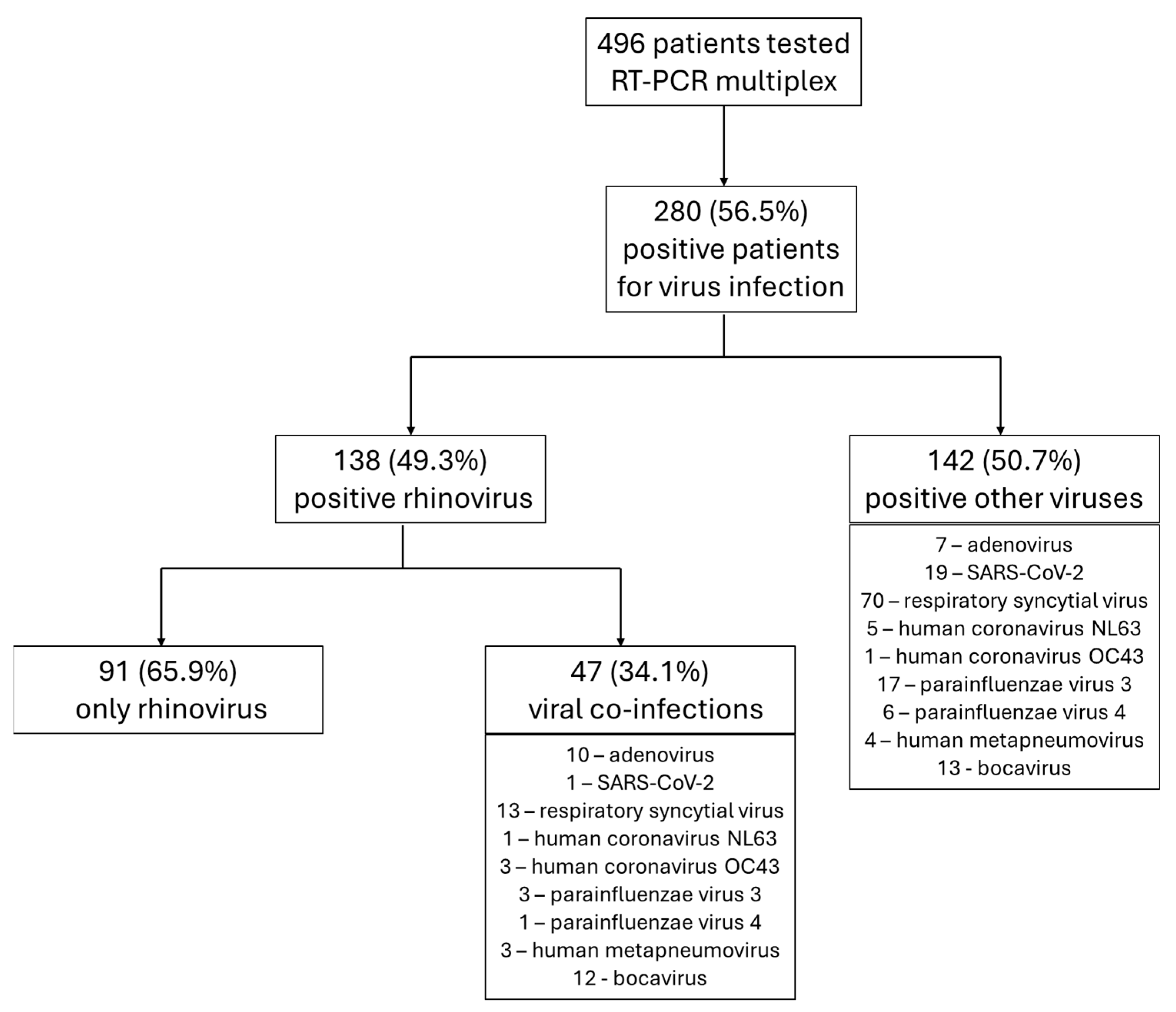
3.1. General Data Analysis

A total of 496 children hospitalized with ILI symptoms underwent RT-PCR testing during the period under review. The positivity rate for at least one viral pathogen was 58.5% (n = 280). Rhinovirus was identified in 138 patients, representing 27.8% of all children tested and 49.3% of all positive samples. In 34.1% (n = 47) of rhinovirus-positive cases, co-infections with other respiratory viruses were identified. The distribution of cases and RT-PCR results is illustrated in Figure 1.

3.2. Specific and Comparative Characteristics of Rhinovirus-Positive Cases

Of the 138 children identified with rhinovirus infection, 55.8% (n = 77) were male, and the median age was 12.5 months (IQR: 2, 41.8 months). The temporal distribution of rhinovirus cases, as illustrated in Figure 2, provides a comprehensive overview of the virus’s circulation patterns throughout the year.
Cough (63.7%) and dyspnea (51.6%) were the most prevalent symptoms among children hospitalized for rhinovirus infection. However, significant differences were observed in the clinical presentation between the analyzed groups (Table 1). In comparison to negative cases, children with rhinovirus were 4.90 times more likely to present with cough (OR = 4.90, 95% CI: 2.90–8.27), 5.25 times more likely to present with nasal congestion (OR = 5.25, 95% CI: 2.94–9.36), and 3.96 times more likely to present with dyspnea (OR = 3.94, 95% CI: 2.33–6.67), as illustrated in Figure 3 and Table 1 In comparison to children who were positive for other respiratory viruses, the likelihood of cough (OR = 0.31, 95% CI: 0.16–0.57), nasal congestion (OR = 0.53, 95% CI: 0.31–0.90), and dyspnea (OR = 4.90, 95% CI: 2.90–8.27) was significantly lower in those with a rhinovirus infection (Figure 3). The presence of another respiratory virus in conjunction with rhinovirus was associated with an increased frequency of cough (63.7% vs. 85.1%, p = 0.009, OR = 0.30, 95% CI: 0.12–0.76) and dyspnea (51.6% vs. 70.2%, p = 0.036, OR = 0.45, 95% CI: 0.21–0.96). The prevalence of fever was 47.3% among children with rhinovirus, which did not differ significantly from the other three groups (p > 0.05 for each, Table 1, Figure 3).
Analysis of blood test results revealed a 3.52-fold greater likelihood of having increased WBC (OR = 3.52, 95%CI: 1.92–6.45) and a 3.25-fold greater likelihood of having increased neutrophils (OR = 3.25, 95%CI: 1.74–6.06) in children with rhinovirus compared to those who tested positive for other respiratory viruses. Additionally, the risk of elevated CRP was 2.02 times higher (OR = 2.02, 95%CI: 1.18–3.44) in those with rhinovirus than those with other viruses.
The odds of receiving an antibiotic during hospitalization were found to be 1.92 times lower (OR = 0.52, 95% CI: 0.31–0.86, p = 0.011) in children who tested positive for rhinovirus compared to those with negative RT-PCR results. A high proportion of children with rhinovirus (63.7%) received inhalation therapy, and the rate of use increased in children who tested positive for other respiratory viruses (80.3%, p = 0.005) and in rhinovirus co-infections (70.2%, p = 0.446). Corticosteroids were administered to 42.9% of children with rhinovirus, a frequency that was higher than in those who tested negative (23.1%) or positive for other respiratory viruses (35.9%), but lower than in those with co-infections (55.3%).
The risk of acute bronchitis/bronchiolitis was 16.7 times higher in children with rhinovirus compared to children who tested negative (OR = 16.70, 95% CI: 8.02–34.75). In the case of co-infection, the risk decreased by 0.42 times for children infected with rhinovirus (OR = 0.42, 95% CI: 0.20–0.88, Table 1, Figure 4). Similarly, the risk of respiratory failure was 4.40 times higher in rhinovirus-positive children compared to those who tested negative (OR = 4.40, 95% CI: 2.47–7.83), but decreased compared to cases positive for other respiratory viruses (OR = 0.60, 95% CI: 0.35–1.08) or to cases with co-infections (OR = 0.44, 95% CI: 0.21–0.91). Additionally, the risks of acute otitis media (OR = 2.83, 95% CI: 1.11–7.24) and acute laryngitis (OR = 3.74, 95% CI: 1.03–13.59) were significantly elevated in comparison to those without identified viruses upon RT-PCR testing. No differences in the risk of acute pneumonia or acute dehydration were identified (Figure 4, Table 1).

4. Discussion

In this retrospective study conducted in one of the largest pediatric hospitals in the capital of Romania, we sought to highlight the clinical characteristics and outcomes associated with rhinovirus infection in children, comparing them with those of children who tested negative or positive for other respiratory viruses. During our analysis, we identified instances of positive infection with the rhinovirus, which occurred concurrently with the advent of the SARS-CoV-2 pandemic. This finding is particularly significant in the context of the broader changes in the epidemiology of respiratory viruses during this period. The SARS-CoV-2 pandemic led to an unprecedented shift in the circulation dynamics of respiratory pathogens, driven largely by public health interventions such as lockdowns, social distancing, and widespread use of masks. Notably, epidemiological surveillance studies have documented a dramatic decline in the incidence of traditionally prevalent respiratory viruses, most strikingly the influenza virus, which saw its incidence drop to near-zero levels in many regions. This shift highlights the complex interplay between rhinovirus and other respiratory viruses during the pandemic and underscores the need for continued monitoring to understand the long-term impacts on viral circulation and infection patterns in pediatric populations [14,15]. Similarly, the incidence of RSV, human coronaviruses, human metapneumovirus, and influenza viruses decreased. As for rhinovirus, a notable decrease in cases only occurred between March and May 2020, with the weekly percentage of positive results for rhinovirus being approximately 14.9% at the end of March and only 3.2% at the end of May. Thereafter, circulation levels increased to pre-pandemic values, with the percentage of positive results in October 2020 being similar to results in other years [16,17,18]. The study conducted by Smedberg et al. in the first part of 2021 demonstrated that 90% of patients presenting with respiratory symptoms specific to a viral infection and testing negative for SARS-CoV-2 but positive for at least one other respiratory pathogen were positive for rhinovirus [19]. In our study, rhinovirus was identified in 49.3% of the pediatric subjects with a positive multiplex RT-PCR result. The closure of educational institutions and the limitation of access to children’s recreational areas also appeared to contribute to the reduction in rhinovirus infections at the onset of the pandemic. However, in the long term, this did not prove to be a significant factor, as an increase in incidence was observed even before the resumption of academic activities [20]. The epidemiological and natural course of respiratory viral infections may be altered by interactions between viruses in cases of co-infection. This occurs through the alteration of receptor expression or other host cell factors that are necessary for replication. Studies have demonstrated that SARS-CoV-2 exhibits a markedly slower growth rate than rhinovirus. In the event of co-infection between the two viruses, the latter will be suppressed. Nevertheless, if rhinovirus infection occurs subsequently, the viral load of SARS-CoV-2 in bronchial epithelial cells is diminished, thereby attenuating the severity of symptoms associated with SARS-CoV-2 [21,22].
In our study, the clinical features of rhinovirus infection were characterized by respiratory manifestations that are typical of a viral upper respiratory tract infection. Cough was identified as the most prevalent symptom associated with rhinovirus infection. In comparison to patients infected with other respiratory viruses, children with rhinovirus infection exhibited a significantly reduced likelihood of developing additional respiratory symptoms, including rhinorrhea, wheezing, dyspnea, and fever. These observations are in accordance with the existing literature, which emphasizes that rhinovirus infection is not associated with distinctive pathognomonic signs or symptoms. Consequently, the diagnosis of this infection remains a significant clinical challenge, necessitating the integration of clinical, epidemiological, and laboratory criteria to ensure an accurate diagnosis [6,23,24]. Moreover, instances of asymptomatic rhinovirus infection have been documented, with a prevalence of up to 32% in children under four years of age, and this prevalence decreases with increasing age. However, the concept of asymptomatic rhinovirus infection requires careful interpretation, as advances in molecular diagnostic methods, such as RT-PCR, have revealed the presence of viral genetic material for prolonged periods after remission of the acute clinical episode. Therefore, the identification of viral RNA in an asymptomatic clinical context does not inherently signify an active infection. Rather, it may suggest the persistence of residual viral fragments [23,25]. This finding underlines the importance of correlating laboratory results with the patient`s clinical features to avoid overdiagnosis and mismanagement of pediatric patients.
The analysis of blood tests yielded no significant associations between identified items and the diagnosis of rhinovirus. The frequency of inflammatory syndrome was found to be higher among children with rhinovirus infections than among those with infections from other respiratory viruses. Nevertheless, other studies have demonstrated that rhinovirus monoinfections may be associated with mild to moderate elevations in serum CRP values [26], and that very high increases in CRP may indicate the possibility of a bacterial co-infection or superinfection.
The findings of our study indicate that a positive result on the RT-PCR test, particularly for rhinovirus, was associated with a notable reduction in the rate of antibiotic administration. It is well established that the majority of acute respiratory infections in children are of viral etiology [27,28]. In this context, the excessive and inappropriate use of antibiotics in viral infections, in the absence of bacterial co-infection, contributes significantly to the global phenomenon of antimicrobial resistance, which is a major public health problem [29,30].
The rapid differentiation between viral and bacterial infections is of crucial importance in clinical practice, as it ensures the appropriate administration of treatment. Multiplex RT-PCR assays have been demonstrated to be a valuable tool for this purpose [31]. Multiplex RT-PCR assays allow simultaneous identification of a broad spectrum of respiratory pathogens with high sensitivity and specificity, thereby reducing the need for empiric antibiotic administration in cases of unknown etiology [31]. For instance, a positive result for rhinovirus unambiguously indicates the presence of a viral pathogen and suggests that antibiotics are not indicated unless there is a high clinical suspicion of bacterial superinfection. Numerous studies have shown that, in the absence of molecular diagnostic tools, viral infections are often inappropriately managed with antibiotics, primarily due to the clinical challenges in distinguishing viral from bacterial infections based solely on symptoms and standard laboratory investigations. This diagnostic uncertainty leads to the overprescription of antibiotics, despite the lack of evidence for bacterial involvement, highlighting the critical need for more accurate diagnostic methods to guide appropriate treatment decisions [29,32,33]. The implementation of multiplex RT-PCR technologies in pediatric and emergency medicine units has led to a significant decrease in antibiotic prescriptions, while reducing hospitalization length and healthcare costs [31,34]. In particular, the identification of rhinovirus, one of the most common viral agents causing respiratory infections in children, has been associated with a reduction in antibiotic use of over 30% in cases of upper and lower respiratory tract infections [35], similar to our study, where we identified a reduction in antibiotic consumption compared to children with negative RT-PCR results. This molecular diagnostic approach offers significant advantages at both the individual and population levels. At the individual level, it helps to avoid adverse effects associated with unwarranted antibiotic treatment. At the population level, it contributes to maintaining the efficacy of antibiotics and preventing the spread of antimicrobial resistance.
Finally, our findings revealed that children with rhinovirus infection exhibited a heightened susceptibility to developing acute otitis media, acute respiratory failure, and acute bronchiolitis or bronchitis compared to those who tested negative. Nevertheless, the aforementioned risks are significantly diminished when compared to children who have tested positive for other respiratory viruses. It has been demonstrated that rhinovirus is one of the most prevalent viral pathogens associated with respiratory complications in children, exerting a significant influence on respiratory morbidity in this vulnerable population [36,37]. Although the risk of complications is higher in the presence of rhinovirus than in its absence, this risk is still lower when compared with other respiratory viral infections, such as those caused by RSV or influenza viruses [18,38,39]. In particular, rhinovirus infections are associated with an increased risk of developing acute otitis media, especially in children under the age of 3 years, which may lead to the need for therapeutic intervention and, in some cases, further complications if not treated appropriately [40,41]. Furthermore, clinical data indicate that pediatric patients with rhinovirus-positive respiratory viral infections are more likely to require hospitalization due to respiratory failure, though this likelihood is lower than in cases of RSV infections [42]. Consequently, in this context, the early identification of viral pathogens via advanced molecular assays, such as multiplex RT-PCR, is of paramount importance in order to ascertain the risk of complications and to customize therapeutic management in order to reduce morbidity. The implementation of these tests helps to prevent the overuse of antibiotics and reduce the length of hospitalization, which has a positive impact on the prognosis of pediatric patients.
It is important to acknowledge the inherent limitations of our study in order to contextualize the results. Firstly, the retrospective nature of the analysis may introduce a potential bias in the interpretation of the data, particularly in the assessment of the clinical picture and the evolution of the patients, as the available data may have been subject to variability in the way it was documented. Furthermore, it was not feasible to entirely negate the impact of potential discrepancies in the methodology employed for RT-PCR testing, in addition to the variations in therapeutic strategies applied to the children included in the study. These factors could potentially influence the comparability of the results. Notwithstanding these limitations, our study remains pertinent in terms of the large number of cases analyzed and the rigor of the evaluations performed, thereby contributing to a more nuanced understanding of the clinical and developmental differences between the groups analyzed according to the RT-PCR results.

5. Conclusions

We have shown in this study that rhinovirus is one of the most important viral agents involved in acute respiratory tract pathology in children. Clinical and blood test data are not suggestive for positive diagnosis of rhinovirus, and the use of multiplex RT-PCR molecular tests is helpful in clearly establishing the etiology. The implementation of these advanced diagnostic methods can bring significant benefits in practice, as a positive rhinovirus result helps reduce unnecessary antibiotic administration and optimizes patient management, thereby decreasing the risk of severe complications such as acute respiratory failure and acute otitis media.

Author Contributions

Conceptualization, S.C.; methodology, S.C. and M.C.; formal analysis, S.C., C.F. and M.C.; investigation, S.C. and C.F.; data curation, S.C. and C.F.; writing—original draft preparation, S.C.; writing—review and editing, S.C., C.F. and M.C.; supervision, M.C. All authors have read and agreed to the published version of the manuscript.

Funding

This research received no external funding.

Institutional Review Board Statement

The study was conducted in accordance with the Declaration of Helsinki and was approved by the Institutional Ethics Committee, number 15643/15 June 2024.

Informed Consent Statement

The study was conducted with a waiver for informed consent given the retrospective nature of the data, and the Ethics Committee approved this waiver.

Data Availability Statement

The data are available through reasonable request to the corresponding author.

Acknowledgments

Publication of this paper was supported by the University of Medicine and Pharmacy Carol Davila through the institutional program Publish not Perish.

Conflicts of Interest

The authors declare no conflicts of interest.

References

  1. Watson, A.; Wilkinson, T.M.A. Respiratory viral infections in the elderly. Ther. Adv. Respir. Dis. 2021, 15, 1753466621995050. [Google Scholar] [CrossRef] [PubMed]
  2. Leotte, J.; Trombetta, H.; Faggion, H.Z.; Almeida, B.M.; Nogueira, M.B.; Vidal, L.R.; Raboni, S.M. Impact and seasonality of human rhinovirus infection in hospitalized patients for two consecutive years. J. Pediatr. 2017, 93, 294–300. [Google Scholar] [CrossRef] [PubMed]
  3. Ljubin-Sternak, S.; Mestrovic, T. Rhinovirus-A True Respiratory Threat or a Common Inconvenience of Childhood? Viruses 2023, 15, 825. [Google Scholar] [CrossRef] [PubMed]
  4. Gern, J.E. The ABCs of rhinoviruses, wheezing, and asthma. J. Virol. 2010, 84, 7418–7426. [Google Scholar] [CrossRef] [PubMed]
  5. Vandini, S.; Biagi, C.; Fischer, M.; Lanari, M. Impact of Rhinovirus Infections in Children. Viruses 2019, 11, 521. [Google Scholar] [CrossRef]
  6. To, K.K.W.; Yip, C.C.Y.; Yuen, K.Y. Rhinovirus—From bench to bedside. J. Formos. Med. Assoc. 2017, 116, 496–504. [Google Scholar] [CrossRef]
  7. Winther, B. Rhinovirus infections in the upper airway. Proc. Am. Thorac. Soc. 2011, 8, 79–89. [Google Scholar] [CrossRef]
  8. Yin-Murphy, M.; Almond, J.W. Picornaviruses. In Medical Microbiology; University of Texas Medical Branch at Galveston: Galveston, TX, USA, 1996. [Google Scholar]
  9. Cafferkey, J.; Coultas, J.A.; Mallia, P. Human rhinovirus infection and COPD: Role in exacerbations and potential for therapeutic targets. Expert Rev. Respir. Med. 2020, 14, 777–789. [Google Scholar] [CrossRef]
  10. Atmar, R.L. Uncommon(ly considered) manifestations of infection with rhinovirus, agent of the common cold. Clin. Infect. Dis. 2005, 41, 266–267. [Google Scholar] [CrossRef]
  11. Kennedy, J.L.; Turner, R.B.; Braciale, T.; Heymann, P.W.; Borish, L. Pathogenesis of rhinovirus infection. Curr. Opin. Virol. 2012, 2, 287–293. [Google Scholar] [CrossRef]
  12. Miron, V.D.; Gunsahin, D.; Filimon, C.; Bar, G.; Craiu, M. Pediatric Emergencies and Hospital Admissions in the First Six Months of the COVID-19 Pandemic in a Tertiary Children’s Hospital in Romania. Children 2022, 9, 513. [Google Scholar] [CrossRef]
  13. Sandulescu, O.; Draganescu, A.; Pitigoi, D. Influenza redefined-clinical and epidemiological insight. Germs 2019, 9, 60. [Google Scholar] [CrossRef] [PubMed]
  14. Sandulescu, M.; Sandulescu, O. Changing clinical patterns and ear-nose-throat complications of seasonal viral respiratory tract infections. Germs 2023, 13, 311–313. [Google Scholar] [CrossRef] [PubMed]
  15. Draganescu, A.C.; Miron, V.D.; Sandulescu, O.; Bilasco, A.; Streinu-Cercel, A.; Sandu, R.G.; Marinescu, A.; Gunsahin, D.; Hoffmann, K.I.; Horobet, D.S.; et al. Omicron in Infants-Respiratory or Digestive Disease? Diagnostics 2023, 13, 421. [Google Scholar] [CrossRef] [PubMed]
  16. Olsen, S.J.; Winn, A.K.; Budd, A.P.; Prill, M.M.; Steel, J.; Midgley, C.M.; Kniss, K.; Burns, E.; Rowe, T.; Foust, A.; et al. Changes in Influenza and Other Respiratory Virus Activity During the COVID-19 Pandemic—United States, 2020–2021. Morb. Mortal. Wkly. Rep. 2021, 70, 1013–1019. [Google Scholar] [CrossRef] [PubMed]
  17. Miron, V.D.; Bar, G.; Filimon, C.; Craiu, M. From COVID-19 to Influenza-Real-Life Clinical Practice in a Pediatric Hospital. Diagnostics 2022, 12, 1208. [Google Scholar] [CrossRef]
  18. Miron, V.D.; Banica, L.; Sandulescu, O.; Paraschiv, S.; Surleac, M.; Florea, D.; Vlaicu, O.; Milu, P.; Streinu-Cercel, A.; Bilasco, A.; et al. Clinical and molecular epidemiology of influenza viruses from Romanian patients hospitalized during the 2019/20 season. PLoS ONE 2021, 16, e0258798. [Google Scholar] [CrossRef]
  19. Smedberg, J.R.; DiBiase, L.M.; Hawken, S.E.; Allen, A.; Mohan, S.; Santos, C.; Smedberg, T.; Barzin, A.H.; Wohl, D.A.; Miller, M.B. Reduction and persistence of co-circulating respiratory viruses during the SARS-CoV-2 pandemic. Am. J. Infect. Control. 2022, 50, 1064–1066. [Google Scholar] [CrossRef]
  20. Haapanen, M.; Renko, M.; Artama, M.; Kuitunen, I. The impact of the lockdown and the re-opening of schools and day cares on the epidemiology of SARS-CoV-2 and other respiratory infections in children–A nationwide register study in Finland. EClinicalMedicine 2021, 34, 100807. [Google Scholar] [CrossRef]
  21. Pinky, L.; Dobrovolny, H.M. SARS-CoV-2 coinfections: Could influenza and the common cold be beneficial? J. Med. Virol. 2020, 92, 2623–2630. [Google Scholar] [CrossRef]
  22. Esneau, C.; Duff, A.C.; Bartlett, N.W. Understanding Rhinovirus Circulation and Impact on Illness. Viruses 2022, 14, 141. [Google Scholar] [CrossRef] [PubMed]
  23. Jacobs, S.E.; Lamson, D.M.; St George, K.; Walsh, T.J. Human rhinoviruses. Clin. Microbiol. Rev. 2013, 26, 135–162. [Google Scholar] [CrossRef] [PubMed]
  24. Miron, V.D.; Craiu, M. Red throat or acute pharyngitis-challenges in real life clinical practice. Germs 2021, 11, 351–353. [Google Scholar] [CrossRef] [PubMed]
  25. Peltola, V.; Waris, M.; Osterback, R.; Susi, P.; Hyypia, T.; Ruuskanen, O. Clinical effects of rhinovirus infections. J. Clin. Virol. 2008, 43, 411–414. [Google Scholar] [CrossRef] [PubMed]
  26. Li, Y.; Min, L.; Zhang, X. Usefulness of procalcitonin (PCT), C-reactive protein (CRP), and white blood cell (WBC) levels in the differential diagnosis of acute bacterial, viral, and mycoplasmal respiratory tract infections in children. BMC Pulm. Med. 2021, 21, 386. [Google Scholar] [CrossRef] [PubMed]
  27. Debes, S.; Haug, J.B.; De Blasio, B.F.; Lindstrom, J.C.; Jonassen, C.M.; Dudman, S.G. Antibiotic Consumption in a Cohort of Hospitalized Adults with Viral Respiratory Tract Infection. Antibiotics 2023, 12, 788. [Google Scholar] [CrossRef]
  28. Diac, I.; Dogaroiu, C.; Keresztesi, A.A.; Horumba, M. Antimicrobial resistance trends-a single-center retrospective study of healthcare-associated pathogens-postmortem sampling from medico-legal autopsies in Bucharest. Germs 2022, 12, 352–360. [Google Scholar] [CrossRef]
  29. Schaut, M.; Schaefer, M.; Trost, U.; Sander, A. Integrated antibiotic clinical decision support system (CDSS) for appropriate choice and dosage: An analysis of retrospective data. Germs 2022, 12, 203–213. [Google Scholar] [CrossRef]
  30. Săndulescu, O.; Viziteu, I.; Streinu-Cercel, A.; Miron, V.D.; Preoțescu, L.L.; Chirca, N.; Albu, S.E.; Craiu, M.; Streinu-Cercel, A. Novel Antimicrobials, Drug Delivery Systems and Antivirulence Targets in the Pipeline—From Bench to Bedside. Appl. Sci. 2022, 12, 11615. [Google Scholar] [CrossRef]
  31. Rao, S.; Lamb, M.M.; Moss, A.; Mistry, R.D.; Grice, K.; Ahmed, W.; Santos-Cantu, D.; Kitchen, E.; Patel, C.; Ferrari, I.; et al. Effect of Rapid Respiratory Virus Testing on Antibiotic Prescribing Among Children Presenting to the Emergency Department with Acute Respiratory Illness: A Randomized Clinical Trial. JAMA Netw. Open 2021, 4, e2111836. [Google Scholar] [CrossRef]
  32. Tarciuc, P.; Plesca, D.A.; Duduciuc, A.; Gimiga, N.; Tataranu, E.; Herdea, V.; Ion, L.M.; Diaconescu, S. Self-Medication Patterns during a Pandemic: A Qualitative Study on Romanian Mothers’ Beliefs toward Self-Treatment of Their Children. Healthcare 2022, 10, 1602. [Google Scholar] [CrossRef] [PubMed]
  33. Plesca, V.S.; Miron, V.D.; Marinescu, A.G.; Draganescu, A.C.; Plesca, A.D.; Sandulescu, O.; Voiosu, C.; Hainarosie, R.; Streinu-Cercel, A. Hospitalizations for Acute Otitis and Sinusitis in Patients Living with HIV: A Retrospective Analysis of a Tertiary Center in Romania. J. Clin. Med. 2024, 13, 3346. [Google Scholar] [CrossRef] [PubMed]
  34. Fang, P.; Elena, A.X.; Kunath, M.A.; Berendonk, T.U.; Klumper, U. Reduced selection for antibiotic resistance in community context is maintained despite pressure by additional antibiotics. ISME Commun. 2023, 3, 52. [Google Scholar] [CrossRef] [PubMed]
  35. van Houten, C.B.; Cohen, A.; Engelhard, D.; Hays, J.P.; Karlsson, R.; Moore, E.; Fernandez, D.; Kreisberg, R.; Collins, L.V.; de Waal, W.; et al. Antibiotic misuse in respiratory tract infections in children and adults–A prospective, multicentre study (TAILORED Treatment). Eur. J. Clin. Microbiol. Infect. Dis. 2019, 38, 505–514. [Google Scholar] [CrossRef]
  36. Wildenbeest, J.G.; van der Schee, M.P.; Hashimoto, S.; Benschop, K.S.; Minnaar, R.P.; Sprikkelman, A.B.; Haarman, E.G.; van Aalderen, W.M.; Sterk, P.J.; Pajkrt, D.; et al. Prevalence of rhinoviruses in young children of an unselected birth cohort from the Netherlands. Clin. Microbiol. Infect. 2016, 22, 736.e9–736.e15. [Google Scholar] [CrossRef]
  37. Plesca, V.S.; Streinu-Cercel, A.; Sandulescu, O.; Draganescu, A.C.; Hainarosie, R.; Plesca, A.D. Incidence and Characteristics of Pediatric Patients with Acute Otitis Hospitalized in a Romanian Infectious Diseases Hospital. Children 2024, 11, 832. [Google Scholar] [CrossRef]
  38. Stewart, C.J.; Hasegawa, K.; Wong, M.C.; Ajami, N.J.; Petrosino, J.F.; Piedra, P.A.; Espinola, J.A.; Tierney, C.N.; Camargo, C.A., Jr.; Mansbach, J.M. Respiratory Syncytial Virus and Rhinovirus Bronchiolitis Are Associated with Distinct Metabolic Pathways. J. Infect. Dis. 2018, 217, 1160–1169. [Google Scholar] [CrossRef]
  39. Draganescu, A.C.; Miron, V.D.; Streinu-Cercel, A.; Florea, D.; Vlaicu, O.; Bilasco, A.; Otelea, D.; Luminos, M.L.; Pitigoi, D.; Streinu-Cercel, A.; et al. Circulation of influenza A viruses among patients hospitalized for severe acute respiratory infection in a tertiary care hospital in Romania in the 2018/19 season: Results from an observational descriptive epidemiological study. Medicine 2021, 100, e28460. [Google Scholar] [CrossRef]
  40. Chantzi, F.M.; Papadopoulos, N.G.; Bairamis, T.; Tsiakou, M.; Bournousouzis, N.; Constantopoulos, A.G.; Liapi, G.; Xatzipsalti, M.; Kafetzis, D.A. Human rhinoviruses in otitis media with effusion. Pediatr. Allergy Immunol. 2006, 17, 514–518. [Google Scholar] [CrossRef]
  41. Plesca, V.S.; Marinescu, A.G.; Voiosu, C.; Draganescu, A.C.; Streinu-Cercel, A.; Vilaia, A.; Hainarosie, R.; Plesca, D.A.; Sandulescu, O. Occurrence of acute otitis and sinusitis in patients hospitalized for influenza. Germs 2024, 14, 38–44. [Google Scholar] [CrossRef]
  42. Rankin, D.A.; Spieker, A.J.; Perez, A.; Stahl, A.L.; Rahman, H.K.; Stewart, L.S.; Schuster, J.E.; Lively, J.Y.; Haddadin, Z.; Probst, V.; et al. Circulation of Rhinoviruses and/or Enteroviruses in Pediatric Patients with Acute Respiratory Illness Before and During the COVID-19 Pandemic in the US. JAMA Netw. Open 2023, 6, e2254909. [Google Scholar] [CrossRef] [PubMed]
Figure 1. Distribution of cases in the study.
Children 11 01303 g001
Figure 2. Distribution of rhinovirus cases by month.
Children 11 01303 g002
Figure 3. Probability of fever, cough, nasal congestion, and dyspnea in patients with rhinovirus infection compared to other groups.
Children 11 01303 g003
Figure 4. Probability of developing complications in patients with rhinovirus infection compared to other groups.
Children 11 01303 g004
Table 1. Characteristics of children with rhinovirus and comparison with other groups.
CharacteristicsOnly Rhinovirus InfectionNegative Patientsp-ValueOther Respiratory Virusesp-ValueRhinovirus Co-Infectionp-Value
N = 91N = 216N = 142N = 47
Demographic data
Male49 (53.8)114 (52.8)0.86278 (54.9)0.86228 (59.6)0.521
Age (months), median (IQR)12 (2, 53)6.5 (0, 58.5)<0.0013 (0, 24.5)<0.00113 (4, 32)0.457
Clinical features
Malaise38 (41.8)73 (33.8)0.18456 (39.4)0.72919 (40.4)0.887
Fever43 (47.3)92 (42.6)0.45468 (47.9)0.92030 (63.8)0.064
Cough58 (63.7)57 (26.4)<0.001121 (85.2)<0.00140 (85.1)0.009
Nasal congestion39 (42.9)27 (12.5)<0.00183 (58.5)0.02023 (48.9)0.497
Dyspnea47 (51.6)46 (21.3)<0.00195 (66.9)0.01933 (70.2)0.036
Diarrhea18 (19.8)57 (26.4)0.21918 (12.7)0.1436 (12.8)0.303
Vomiting17 (18.7)34 (15.7)0.10515 (10.6)0.0792 (4.3)0.019
Laboratory findings
Increased WBC38 (41.8)66 (30.6)0.05824 (16.9)<0.00114 (29.8)0.169
Decreased WBC3 (3.3)16 (7.4)0.17210 (7.0)0.2230 (0.0)NA
Increased neutrophils 34 (37.4)64 (29.6)0.18422 (15.5)<0.00113 (27.7)0.254
Decreased neutrophils1 (1.1)14 (6.5)0.0766 (4.2)0.2511 (2.1)NA
Increased monocyte55 (60.4)125 (57.9)0.68078 (54.9)0.40631 (66.0)0.527
Increased lymphocyte9 (9.9)24 (11.1)0.75117 (12.0)0.6244 (8.5)0.828
Decreased lymphocyte22 (24.2)36 (16.7)0.12434 (23.9)0.9209 (19.1)0.502
Elevated CRP51 (56.0)96 (44.4)0.06355 (38.7)0.00920 (42.6)0.132
Treatment
Antibiotics34 (37.4)115 (53.2)0.01171 (50.0)0.05821 (44.7)0.350
Aerosol therapy58 (63.7)52 (24.9)<0.001114 (80.3)0.00533 (70.2)0.446
Isotonic saline solution *9 (9.9)7 (3.2)0.02319 (13.4)0.4235 (10.6)0.892
Hypertonic saline solution *15 (16.5)25 (11.6)0.24312 (8.5)0.0612 (4.3)0.041
Salbutamol *35 (38.5)25 (11.6)<0.00130 (21.1)0.00314 (29.8)0.312
Adrenaline *24 (26.4)20 (9.3)<0.00179 (55.6)<0.00121 (44.7)0.029
Cortisone #39 (42.9)50 (23.1)<0.00151 (35.9)0.28726 (55.3)0.164
Complications
AOM10 (11.0)9 (4.2)0.0238 (5.6)0.1355 (10.6)0.920
Acute bronchitis or bronchiolitis43 (47.3)11 (5.1)<0.00183 (58.5)0.09432 (68.1)0.019
ARF36 (39.6)28 (13.0)<0.00174 (52.1)0.06028 (59.6)0.025
Acute laryngitis6 (6.6)4 (1.9)0.0425 (3.5)0.3464 (8.5)0.734
Acute pneumonia7 (7.7)16 (7.4)0.92013 (9.2)0.7185 (10.6)0.751
Acute dehydration35 (38.5)81 (37.5)0.86250 (35.2)0.61721 (44.7)0.479
*—these treatments were administered as aerosol therapy (wet nebulization); #—oral or intravenous administration; IQR—interquartile range; ARF—acute respiratory failure; WBC—white blood cells; CRP—C-reactive protein; AOM—acute otitis media; NA—not applicable.
Disclaimer/Publisher’s Note: The statements, opinions and data contained in all publications are solely those of the individual author(s) and contributor(s) and not of MDPI and/or the editor(s). MDPI and/or the editor(s) disclaim responsibility for any injury to people or property resulting from any ideas, methods, instructions or products referred to in the content.

