
Evaluation of the Impact of Serum Vitamin D Levels on the Scoring Atopic Dermatitis Index in Pediatric Atopic Dermatitis

Abstract
1. Introduction
2. Materials and Methods
2.1. Skin Prick Tests
2.2. Total Eosinophil Count, Specific IgE in Serum, Total IgE, and Atopy
2.3. 25-Hydroxyvitamin D3 Levels
2.4. Statistical Analysis
2.5. Ethics
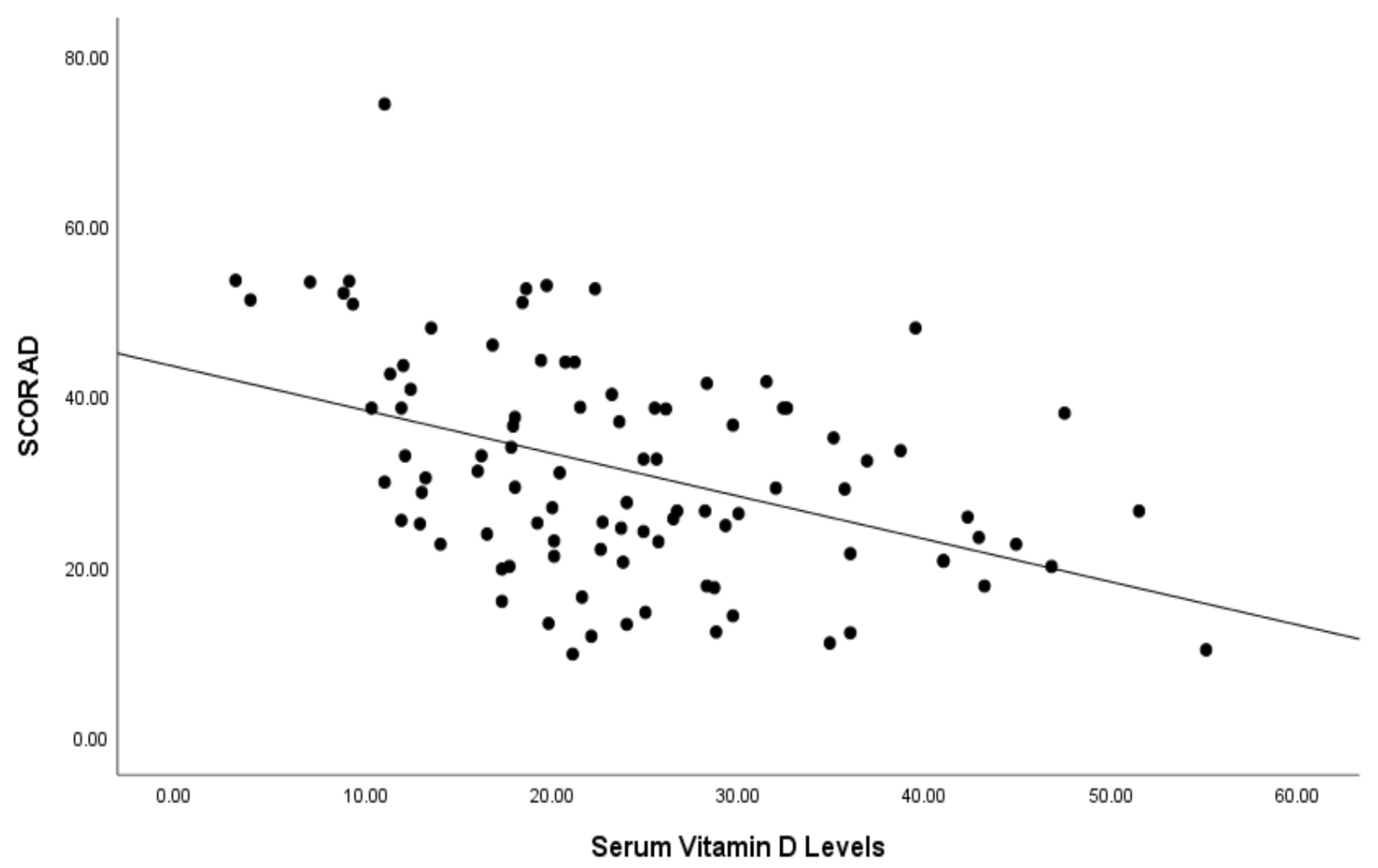
3. Results
4. Discussion
Limitations and Strengths
5. Conclusions
Author Contributions
Funding
Institutional Review Board Statement
Informed Consent Statement
Data Availability Statement
Conflicts of Interest
References
- Mesjasz, A.; Zawadzka, M.; Chałubiński, M.; Trzeciak, M. Is Atopic Dermatitis Only a Skin Disease? Int. J. Mol. Sci. 2023, 24, 837. [Google Scholar] [CrossRef] [PubMed]
- Su, O.; Bahalı, A.G.; Demir, A.D.; Ozkaya, D.B.; Uzuner, S.; Dizman, D.; Onsun, N. The relationship between severity of disease and vitamin D levels in children with atopic dermatitis. Postepy Dermatol. Alergol. 2017, 34, 224–227. [Google Scholar] [CrossRef]
- Lara-Corrales, I.; Huang, C.M.; Parkin, P.C.; Rubio-Gomez, G.A.; Posso-De Los Rios, C.J.; Maguire, J.; Pope, E. Vitamin D level and supplementation in pediatric atopic dermatitis: A randomized controlled trial. J. Cutan. Med. Surg. 2019, 23, 44–49. [Google Scholar] [CrossRef] [PubMed]
- Bulut, M.; Ozceker, D.; Tamay, Z. The relationship between serum vitamin D levels and childhood atopic dermatitis. Ann. Med. Res. 2020, 27, 1722–1727. [Google Scholar] [CrossRef]
- Wang, S.S.; Hon, K.L.; Kong, A.P.; Pong, H.N.; Wong, G.W.; Leung, T.F. Vitamin D deficiency is associated with diagnosis and severity of childhood atopic dermatitis. Pediatr. Allergy Immunol. 2014, 25, 30–35. [Google Scholar] [CrossRef] [PubMed]
- Bieber, T. Atopic dermatitis. Ann. Dermatol. 2010, 22, 125–137. [Google Scholar] [CrossRef] [PubMed]
- Kowalska-Olędzka, E.; Czarnecka, M.; Baran, A. Epidemiology of atopic dermatitis in Europe. J. Drug Assess. 2019, 8, 126–128. [Google Scholar] [CrossRef] [PubMed]
- Munawwarah, L.; Evalina, R.; Sofyani, S. Serum 25-hydroxyvitamin-D level and atopic dermatitis severity in children. Paediatr. Indones. 2017, 57, 234–238. [Google Scholar] [CrossRef][Green Version]
- Oranje, A.P.; Glazenburg, E.J.; Wolkerstorfer, A.; de Waard-van der Spek, F.B. Practical issues on interpretation of scoring atopic dermatitis: The SCORAD index, objective SCORAD and the three-item severity score. Br. J. Dermatol. 2007, 157, 645–648. [Google Scholar] [CrossRef]
- Umar, M.; Sastry, K.S.; Al Ali, F.; Al-Khulaifi, M.; Wang, E.; Chouchane, A.I. Vitamin D and the Pathophysiology of Inflammatory Skin Diseases. Skin Pharmacol. Physiol. 2018, 31, 74–86. [Google Scholar] [CrossRef]
- Bergler-Czop, B.; Brzezińska-Wcisło, L. Serum vitamin D level—The effect on the clinical course of psoriasis. Postepy Dermatol. Alergol. 2016, 33, 445–449. [Google Scholar] [CrossRef]
- Ao, T.; Kikuta, J.; Ishii, M. The Effects of Vitamin D on Immune System and Inflammatory Diseases. Biomolecules 2021, 11, 1624. [Google Scholar] [CrossRef] [PubMed]
- L Bishop, E.; Ismailova, A.; Dimeloe, S.; Hewison, M.; White, J.H. Vitamin D and Immune Regulation: Antibacterial, Antiviral, Anti-Inflammatory. JBMR Plus 2020, 5, e10405. [Google Scholar] [CrossRef]
- Karampinis, E.; Goudouras, G.; Ntavari, N.; Bogdanos, D.P.; Roussaki-Schulze, A.V.; Zafiriou, E. Serum vitamin D levels can be predictive of psoriasis flares up after COVID-19 vaccination: A retrospective case control study. Front. Med. 2023, 10, 1203426. [Google Scholar] [CrossRef]
- Reinholz, M.; Ruzicka, T.; Schauber, J. Vitamin D and its role in allergic disease. Clin. Exp. Allergy 2012, 42, 817–826. [Google Scholar] [CrossRef] [PubMed]
- Peroni, D.G.; Piacentini, G.L.; Cametti, E.; Chinellato, I.; Boner, A.L. Correlation between serum 25-hydroxyvitamin D levels and severity of atopic dermatitis in children. Br. J. Dermatol. 2011, 164, 1078–1082. [Google Scholar] [CrossRef] [PubMed]
- Robl, R.; Uber, M.; Abagge, K.T.; Lima, M.N.; Carvalho, V.O. Serum Vitamin D Levels Not Associated with Atopic Dermatitis Severity. Pediatr. Dermatol. 2016, 33, 283–288. [Google Scholar] [CrossRef]
- Chiu, Y.E.; Havens, P.L.; Siegel, D.H.; Ali, O.; Wang, T.; Holland, K.E.; Galbraith, S.S.; Lyon, V.B.; Drolet, B.A. Serum 25-hydroxyvitamin D concentration does not correlate with atopic dermatitis severity. J. Am. Acad. Dermatol. 2013, 69, 40–46. [Google Scholar] [CrossRef]
- Chalcraft, J.R.; Cardinal, L.M.; Wechsler, P.J.; Hollis, B.W.; Gerow, K.G.; Alexander, B.M.; Keith, J.F.; Larson-Meyer, D.E. Vitamin D Synthesis Following a Single Bout of Sun Exposure in Older and Younger Men and Women. Nutrients 2020, 12, 2237. [Google Scholar] [CrossRef]
- Ahn, J.Y.; Choi, B.S. Clinical Evaluation of Specific Immunoglobulin E in Sputum in Pediatric Patients. Pediatr. Allergy Immunol. Pulmonol. 2018, 31, 73–77. [Google Scholar] [CrossRef]
- Holick, M.F. Vitamin D status: Measurement, interpretation, and clinical application. Ann. Epidemiol. 2009, 19, 73–78. [Google Scholar] [CrossRef]
- Grant, W.B.; Boucher, B.J.; Bhattoa, H.P.; Lahore, H. Why vitamin D clinical trials should be based on 25-hydroxyvitamin D concentrations. J. Steroid Biochem. Mol. Biol. 2018, 177, 266–269. [Google Scholar] [CrossRef] [PubMed]
- Kennel, K.A.; Drake, M.T.; Hurley, D.L. Vitamin D deficiency in adults: When to test and how to treat. Mayo Clin. Proc. 2010, 85, 752–758. [Google Scholar] [CrossRef]
- Altaş, U.; Akgün Ünlü, D.; Güllüce, H.; Tunce, E.; Altaş, Z.M.; Söğütlü, Y.; Özkars, M.Y. Evaluation of the relationship between laboratory parameters and allergy tests in children with atopic dermatitis. Chron. Precis. Med. Res. 2023, 4, 168–171. [Google Scholar]
- Di Rosa, M.; Malaguarnera, M.; Nicoletti, F.; Malaguarnera, L. Vitamin D3: A helpful immuno-modulator. Immunology 2011, 134, 123–139. [Google Scholar] [CrossRef] [PubMed]
- Akdis, C.A.; Arkwright, P.D.; Brüggen, M.C.; Busse, W.; Gadina, M.; Guttman-Yassky, E.; Kabashima, K.; Mitamura, Y.; Vian, L.; Wu, J.; et al. Type 2 immunity in the skin and lungs. Allergy 2020, 75, 1582–1605. [Google Scholar] [CrossRef] [PubMed]
- Erem, A.S.; Razzaque, M.S. Vitamin D-independent benefits of safe sunlight exposure. J. Steroid Biochem. Mol. Biol. 2021, 213, 105957. [Google Scholar] [CrossRef]
- Fenner, J.; Silverberg, N.B. Oral supplements in atopic dermatitis. Clin. Dermatol. 2018, 36, 653–658. [Google Scholar] [CrossRef]
- Mutgi, K.; Koo, J. Update on the role of systemic vitamin D in atopic dermatitis. Pediatr. Dermatol. 2013, 30, 303–307. [Google Scholar] [CrossRef]
- Zhang, Y.; Leung, D.Y.; Richers, B.N.; Liu, Y.; Remigio, L.K.; Riches, D.W.; Goleva, E. Vitamin D inhibits monocyte/macrophage proinflammatory cytokine production by targeting MAPK phosphatase-1. J. Immunol. 2012, 188, 2127–2135. [Google Scholar] [CrossRef]
- Luger, T.; Amagai, M.; Dreno, B.; Dagnelie, M.A.; Liao, W.; Kabashima, K.; Schikowski, T.; Proksch, E.; Elias, P.M.; Simon, M.; et al. Atopic dermatitis: Role of the skin barrier, environment, microbiome, and therapeutic agents. J. Dermatol. Sci. 2021, 102, 142–157. [Google Scholar] [CrossRef] [PubMed]
- El Taieb, M.A.; Fayed, H.M.; Aly, S.S.; Ibrahim, A.K. Assessment of serum 25-hydroxyvitamin d levels in children with atopic dermatitis: Correlation with SCORAD index. Dermatitis 2013, 24, 296–301. [Google Scholar] [CrossRef] [PubMed]
- Cheon, B.R.; Shin, J.E.; Kim, Y.J.; Shim, J.W.; Kim, D.S.; Jung, H.L.; Park, M.S.; Shim, J.Y. Relationship between serum 25-hydroxyvitamin D and interleukin-31 levels, and the severity of atopic dermatitis in children. Korean J. Pediatr. 2015, 58, 96–101. [Google Scholar] [CrossRef]
- Raj, K.A.P.; Handa, S.; Narang, T.; Sachdeva, N.; Mahajan, R. Correlation of serum vitamin D levels with severity of pediatric atopic dermatitis and the impact of vitamin D supplementation on treatment outcomes. J. Dermatolog. Treat. 2022, 33, 1397–1400. [Google Scholar] [CrossRef] [PubMed]
- Hattangdi-Haridas, S.R.; Lanham-New, S.A.; Wong, W.H.S.; Ho, M.H.K.; Darling, A.L. Vitamin D Deficiency and Effects of Vitamin D Supplementation on Disease Severity in Patients with Atopic Dermatitis: A Systematic Review and Meta-Analysis in Adults and Children. Nutrients 2019, 11, 1854. [Google Scholar] [CrossRef] [PubMed]
- Kim, M.J.; Kim, S.-N.; Lee, Y.W.; Choe, Y.B.; Ahn, K.J. Vitamin D Status and Efficacy of Vitamin D Supplementation in Atopic Dermatitis: A Systematic Review and Meta-Analysis. Nutrients 2016, 8, 789. [Google Scholar] [CrossRef] [PubMed]
- Lubis, H.H.; Nababan, K.A.; Paramita, D.A. Correlation of low vitamin D status with atopic dermatitis severity in children. Bali Med. J. 2021, 10, 291–295. [Google Scholar] [CrossRef]
- Lee, S.A.; Hong, S.; Kim, H.J.; Lee, S.H.; Yum, H.Y. Correlation between serum vitamin d level and the severity of atopic dermatitis associated with food sensitization. Allergy Asthma Immunol. Res. 2013, 5, 207–210. [Google Scholar] [CrossRef]
- Akan, A.; Azkur, D.; Ginis, T.; Toyran, M.; Kaya, A.; Vezir, E.; Ozcan, C.; Ginis, Z.; Kocabas, C.N. Vitamin D level in children is correlated with severity of atopic dermatitis but only in patients with allergic sensitizations. Pediatr. Dermatol. 2013, 30, 359–363. [Google Scholar] [CrossRef]
- Liu, X.; Wang, G.; Hong, X.; Wang, D.; Tsai, H.J.; Zhang, S.; Arguelles, L.; Kumar, R.; Wang, H.; Liu, R.; et al. Gene-vitamin D interactions on food sensitization: A prospective birth cohort study. Allergy 2011, 66, 1442–1448. [Google Scholar] [CrossRef]
- Suaini, N.H.A.; Zhang, Y.; Vuillermin, P.J.; Allen, K.J.; Harrison, L.C. Immune Modulation by Vitamin D and Its Relevance to Food Allergy. Nutrients 2015, 7, 6088–6108. [Google Scholar] [CrossRef] [PubMed]
- Baek, J.H.; Shin, Y.H.; Chung, I.H.; Kim, H.J.; Yoo, E.G.; Yoon, J.W.; Jee, H.M.; Chang, Y.E.; Han, M.Y. The link between serum vitamin D level, sensitization to food allergens, and the severity of atopic dermatitis in infancy. J. Pediatr. 2014, 165, 849–854.e1. [Google Scholar] [CrossRef] [PubMed]
- Spencer, L.A.; Weller, P.F. Eosinophils and Th2 immunity: Contemporary insights. Immunol. Cell Biol. 2010, 88, 250–256. [Google Scholar] [CrossRef]
- Skrobot, A.; Demkow, U.; Wachowska, M. Immunomodulatory Role of Vitamin D: A Review. Adv. Exp. Med. Biol. 2018, 1108, 13–23. [Google Scholar]
- Ethier, C.; Yu, Y.; Cameron, L.; Lacy, P.; Davoine, F. Calcitriol Reduces Eosinophil Necrosis Which Leads to the Diminished Release of Cytotoxic Granules. Int. Arch. Allergy Immunol. 2016, 171, 119–129. [Google Scholar] [CrossRef]
- Souto Filho, J.T.D.; de Andrade, A.S.; Ribeiro, F.M.; Alves, P.A.S.; Simonini, V.R.F. Impact of vitamin D deficiency on increased blood eosinophil counts. Hematol. Oncol. Stem Cell Ther. 2018, 11, 25–29. [Google Scholar] [CrossRef] [PubMed]
- De Groot, J.C.; van Roon, E.N.; Storm, H.; Veeger, N.J.; Zwinderman, A.H.; Hiemstra, P.S.; Bel, E.H.; ten Brinke, A. Vitamin D reduces eosinophilic airway inflammation in nonatopic asthma. J. Allergy Clin. Immunol. 2015, 135, 670–675.e3. [Google Scholar] [CrossRef] [PubMed]
- Dogru, M.; Kirmizibekmez, H.; Yesiltepe Mutlu, R.G.; Aktas, A.; Ozturkmen, S. Clinical effects of vitamin D in children with asthma. Int. Arch. Allergy Immunol. 2014, 164, 319–325. [Google Scholar] [CrossRef]


| Group | |||
|---|---|---|---|
| Control n = 90 | Patient n = 96 | p Value | |
| Gender, n (%) | |||
| Female | 43 (47.8%) | 45 (46.9%) | 0.999 & |
| Male | 47 (52.2%) | 51 (53.1%) | |
| Age (years) | 4 (2–8) | 4 (3–6) | 0.855 |
| Total eosinophil count (103/mm3) | 287.4 ± 45.2 | 444.7 ± 43.8 | 0.018 † |
| Log total IgE | 1.71 ± 0.080 | 1.99 ± 0.07 | 0.018 † |
| 25-hydroxyvitamin D3 (ng/mL) | 29.71 ± 1.19 | 23.68 ± 1.15 | 0.001 † |
| SCORAD Index | |||
|---|---|---|---|
| n (%) | M (IQR) | p Value | |
| Gender | |||
| Girls | 45 (46.9) | 33.0 (18.1) | 0.072 † |
| Boys | 51 (53.1) | 26.5 (17.3) | |
| Vitamin D status | |||
| Severe (≤10 ng/mL) | 6 (6.2) | 52.7 (2.3) a | |
| Mild to moderate (10.1–24 ng/mL) | 50 (52.1) | 30.7 (18.3) b | <0.001 ‡ |
| Optimal (24.1–80 ng/mL) | 40 (41.7) | 26.0 (14.5) b | |
| Sensitization | |||
| None | 50 (52.1) | 25.6 (14.3) | 0.002 † |
| Positive | 46 (47.9) | 36.5 (18.8) | |
| Food allergy | |||
| None | 81 (84.4) | 26.9 (17.9) | 0.002 † |
| Positive | 15 (15.6) | 40.8 (22.6) | |
| Aeroallergen sensitivity | |||
| None | 62 (64.6) | 29.1 (17.5) | 0.285 † |
| Positive | 34 (35.4) | 31.7 (17.7) |
| SCORAD İndex | ||
|---|---|---|
| rho | p | |
| Age | −0.190 | 0.064 |
| Gender | −0.178 | 0.098 |
| Total eosinophil count | 0.336 | 0.001 |
| Log total IgE | 0.134 | 0.194 |
| Regression Coefficients * | |||||||
|---|---|---|---|---|---|---|---|
| β | se | Zβ | t | p | 95.0% Confidence Interval for β | ||
| Lower Bound | Upper Bound | ||||||
| Constant | 39.198 | 3.665 | 10.694 | <0.001 | 31.918 | 46.479 | |
| Serum vitamin D levels | −0.449 | 0.103 | −0.383 | −4.360 | <0.001 | −0.653 | −0.244 |
| Eosinophil Count | 0.009 | 0.002 | 0.361 | 4.146 | <0.001 | 0.005 | 0.013 |
| 25(OH)D, (ng/mL) | |||
|---|---|---|---|
| n (%) | M (IQR) | p Value | |
| AD severity (SCORAD) | <0.001 ‡ | ||
| Mild (≤25) | 33 (34.3) | 25.0 (15.4) a | |
| Moderate (25–50) | 52 (54.2) | 22.1 (13.8) a | |
| Severe (>50) | 11 (11.5) | 9.3 (11.6) b |
Disclaimer/Publisher’s Note: The statements, opinions and data contained in all publications are solely those of the individual author(s) and contributor(s) and not of MDPI and/or the editor(s). MDPI and/or the editor(s) disclaim responsibility for any injury to people or property resulting from any ideas, methods, instructions or products referred to in the content. |
© 2023 by the authors. Licensee MDPI, Basel, Switzerland. This article is an open access article distributed under the terms and conditions of the Creative Commons Attribution (CC BY) license (https://creativecommons.org/licenses/by/4.0/).
Share and Cite
Çiçek, F.; Köle, M.T. Evaluation of the Impact of Serum Vitamin D Levels on the Scoring Atopic Dermatitis Index in Pediatric Atopic Dermatitis. Children 2023, 10, 1522. https://doi.org/10.3390/children10091522
Çiçek F, Köle MT. Evaluation of the Impact of Serum Vitamin D Levels on the Scoring Atopic Dermatitis Index in Pediatric Atopic Dermatitis. Children. 2023; 10(9):1522. https://doi.org/10.3390/children10091522
Chicago/Turabian StyleÇiçek, Fatih, and Mehmet Tolga Köle. 2023. "Evaluation of the Impact of Serum Vitamin D Levels on the Scoring Atopic Dermatitis Index in Pediatric Atopic Dermatitis" Children 10, no. 9: 1522. https://doi.org/10.3390/children10091522
APA StyleÇiçek, F., & Köle, M. T. (2023). Evaluation of the Impact of Serum Vitamin D Levels on the Scoring Atopic Dermatitis Index in Pediatric Atopic Dermatitis. Children, 10(9), 1522. https://doi.org/10.3390/children10091522