Article

Impact of Frailty on Hippocampal Volume in Patients with Chronic Obstructive Pulmonary Disease

1
Department of Neuropsychiatry, Wakayama Medical University, Wakayama 641-0012, Japan
2
Department of Respiratory Medicine and Infectious Disease, Graduate School of Medicine, Yamaguchi University, Ube 755-8505, Japan
3
Department of System Neurophysiology, Wakayama Medical University, Wakayama 641-0012, Japan
4
Department of Radiology, St. Hill Hospital, Ube 755-0155, Japan
5
Department of Medicine and Clinical Science, Graduate School of Medicine, Yamaguchi University, Ube 755-8505, Japan
*
Author to whom correspondence should be addressed.
Biomedicines 2021, 9(9), 1103; https://doi.org/10.3390/biomedicines9091103
Submission received: 18 July 2021 / Revised: 22 August 2021 / Accepted: 25 August 2021 / Published: 28 August 2021

Abstract

:
Brain frailty may be related to the pathophysiology of poor clinical outcomes in chronic obstructive pulmonary disease (COPD). This study examines the relationship between hippocampal subfield volumes and frailty and depressive symptoms, and their combined association with quality of life (QOL) in patients with COPD. The study involved 40 patients with COPD. Frailty, depressive symptoms and QOL were assessed using Kihon Checklist (KCL), Hospital Anxiety and Depression Scale (HADS), and World Health Organization Quality of Life Assessment (WHO/QOL-26). Anatomical MRI data were acquired, and volumes of the hippocampal subfields were obtained using FreeSurfer (version 6.0). Statistically, HADS score had significant association with WHO/QOL-26 and KCL scores. KCL scores were significantly associated with volumes of left and right whole hippocampi, presubiculum and subiculum, but HADS score had no significant association with whole hippocampi or hippocampal subfield volumes. Meanwhile, WHO/QOL-26 score was significantly associated with volume of the left CA1. There was a significant association between frailty, depression, and QOL. Hippocampal pathology was related to frailty and, to some extent, with QOL in patients with COPD. Our results suggest the impact of frailty on hippocampal volume and their combined associations with poor QOL in COPD.

1. Introduction

Chronic obstructive pulmonary disease (COPD) is a highly prevalent chronic disease, the progressive course of which impacts upon mortality and disturbs various aspects of the patient’s life [1,2]. Chronic systemic inflammatory reaction causes extra-pulmonary manifestation in COPD, such as motor and psychiatric symptoms [3,4,5,6,7]. Systematic review and meta-analyses have reported the prevalence of frailty and depression in patients with COPD as 19% [8] and 27.1% [9], respectively.
In patients with COPD, poor clinical outcomes and a higher rate of mortality are associated with frailty [10,11,12] and co-morbid depression [13,14,15,16,17,18]. COPD has been shown in longitudinal studies to increase the risk of developing depression, although there is also an inverse relationship: depression adversely affects the prognosis of COPD [19]. Similarly, individuals with frailty have been reported to have an increased likelihood of developing depression, and vice versa [20].
Hypoxia, inflammatory mediators and cerebral small vessel disease are leading brain pathologies of patients with COPD [21,22]. The hippocampus is a plastic and vulnerable region of the brain that is especially susceptible to damage [23]. Hippocampal volume loss has been reported in patients with COPD [24,25], in frailty [26,27] and in patients with depression [28].
We hypothesized that volume loss in hippocampal subfields might be associated with poor clinical outcomes in patients with COPD. We, therefore, examined the relationship between hippocampal subfield volumes and frailty, depressive symptoms and quality of life (QOL). We also aimed to elucidate the association between frailty and depressive symptoms and QOL in patients with COPD.

2. Materials and Methods

2.1. Subjects

Forty patients with COPD diagnosed by a pulmonologist and recruited from the Yamaguchi Medical University Hospital were included in the study (Table 1). All patients were diagnosed and treated based on the Global Initiative for Chronic Obstructive Lung Disease (GOLD) guidelines (Global Initiative for Chronic Obstructive Lung Disease. 2021, available online: www.goldcopd.inc. (accessed on 10 June 2021)). They were all in a stable condition and had no exacerbations for at least three months prior to the study. We excluded patients with other pulmonary diseases, such as interstitial lung disease, and those with disorders that would prevent them from completing the study assessments. This study was approved by the ethics committees at Yamaguchi University (6 June 2016) and Wakayama Medical University (19 August 2016), and the trial was registered at UMIN-ICDR (UMIN000024645, 13 October 2016). Written informed consent was obtained from all patients.

2.2. Clinical Assessments

Frailty, depressive symptoms and QOL were assessed using Kihon Checklist (KCL), Hospital Anxiety and Depression Scale (HADS) and WHO/QOL-26, respectively. KCL is a self-reported comprehensive health checklist designed by a study group from the Japanese Ministry of Health, Labor and Welfare. It is used as a screening tool of frailty for community-dwelling older adults [29] and patients with COPD [30,31,32].

2.3. Neuroimaging Analysis

Anatomical MRI data on a 3.0T MR scanner (Achieva 3.0T Quasar Dual; Philips Medical Systems) was acquired using an 8ch SENSE head coil. A 3D fastfield echo T1-weighted sequence was used for anatomical MRI (TR/TE = 7.0/3.3 ms, FOV = 256 mm, 200 slices, acquisition voxel size = 1.00 × 1.00 × 1.00 mm, and a slice thickness = 1.0 mm). Volumes of the hippocampal subfields and estimated total intracranial volume (eTIV) in each hemisphere were obtained using FreeSurfer software (version 6.0) (https://surfer.nmr.mgh.harvard.edu, accessed on 10 June 2021) (Figure S1). In segmentation of the hippocampus, to reduce multiple comparisons, we focused on seven main subfields (CA1, CA3, CA4, parasubiculum, presubiculum, subiculum, GC-ML-DG). The quality of preprocessed images was visually checked by S.T. and K.Y.

2.4. Statistics

All statistical analyses were performed using IBM SPSS Statistics for Windows, Version 22 (IBM Japan, Ltd., Tokyo, Japan). Normality of data was assessed using the Shapiro–Wilk test with statistical significance set at p < 0.05. There were non-normal distributions of volumes of left and right GC-ML-GD, right whole hippocampus, right CA1, right CA3, right CA4, and KCL score. The other hippocampal subfield volumes, age, HADS score, WHO/QOL-26 score, and values of respiratory function (%VC, %FVC, %FEV1, %DLco/VA) had normal distribution. Subjects were grouped into high, moderate, and low QOL groups according to top and bottom 25th percentile point of WHO/QOL-26 (high QOL: 4.38–3.50, moderate QOL: 3.42–2.69, low QOL: 2.65–1.88). They were also grouped into non-frail, pre-frail, and frail groups by KCL score cutoff points (non-frail: <3, pre-frail: 4–7, frail: >7) [33], and also grouped into non-depressive, moderate depressive, and depressive groups by HADS score cutoff points (non-depressive: <11, moderate depressive: 11–19, depressive: >19) [34]. In categorical analyses, differences in HADS or KCL scores between groups divided by WHO/QOL-26 scores were analyzed using Mann–Whitney U tests. Differences in KCL scores between groups divided by HADS score and differences in HADS scores between groups divided by KCL score were analyzed using Mann–Whitney U tests. In dimensional analyses, correlations of WHO/QOL-26 scores with HADS and KCL scores and correlations between HADS and KCL scores were analyzed using Spearman’s partial rank correlation tests with age as a covariate. Differences in volume of whole hippocampi or hippocampal subfields between groups divided by HADS or KCL scores were analyzed using Mann–Whitney U tests. There were significant differences in categorical analysis, these associations were confirmed using Spearman’s partial rank correlation tests with age and eTIV as covariates. To evaluate direct association between hippocampal volume and QOL, differences in volume of whole hippocampi or hippocampal subfields between groups divided by WHO/QOL-26 scores were analyzed using Mann–Whitney U tests. When significant differences were found in categorical analysis, these associations were confirmed using Spearman’s partial rank correlation tests with age and eTIV as covariates. Correlations between values of respiratory function (%VC, %FVC, %FEV1, %DLco/VA) and scores of WHO/QOL-26, HADS, KCL were analyzed using Spearman’s partial rank correlation tests with age as a covariate. Correlations between values of respiratory function (%VC, %FVC, %FEV1, %DLco/VA) and volume of whole hippocampi or hippocampal subfields were examined using Spearman’s partial rank correlation tests with age and eTIV as covariates. To evaluate direct and indirect relation, the factors which showed significant correlation were analyzed using step-wise multiple regression analysis. Statistical significance was set at p < 0.05 in all analyses.

3. Results

3.1. Association between QOL and Frailty and Depressive Symptoms

Subjects were divided into high QOL (n = 11), moderate QOL (n = 19), and low QOL (n = 10) groups (Table S1). HADS and KCL scores were significantly higher in the low QOL group compared with those in the high and moderate QOL groups (Table S1). In correlation analysis, WHO/QOL-26 scores were significantly negatively correlated with KCL (r = −0.596; p < 0.001) and HADS (r = −0.723; p < 0.001) scores (Table 2).

3.2. Association between Frailty and Depressive Symptoms

The patients were divided into non-frail (n = 9), pre-frail (n = 11), and frail (n = 20) groups (Table S2) and divided into non-depressive (n = 15), moderate depressive (n = 18), and depressive (n = 7) groups (Table S3). HADS scores were significantly higher in the frail group compared with the non-frail and pre-frail groups (Table S2). KCL scores were significantly higher in the depressive group compared with the non-depressive and moderate depressive groups (Table S3). In correlation analysis, there was significant correlation between HADS and KCL scores (r = 0.560; p < 0.001) (Table 2).

3.3. Association between Volume of Hippocampal Subfields and Frailty and Depressive Symptoms

Volume of the left and right whole hippocampi, subiculum, and presubiculum were significantly lower in the frail group compared with non-frail groups (Table S4). Volume of the left and right whole hippocampi, left subiculum, and right presubiculum were significantly lower in the frail group compared with pre-frail groups (Table S4). None of the hippocampal subfield volumes had significant difference between groups divided by depressive symptoms (Table S5). In correlation analysis, KCL scores were significantly negatively correlated with volume of left and right whole hippocampi, subiculum and presubiculum (Table 3, Figure 1a,b).

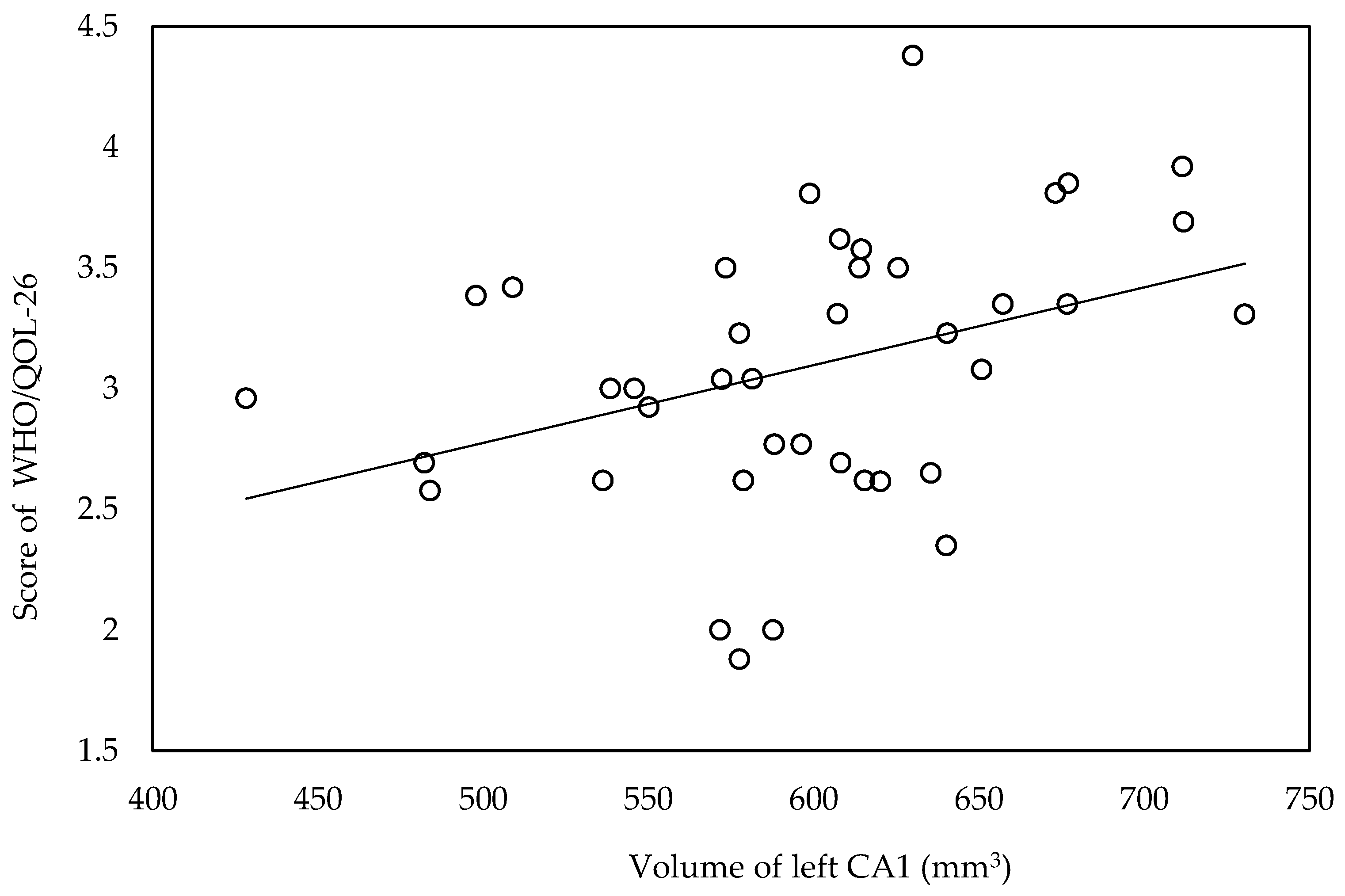
3.4. Association between Hippocampal Subfield Volume and QOL

Volume of left CA1 was significantly lower in moderate and low QOL groups compared with high QOL group (Table S6). In correlation analysis, WHO/QOL-26 scores were significantly correlated with volume of left CA1 (r = 0.401, p = 0.013) (Figure 2).

3.5. Association between Respiratory Values with QOL, Frailty, Depressive Symptoms and Hippocampal Subfield Volume

KCL scores were significantly correlated with values of %VC (r = −0.411, p = 0.009) and %FVC (r = −0.426, p = 0.007). There were no significant correlations between respiratory values and the volume of whole hippocampi and hippocampal subfields.

3.6. Step-Wise Multiple Regression Analyses for the Correlated Factors
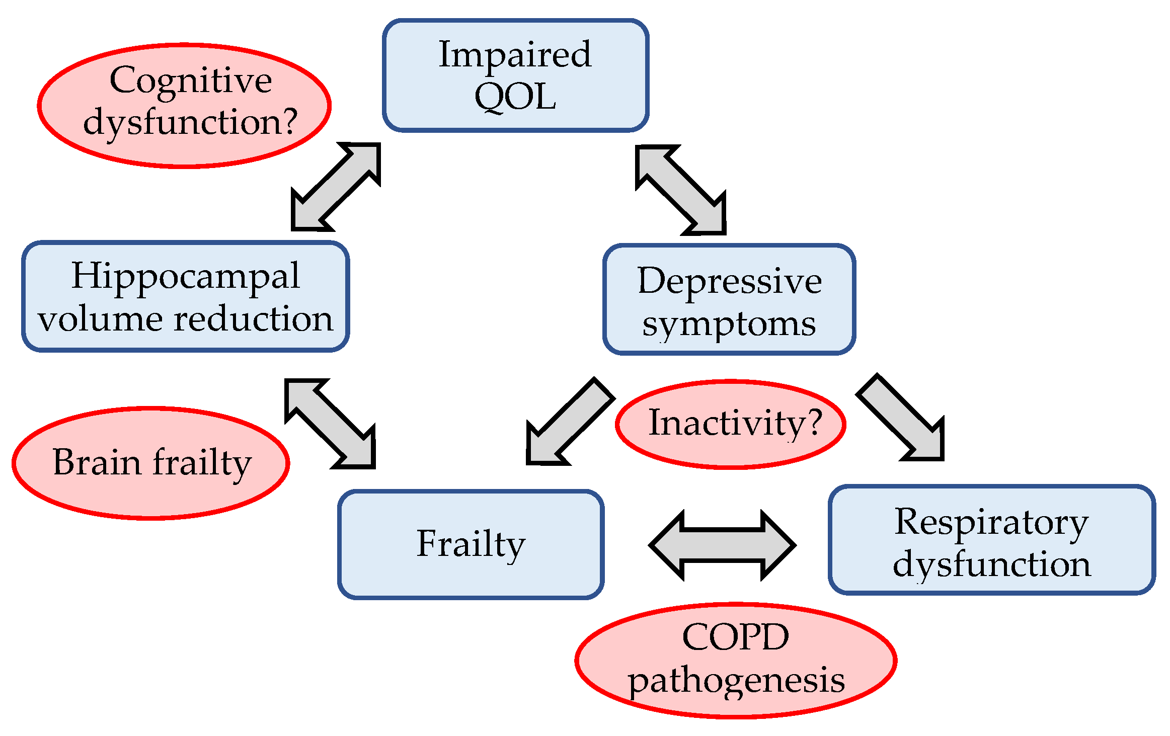
Step-wise multiple regression analyses revealed that WHO/QOL-26 score was sig-nificantly related to HADS score (β = −0.712, p < 0.001) and volume of left CA1 (β = 0.252, p = 0.017) (Table S7); HADS score was significantly related to WHO/QOL-26 score (β = −0.755, p < 0.001) (Table S7); KCL score was significantly related to HADS score (β = 0.565, p < 0.001), volume of left whole hippocampus (β = −0.394, p < 0.001) and value of %FVC (β = −0.353, p = 0.001) (Table S7); volume of left and right whole hippocampi, subiculum and presubiculum were significantly related to KCL scores (Table S8); volume of left CA1 was significantly related to WHO/QOL-26 score (β = 0.376, p = 0.017) (Table S8); value of %VC was significantly related with KCL score (β = −0.588, p = 0.003) and HADS score (β = 0.419, p = 0.029) (Table S9); value of %FVC was significantly related to KCL score (β = −0.607, p = 0.002) and HADS score (β = 0.411, p = 0.031) (Table S9). Associations revealed by step-wise multiple regression analyses are shown in Figure 3.

4. Discussion

This study confirmed association between poor QOL with frailty and with depressive symptoms. Depressive symptoms had impact on frailty and respiratory dysfunction. Interestingly, hippocampal volume was associated with frailty and QOL in patients with COPD, but not with depressive symptoms.
To our knowledge, this is the first study to show statistically significant direct association between volume reduction in the hippocampi and frailty, and therefore QOL in patients with COPD. It suggests a significant role of brain pathology in COPD prognosis. A high prevalence of frailty in patients with COPD (> 50%) was reported in a meta-analysis of an observational study [8] and in a national survey based in the United States [35]. Frailty is one of the worst prognostic factors related to increased rate of hospitalization [10], high mortality [10,11], greater disability [12,35], and to poor patient-reported outcomes and QOL [10,30]. Volume reduction in the hippocampi of patients with COPD has been reported [24,25], and relationships have been suggested between hippocampal volume loss in older adults with frailty [26] and with weakness and slowness [27]. Figure S1 shows the hippocampal subfield segmentation which was focused on in this study. Left and right subiculum and presubiculum volumes showed significant negative correlation with KCL scores. The subiculum and presubiculum are structures located within the hippocampus proper and entorhinal and other cortices, and are, therefore, a major output structure of the hippocampus [36,37,38]. Further research is needed to elucidate the relationship between frailty and reduced hippocampal volume and cognitive decline in patients with COPD.
Contrary to our expectations, the volume of hippocampal subfields did not have significant correlation with HADS scores. Among our patients with COPD, the number who scored above the “depressive” cutoff point in HADS was 7/40, while 20/40 scored above the “frail” cutoff point in KCL, suggesting lower severity of depressive symptoms than those of frailty. Moreover, while some studies have highlighted hippocampal volume reduction in patients with depression [39,40,41,42,43,44], depressive symptoms in COPD may have complex biological mechanisms arising from systemic hypoxia [45,46] and inflammation [45,46,47,48,49,50], contributing to the lack of statistically significant correlation between hippocampal volume and depressive symptoms in our patients with COPD.
While the volume of the whole hippocampi or of several hippocampal subfields were associated with frailty, the volume of the left CA1 was significantly associated solely with the QOL score. This suggests hippocampal volume had a weak association with QOL compared with frailty. Interestingly, CA1 was significantly associated with QOL, but not with frailty. CA1 is located between CA3 and the subiculum and modulate hippocampal circuitry [51,52], so further research is needed to elucidate any association between hippocampal circuit activity and QOL in patients with COPD.
Concerning the results of step-wise multiple regression analyses, our patients with COPD had a significant association between QOL and depressive symptoms and hippocampal volume reduction. Among our subjects, frailty was not directly associated with QOL, but it may impact upon QOL via reduction in hippocampal volume (brain frailty). Our results are in line with previous studies that reported a relationship between impaired QOL in patients with COPD and comorbid depression [13,16,53], as well as frailty [10]. QOL has been recognized as an important outcome in COPD because it is a predictor of subsequent exacerbations of COPD, hospitalization, and all-course mortality [2,54]. Association between depression and low physical activity has been reported in some cross-sectional studies [55,56], and a previous study with prospective design reported association between depressive symptoms and a reduction in physical activity six months later [57]. Bidirectional association between depression and clinical outcomes in COPD has been demonstrated in a meta-analysis of longitudinal studies [19]. In a longitudinal study of community-dwelling older adults, frailty was reported to predict persistent depressive symptoms [58]. Our results and those of these previous studies highlight the importance of intervention against frailty or depressive symptoms to improve QOL in patients with COPD.
Interestingly, in categorical analyses, patients in the low-QOL group showed worse frailty and depressive symptoms than patients in the moderate- and high-QOL groups, but there was no difference between high- and moderate-QOL groups. Similarly, patients in the frail group showed worse depressive symptoms than patients in the non-frail and pre-frail groups, but there was no difference between patients in the non-frail and pre-frail group. Patients in the depressive group showed worse frailty than patients in the non-depressive and moderate depressive groups, but there was no difference between patients in the non-depressive and moderate depressive groups. This suggests that early intervention during pre-frailty or moderate depressive symptoms could prevent the decline of QOL in patients with COPD. Our results also showed that frailty could be the cause of hippocampal volume reduction in patients with COPD. Previous studies have demonstrated reversibility of hippocampal volume reduction by increased physical activity [59,60]. This evidence suggests that early intervention for frailty may target brain frailty in patients with COPD, so assessment of the hippocampus, such as in our study could be useful for the management of COPD.
Our study has some limitations, and the results should be interpreted cautiously. First, healthy controls were not included in this study. While we found COPD pathogenic relation between respiratory dysfunction and frailty and their combined impacts upon hippocampal volume reduction and QOL, these associations were not compared with healthy controls. However, since healthy subjects usually do not have respiratory dysfunction, we have believed that our results are specific for COPD. Second, WHO/QOL-26 is not a QOL assessment tool specifically for patients with COPD. However, assessment by WHO/QOL has been widely used in disease groups, including for patients with COPD, in previous studies [61,62]. Third, treatment for COPD or several comorbidities with COPD might influence frailty, hippocampal volume, depressive symptoms and QOL. Notably, overlap with obstructive sleep apnea syndrome (OSAS) should be screened in future studies because some previous studies have reported association between OSAS and impaired QOL [63,64] and hippocampal volume reduction [65].
In conclusion, in this study there was significant association between frailty, depressive symptoms, and poor QOL. Hippocampal pathology could be related to frailty and to some extent with QOL in patients with COPD. Brain frailty accompanied by reduction in hippocampal volume is suggested to be a cause of poor QOL. Further research with a longitudinal design is needed to elucidate the causal relationship of our findings.

Supplementary Materials

The following are available online at https://www.mdpi.com/article/10.3390/biomedicines9091103/s1, Figure S1: Hippocampal subfield segmentation, Table S1: Differences of HADS or KCL scores between groups divided by WHO/QOL-26, Table S2: Differences of HADS scores between groups divided by KCL score, Table S3: Differences of KCL scores between groups divided by HADS score, Table S4: Differences in volume of whole hippocampi or hippocampal subfields between groups divided by KCL score, Table S5: Differences in volume of whole hippocampi or hippocampal subfields between groups divided by HADS score, Table S6: Differences in volume of whole hippocampi or hippocampal subfields between groups divided by WHO/QOL-26 score, Table S7: Step-wise multiple regression analysis of the relationship among WHO/QOL-26, KCL and HADS scores, volume of whole hippocampi or hippocampal subfields, and values of respiratory function, Table S8: Step-wise multiple regression analysis of the relationship between volume of whole hippocampi or hippocampal subfields with WHO/QOL-26, HADS and KCL scores and values of respiratory function, Table S9: Step-wise multiple regression analysis of the relationship between values of respiratory function with WHO/QOL-26, KCL and HADS scores and volume of whole hippocampi or hippocampal subfields.

Author Contributions

S.T., T.H. and K.M. designed the study. T.H, K.S., K.O., S.O., Y.M., Y.Y., M.A.-N. and N.E. oversaw subject recruitment. T.H. and K.D. produced and organized the datasets. S.T., K.Y. and T.D. conducted neuroimaging data processing. S.T. performed the initial analysis and wrote the first draft of the paper. T.H. and K.Y. assisted in additional analyses and in writing the manuscript. S.T., T.H. and K.M. provided feedback about the study design during the study implementation. All authors have read and agreed to the published version of the manuscript.

Funding

This research was funded by KAKENHI, grant number 21K08180.

Institutional Review Board Statement

This study was approved by the ethics committees at Yamaguchi University (No.H28-031-10) and Wakayama Medical University (No.1843), and the trial was registered at UMIN-ICDR (UMIN000024645).

Informed Consent Statement

Informed consent was obtained from all subjects involved in the study.

Data Availability Statement

Research data are not shared.

Acknowledgments

The authors would like to thank Akio Nakajima, Hiroe Hotta, and Kumiko Teramoto for their contributions to this study. We also acknowledge proofreading and editing by Benjamin Phillis at the Clinical Study Support Center at Wakayama Medical University.

Conflicts of Interest

The authors declare no conflict of interest.

References

  1. Rabe, K.F.; Watz, H. Chronic obstructive pulmonary disease. Lancet 2017, 389, 1931–1940. [Google Scholar] [CrossRef]
  2. Yorgancioglu, A.; Havlucu, Y.; Celik, P.; Dinc, G.; Saka, A. Relation between quality of life and morbidity and mortality in COPD patients: Two-year follow-up study. COPD 2010, 7, 248–253. [Google Scholar] [CrossRef]
  3. Agusti, A.; Soriano, J.B. COPD as a systemic disease. COPD 2008, 5, 133–138. [Google Scholar] [CrossRef] [PubMed]
  4. Decramer, M.; Janssens, W.; Miravitlles, M. Chronic obstructive pulmonary disease. Lancet 2012, 379, 1341–1351. [Google Scholar] [CrossRef]
  5. Hirano, T.; Doi, K.; Matsunaga, K.; Takahashi, S.; Donishi, T.; Suga, K.; Oishi, K.; Yasuda, K.; Mimura, Y.; Harada, M.; et al. A novel role of growth differentiation factor (GDF)-15 in overlap with sedentary lifestyle and cognitive risk in COPD. J. Clin. Med. 2020, 9, 2737. [Google Scholar] [CrossRef] [PubMed]
  6. Matsunaga, K.; Harada, M.; Suizu, J.; Oishi, K.; Asami-Noyama, M.; Hirano, T. Comorbid conditions in chronic obstructive pulmonary disease: Potential therapeutic targets for unmet needs. J. Clin. Med. 2020, 9, 3078. [Google Scholar] [CrossRef]
  7. Oishi, K.; Matsunaga, K.; Harada, M.; Suizu, J.; Murakawa, K.; Chikumoto, A.; Ohteru, Y.; Matsuda, K.; Uehara, S.; Hamada, K.; et al. A new dyspnea evaluation system focusing on patients’ perceptions of dyspnea and their living disabilities: The linkage between COPD and frailty. J. Clin. Med. 2020, 9, 3580. [Google Scholar] [CrossRef]
  8. Marengoni, A.; Vetrano, D.L.; Manes-Gravina, E.; Bernabei, R.; Onder, G.; Palmer, K. The relationship between COPD and frailty: A systematic review and meta-analysis of observational studies. Chest 2018, 154, 21–40. [Google Scholar] [CrossRef]
  9. Matte, D.L.; Pizzichini, M.M.; Hoepers, A.T.; Diaz, A.P.; Karloh, M.; Dias, M.; Pizzichini, E. Prevalence of depression in COPD: A systematic review and meta-analysis of controlled studies. Respir Med. 2016, 117, 154–161. [Google Scholar] [CrossRef]
  10. Kennedy, C.C.; Novotny, P.J.; LeBrasseur, N.K.; Wise, R.A.; Sciurba, F.C.; Benzo, R.P. Frailty and clinical outcomes in chronic obstructive pulmonary disease. Ann. Am. Thorac. Soc. 2019, 16, 217–224. [Google Scholar] [CrossRef]
  11. Lahousse, L.; Ziere, G.; Verlinden, V.J.; Zillikens, M.C.; Uitterlinden, A.G.; Rivadeneira, F.; Tiemeier, H.; Joos, G.F.; Hofman, A.; Ikram, M.A.; et al. Risk of frailty in elderly with COPD: A population-based study. J. Gerontol. A Biol. Sci. Med. Sci. 2016, 71, 689–695. [Google Scholar] [CrossRef] [PubMed]
  12. Bernabeu-Mora, R.; Oliveira-Sousa, S.L.; Sánchez-Martínez, M.P.; García-Vidal, J.A.; Gacto-Sánchez, M.; Medina-Mirapeix, F. Frailty transitions and associated clinical outcomes in patients with stable COPD: A longitudinal study. PLoS ONE 2020, 15, e0230116. [Google Scholar] [CrossRef]
  13. Yohannes, A.M.; Willgoss, T.G.; Baldwin, R.C.; Connolly, M.J. Depression and anxiety in chronic heart failure and chronic obstructive pulmonary disease: Prevalence, relevance, clinical implications and management principles. Int. J. Geriatr. Psychiatry 2010, 25, 1209–1221. [Google Scholar] [CrossRef]
  14. Yohannes, A.M.; Alexopoulos, G.S. Depression and anxiety in patients with COPD. Eur Respir Rev. 2014, 23, 345–349. [Google Scholar] [CrossRef] [PubMed] [Green Version]
  15. Lou, P.; Chen, P.; Zhang, P.; Yu, J.; Wang, Y.; Chen, N.; Zhang, L.; Wu, H.; Zhao, J. Effects of smoking, depression, and anxiety on mortality in COPD patients: A prospective study. Respir. Care 2014, 59, 54–61. [Google Scholar] [CrossRef] [Green Version]
  16. Panagioti, M.; Scott, C.; Blakemore, A.; Coventry, P.A. Overview of the prevalence, impact, and management of depression and anxiety in chronic obstructive pulmonary disease. Int. J. Chron. Obstruct. Pulmon. Dis. 2014, 9, 1289–1306. [Google Scholar] [PubMed] [Green Version]
  17. Connolly, M.J.; Yohannes, A.M. The impact of depression in older patients with chronic obstructive pulmonary disease and asthma. Maturitas 2016, 92, 9–14. [Google Scholar] [CrossRef]
  18. Rivera, C.M.; Galicia, J.C.; Navarrete, B.A.; Garcia-Polo, C.; Iturriaga, L.A.R.; Herrejón, A.; Lucas, J.A.R.; García-Sidro, P.; Tirado-Conde, G.; López-Campos, J.L.; et al. Factors associated with depression in COPD: A multicenter study. Lung 2016, 194, 335–343. [Google Scholar] [CrossRef]
  19. Atlantis, E.; Fahey, P.; Cochrane, B.; Smith, S. Bidirectional associations between clinically relevant depression or anxiety and COPD: A systematic review and meta-analysis. Chest 2013, 144, 766–777. [Google Scholar] [CrossRef]
  20. Soysal, P.; Veronese, N.; Thompson, T.; Kahl, K.G.; Fernandes, B.S.; Prina, A.M.; Solmi, M.; Schofield, P.; Koyanagi, A.; Tseng, P.T.; et al. Relationship between depression and frailty in older adults: A systematic review and meta-analysis. Ageing Res. Rev. 2017, 36, 78–87. [Google Scholar] [CrossRef] [Green Version]
  21. Lahousse, L.; Tiemeier, H.; Ikram, M.A.; Brusselle, G.G. Chronic obstructive pulmonary disease and cerebrovascular disease: A comprehensive review. Respir. Med. 2015, 109, 1371–1380. [Google Scholar] [CrossRef] [PubMed] [Green Version]
  22. Yin, M.; Wang, H.; Hu, X.; Li, X.; Fei, G.; Yu, Y. Patterns of brain structural alteration in COPD with different levels of pulmonary function impairment and its association with cognitive deficits. BMC Pulm. Med. 2019, 19, 203. [Google Scholar] [CrossRef] [Green Version]
  23. Kim, E.J.; Pellman, B.; Kim, J.J. Stress effects on the hippocampus: A critical review. Learn. Mem. 2015, 22, 411–416. [Google Scholar] [CrossRef] [PubMed] [Green Version]
  24. Esser, R.W.; Stoeckel, M.C.; Kirsten, A.; Watz, H.; Taube, K.; Lehmann, K.; Petersen, S.; Magnussen, H.; von Leupoldt, A. Structural Brain Changes in Patients With COPD. Chest 2016, 149, 426–434. [Google Scholar] [CrossRef]
  25. Li, J.; Fei, G.H. The unique alterations of hippocampus and cognitive impairment in chronic obstructive pulmonary disease. Respir. Res. 2013, 14, 140. [Google Scholar] [CrossRef] [Green Version]
  26. Chen, W.T.; Chou, K.H.; Liu, L.K.; Lee, P.L.; Lee, W.J.; Chen, L.K.; Wang, P.N.; Lin, C.P. Reduced cerebellar gray matter is a neural signature of physical frailty. Hum. Brain Mapp. 2015, 36, 3666–3676. [Google Scholar] [CrossRef]
  27. Nishita, Y.; Nakamura, A.; Kato, T.; Otsuka, R.; Iwata, K.; Tange, C.; Ando, F.; Ito, K.; Shimokata, H.; Arai, H. Links Between Physical Frailty and Regional Gray Matter Volumes in Older Adults: A Voxel-Based Morphometry Study. J. Am. Med. Dir. Assoc. 2019, 20, 1587–1592. [Google Scholar] [CrossRef] [PubMed] [Green Version]
  28. Videbech, P.; Ravnkilde, B. Hippocampal volume and depression: A meta-analysis of MRI studies. Am. J. Psychiatry 2004, 161, 1957–1966. [Google Scholar] [CrossRef]
  29. Sewo Sampaio, P.Y.; Sampaio, R.A.; Yamada, M.; Arai, H. Systematic review of the Kihon Checklist: Is it a reliable assessment of frailty? Geriatr. Gerontol. Int. 2016, 16, 893–902. [Google Scholar] [CrossRef] [PubMed]
  30. Kusunose, M.; Oga, T.; Nakamura, S.; Hasegawa, Y.; Nishimura, K. Frailty and patient-reported outcomes in subjects with chronic obstructive pulmonary disease: Are they independent entities? BMJ Open Respir. Res. 2017, 4, e000196. [Google Scholar] [CrossRef] [Green Version]
  31. Guan, C.; Niu, H. Frailty assessment in older adults with chronic obstructive respiratory diseases. Clin. Interv. Aging 2018, 13, 1513–1524. [Google Scholar] [CrossRef] [PubMed] [Green Version]
  32. Hirai, K.; Tanaka, A.; Homma, T.; Kaneko, K.; Uno, T.; Sato, H.; Manabe, R.; Ohta, S.; Kusumoto, S.; Yamaguchi, F.; et al. Comparison of three frailty models and a sarcopenia model in elderly patients with chronic obstructive pulmonary disease. Geriatr. Gerontol. Int. 2019, 19, 896–901. [Google Scholar] [CrossRef]
  33. Satake, S.; Senda, K.; Hong, Y.J.; Miura, H.; Endo, H.; Sakurai, T.; Kondo, I.; Toba, K. Validity of the Kihon Checklist for assessing frailty status. Geriatr. Gerontol. Int. 2016, 16, 709–715. [Google Scholar] [CrossRef]
  34. Kugaya, A.; Akechi, T.; Okuyama, T.; Okamura, H.; Uchitomi, Y. Screening for psychological distress in Japanese cancer patients. Jpn. J. Clin. Oncol. 1998, 28, 333–338. [Google Scholar] [CrossRef]
  35. Park, S.K.; Richardson, C.R.; Holleman, R.G.; Larson, J.L. Frailty in people with COPD, using the National Health and Nutrition Evaluation Survey dataset (2003-2006). Heart Lung 2013, 42, 163–170. [Google Scholar] [CrossRef] [PubMed] [Green Version]
  36. O’Mara, S. The subiculum: What it does, what it might do, and what neuroanatomy has yet to tell us. J. Anat. 2005, 207, 271–282. [Google Scholar] [CrossRef]
  37. Honda, Y.; Fururta, T. Multiple Patterns of Axonal Collateralization of Single Layer III Neurons of the Rat Presubiculum. Front. Neural. Circuits 2019, 13, 45. [Google Scholar] [CrossRef] [PubMed] [Green Version]
  38. Dalton, M.A.; Maguire, E.A. The pre/parasubiculum: A hippocampal hub for scene-based cognition? Curr. Opin. Behav. Sci. 2017, 17, 34–40. [Google Scholar] [CrossRef] [PubMed]
  39. Campbell, S.; Macqueen, G. The role of the hippocampus in the pathophysiology of major depression. J. Psychiatry Neurosci. 2004, 29, 417–426. [Google Scholar]
  40. Roddy, D.W.; Farrell, C.; Doolin, K.; Roman, E.; Tozzi, L.; Frodl, T.; O’Keane, V.; O’Hanlon, E. The Hippocampus in Depression: More Than the Sum of Its Parts? Advanced Hippocampal Substructure Segmentation in Depression. Biol. Psychiatry 2019, 85, 487–497. [Google Scholar] [CrossRef]
  41. MacQueen, G.; Frodl, T. The hippocampus in major depression: Evidence for the convergence of the bench and bedside in psychiatric research? Mol. Psychiatry 2011, 16, 252–264. [Google Scholar] [CrossRef] [Green Version]
  42. Watanabe, R.; Kakeda, S.; Watanabe, K.; Liu, X.; Katsuki, A.; Umeno-Nakano, W.; Hori, H.; Abe, O.; Yoshimura, R.; Korogi, Y. Relationship between the hippocampal shape abnormality and serum cortisol levels in first-episode and drug-naïve major depressive disorder patients. Depress. Anxiety 2017, 34, 401–409. [Google Scholar] [CrossRef] [PubMed]
  43. Han, K.M.; Won, E.; Sim, Y.; Tae, W.S. Hippocampal subfield analysis in medication-naive female patients with major depressive disorder. J. Affect. Disord. 2016, 194, 21–29. [Google Scholar] [CrossRef] [PubMed]
  44. Han, K.M.; Kim, A.; Kang, W.; Kang, Y.; Kang, J.; Won, E.; Tae, W.S.; Ham, B.J. Hippocampal subfield volumes in major depressive disorder and bipolar disorder. Eur. Psychiatry 2019, 57, 70–77. [Google Scholar] [CrossRef]
  45. Pumar, M.I.; Gray, C.R.; Walsh, J.R.; Yang, I.A.; Rolls, T.A.; Ward, D.L. Anxiety and depression-Important psychological comorbidities of COPD. J. Thorac. Dis. 2014, 6, 1615–1631. [Google Scholar] [PubMed]
  46. Tselebis, A.; Pachi, A.; Ilias, I.; Kosmas, E.; Bratis, D.; Moussas, G.; Tzanakis, N. Strategies to improve anxiety and depression in patients with COPD: A mental health perspective. Neuropsychiatr Dis Treat. 2016, 12, 297–328. [Google Scholar] [CrossRef] [PubMed] [Green Version]
  47. Al-Shair, K.; Kolsum, U.; Dockry, R.; Morris, J.; Singh, D.; Vestbo, J. Biomarkers of systemic inflammation and depression and fatigue in moderate clinically stable COPD. Respir. Res. 2011, 12, 3. [Google Scholar] [CrossRef] [PubMed] [Green Version]
  48. Eagan, T.M.; Ueland, T.; Wagner, P.D.; Hardie, J.A.; Mollnes, T.E.; Damås, J.K.; Aukrust, P.; Bakke, P.S. Systemic inflammatory markers in COPD: Results from the Bergen COPD Cohort Study. Eur. Respir. J. 2010, 35, 540–548. [Google Scholar] [CrossRef] [Green Version]
  49. Rybka, J.; Korte, S.M.; Czajkowska-Malinowska, M.; Wiese, M.; Kędziora-Kornatowska, K.; Kędziora, J. The links between chronic obstructive pulmonary disease and comorbid depressive symptoms: Role of IL-2 and IFN-gamma. Clin. Exp. Med. 2016, 16, 493–502. [Google Scholar] [CrossRef] [Green Version]
  50. Lu, Y.; Feng, L.; Feng, L.; Nyunt, M.S.; Yap, K.B.; Ng, T.P. Systemic inflammation, depression and obstructive pulmonary function: A population-based study. Respir. Res. 2013, 14, 53. [Google Scholar] [CrossRef] [Green Version]
  51. Xu, X.; Sun, Y.; Holmes, T.C.; López, A.J. Noncanonical connections between the subiculum and hippocampal CA1. J. Comp. Neurol. 2016, 524, 3666–3673. [Google Scholar] [CrossRef] [PubMed]
  52. Matsumoto, N.; Kitanishi, T.; Mizuseki, K. The subiculum: Unique hippocampal hub and more. Neurosci. Res. 2019, 143, 1–12. [Google Scholar] [CrossRef] [PubMed]
  53. Al-Gamal, E. Quality of life, anxiety and depression among patients with chronic obstructive pulmonary disease and their spouses. Issues Ment Health Nurs. 2014, 35, 761–767. [Google Scholar] [CrossRef] [PubMed]
  54. Fan, V.S.; Curtis, J.R.; Tu, S.P.; McDonell, M.B.; Fihn, S.D.; Ambulatory Care Quality Improvement Project Investigators. Using quality of life to predict hospitalization and mortality in patients with obstructive lung diseases. Chest 2002, 122, 429–436. [Google Scholar] [CrossRef]
  55. Altenburg, W.A.; Bossenbroek, L.; de Greef, M.H.; Kerstjens, H.A.; ten Hacken, N.H.; Wempe, J.B. Functional and psychological variables both affect daily physical activity in COPD: A structural equations model. Respir. Med. 2013, 107, 1740–1747. [Google Scholar] [CrossRef] [PubMed] [Green Version]
  56. Miravitlles, M.; Cantoni, J.; Naberan, K. Factors associated with a low level of physical activity in patients with chronic obstructive pulmonary disease. Lung 2014, 192, 259–265. [Google Scholar] [CrossRef] [PubMed]
  57. Dueñas-Espín, I.; Demeyer, H.; Gimeno-Santos, E.; Polkey, M.I.; Hopkinson, N.S.; Rabinovich, R.A.; Dobbels, F.; Karlsson, N.; Troosters, T.; Garcia-Aymerich, J. Depression symptoms reduce physical activity in COPD patients: A prospective multicenter study. Int. J. Chron. Obstruct. Pulmon. Dis. 2016, 11, 1287–1295. [Google Scholar] [CrossRef] [Green Version]
  58. Feng, L.; Nyunt, M.S.; Feng, L.; Yap, K.B.; Ng, T.P. Frailty predicts new and persistent depressive symptoms among community-dwelling older adults: Findings from Singapore longitudinal aging study. J. Am. Med. Dir. Assoc. 2014, 15, 76.e7–76.e12. [Google Scholar] [CrossRef]
  59. Erickson, K.I.; Voss, M.W.; Prakash, R.S.; Basak, C.; Szabo, A.; Chaddock, L.; Kim, J.S.; Heo, S.; Alves, H.; White, S.M.; et al. Exercise training increases size of hippocampus and improves memory. Proc. Natl. Acad. Sci. USA 2011, 108, 3017–3022. [Google Scholar] [CrossRef] [Green Version]
  60. Erickson, K.I.; Leckie, R.L.; Weinstein, A.M. Physical activity, fitness, and gray matter volume. Neurobiol. Aging 2014, 35 (Suppl. 2), S20–S28. [Google Scholar] [CrossRef] [Green Version]
  61. Andenaes, R.; Moum, T.; Kalfoss, M.H.; Wahl, A.K. Changes in health status, psychological distress, and quality of life in COPD patients after hospitalization. Qual. Life Res. 2006, 15, 249–257. [Google Scholar] [CrossRef]
  62. Liang, W.M.; Chen, J.J.; Chang, C.H.; Chen, H.W.; Chen, S.L.; Hang, L.W.; Wang, J.D. An empirical comparison of the WHOQOL-BREF and the SGRQ among patients with COPD. Qual. Life Res. 2008, 17, 793–800. [Google Scholar] [CrossRef] [PubMed] [Green Version]
  63. Akashiba, T.; Kawahara, S.; Akahoshi, T.; Omori, C.; Saito, O.; Majima, T.; Horie, T. Relationship between quality of life and mood or depression in patients with severe obstructive sleep apnea syndrome. Chest 2002, 122, 861–865. [Google Scholar] [CrossRef] [PubMed]
  64. Pauletto, P.; Réus, J.C.; Bolan, M.; Massignan, C.; Flores-Mir, C.; Maia, I.; Gozal, D.; Hallal, A.L.C.; Porporatti, A.L.; Canto, G.L. Association between obstructive sleep apnea and health-related quality of life in untreated adults: A systematic review. Sleep Breath 2021, in press. [Google Scholar] [CrossRef] [PubMed]
  65. Dusak, A.; Ursavas, A.; Hakyemez, B.; Gokalp, G.; Taskapilioglu, O.; Parlak, M. Correlation between hippocampal volume and excessive daytime sleepiness in obstructive sleep apnea syndrome. Eur. Rev. Med. Pharmacol. Sci. 2013, 17, 1198–1204. [Google Scholar] [PubMed]
Figure 1. Scattergram for the association between KCL score and volume of whole hippocampus in the left (a) and right (b) hemispheres.
Biomedicines 09 01103 g001
Figure 2. Scattergram for the association between WHO/QOL-26 score and volume of left CA1.
Biomedicines 09 01103 g002
Figure 3. Association between QOL, frailty, depressive symptoms and hippocampal volume in patients with COPD. “Cognitive dysfunction” and “Inactivity” are presumption for future studies.
Biomedicines 09 01103 g003
Table 1. Characteristics of the subjects.
MeasureMeanS.D.
Gender (male/female)39/1
Age (years)70.63 8.21
BMI (kg/m2)23.26 3.29
Smoking status (Cu/Ex/Non)12/28/0
Pack years46.6025.49
%VC (%)97.00 18.67
%FVC (%)98.70 19.26
%FEV1 (%)73.96 18.07
%DLco/VA (%)77.84 24.10
HADS12.83 7.09
KCL8.30 5.08
WHO/QOL-263.09 0.56
Abbreviations: S.D. = standard deviation, BMI = body mass index, Cu = current smoker, Ex = ex-smoker, Non = non-smoker, VC = vital capacity; FVC = forced vital capacity, FEV1 = forced expiratory volume in 1 s, DLco = diffusing capacity of lung carbon monoxide, VA = alveolar volume, HADS = Hospital Anxiety and Depression Scale, KCL = Kihon Checklist, WHO/QOL-26 = World Health Organization Quality of Life Assessment.
Table 2. Correlations among scores of WHO/QOL-26, KCL and HADS.
WHO/QOL-26KCLHADS
WHO/QOL-261
KCL−0.596 *1
HADS−0.723 *0.560 *1
Abbreviation: HADS = Hospital Anxiety and Depression Scale, KCL = Kihon Checklist, WHO/QOL-26 = World Health Organization Quality of Life Assessment. * p < 0.001.
Table 3. Correlations between volume of hippocampal subfields and KCL score.
KCL
rp
Left whole hippocampus −0.4580.004
Right whole hippocampus −0.4100.011
Left subiculum −0.5180.001
Left presubiculum −0.4010.013
Right subiculum −0.4970.001
Right presubiculum −0.4930.002
Abbreviation: KCL = Kihon Checklist.
Publisher’s Note: MDPI stays neutral with regard to jurisdictional claims in published maps and institutional affiliations.

