Article

TERT Promoter Mutations Increase Sense and Antisense Transcription from the TERT Promoter

1
Department of Infection and Immunity, Luxembourg Institute of Health, 29, Rue Henri Koch, L-4354 Esch-sur-Alzette, Luxembourg
2
Faculty of Science, Technology and Medicine, University of Luxembourg, L-4365 Esch-sur-Alzette, Luxembourg
3
Immune Endocrine and Epigenetics Research Group, Department of Infection and Immunity, Luxembourg Institute of Health, 29, Rue Henri Koch, L-4354 Esch-sur-Alzette, Luxembourg
4
HIV Clinical and Translational Research Group, Department of Infection and Immunity, Luxembourg Institute of Health, 29, Rue Henri Koch, L-4354 Esch-sur-Alzette, Luxembourg
*
Author to whom correspondence should be addressed.
Biomedicines 2021, 9(12), 1773; https://doi.org/10.3390/biomedicines9121773
Submission received: 7 October 2021 / Revised: 19 November 2021 / Accepted: 22 November 2021 / Published: 26 November 2021
(This article belongs to the Special Issue Telomerase: Role in Health and Aging)

Abstract

:
Background: Chief among mechanisms of telomerase reverse transcriptase (TERT) reactivation is the appearance of mutations in the TERT promoter. The two main TERT promoter mutations are C>T transitions located −146C>T and −124C>T upstream from the translational start site. They generate a novel Ets/TCF binding site. Both mutations are mutually exclusive and −124C>T is strikingly overrepresented in most cancers. We investigated whether this mutational bias and mutual exclusion could be due to transcriptional constraints. Methods: We compared sense and antisense transcription of a panel of TERT promoter-luciferase vectors harboring the −124C>T and -146C>T mutations alone or together. lncRNA TAPAS levels were measured by RT-PCR. Results: Both mutations generally increased TERT transcription by 2–4-fold regardless of upstream and downstream regulatory elements. The double mutant increased transcription in an additive fashion, arguing against a direct transcriptional constraint. The −146C>T mutation, alone or in combination with −124C>T, also unleashed antisense transcription. In line with this finding, lncRNA TAPAS was higher in cells with mutated TERT promoter (T98G and U87) than in cells with wild-type promoter, suggesting that lncRNA TAPAS may balance the effect of TERT promoter mutations. Conclusions: −146C>T and −124C>T TERT promoter mutations increase TERT sense and antisense transcription, and the double mutant features higher transcription levels. Increased antisense transcription may contain TERT expression within sustainable levels.

Graphical Abstract

1. Introduction

Telomeres are DNA–protein complexes which act as protective caps for chromosomal ends. They consist of tandem repeats of the sequence TTAGGG bound by Shelterin enzymes. They shield chromosomal ends from inappropriate DNA damage responses, end-to-end fusion events and erosion, maintaining genomic integrity [1,2,3,4]. Telomeres progressively shorten with each cell division. Critically short telomeres which are no longer bound by Shelterins are recognized as double stranded DNA breaks (DSBs), leading to the activation of DNA damage responses, permanent growth arrest, and apoptosis. Therefore, telomeric erosion ensures a finite number of cell divisions [5]. Cancer cells override senescence and apoptosis by restoring telomeres [6,7]. The main mechanism of telomere maintenance (85–90% of cancers) is reactivation of Telomerase, the enzyme that extends telomeres [8,9,10,11] and particularly of its reverse transcriptase subunit (TERT) [8,10,11,12].
TERT transcription is tightly regulated. Promoter characterization studies have localized the core promoter within 300 base pairs (bp) uptream from the translational start site (TSS). The TERT core promoter is essential for transcriptional activation as it harbors binding sites for numerous activating transcription factors including c-Myc (E-box), Sp1, HIF-1α, STAT, Ets/CTF, and HSP90 [10,13,14]. Binding of CTCF downstream from the core promoter to the region spanning exon 1, intron 1 and the beginning of exon 2 impairs transcription [15,16]. The proximal upstream region (positions −600 to −300 from the TSS) plays a role in regulation and through methylation [15,17]. Hypomethylation of the TERT hypermethylated oncological region (THOR, position −649 to −217 from the TSS) allows binding of transcription factors and is associated with TERT reactivation in cancer cell lines [17,18]. Others in contrast report that unmethylated THOR represses TERT promoter activity [19]. Finally, the distal upstream promoter region (nucleotides −1800 to −800 from the TSS) contains elements involved in the regulation of alternative splicing of TERT mRNA [15]. Like many promoters lacking a TATA- or a CAAT-box and initiator elements, the TERT promoter is bidirectional and antisense activity is silenced by the upstream proximal promoter region [15]. Antisense transcription leads to a long non-coding RNA (lncRNA) named TAPAS, which has been suggested to negatively regulate TERT expression in a negative feedback loop [20].
In 31% of cancers, TERT reactivation is due to mutations in the TERT promoter [21]. The most common TERT promoter mutations are located at positions 1295228 (C228T) and 1295250 (C250T) on chromosome 5 and generate C>T transitions −124 and −146 base pairs upstream from the TERT TSS. They are highly prevalent in primary gliomas and glioblastoma multiforme (GBM), oligodendrogliomas and astrocytomas [11,22,23,24,25,26,27,28,29,30,31,32,33,34,35,36,37,38,39,40], melanomas, cutaneous basal cell carcinoma and squamous cell carcinoma (SCC) [23,41,42,43,44,45,46,47,48,49], myxoid liposarcomas [22], urothelial bladder carcinoma [24,32,41,50,51,52], hepatocellular carcinoma (HCC) [32,41,53,54,55,56], thyroid cancers [57,58,59,60,61,62,63,64,65], as well as oral and cervical SCC [32,66,67]. Other less frequent tandem mutations at positions −125/−124 CC>TT and −139/−138 CC>TT or germline mutations at position −57C>T are frequently found in cutaneous tumors [44,49]. For unknown reasons, the −124C>T mutation is strongly overrepresented in all cancers, with the exception of skin cancers, where both hotspots are mutated with comparable frequencies [21].
Both mutations generate identical sequences and increase TERT transcription by ~2–6 fold by qRT-PCR and reporter assays in vitro [41,68] and in patient tumors [23,25,26,27,28,29,31,41,67,68,69]. They generate novel binding sites (GGAA, or TTCC on the opposite strand) for the Ets/TCF transcription factor family [70,71,72]. Among the 30 members of this transcription factor family, GABP and Ets-1/2 bind TERT promoter mutations. GABP selectively binds the −124C>T and −146C>T mutations in GBM, melanoma, and urothelial bladder cancer cell lines [70,71,72]. Unlike the other Ets/TCF family members, GABP is a tetramer composed of GABPA and GABPB dimers. To bind, it needs two nearby in-phase GGAA sites [73,74,75,76] positioned 1, 2, or n helical turns away from each other [70], or brought close together by DNA looping [71]. Ets-1/2 bind the −146C>T mutation and activate transcription in conjunction with the non-canonical NF-κB p52 [69]. TERT promoter mutations have been associated with epigenetically active chromatin [18,27,70,77,78].
TERT promoter mutations and TERT reactivation are generally monoallelic [18,27], suggesting that TERT reactivation on one allele is sufficient to counteract telomere attrition in cancer cells [27]. Furthermore, TERT promoter mutations are mutually exclusive [21,41]. They are also generally absent from cancers where TERT transcription is reactivated by other mechanisms such as copy number duplications [79,80] or viral oncogenes [53,54,55,66,67,79,81,82,83,84] reviewed in [21,80]. The reasons why TERT reactivation tends to rely on one single mechanism remain elusive. In this work, we asked whether a transcriptional constraint could restrict the appearance of both mutations concomitantly. To address this question, we engineered a series of TERT promoter-luciferase reporter constructs spanning different upstream and downstream regulatory regions of the TERT promoter and carrying the −124C>T and −146C>T mutations alone or in combination (−124C>T + −146C>T). We found that the −124C>T and −146C>T mutations lead to similar increases in transcription, in line with previous reports [41,68] and that both mutations together increase transcription in an additive fashion. Moreover, antisense transcription was increased by the −146C>T mutation alone or in combination with −124C>T. As antisense transcription leads to overexpression of the inhibitory lncRNA TAPAS [20], the −146C>T mutation, alone or in combination with −124C>T, may lead to an overall lower level of TERT, explaining its lower prevalence and the absence of the double mutant in vivo.

2. Materials and Methods

2.1. Cell Lines and Cell Culture

All cell lines (HEK293T, HeLa, T98G and U87) were maintained in DMEM supplemented with 10% fetal calf serum (FCS) (Lonza, Verviers, Belgium), 2 mM L-Glutamine, 50 µg/mL Penicillin and 50 µg/mL Streptomycin (all from Invitrogen, Merelbeke, Belgium).

2.2. Construction of TERT Promoter-Luciferase Reporter Expression Vectors

A panel of reporter vectors expressing different portions of the wild type (wt) or mutated TERT promoter upstream of the firefly luciferase gene were constructed. To that aim, a DNA fragment spanning the TERT promoter from position −1822 to +359 relative to the TSS was amplified from HEK293T genomic DNA (gDNA) using the following forward primer: TERTpF-1822: 5′-GAAGCTCACCCCACTCAAGTGTT-3′ and reverse primer: TERTpR+359: 5′-CACTCGGGCCACCAGCTCCTT-3′ and TA-cloned in pCR2.1 (Invitrogen, Merelbeke, Belgium). The −146C>T and −124C>T mutations were introduced in the TERT promoter construct individually or in combination by overlapping PCR using the same external primers and internal primers: TERT-Fmut−146: 5′-CGACCCCTTCCGGGTCCC-3′ and TERT-Rmut-146C>T: 5′-GGGACCCGGAAGGGGTCG-3′ for −146C>T and TERT- Fmut-124: 5′-CCCAGCCCCTTCCGGGCC -3′ and TERT-Rmut-124: 5′-GGCCCGGAAGGGGCTGGG-3′ for −124C>T. The mutated base is highlighted in bold. Smaller PCR fragments spanning different portions of the TERT promoter were amplified using different combinations of forward and reverse primers. The complete list of primers used for cloning and mutagenesis is reported in Supplementary Table S1. The following TERT promoter regions were amplified: TERT −1822 to +359 encompassing the full promoter; TERT −1822 to −23 spanning the core promoter and the distal and proximal upstream regulatory regions, TERT −499 to −23 spanning the core promoter and part of the proximal upstream promoter, TERT −297 to −23 spanning the core promoter, TERT −297 to +81 spanning the core promoter and exon 1, TERT −297 to +359 spanning the core promoter and the downstream regulatory region (exon 1 + intron 1 + 38 nucleotides of exon 2), TERT −499 to +81 spanning the proximal upstream promoter down to exon 1 and TERT −499 to +359 spanning the proximal upstream promoter and the downstream regulatory region. The PCR products were TA-sub-cloned into pCR2.1 and sequenced. Finally, the fragments were then cloned into pGL4.10 Luc2 (Promega, Leiden, The Netherlands) upstream of the firefly luciferase reporter gene in sense and antisense (AS) orientation, using Kpn I and EcoR V or Xho I and Hind III, depending on the orientation of the promoter in pCR2.1. All TERT promoter-luciferase constructs are summarized in (Figure 1A–C). The luciferase TSS was kept in order to assess the contribution of mutations to antisense transcription.

2.3. Transfection of Cells

HEK293T cells (1.2 × 10⁵/well in a 48-well plate) or HeLa cells (5 × 10⁴ cells/well in a 48-well plate) were transfected with 200 ng of each of the wt or mutated pGL4.F10Luc-TERT promoter-luciferase constructs using JetPRIME (Polyplus, via Westburg, Leusden, The Netherlands) following the manufacturer’s recommendations. To normalize for transfection efficiency, a plasmid expressing HSV-TK-Renilla-luciferase (Promega pGL4.74 hRLuc) (10 ng in HEK293T and 20 ng in HeLa cells respectively) was included in all experiments. Using 10 ng of pGL4.74 hRLuc gave high signals in HEK293T but too low signals in HeLa and thus had to be increased. For some experiments, T98G (8 × 10⁴ cells/well in a 48-well plate) and U87 (5 × 10⁴ cells/well in a 48-well plate) were used. They were transfected with 400 ng of each of the wt plasmids and 100 ng of HSV-TK-Renilla-luciferase for normalization. Lower amounts of DNA did not give exploitable Fluc or RLuc signals in these cell lines. Cells were lysed after 48 h and firefly luciferase induction was normalized to the corresponding Renilla Luciferase signal measured using the Dual Glo Luciferase Assay (Promega, Leiden, The Netherlands) using a Mithras LB950 luminometer (Berthold Technologies, Thoiry, France). For mutant clones, the fold-change relative to the corresponding wt construct was calculated. All transfections were performed in duplicate wells and repeated in at least 3 independent experiments for HEK293T and HeLa and two independent experiments for T98G and U87. Error bars represent standard error.

2.4. RNA Extraction and RT-qPCR

Total RNA was extracted from HEK293T (2 × 10⁵ cells/well), HeLa (2 × 10⁵ cells/well), T98G (2 × 10⁵ cells/well) and U87 (2 × 10⁵ cells/well) cells cultured in 24-well plates for 48 h using Trizol. One µg of total RNA was reverse transcribed with the QuantiTect reverse transcription kit (Qiagen, Venlo, The Netherlands). qPCR for lncRNA TAPAS and GAPDH was performed in duplicate using Takyon Rox dTT Blue 2X Master Mix (Eurogentec, Seraing, Belgium), and primers described in [20] or the Applied Biosystems kit for GAPDH. lncRNA TAPAS was normalized to GAPDH according to the 2ΔΔCt method [85] and relative expression levels were calculated by comparison with HEK293T cells.

2.5. Statistical Analyses

Statistical analyses were performed using GraphPad Prism v5. Differences in TERT promoter-luciferase expression were compared using a one-way ANOVA followed by Newman–Keuls multiple comparison post-hoc test. Equal variance between groups was verified through Bartletts’ test. p-values < 0.05 were considered significant.

3. Results

3.1. Regulation of Sense and Antisense Transcription from the TERT Promoter

3.1.1. Regulation of Sense Expression from the TERT Promoter

Most studies to date addressing the impact of mutations −146C>T and −124C>T on TERT reactivation have been performed using reporter vectors restrained to the core promoter [25,32,41,68,70]. Furthermore, no study has investigated why the mutations are mutually exclusive. To investigate whether transcriptional constraints underlie these observations, we engineered a panel of firefly luciferase reporter vectors controlled by different regions of the TERT promoter: the core promoter only (TERTcore spanning regions −295 to −23 relative to the TSS), the core promoter with upstream regulatory regions (intermediate upstream promoter (TERT−499 to −23) or distal upstream promoter (TERT−1822 to −23), the core promoter with downstream regulatory regions (TERT−295 to +81 spanning exon 1 down to position +81 or TERT−295 to +359 spanning exon 1 + intron 1 + 38 nucleotides of exon 2, down to nucleotide +359 from the TSS), or the core promoter with both upstream and downstream regions (TERT−1822 to +359). The regulatory regions of the TERT promoter are shown in Figure 1A and the TERT promoter-luciferase (TERTp-luciferase) constructs are shown in Figure 1B.
Transient transfection of the TERTp-luciferase constructs in HEK293T and HeLa cells showed high expression levels of all constructs containing the TERTcore promoter alone or the core promoter and its upstream regulatory regions as long as they lacked downstream regions (Figure 1D,E). This held true for the distal upstream promoter (TERT−1822 to −23) and for the proximal upstream promoter (TERT−499 to −23). In contrast, all TERTp-luciferase constructs containing regions downstream from the core promoter, including the full TERT promoter (TERT−1822 to +359), led to very low or undetectable transcription levels (p < 0.001 in both cell lines) (Figure 1D,E). In agreement with previous reports [15,16,86], the region spanning exon 1 + intron 1 (i.e., constructs including regions down to +81 or to +359 from the TSS) nearly abrogated TERT expression compared with the core promoter alone (TERTpcore-luciferase) (p < 0.001 for HEK293T and HeLa cells) (Figure 1D,E) and compared with the proximal upstream (TERT−499 to −295) and distal upstream (TERT−1822 to −295) regions in HEK293T cells (p < 0.001). Therefore, the regions encompassing nucleotides down to position −23 relative to the TSS contain the positive regulatory elements enabling TERT expression, while the region spanning exon 1 + intron 1 (i.e., constructs including regions down to +81 or to +359 from the TSS) acts to negatively regulate TERT expression. Notably, the distal upstream 5′ region (TERT−1822 to −295) moderately decreased expression compared with the TERT core promoter in HeLa cells only (p < 0.05) (Figure 1E) to levels comparable with those reported previously [86]. Transfection of the same TERTp-luciferase constructs in two GBM cell lines carrying TERT promoter mutations (T98G and U87), displayed similar profiles (Supplementary Figure S1), in agreement with a previous study showing that the downstream exonic region of the TERT promoter inhibits TERT expression in a cell-independent manner [16]. Slight modulations in TERT expression levels between cell lines have been documented previously [16,86] and likely reflect differential expression of transcription factors between cell lines.

3.1.2. Regulation of Antisense Expression from the TERT Promoter

The TERT promoter was reported to be bidirectional [15], generating a lncRNA TAPAS in antisense direction, which negatively regulates TERT expression [20]. We therefore also cloned each of the TERT promoter regions described above in antisense orientation upstream of the firefly luciferase reporter (Figure 1C) generating a mirror series of TERTp-AS-luciferase constructs. As shown in Figure 1F,G, antisense transcription was recorded for the TERTpcore-AS-luciferase construct, in agreement with previous reports [15,20], as well as for TERTp-AS-luciferase constructs which contained downstream regions spanning exon 1, intron 1 and the first 38 nucleotides of exon 2. Of note, the TERTpcore-AS-luciferase construct harbors the lncRNA TAPAS transcription initiation site, located at position −167 from the ATG, as well as the ATG from the luciferase gene, both of which could be used to initiate transcription. In contrast, when upstream regulatory regions were present, antisense transcription was fully repressed in both cell lines (see transcription levels of the full promoter TERTp−1822 to +359-AS-luciferase, TERTp−1822 to −23-AS-luciferase, TERTp−499 to −23-AS-luciferase, TERTp−499 to +81-AS-luciferase and TERTp-499 to +359-AS-luciferase) (Figure 1F,G), indicating that the region spanning the proximal upstream promoter between nucleotides −499 and −295 is responsible for inhibiting antisense transcription.
Taken together, these results confirm that the TERT promoter is bidirectional, that the region spanning exon 1 + intron 1 + the first 38 nucleotides of exon 2 leashes TERT transcription while the proximal upstream promoter (−499 to −295 from the TSS) represses antisense transcription from the TERT promoter.

3.2. TERT Promoter Mutations −146C>T and −124C>T Increase TERT Transcription in an Additive Fashion

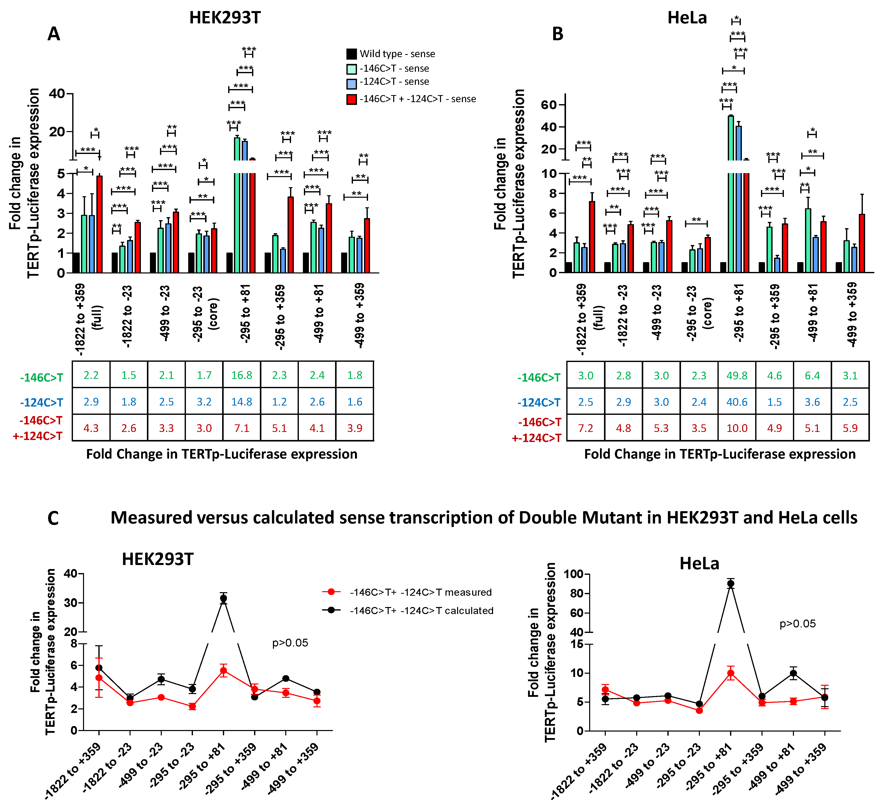
To assess the impact of the two main TERT promoter mutations −146C>T + −124C>T on TERT transcription, we introduced the mutations alone and in combination in each of the sense TERTp-luciferase reporter vectors, generating 4 constructs for each TERT promoter region: wt, −146C>T mutant, −124C>T mutant and −146C>T + −124C>T (double mutant). As shown in Figure 2A, the −146C>T mutation increased TERT promoter-driven expression by 1.5 to 2.4-fold and the −124C>T mutation by 1.6 to 3.2-fold in HEK293T cells (Figure 2A). This increase reached statistical significance in most cases. Similarly in HeLa cells, the −146C>T increased transcription by at least 3-fold and the −124C>T mutation by >3-fold with statistical support (Figure 2B). Despite variations between cell types, the increase in TERT promoter-driven luciferase was recorded consistently for most constructs. Furthermore, there was no statistical difference in the increase mediated by −146C>T and −124C>T. Of note, when expressed in the TERTp−295 to +81-luciferase construct, the single mutations boosted expression by over 15-fold in HEK293T cells and by over 40-fold in HeLa cells (p < 0.001 in both cases) (Figure 1A,B). Since the first exon covered by this promoter fragment was described to be epigenetically regulated and to contain a binding site for CTCF [16], this result suggests that the presence of either of the two mutations might affect the binding of transcriptional repressors. Yet, although TERTp-luciferase was increased by each of the mutations, this trend did not reach statistical significance when the region encompassing exon 1 + intron 1 + 38 bp of exon 2 was present, in both HEK293T cells and in HeLa cells, illustrating the strong transcriptional repression exerted by intron 1 + exon 1 + 38 bp of exon 2.
The double mutant enabled significantly higher transcription levels compared with the wt TERT promoter (p < 0.01 or p < 0.001) and compared to each of the single mutants for all constructs in both cell lines (Figure 2A,B). Generally, the two mutations together showed an additive increase in TERT expression levels (Figure 2C). This finding held true for all constructs which included upstream distal or proximal regulatory regions and for constructs containing the downstream regulatory region spanning exon 1 + intron 1 + the first 38 nucleotides of exon 2. For the TERTpcore-luciferase construct, a similar trend was recorded, although statistical support was reached only in HEK293T cells for the −146C>T mutant compared to the double mutant (Figure 2A,B), suggesting that the transcriptional advantage linked to the two mutations involves elements contained in the upstream regulatory regions. Intriguingly, the two constructs which included the first exon (TERTp−295 to +81-luciferase and TERTp−499 to +81-luciferase), had similar or lower transcription when the two mutations were present compared with the single mutants (Figure 2A–C). While the single mutations individually relieve the binding of inhibitory factors to exon 1, the two mutations concomitantly may lead to unfavorable chromatin looping or structures that ultimately decrease transcription over the single mutations. Overall, our results indicate that the double mutant confers a transcriptional advantage over the single mutants and argue against a transcriptional constraint being the main reason for the mutual exclusion of TERT promoter mutations in vivo.

3.3. TERT Promoter Mutations Increase Antisense Transcription from the TERT Promoter

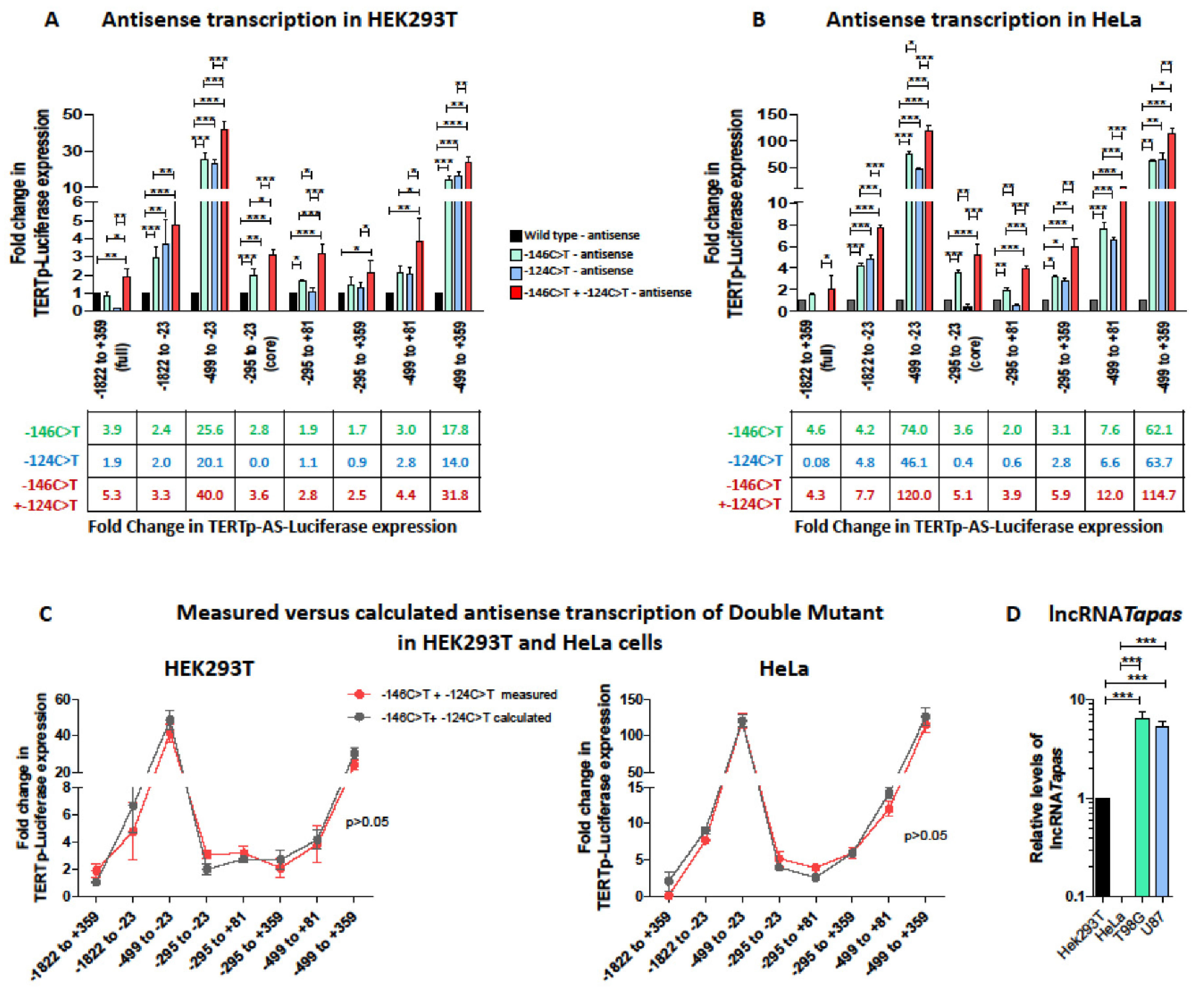
Next, we investigated whether TERT promoter mutations impacted antisense transcription from the TERT promoter, introducing the −146C>T and −124C>T mutations alone or together in each of the TERTp-AS-luciferase constructs. As shown in Figure 3A,B, antisense transcription from the full TERT promoter (TERT−1822 to +359) and from the TERTcore promoter was upregulated by the −146C>T mutation, but not by the −124C>T mutation in both cell lines, suggesting that the latter mutation only impacts sense transcription. In the presence of downstream regulatory elements (exon 1 only or exon 1 + intron 1 + 38 bp of exon 2), the −124C>T slightly increased antisense transcription compared with the wt promoter, but less than the −146C>T. However, when the upstream regulatory elements were present, antisense transcription was increased comparably by both mutations, to levels ranging from 2-fold to 63-fold (Figure 3A,B). Remarkably, the transcriptional increase was particularly striking in constructs including only the proximal upstream regulatory sequences and lacking the distal upstream regulatory sequences (i.e., downstream from position −499 from the TSS), while it ranged from 2- to 4.6-fold only when the distal upstream regulatory sequences were present. This observation suggests that repressor elements which are not affected by TERT promoter mutations bind in the distal upstream promoter region. The presence of the first exon also mitigated the impact of TERT promoter mutations on antisense transcription to an intermediate level, perhaps due to occupational interference. Overall, since the full TERT promoter (TERTp−1822 to +359-AS-luciferase), which harbors both upstream and downstream regulatory elements, and the TERTcore promoter consistently show upregulation of antisense transcription by the −146C>T mutation only, it is likely that in vivo the −146C>T mutation more efficiently allows an overcoming of the repression of antisense transcription.
As for sense transcription, the double mutant led to an additive transcriptional increase in both cell lines (Figure 3C). In line with this finding, in constructs where only −146C>T had an impact, the double mutant had a similar impact to the −146C>T single mutant.
Next, we assessed whether the increase in antisense transcription from the TERT promoter by TERT promoter mutations translates into increased lncRNA TAPAS levels. To that aim, we measured lncRNA TAPAS levels in two GBM cell lines harboring the monoallelic −146C>T (T98G) and −124C>T (U87) TERT promoter mutations. We detected much lower levels of lncRNA TAPAS in HeLa than in HEK293T cells, again highlighting cell-type specificities, and significantly higher levels of lncRNA TAPAS in cells with TERT promoter mutations compared with HEK293T and HeLa (Figure 3D). Because there are no cell lines harboring the two mutations, we could not measure lncRNA TAPAS levels expressed by a promoter harboring the two mutations.
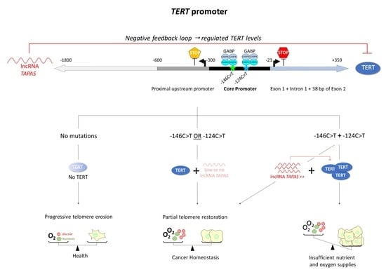
Together, these results show that sense and antisense transcription from the TERT promoter are linked, and that the −146C>T mutation upregulates both TERT expression and antisense transcription, generating lncRNA TAPAS. Therefore, while TERT transcription is increased by C-146C>T, the concomitant increase of lncRNA TAPAS may tame TERT levels in a negative feedback loop. As lncRNA TAPAS levels are inversely correlated with TERT levels, it is tempting to hypothesize that higher lncRNA TAPAS associated with the −146C>T mutation may explain the higher prevalence of −124C>T in vivo. In this scenario, the two mutations concomitantly would increase lncRNA TAPAS levels similarly or even further, ultimately mitigating the advantage of a single TERT promoter mutation. Physiologically, this dual regulation may help keep TERT levels below a given threshold.

4. Discussion

In this study, we investigated the impact of the two most common TERT promoter mutations, alone or in combination, on sense and antisense transcription from the TERT promoter. Our aim was to assess whether a transcriptional constraint could explain why TERT promoter mutations are mutually exclusive. In line with previous reports based on the TERT core promoter [23,25,26,27,28,29,31,41,67,68,69], we confirm that the mutations increase TERT transcription levels from the core promoter by 2–6-fold and that both mutations increase transcription similarly. We extend these findings by showing that overall the transcriptional increase was comparable across most constructs, regardless of whether they include upstream or downstream regulatory elements (Figure 2). This observation suggests that the presence of TERT promoter mutations is sufficient to overcome the transcriptional silencing of TERT in normal cells. The fact that the two mutations led to a comparable increase in sense transcription argues against the hypothesis that a transcriptional advantage selects for the overwhelming predominance of the −124C>T mutation in vivo. One possible explanation could be that this mutational bias reflects different aetiologies. The cytosine located −124 bp upstream from the TSS could be more accessible to enzymes, including APOBEC3 enzymes, and may therefore be more easily deaminated. In specific malignancies such as skin cancers, in contrast, the two mutations are equally frequent [42,46,47] and may be caused by UV radiation and C>T mutations at CC dinucleotides. Therefore, the differential distribution of TERT promoter mutations across cancer types may reflect different aetiologies, APOBEC3- and aging-induced deamination on one hand, versus UV-induced mutations on the other [21].
Our findings further reveal that the −146C>T mutation also unleashes antisense transcription from the TERT promoter (Figure 3A,B) and that lncRNA TAPAS is higher in prototypical cell lines harboring monoallelic TERT promoter mutations (Figure 3D). In a previous study, Malhotra et al. had recorded no impact of the −124C>T mutation on antisense transcription driven by the TERT core promoter [20]. Our results confirm that −124C>T does not affect antisense transcription from the TERT promoter but importantly, they reveal that the −146C>T mutation consistently reactivates antisense transcription from the TERT promoter, be it restricted to the TERT core promoter or inclusive of upstream and downstream regulatory elements (Figure 3). Moreover, we show that both mutations upregulate antisense transcription to equivalent levels when upstream regulatory elements only are present. The fact that the −124C>T mutation decreases antisense transcription from the TERTcore promoter compared with the wt and to all other constructs suggests that the transcription factor that binds the novel GGAA site at position −124 needs to dimerize and thus concomitantly binds to another GGAA site beyond the boundaries of the core promoter. One candidate could be GABPA [70]. NF-κB is a less likely candidate as it preferentially binds the −146C>T [69]. In this case, binding to −124C>T would be possible when downstream or proximal upstream regulatory sequences are present. However, when all regulatory elements are present (TERT−1822 to +359), this binding site seems to no longer be exploited or accessible. One possible reason could be DNA looping resulting in ternary DNA structures that do not allow the binding of transcription factors to GGAA at position −124 and even at neighboring GGAA sites. In this scenario, the –146 position would, a contrario, still remain accessible, and thus not be impacted by the DNA looping. Alternatively, access to RNA Pol II to the antisense transcriptional initiation site may be affected by the ternary structure adopted by DNA in the presence of the −124C>T mutation. Answers to these questions warrant further investigation. These observations suggest that the −146C>T mutation upregulates antisense transcription and thereby lncRNA TAPAS concomitantly to TERT, and might thus negatively affect TERT levels. How lncRNA TAPAS impacts TERT levels remains elusive, as only an inverse correlation has been described to date [20]. Yet, it is tempting to hypothesize that higher lncRNA TAPAS decrease TERT and ultimately contribute to the selective advantage of −124C>T and its strong overrepresentation across cancers. In this study, we only investigated the effect of the two most frequent TERT promoter mutations, −146C>T and −124C>T. Other mutations which create novel Ets/TCF binding sites pepper the TERT promoter [44,49]. They all have similar effects on TERT transcription [23,25,26,27,28,29,31,32,41,42,53,67,68,69,87,88]. Since they are much less prevalent than the −124C>T and −146C>T mutation, it is possible that they may also increase antisense transcription. Further studies would be needed to verify the impact of these mutations on antisense transcription and on lncRNA TAPAS levels.
The strongest impact of both TERT promoter mutations, alone or in combination, on antisense transcription was recorded for constructs containing the proximal upstream promoter region appended to the TERT core promoter in both cell lines (Figure 3A,B, compare TERTpcore-AS-luciferase to TERTp−499 to −23-AS-luciferase or TERTp−295 to +359-AS-luciferase to TERTp−499 to +359-AS-luciferase). This region hosts binding sites for strong repressors such as CTCF [15,16], and is known to negatively regulate antisense transcription from the TERT promoter ([15,17] and Figure 1F,G). It also overlaps the THOR sequence, which contains 52 CpG islands and is subject to epigenetic modifications which affect TERT sense expression [15,17,18,19]. Our results pinpoint this region spanning nucleotides −499 and −295 from the TSS as the region most responsive to derepression by TERT promoter mutations and suggest that the −146C>T mutation may relieve the binding of these repressors. The distal upstream TERT promoter region tames this upregulation and thus further contributes to negatively regulate antisense transcription from the TERT promoter (Figure 3A,B, compare TERTp−499 to −23-AS-luciferase to TERTp−1822 to −23-AS-luciferase or TERTp−499 to +359-AS-luciferase to TERTp−1822 to +359-AS-luciferase), but its impact is much less directly affected by the presence of TERT promoter mutations. Together, these results suggest that repressors binding to the region encompassing the proximal upstream regulatory region (−499 to −295 from the TSS) can be displaced by transcription factors such as GABPA or Ets/TCF binding to mutations in the TERT promoter, while the distal upstream regulatory region is less affected by such a mechanism. It is noteworthy that epigenetic tuning of transcription cannot be investigated using TERTp-luciferase reporters, and only the balance between activator and repressor transcription factors is measured using this system. Further studies would be needed to ascertain whether methylation patterns and lncRNA TAPAS expression are affected by TERT promoter mutations.
Most interestingly, when the two mutations were expressed together, the increase in TERT sense and antisense transcription was significantly higher than the increase enabled to the single mutations. Furthermore, it corresponded to the additive effect of each of the single mutations alone (Figure 2C and Figure 3C). Therefore, the double mutant would a priori provide a direct transcriptional advantage over the single mutants. Yet, the higher transcription from the double mutant TERT promoter may also increase antisense transcription driven by the −146C>T mutation and thereby increase levels of lncRNA TAPAS. As lncRNA TAPAS and TERT expression are negatively correlated, lncRNA TAPAS has been proposed to act as a negative regulator of TERT expression in a negative feedback loop [20]. High antisense transcription mediated by −146C>T mutation could thus increase lncRNA TAPAS levels and thereby lead to an overall decrease in TERT expression in a negative feedback loop. Such a self-regulatory mechanism could explain the mutual exclusion of TERT promoter mutations in vivo. We could not measure the impact of both mutations on lncRNA TAPAS levels because there are no cell lines harboring both mutations concomitantly.
TERT promoter mutations are generally mutually exclusive with each other [41] as well as with other telomere maintenance mechanisms such as copy number duplications [79,80], viral oncogenes [21,53,54,55,66,67,79,81,82,83,84] or alternative lengthening of telomeres (ALT) [22,33,34,57]. The reasons for this strict exclusion remain speculative to date but it is thought that it reflects a narrow window in terms of TERT homeostasis. Indeed, TERT levels are exquisitely balanced [21]. TERT promoter mutations generally occur late in tumor genesis, after the appearance of driver mutations in other oncogenes such as RAS/BRAF, EGFR, which lead to constitutive cell division and thus accelerate telomere erosion. TERT reactivation regenerates telomeres in cells with critically short telomeres sufficiently to maintain them above the critical threshold and to stabilize the tumor genome, thereby maintaining self-renewal potential [3,89,90,91,92]. Moreover, TERT also exerts other functions which are unrelated to telomere elongation but alter cell turnover. It contributes to cell survival and proliferation by facilitating Wnt/β-catenin-dependent [93,94], c-Myc-dependent [95,96], and NF-κB-dependent gene transcription [97,98], thereby sustaining both oncogenic signaling pathways and its own transcription in a feedforward loop [99,100]. The proliferative advantage conferred by TERT reactivation however also has a cost in terms of metabolic alterations, oxygen and nutrient supplies and the tumor microenvironment. A modest increase in TERT expression (2- to 4-fold) and a single genetic mechanism of telomere elongation enable sufficient telomere healing to avoid telomere crisis and cell death [11,22,32,39,57,60,100,101], yet remain sustainable in terms of oxygen and nutrient supplies. TERT homeostasis is thus likely an exquisite balance between escape from apoptosis resulting from telomere attrition and genomic instability on one hand, and cell sustainability in terms of oxygen and nutrient supplies on the other. In line with this view, it is remarkable that TERT promoter mutations are generally found in solid cancers with relatively low self-renewal rate [22], while they are rare (<15%) in cancers with high turnover and intrinsic TERT expression, such as hematopoietic, lymphoid, or gastrointestinal malignancies [3,22,32,80,102]. We suggest a model whereby TERT promoter mutations increase both sense (TERT) and antisense (lncRNA TAPAS) transcription to maintain TERT protein levels within a sustainable threshold. Disproportionate levels of one transcription product would shift the balance towards insufficient or excessive TERT, eventually leading to cell death by apoptosis or starvation. Such a self-regulatory mechanism where TERT promoter reactivation promotes TERT expression together with a TERT auto-regulatory mechanism is an extremely elegant means to ensure this equilibrium is maintained. Further studies will be needed to assess whether lncRNA TAPAS is the only mechanism ensuring TERT transcriptional homeostasis.

Supplementary Materials

The following are available online at https://www.mdpi.com/article/10.3390/biomedicines9121773/s1, Table S1: List of primers used for TERT promoter cloning and mutagenesis. Table S2: List of primers used for qRT-PCR. Supplementary Figure S1: TERT promoter-driven transcription in T98G and U87 GBM cell lines.

Author Contributions

Formal analysis, F.H., J.D.T. and D.P.-B.; funding acquisition, D.P.-B.; investigation, F.H., L.J. and M.L.; methodology, F.H. and D.P.-B.; project administration, D.P.-B.; resources, D.P.-B.; supervision, D.P.-B.; writing—original draft, F.H., J.D.T. and D.P.-B. All authors have read and agreed to the published version of the manuscript.

Funding

This work was supported by the Ministère de l’Education et de la Recherche du Luxembourg; FH is supported by the Fonds National de la Recherche du Luxembourg FNR-PRIDE scheme (PRIDE/11012546/NEXTIMMUNE).

Institutional Review Board Statement

Not applicable.

Informed Consent Statement

Not applicable.

Conflicts of Interest

The authors declare no conflict of interest.

References

  1. Greider, C.W. Telomere length regulation. Annu. Rev. Biochem. 1996, 65, 337–365. [Google Scholar] [CrossRef]
  2. Robles-Espinoza, C.D.; Velasco-Herrera Mdel, C.; Hayward, N.K.; Adams, D.J. Telomere-regulating genes and the telomere interactome in familial cancers. Mol. Cancer Res. 2015, 13, 211–222. [Google Scholar] [CrossRef] [Green Version]
  3. Heidenreich, B.; Kumar, R. TERT promoter mutations in telomere biology. Mutat. Res. 2017, 771, 15–31. [Google Scholar] [CrossRef] [PubMed]
  4. Leao, R.; Apolonio, J.D.; Lee, D.; Figueiredo, A.; Tabori, U.; Castelo-Branco, P. Mechanisms of human telomerase reverse transcriptase (hTERT) regulation: Clinical impacts in cancer. J. Biomed. Sci. 2018, 25, 22. [Google Scholar] [CrossRef]
  5. Allsopp, R.C.; Vaziri, H.; Patterson, C.; Goldstein, S.; Younglai, E.V.; Futcher, A.B.; Greider, C.W.; Harley, C.B. Telomere length predicts replicative capacity of human fibroblasts. Proc. Natl. Acad. Sci. USA 1992, 89, 10114–10118. [Google Scholar] [CrossRef] [PubMed] [Green Version]
  6. Wright, W.E.; Piatyszek, M.A.; Rainey, W.E.; Byrd, W.; Shay, J.W. Telomerase activity in human germline and embryonic tissues and cells. Dev. Genet. 1996, 18, 173–179. [Google Scholar] [CrossRef]
  7. Hanahan, D.; Weinberg, R.A. Hallmarks of cancer: The next generation. Cell 2011, 144, 646–674. [Google Scholar] [CrossRef] [PubMed] [Green Version]
  8. Holt, S.E.; Wright, W.E.; Shay, J.W. Regulation of telomerase activity in immortal cell lines. Mol. Cell. Biol. 1996, 16, 2932–2939. [Google Scholar] [CrossRef] [Green Version]
  9. Yasumoto, S.; Kunimura, C.; Kikuchi, K.; Tahara, H.; Ohji, H.; Yamamoto, H.; Ide, T.; Utakoji, T. Telomerase activity in normal human epithelial cells. Oncogene 1996, 13, 433–439. [Google Scholar]
  10. Akincilar, S.C.; Unal, B.; Tergaonkar, V. Reactivation of telomerase in cancer. Cell. Mol. Life Sci. 2016, 73, 1659–1670. [Google Scholar] [CrossRef] [Green Version]
  11. Barthel, F.P.; Wei, W.; Tang, M.; Martinez-Ledesma, E.; Hu, X.; Amin, S.B.; Akdemir, K.C.; Seth, S.; Song, X.; Wang, Q.; et al. Systematic analysis of telomere length and somatic alterations in 31 cancer types. Nat. Genet. 2017, 49, 349–357. [Google Scholar] [CrossRef]
  12. Kim, N.W.; Piatyszek, M.A.; Prowse, K.R.; Harley, C.B.; West, M.D.; Ho, P.L.; Coviello, G.M.; Wright, W.E.; Weinrich, S.L.; Shay, J.W. Specific association of human telomerase activity with immortal cells and cancer. Science 1994, 266, 2011–2015. [Google Scholar] [CrossRef]
  13. Ramlee, M.K.; Wang, J.; Toh, W.X.; Li, S. Transcription Regulation of the Human Telomerase Reverse Transcriptase (hTERT) Gene. Genes 2016, 7, 50. [Google Scholar] [CrossRef]
  14. Yuan, X.; Larsson, C.; Xu, D. Mechanisms underlying the activation of TERT transcription and telomerase activity in human cancer: Old actors and new players. Oncogene 2019, 38, 6172–6183. [Google Scholar] [CrossRef] [Green Version]
  15. Renaud, S.; Bosman, F.T.; Benhattar, J. Implication of the exon region in the regulation of the human telomerase reverse transcriptase gene promoter. Biochem. Biophys. Res. Commun. 2003, 300, 47–54. [Google Scholar] [CrossRef]
  16. Renaud, S.; Loukinov, D.; Bosman, F.T.; Lobanenkov, V.; Benhattar, J. CTCF binds the proximal exonic region of hTERT and inhibits its transcription. Nucleic Acids Res. 2005, 33, 6850–6860. [Google Scholar] [CrossRef] [PubMed]
  17. Renaud, S.; Loukinov, D.; Abdullaev, Z.; Guilleret, I.; Bosman, F.T.; Lobanenkov, V.; Benhattar, J. Dual role of DNA methylation inside and outside of CTCF-binding regions in the transcriptional regulation of the telomerase hTERT gene. Nucleic Acids Res. 2007, 35, 1245–1256. [Google Scholar] [CrossRef] [Green Version]
  18. Rowland, T.J.; Bonham, A.J.; Cech, T.R. Allele-specific proximal promoter hypomethylation of the telomerase reverse transcriptase gene (TERT) associates with TERT expression in multiple cancers. Mol. Oncol. 2020, 14, 2358–2374. [Google Scholar] [CrossRef] [PubMed]
  19. Lee, D.D.; Leao, R.; Komosa, M.; Gallo, M.; Zhang, C.H.; Lipman, T.; Remke, M.; Heidari, A.; Nunes, N.M.; Apolonio, J.D.; et al. DNA hypermethylation within TERT promoter upregulates TERT expression in cancer. J. Clin. Investig. 2019, 129, 223–229. [Google Scholar] [CrossRef] [Green Version]
  20. Malhotra, S.; Freeberg, M.A.; Winans, S.J.; Taylor, J.; Beemon, K.L. A Novel Long Non-Coding RNA in the hTERT Promoter Region Regulates hTERT Expression. Noncoding RNA 2017, 4, 1. [Google Scholar] [CrossRef] [Green Version]
  21. Hafezi, F.; Perez Bercoff, D. The Solo Play of TERT Promoter Mutations. Cells 2020, 9, 749. [Google Scholar] [CrossRef] [Green Version]
  22. Killela, P.J.; Reitman, Z.J.; Jiao, Y.; Bettegowda, C.; Agrawal, N.; Diaz, L.A., Jr.; Friedman, A.H.; Friedman, H.; Gallia, G.L.; Giovanella, B.C.; et al. TERT promoter mutations occur frequently in gliomas and a subset of tumors derived from cells with low rates of self-renewal. Proc. Natl. Acad. Sci. USA 2013, 110, 6021–6026. [Google Scholar] [CrossRef] [Green Version]
  23. Vinagre, J.; Almeida, A.; Populo, H.; Batista, R.; Lyra, J.; Pinto, V.; Coelho, R.; Celestino, R.; Prazeres, H.; Lima, L.; et al. Frequency of TERT promoter mutations in human cancers. Nat. Commun. 2013, 4, 2185. [Google Scholar] [CrossRef] [PubMed] [Green Version]
  24. Liu, X.; Wu, G.; Shan, Y.; Hartmann, C.; von Deimling, A.; Xing, M. Highly prevalent TERT promoter mutations in bladder cancer and glioblastoma. Cell Cycle 2013, 12, 1637–1638. [Google Scholar] [CrossRef] [PubMed] [Green Version]
  25. Chen, C.; Han, S.; Meng, L.; Li, Z.; Zhang, X.; Wu, A. TERT promoter mutations lead to high transcriptional activity under hypoxia and temozolomide treatment and predict poor prognosis in gliomas. PLoS ONE 2014, 9, e100297. [Google Scholar] [CrossRef] [Green Version]
  26. Park, C.K.; Lee, S.H.; Kim, J.Y.; Kim, J.E.; Kim, T.M.; Lee, S.T.; Choi, S.H.; Park, S.H.; Kim, I.H. Expression level of hTERT is regulated by somatic mutation and common single nucleotide polymorphism at promoter region in glioblastoma. Oncotarget 2014, 5, 3399–3407. [Google Scholar] [CrossRef] [Green Version]
  27. Huang, F.W.; Bielski, C.M.; Rinne, M.L.; Hahn, W.C.; Sellers, W.R.; Stegmeier, F.; Garraway, L.A.; Kryukov, G.V. TERT promoter mutations and monoallelic activation of TERT in cancer. Oncogenesis 2015, 4, e176. [Google Scholar] [CrossRef] [Green Version]
  28. Spiegl-Kreinecker, S.; Lotsch, D.; Ghanim, B.; Pirker, C.; Mohr, T.; Laaber, M.; Weis, S.; Olschowski, A.; Webersinke, G.; Pichler, J.; et al. Prognostic quality of activating TERT promoter mutations in glioblastoma: Interaction with the rs2853669 polymorphism and patient age at diagnosis. Neuro Oncol. 2015, 17, 1231–1240. [Google Scholar] [CrossRef] [PubMed] [Green Version]
  29. Arita, H.; Narita, Y.; Fukushima, S.; Tateishi, K.; Matsushita, Y.; Yoshida, A.; Miyakita, Y.; Ohno, M.; Collins, V.P.; Kawahara, N.; et al. Upregulating mutations in the TERT promoter commonly occur in adult malignant gliomas and are strongly associated with total 1p19q loss. Acta Neuropathol. 2013, 126, 267–276. [Google Scholar] [CrossRef]
  30. Arita, H.; Narita, Y.; Takami, H.; Fukushima, S.; Matsushita, Y.; Yoshida, A.; Miyakita, Y.; Ohno, M.; Shibui, S.; Ichimura, K. TERT promoter mutations rather than methylation are the main mechanism for TERT upregulation in adult gliomas. Acta Neuropathol. 2013, 126, 939–941. [Google Scholar] [CrossRef]
  31. Johanns, T.M.; Fu, Y.; Kobayashi, D.K.; Mei, Y.; Dunn, I.F.; Mao, D.D.; Kim, A.H.; Dunn, G.P. High incidence of TERT mutation in brain tumor cell lines. Brain Tumor Pathol. 2016, 33, 222–227. [Google Scholar] [CrossRef] [PubMed] [Green Version]
  32. Huang, D.S.; Wang, Z.; He, X.J.; Diplas, B.H.; Yang, R.; Killela, P.J.; Meng, Q.; Ye, Z.Y.; Wang, W.; Jiang, X.T.; et al. Recurrent TERT promoter mutations identified in a large-scale study of multiple tumour types are associated with increased TERT expression and telomerase activation. Eur. J. Cancer 2015, 51, 969–976. [Google Scholar] [CrossRef] [Green Version]
  33. Pekmezci, M.; Rice, T.; Molinaro, A.M.; Walsh, K.M.; Decker, P.A.; Hansen, H.; Sicotte, H.; Kollmeyer, T.M.; McCoy, L.S.; Sarkar, G.; et al. Adult infiltrating gliomas with WHO 2016 integrated diagnosis: Additional prognostic roles of ATRX and TERT. Acta Neuropathol. 2017, 133, 1001–1016. [Google Scholar] [CrossRef]
  34. Eckel-Passow, J.E.; Lachance, D.H.; Molinaro, A.M.; Walsh, K.M.; Decker, P.A.; Sicotte, H.; Pekmezci, M.; Rice, T.; Kosel, M.L.; Smirnov, I.V.; et al. Glioma Groups Based on 1p/19q, IDH, and TERT Promoter Mutations in Tumors. N. Engl. J. Med. 2015, 372, 2499–2508. [Google Scholar] [CrossRef] [PubMed] [Green Version]
  35. Simon, M.; Hosen, I.; Gousias, K.; Rachakonda, S.; Heidenreich, B.; Gessi, M.; Schramm, J.; Hemminki, K.; Waha, A.; Kumar, R. TERT promoter mutations: A novel independent prognostic factor in primary glioblastomas. Neuro Oncol. 2015, 17, 45–52. [Google Scholar] [CrossRef] [PubMed]
  36. Arita, H.; Yamasaki, K.; Matsushita, Y.; Nakamura, T.; Shimokawa, A.; Takami, H.; Tanaka, S.; Mukasa, A.; Shirahata, M.; Shimizu, S.; et al. A combination of TERT promoter mutation and MGMT methylation status predicts clinically relevant subgroups of newly diagnosed glioblastomas. Acta Neuropathol. Commun. 2016, 4, 79. [Google Scholar] [CrossRef] [Green Version]
  37. Yang, P.; Cai, J.; Yan, W.; Zhang, W.; Wang, Y.; Chen, B.; Li, G.; Li, S.; Wu, C.; Yao, K.; et al. Classification based on mutations of TERT promoter and IDH characterizes subtypes in grade II/III gliomas. Neuro Oncol. 2016, 18, 1099–1108. [Google Scholar] [CrossRef] [Green Version]
  38. Mosrati, M.A.; Malmstrom, A.; Lysiak, M.; Krysztofiak, A.; Hallbeck, M.; Milos, P.; Hallbeck, A.L.; Bratthall, C.; Strandeus, M.; Stenmark-Askmalm, M.; et al. TERT promoter mutations and polymorphisms as prognostic factors in primary glioblastoma. Oncotarget 2015, 6, 16663–16673. [Google Scholar] [CrossRef] [Green Version]
  39. Batista, R.; Cruvinel-Carloni, A.; Vinagre, J.; Peixoto, J.; Catarino, T.A.; Campanella, N.C.; Menezes, W.; Becker, A.P.; de Almeida, G.C.; Matsushita, M.M.; et al. The prognostic impact of TERT promoter mutations in glioblastomas is modified by the rs2853669 single nucleotide polymorphism. Int. J. Cancer 2016, 139, 414–423. [Google Scholar] [CrossRef] [Green Version]
  40. Williams, E.A.; Miller, J.J.; Tummala, S.S.; Penson, T.; Iafrate, A.J.; Juratli, T.A.; Cahill, D.P. TERT promoter wild-type glioblastomas show distinct clinical features and frequent PI3K pathway mutations. Acta Neuropathol. Commun. 2018, 6, 106. [Google Scholar] [CrossRef] [Green Version]
  41. Huang, F.W.; Hodis, E.; Xu, M.J.; Kryukov, G.V.; Chin, L.; Garraway, L.A. Highly recurrent TERT promoter mutations in human melanoma. Science 2013, 339, 957–959. [Google Scholar] [CrossRef] [Green Version]
  42. Griewank, K.G.; Murali, R.; Schilling, B.; Schimming, T.; Moller, I.; Moll, I.; Schwamborn, M.; Sucker, A.; Zimmer, L.; Schadendorf, D.; et al. TERT promoter mutations are frequent in cutaneous basal cell carcinoma and squamous cell carcinoma. PLoS ONE 2013, 8, e80354. [Google Scholar] [CrossRef]
  43. Griewank, K.G.; Murali, R.; Schilling, B.; Scholz, S.; Sucker, A.; Song, M.; Susskind, D.; Grabellus, F.; Zimmer, L.; Hillen, U.; et al. TERT promoter mutations in ocular melanoma distinguish between conjunctival and uveal tumours. Br. J. Cancer 2013, 109, 497–501. [Google Scholar] [CrossRef] [Green Version]
  44. Horn, S.; Figl, A.; Rachakonda, P.S.; Fischer, C.; Sucker, A.; Gast, A.; Kadel, S.; Moll, I.; Nagore, E.; Hemminki, K.; et al. TERT promoter mutations in familial and sporadic melanoma. Science 2013, 339, 959–961. [Google Scholar] [CrossRef] [Green Version]
  45. Heidenreich, B.; Nagore, E.; Rachakonda, P.S.; Garcia-Casado, Z.; Requena, C.; Traves, V.; Becker, J.; Soufir, N.; Hemminki, K.; Kumar, R. Telomerase reverse transcriptase promoter mutations in primary cutaneous melanoma. Nat. Commun. 2014, 5, 3401. [Google Scholar] [CrossRef]
  46. Populo, H.; Boaventura, P.; Vinagre, J.; Batista, R.; Mendes, A.; Caldas, R.; Pardal, J.; Azevedo, F.; Honavar, M.; Guimaraes, I.; et al. TERT promoter mutations in skin cancer: The effects of sun exposure and X-irradiation. J. Investig. Dermatol. 2014, 134, 2251–2257. [Google Scholar] [CrossRef] [Green Version]
  47. Scott, G.A.; Laughlin, T.S.; Rothberg, P.G. Mutations of the TERT promoter are common in basal cell carcinoma and squamous cell carcinoma. Mod. Pathol. 2014, 27, 516–523. [Google Scholar] [CrossRef] [PubMed] [Green Version]
  48. Ofner, R.; Ritter, C.; Heidenreich, B.; Kumar, R.; Ugurel, S.; Schrama, D.; Becker, J.C. Distribution of TERT promoter mutations in primary and metastatic melanomas in Austrian patients. J. Cancer Res. Clin. Oncol. 2017, 143, 613–617. [Google Scholar] [CrossRef] [PubMed]
  49. Andres-Lencina, J.J.; Rachakonda, S.; Garcia-Casado, Z.; Srinivas, N.; Skorokhod, A.; Requena, C.; Soriano, V.; Kumar, R.; Nagore, E. TERT promoter mutation subtypes and survival in stage I and II melanoma patients. Int. J. Cancer 2018, 144, 1027–1036. [Google Scholar] [CrossRef]
  50. Nguyen, D.; Taheri, D.; Springer, S.; Cowan, M.; Guner, G.; Mendoza Rodriguez, M.A.; Wang, Y.; Kinde, I.; Vanden Bussche, C.J.; Olson, M.T.; et al. High prevalence of TERT promoter mutations in micropapillary urothelial carcinoma. Virchows Arch. 2016, 469, 427–434. [Google Scholar] [CrossRef] [PubMed] [Green Version]
  51. Cowan, M.; Springer, S.; Nguyen, D.; Taheri, D.; Guner, G.; Rodriguez, M.A.; Wang, Y.; Kinde, I.; VandenBussche, C.J.; Olson, M.T.; et al. High prevalence of TERT promoter mutations in primary squamous cell carcinoma of the urinary bladder. Mod. Pathol. 2016, 29, 511–515. [Google Scholar] [CrossRef] [PubMed]
  52. Allory, Y.; Beukers, W.; Sagrera, A.; Flandez, M.; Marques, M.; Marquez, M.; van der Keur, K.A.; Dyrskjot, L.; Lurkin, I.; Vermeij, M.; et al. Telomerase reverse transcriptase promoter mutations in bladder cancer: High frequency across stages, detection in urine, and lack of association with outcome. Eur. Urol. 2014, 65, 360–366. [Google Scholar] [CrossRef] [PubMed] [Green Version]
  53. Nault, J.C.; Mallet, M.; Pilati, C.; Calderaro, J.; Bioulac-Sage, P.; Laurent, C.; Laurent, A.; Cherqui, D.; Balabaud, C.; Zucman-Rossi, J. High frequency of telomerase reverse-transcriptase promoter somatic mutations in hepatocellular carcinoma and preneoplastic lesions. Nat. Commun. 2013, 4, 2218. [Google Scholar] [CrossRef] [Green Version]
  54. Pezzuto, F.; Izzo, F.; Buonaguro, L.; Annunziata, C.; Tatangelo, F.; Botti, G.; Buonaguro, F.M.; Tornesello, M.L. Tumor specific mutations in TERT promoter and CTNNB1 gene in hepatitis B and hepatitis C related hepatocellular carcinoma. Oncotarget 2016, 7, 54253–54262. [Google Scholar] [CrossRef] [PubMed] [Green Version]
  55. Chen, Y.L.; Jeng, Y.M.; Chang, C.N.; Lee, H.J.; Hsu, H.C.; Lai, P.L.; Yuan, R.H. TERT promoter mutation in resectable hepatocellular carcinomas: A strong association with hepatitis C infection and absence of hepatitis B infection. Int. J. Surg. 2014, 12, 659–665. [Google Scholar] [CrossRef] [Green Version]
  56. Cevik, D.; Yildiz, G.; Ozturk, M. Common telomerase reverse transcriptase promoter mutations in hepatocellular carcinomas from different geographical locations. World J. Gastroenterol. 2015, 21, 311–317. [Google Scholar] [CrossRef]
  57. Liu, T.; Wang, N.; Cao, J.; Sofiadis, A.; Dinets, A.; Zedenius, J.; Larsson, C.; Xu, D. The age- and shorter telomere-dependent TERT promoter mutation in follicular thyroid cell-derived carcinomas. Oncogene 2014, 33, 4978–4984. [Google Scholar] [CrossRef] [Green Version]
  58. Liu, X.; Qu, S.; Liu, R.; Sheng, C.; Shi, X.; Zhu, G.; Murugan, A.K.; Guan, H.; Yu, H.; Wang, Y.; et al. TERT promoter mutations and their association with BRAF V600E mutation and aggressive clinicopathological characteristics of thyroid cancer. J. Clin. Endocrinol. Metab. 2014, 99, E1130–E1136. [Google Scholar] [CrossRef] [Green Version]
  59. Landa, I.; Ganly, I.; Chan, T.A.; Mitsutake, N.; Matsuse, M.; Ibrahimpasic, T.; Ghossein, R.A.; Fagin, J.A. Frequent somatic TERT promoter mutations in thyroid cancer: Higher prevalence in advanced forms of the disease. J. Clin. Endocrinol. Metab. 2013, 98, E1562–E1566. [Google Scholar] [CrossRef] [Green Version]
  60. Melo, M.; da Rocha, A.G.; Vinagre, J.; Batista, R.; Peixoto, J.; Tavares, C.; Celestino, R.; Almeida, A.; Salgado, C.; Eloy, C.; et al. TERT promoter mutations are a major indicator of poor outcome in differentiated thyroid carcinomas. J. Clin. Endocrinol. Metab. 2014, 99, E754–E765. [Google Scholar] [CrossRef] [Green Version]
  61. Muzza, M.; Colombo, C.; Rossi, S.; Tosi, D.; Cirello, V.; Perrino, M.; De Leo, S.; Magnani, E.; Pignatti, E.; Vigo, B.; et al. Telomerase in differentiated thyroid cancer: Promoter mutations, expression and localization. Mol. Cell. Endocrinol. 2015, 399, 288–295. [Google Scholar] [CrossRef]
  62. George, J.R.; Henderson, Y.C.; Williams, M.D.; Roberts, D.B.; Hei, H.; Lai, S.Y.; Clayman, G.L. Association of TERT Promoter Mutation, But Not BRAF Mutation, With Increased Mortality in PTC. J. Clin. Endocrinol. Metab. 2015, 100, E1550–E1559. [Google Scholar] [CrossRef] [Green Version]
  63. Shi, X.; Liu, R.; Qu, S.; Zhu, G.; Bishop, J.; Liu, X.; Sun, H.; Shan, Z.; Wang, E.; Luo, Y.; et al. Association of TERT promoter mutation 1,295,228 C>T with BRAF V600E mutation, older patient age, and distant metastasis in anaplastic thyroid cancer. J. Clin. Endocrinol. Metab. 2015, 100, E632–E637. [Google Scholar] [CrossRef] [Green Version]
  64. Bae, J.S.; Kim, Y.; Jeon, S.; Kim, S.H.; Kim, T.J.; Lee, S.; Kim, M.H.; Lim, D.J.; Lee, Y.S.; Jung, C.K. Clinical utility of TERT promoter mutations and ALK rearrangement in thyroid cancer patients with a high prevalence of the BRAF V600E mutation. Diagn. Pathol. 2016, 11, 21. [Google Scholar] [CrossRef] [Green Version]
  65. Song, Y.S.; Lim, J.A.; Choi, H.; Won, J.K.; Moon, J.H.; Cho, S.W.; Lee, K.E.; Park, Y.J.; Yi, K.H.; Park, D.J.; et al. Prognostic effects of TERT promoter mutations are enhanced by coexistence with BRAF or RAS mutations and strengthen the risk prediction by the ATA or TNM staging system in differentiated thyroid cancer patients. Cancer 2016, 122, 1370–1379. [Google Scholar] [CrossRef]
  66. Vinothkumar, V.; Arunkumar, G.; Revathidevi, S.; Arun, K.; Manikandan, M.; Rao, A.K.; Rajkumar, K.S.; Ajay, C.; Rajaraman, R.; Ramani, R.; et al. TERT promoter hot spot mutations are frequent in Indian cervical and oral squamous cell carcinomas. Tumour Biol. 2016, 37, 7907–7913. [Google Scholar] [CrossRef] [PubMed]
  67. Annunziata, C.; Pezzuto, F.; Greggi, S.; Ionna, F.; Losito, S.; Botti, G.; Buonaguro, L.; Buonaguro, F.M.; Tornesello, M.L. Distinct profiles of TERT promoter mutations and telomerase expression in head and neck cancer and cervical carcinoma. Int. J. Cancer 2018, 143, 1153–1161. [Google Scholar] [CrossRef] [PubMed] [Green Version]
  68. Rachakonda, P.S.; Hosen, I.; de Verdier, P.J.; Fallah, M.; Heidenreich, B.; Ryk, C.; Wiklund, N.P.; Steineck, G.; Schadendorf, D.; Hemminki, K.; et al. TERT promoter mutations in bladder cancer affect patient survival and disease recurrence through modification by a common polymorphism. Proc. Natl. Acad. Sci. USA 2013, 110, 17426–17431. [Google Scholar] [CrossRef] [Green Version]
  69. Li, Y.; Zhou, Q.L.; Sun, W.; Chandrasekharan, P.; Cheng, H.S.; Ying, Z.; Lakshmanan, M.; Raju, A.; Tenen, D.G.; Cheng, S.Y.; et al. Non-canonical NF-kappaB signalling and ETS1/2 cooperatively drive C250T mutant TERT promoter activation. Nat. Cell Biol. 2015, 17, 1327–1338. [Google Scholar] [CrossRef] [PubMed]
  70. Bell, R.J.; Rube, H.T.; Kreig, A.; Mancini, A.; Fouse, S.D.; Nagarajan, R.P.; Choi, S.; Hong, C.; He, D.; Pekmezci, M.; et al. Cancer. The transcription factor GABP selectively binds and activates the mutant TERT promoter in cancer. Science 2015, 348, 1036–1039. [Google Scholar] [CrossRef] [Green Version]
  71. Akincilar, S.C.; Khattar, E.; Boon, P.L.; Unal, B.; Fullwood, M.J.; Tergaonkar, V. Long-Range Chromatin Interactions Drive Mutant TERT Promoter Activation. Cancer Discov. 2016, 6, 1276–1291. [Google Scholar] [CrossRef] [PubMed] [Green Version]
  72. Mancini, A.; Xavier-Magalhaes, A.; Woods, W.S.; Nguyen, K.T.; Amen, A.M.; Hayes, J.L.; Fellmann, C.; Gapinske, M.; McKinney, A.M.; Hong, C.; et al. Disruption of the beta1L Isoform of GABP Reverses Glioblastoma Replicative Immortality in a TERT Promoter Mutation-Dependent Manner. Cancer Cell 2018, 34, 513–528.e8. [Google Scholar] [CrossRef] [PubMed] [Green Version]
  73. Sizemore, G.M.; Pitarresi, J.R.; Balakrishnan, S.; Ostrowski, M.C. The ETS family of oncogenic transcription factors in solid tumours. Nat. Rev. Cancer 2017, 17, 337–351. [Google Scholar] [CrossRef] [PubMed]
  74. Thompson, C.C.; Brown, T.A.; McKnight, S.L. Convergence of Ets- and notch-related structural motifs in a heteromeric DNA binding complex. Science 1991, 253, 762–768. [Google Scholar] [CrossRef]
  75. Oikawa, T.; Yamada, T. Molecular biology of the Ets family of transcription factors. Gene 2003, 303, 11–34. [Google Scholar] [CrossRef]
  76. La Marco, K.; Thompson, C.C.; Byers, B.P.; Walton, E.M.; McKnight, S.L. Identification of Ets- and notch-related subunits in GA binding protein. Science 1991, 253, 789–792. [Google Scholar] [CrossRef]
  77. Stern, J.L.; Theodorescu, D.; Vogelstein, B.; Papadopoulos, N.; Cech, T.R. Mutation of the TERT promoter, switch to active chromatin, and monoallelic TERT expression in multiple cancers. Genes Dev. 2015, 29, 2219–2224. [Google Scholar] [CrossRef] [Green Version]
  78. Stern, J.L.; Paucek, R.D.; Huang, F.W.; Ghandi, M.; Nwumeh, R.; Costello, J.C.; Cech, T.R. Allele-Specific DNA Methylation and Its Interplay with Repressive Histone Marks at Promoter-Mutant TERT Genes. Cell Rep. 2017, 21, 3700–3707. [Google Scholar] [CrossRef] [Green Version]
  79. Totoki, Y.; Tatsuno, K.; Covington, K.R.; Ueda, H.; Creighton, C.J.; Kato, M.; Tsuji, S.; Donehower, L.A.; Slagle, B.L.; Nakamura, H.; et al. Trans-ancestry mutational landscape of hepatocellular carcinoma genomes. Nat. Genet. 2014, 46, 1267–1273. [Google Scholar] [CrossRef]
  80. Gaspar, T.B.; Sa, A.; Lopes, J.M.; Sobrinho-Simoes, M.; Soares, P.; Vinagre, J. Telomere Maintenance Mechanisms in Cancer. Genes 2018, 9, 241. [Google Scholar] [CrossRef] [Green Version]
  81. Klingelhutz, A.J.; Foster, S.A.; McDougall, J.K. Telomerase activation by the E6 gene product of human papillomavirus type 16. Nature 1996, 380, 79–82. [Google Scholar] [CrossRef]
  82. Oh, S.T.; Kyo, S.; Laimins, L.A. Telomerase activation by human papillomavirus type 16 E6 protein: Induction of human telomerase reverse transcriptase expression through Myc and GC-rich Sp1 binding sites. J. Virol. 2001, 75, 5559–5566. [Google Scholar] [CrossRef] [PubMed] [Green Version]
  83. Veldman, T.; Horikawa, I.; Barrett, J.C.; Schlegel, R. Transcriptional activation of the telomerase hTERT gene by human papillomavirus type 16 E6 oncoprotein. J. Virol. 2001, 75, 4467–4472. [Google Scholar] [CrossRef] [PubMed] [Green Version]
  84. Kawai-Kitahata, F.; Asahina, Y.; Tanaka, S.; Kakinuma, S.; Murakawa, M.; Nitta, S.; Watanabe, T.; Otani, S.; Taniguchi, M.; Goto, F.; et al. Comprehensive analyses of mutations and hepatitis B virus integration in hepatocellular carcinoma with clinicopathological features. J. Gastroenterol. 2016, 51, 473–486. [Google Scholar] [CrossRef] [PubMed]
  85. Livak, K.J.; Schmittgen, T.D. Analysis of relative gene expression data using real-time quantitative PCR and the 2(-Delta Delta C(T)) Method. Methods 2001, 25, 402–408. [Google Scholar] [CrossRef]
  86. De Wilde, J.; Kooter, J.M.; Overmeer, R.M.; Claassen-Kramer, D.; Meijer, C.J.; Snijders, P.J.; Steenbergen, R.D. hTERT promoter activity and CpG methylation in HPV-induced carcinogenesis. BMC Cancer 2010, 10, 271. [Google Scholar] [CrossRef] [PubMed] [Green Version]
  87. Hosen, I.; Rachakonda, P.S.; Heidenreich, B.; de Verdier, P.J.; Ryk, C.; Steineck, G.; Hemminki, K.; Kumar, R. Mutations in TERT promoter and FGFR3 and telomere length in bladder cancer. Int. J. Cancer 2015, 137, 1621–1629. [Google Scholar] [CrossRef]
  88. Yang, X.; Guo, X.; Chen, Y.; Chen, G.; Ma, Y.; Huang, K.; Zhang, Y.; Zhao, Q.; Winkler, C.A.; An, P.; et al. Telomerase reverse transcriptase promoter mutations in hepatitis B virus-associated hepatocellular carcinoma. Oncotarget 2016, 7, 27838–27847. [Google Scholar] [CrossRef]
  89. Mukherjee, S.; Firpo, E.J.; Wang, Y.; Roberts, J.M. Separation of telomerase functions by reverse genetics. Proc. Natl. Acad. Sci. USA 2011, 108, E1363–E1371. [Google Scholar] [CrossRef] [Green Version]
  90. Liu, T.; Yuan, X.; Xu, D. Cancer-Specific Telomerase Reverse Transcriptase (TERT) Promoter Mutations: Biological and Clinical Implications. Genes 2016, 7, 38. [Google Scholar] [CrossRef]
  91. Chiba, K.; Lorbeer, F.K.; Shain, A.H.; McSwiggen, D.T.; Schruf, E.; Oh, A.; Ryu, J.; Darzacq, X.; Bastian, B.C.; Hockemeyer, D. Mutations in the promoter of the telomerase gene TERT contribute to tumorigenesis by a two-step mechanism. Science 2017, 357, 1416–1420. [Google Scholar] [CrossRef] [Green Version]
  92. Li, C.; Wu, S.; Wang, H.; Bi, X.; Yang, Z.; Du, Y.; He, L.; Cai, Z.; Wang, J.; Fan, Z. The C228T mutation of TERT promoter frequently occurs in bladder cancer stem cells and contributes to tumorigenesis of bladder cancer. Oncotarget 2015, 6, 19542–19551. [Google Scholar] [CrossRef] [Green Version]
  93. Park, J.I.; Venteicher, A.S.; Hong, J.Y.; Choi, J.; Jun, S.; Shkreli, M.; Chang, W.; Meng, Z.; Cheung, P.; Ji, H.; et al. Telomerase modulates Wnt signalling by association with target gene chromatin. Nature 2009, 460, 66–72. [Google Scholar] [CrossRef] [Green Version]
  94. Liu, Z.; Li, Q.; Li, K.; Chen, L.; Li, W.; Hou, M.; Liu, T.; Yang, J.; Lindvall, C.; Bjorkholm, M.; et al. Telomerase reverse transcriptase promotes epithelial-mesenchymal transition and stem cell-like traits in cancer cells. Oncogene 2013, 32, 4203–4213. [Google Scholar] [CrossRef] [PubMed] [Green Version]
  95. Koh, C.M.; Khattar, E.; Leow, S.C.; Liu, C.Y.; Muller, J.; Ang, W.X.; Li, Y.; Franzoso, G.; Li, S.; Guccione, E.; et al. Telomerase regulates MYC-driven oncogenesis independent of its reverse transcriptase activity. J. Clin. Investig. 2015, 125, 2109–2122. [Google Scholar] [CrossRef] [Green Version]
  96. Tang, B.; Xie, R.; Qin, Y.; Xiao, Y.F.; Yong, X.; Zheng, L.; Dong, H.; Yang, S.M. Human telomerase reverse transcriptase (hTERT) promotes gastric cancer invasion through cooperating with c-Myc to upregulate heparanase expression. Oncotarget 2016, 7, 11364–11379. [Google Scholar] [CrossRef] [PubMed]
  97. Ghosh, A.; Saginc, G.; Leow, S.C.; Khattar, E.; Shin, E.M.; Yan, T.D.; Wong, M.; Zhang, Z.; Li, G.; Sung, W.K.; et al. Telomerase directly regulates NF-kappaB-dependent transcription. Nat. Cell Biol. 2012, 14, 1270–1281. [Google Scholar] [CrossRef] [PubMed]
  98. Ding, D.; Xi, P.; Zhou, J.; Wang, M.; Cong, Y.S. Human telomerase reverse transcriptase regulates MMP expression independently of telomerase activity via NF-kappaB-dependent transcription. FASEB J. 2013, 27, 4375–4383. [Google Scholar] [CrossRef] [PubMed]
  99. Li, Y.; Tergaonkar, V. Noncanonical functions of telomerase: Implications in telomerase-targeted cancer therapies. Cancer Res. 2014, 74, 1639–1644. [Google Scholar] [CrossRef] [Green Version]
  100. Pestana, A.; Vinagre, J.; Sobrinho-Simoes, M.; Soares, P. TERT biology and function in cancer: Beyond immortalisation. J. Mol. Endocrinol. 2017, 58, R129–R146. [Google Scholar] [CrossRef]
  101. Rachakonda, S.; Kong, H.; Srinivas, N.; Garcia-Casado, Z.; Requena, C.; Fallah, M.; Heidenreich, B.; Planelles, D.; Traves, V.; Schadendorf, D.; et al. Telomere length, telomerase reverse transcriptase promoter mutations, and melanoma risk. Genes Chromosomes Cancer 2018, 57, 564–572. [Google Scholar] [CrossRef] [PubMed]
  102. Chiba, K.; Johnson, J.Z.; Vogan, J.M.; Wagner, T.; Boyle, J.M.; Hockemeyer, D. Cancer-associated TERT promoter mutations abrogate telomerase silencing. eLife 2015, 4, e07918. [Google Scholar] [CrossRef] [PubMed]
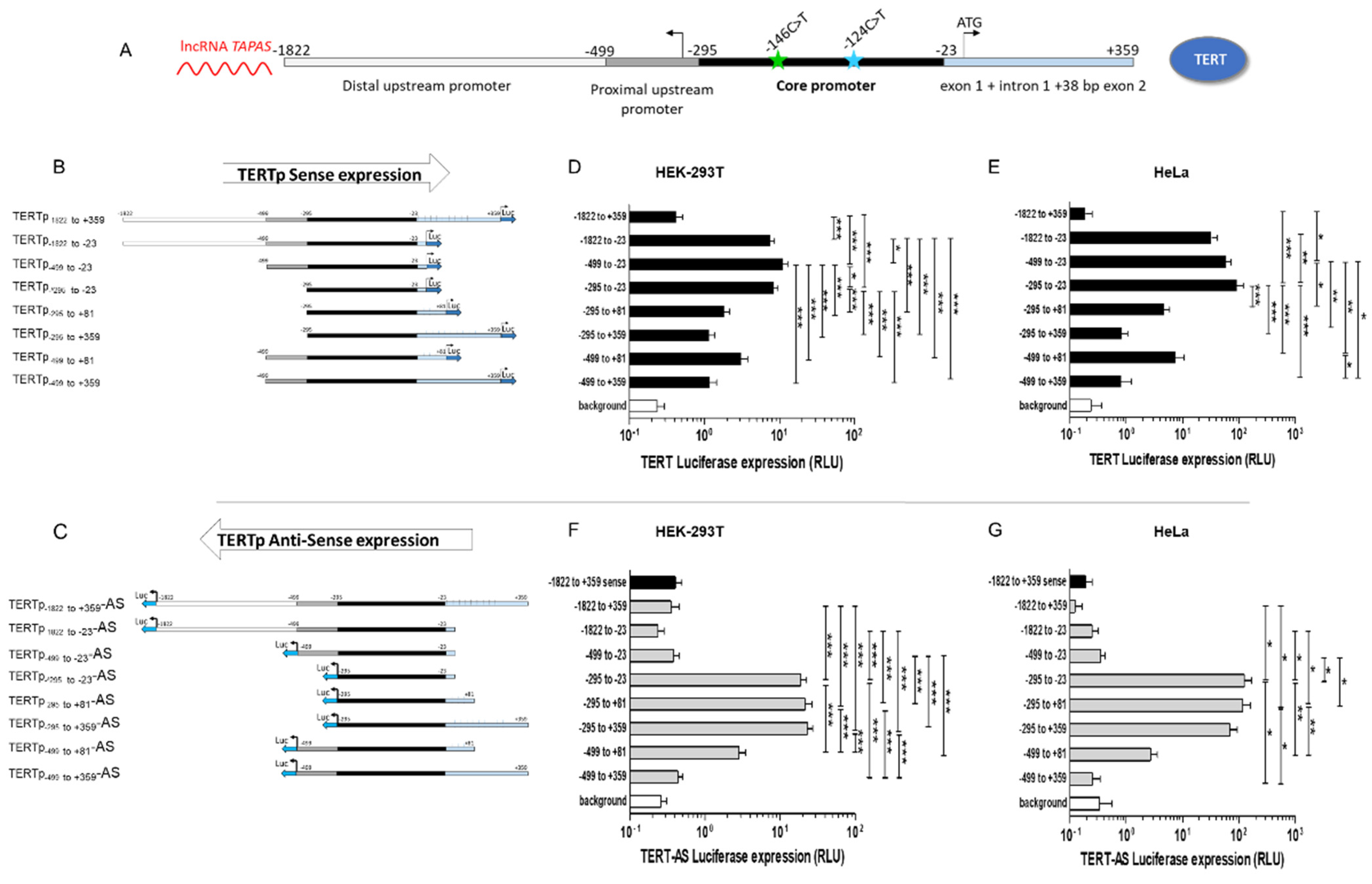
Figure 1. TERT transcription regulation. (A). Schematic representation of the TERT promoter. The TERT promoter comprises the core promoter (−295 to −23 from the TSS (black) and upstream and downstream regulatory regions. The TSS is represented with an arrow. Mutations at position −146 and −124 from the TSS are represented by a green and blue star, respectively. (B). Schematic representation of the TERTp-luciferase reporter plasmids in sense orientation. Different portions of the TERT promoter were amplified from HEK293T cells and cloned upstream of the firefly luciferase reporter gene. (C). Schematic representation of the antisense TERTp-luciferase reporter plasmids. The same PCR-amplified regions of the TERT promoter from HEK293T cells were cloned upstream of the firefly luciferase reporter gene in antisense orientation. (D,E). Luciferase expression in HEK293T (D) and HeLa (E) cells transfected with each of the TERT promoter constructs. HEK293T (1.2 × 10⁵ cells/well) or HeLa (5 × 10⁴ cells/well) were seeded in a 48-well plate and transfected with 200 ng of each of the TERTp-luciferase reporters + HSV-TK-Renilla-luciferase for normalization (10 ng in HEK293T and 20 ng in HeLa). The promoterless luciferase vector was used to assess background. Cells were lysed 48 h post transfection and Firefly and Renilla Luciferase activities were measured. Firefly Luciferase relative light units (RLU) was normalized to Renilla Luciferase RLU. Results are the mean of at least 3 independent experiments carried out in duplicate or triplicate. (F,G). Luciferase expression in HEK293T (F) and HeLa (G) cells transfected with each of the antisense TERTp-AS-luciferase constructs. HEK293T or HeLa cells were transfected with 200 ng of each of the Antisense TERTp-luciferase reporters + HSV-TK-Renilla-luciferase for normalization (10 ng in HEK293T and 20 ng in HeLa) in the same conditions as for panels D and E. In each experiment with TERTp-AS-luciferase vectors, the sense TERTp−1822 to +359-luciferase was included for comparison. Results are the mean of 3 independent experiments carried out in duplicate or in triplicate. Differences were compared using a one-way ANOVA. * indicates p < 0.05, ** indicates p < 0.01, *** indicates p < 0.001.
Biomedicines 09 01773 g001
Figure 2. Impact of TERT promoter mutations −146C>T and −124C>T on TERT transcription. (A,B). Fold-change in TERT expression in HEK293T (A) and HeLa (B) cells transfected with the wt or mutant TERTp-luciferase reporter constructs. HEK293T (1.2 × 10⁵ cells/well) or HeLa (5 × 10⁴ cells/well) were seeded in a 48-well plate and transfected with 200 ng of each of the wt (black) or mutated (green: −146C>T; blue: −124C>T; red: −146C>T + −124C>T) TERTp-luciferase reporters + HSV-TK-Renilla-luciferase for normalization (10 ng in HEK293T and 20 ng in HeLa) for 48 h. Firefly Luciferase RLU was normalized to Renilla Luciferase RLU and transcription levels were further normalized to the wt for each TERT promoter construct. Results are the mean of at least 3 independent experiments carried out in duplicate. Error bars represent standard error. Differences were compared using a one-way ANOVA followed by a Newman–Keuls post-hoc test for pairwise comparisons. * indicates p < 0.05, ** indicates p < 0.01, *** indicates p < 0.001. (C). Comparison of the measured and estimated fold increase in TERT expression for the double mutant. The fold increase in TERTp-luciferase expression measured for the double mutant (red) was compared with the calculated sum of the fold increase due to the −146C>T mutation + −124C>T mutation (black).
Biomedicines 09 01773 g002
Figure 3. Impact of TERT promoter mutations146C>T and124C>T on antisense expression from the TERT promoter. (A,B). Fold-change in TERT expression in HEK293T (A) and HeLa (B) cells transfected with the wt or mutant TERTp-luciferase reporter constructs. HEK293T (1.2 × 10⁵ cells/well) or HeLa (5 × 10⁴ cells/well) were seeded in a 48-well plate and transfected with 200 ng of each of the wt (black) or mutated (green: −146C>T; blue: −124C>T; red: −146C>T + −124C>T) TERTp-AS-luciferase vectors + HSV-TK-Renilla-luciferase for normalization (10 ng in HEK293T and 20 ng in HeLa) for 48 h. Firefly Luciferase RLU was normalized to Renilla Luciferase RLU and transcription levels were further normalized to the wt for each TERT promoter construct. Results are the mean of at least 3 independent experiments carried out in duplicate. Error bars represent standard error. (C). Comparison of the measured and estimated fold increase in TERT antisense expression for the double mutant. The fold increase in TERTp-AS-luciferase expression measured for the double mutant (pink) was compared with the calculated sum of the fold increase due to the −146C>T mutation + −124C>T mutation (grey). (D). Relative expression of lncRNA TAPAS in different cell lines. Total RNA was extracted from cells (2 × 10⁵ cells) cultured for 48 h and 1 μg RNA was reverse transcribed. Relative levels of lncRNA TAPAS were compared in HEK293T, HeLa, T98G (−146C>T) and U87 (−124C>T) cells using the 2ΔΔCt method and relative expression levels were calculated by comparison with HEK293T cells. Experiments were performed in duplicate wells and the mean +/− standard error of two independent experiments are represented. Differences were compared using a one-way ANOVA followed by a Newman–Keuls post-hoc test for pairwise comparison. * indicates p < 0.05, ** indicates p < 0.01, *** indicates p < 0.001.
Biomedicines 09 01773 g003
Publisher’s Note: MDPI stays neutral with regard to jurisdictional claims in published maps and institutional affiliations.