Glutathione-Related Responses to Hyperglycemic and Hyperosmotic Stress in Cultured Human Retinal Pericytes and Endothelial Cells: An Exploratory Study
 , ,
, ,  , , , ,
Abstract
1. Introduction
2. Materials and Methods
2.1. Study Setting, Funding, and Ethics
2.2. Cell Culture
2.3. Hyperglycemic and Mannitol-Associated Hyperosmotic Exposures
2.4. Sample Collection
2.5. Biochemical Endpoints
2.6. Statistical Analysis
3. Results
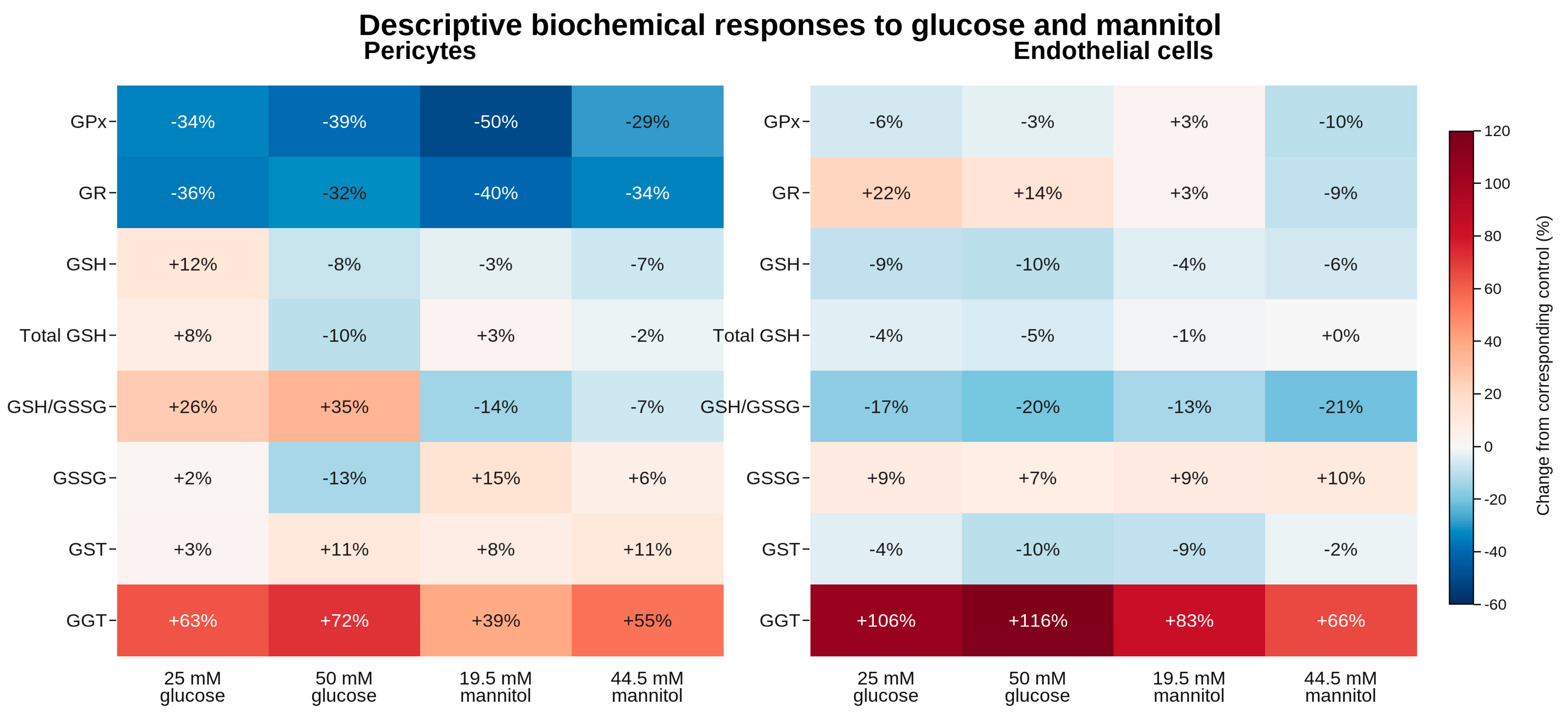
3.1. Pericyte Response Profile


| Marker | Control | G25 | G50 | M19.5 | M44.5 | Welch p | η2 |
|---|---|---|---|---|---|---|---|
| GPx | 881.81 ± 196.08 | 579.75 ± 214.09 | 539.15 ± 175.21 | 441.72 ± 101.79 | 623.60 ± 245.47 | <0.001 | 0.393 |
| GR | 446.59 ± 67.37 | 284.03 ± 117.43 | 301.89 ± 100.86 | 269.74 ± 65.47 | 292.96 ± 55.35 | <0.001 | 0.393 |
| GSH | 387.58 ± 31.74 | 433.20 ± 95.08 | 355.99 ± 35.67 | 374.98 ± 13.31 | 360.90 ± 24.38 | 0.071 | 0.260 |
| Total GSH | 602.82 ± 57.97 | 651.73 ± 183.73 | 543.77 ± 120.82 | 622.37 ± 86.80 | 588.27 ± 109.67 | 0.503 | 0.092 |
| GSH/GSSG | 1.96 ± 0.69 | 2.48 ± 1.07 | 2.64 ± 1.57 | 1.69 ± 0.57 | 1.82 ± 0.69 | 0.226 | 0.137 |
| GSSG | 215.24 ± 63.14 | 218.53 ± 128.62 | 187.78 ± 109.02 | 247.39 ± 85.64 | 227.37 ± 93.21 | 0.763 | 0.041 |
| GST | 74.71 ± 13.66 | 77.19 ± 13.12 | 82.98 ± 15.78 | 80.77 ± 12.47 | 82.70 ± 7.46 | 0.529 | 0.066 |
| GGT | 7.60 ± 1.54 | 12.40 ± 1.59 | 13.05 ± 3.01 | 10.54 ± 2.24 | 11.75 ± 1.33 | <0.001 | 0.498 |
3.2. Endothelial Response Profile
| Marker | Control | G25 | G50 | M19.5 | M44.5 | Welch p | η2 |
|---|---|---|---|---|---|---|---|
| GPx | 1914.64 ± 245.98 | 1809.09 ± 421.55 | 1854.56 ± 274.83 | 1969.86 ± 483.20 | 1727.89 ± 370.42 | 0.696 | 0.054 |
| GR | 1045.02 ± 158.72 | 1271.88 ± 434.46 | 1186.14 ± 378.21 | 1080.74 ± 281.98 | 953.91 ± 270.42 | 0.346 | 0.118 |
| GSH | 406.56 ± 52.72 | 369.72 ± 27.30 | 365.73 ± 11.17 | 389.57 ± 14.28 | 381.14 ± 21.87 | 0.005 | 0.216 |
| Total GSH | 568.37 ± 56.41 | 546.23 ± 36.94 | 540.94 ± 24.36 | 564.01 ± 33.14 | 568.18 ± 47.73 | 0.306 | 0.081 |
| GSH/GSSG | 2.65 ± 0.73 | 2.19 ± 0.59 | 2.13 ± 0.34 | 2.31 ± 0.43 | 2.09 ± 0.35 | 0.296 | 0.147 |
| GSSG | 153.94 ± 25.50 | 167.39 ± 23.52 | 164.86 ± 25.26 | 167.28 ± 23.93 | 169.18 ± 18.11 | 0.670 | 0.057 |
| GST | 71.40 ± 10.59 | 68.37 ± 7.56 | 63.95 ± 9.08 | 65.06 ± 3.72 | 70.02 ± 9.91 | 0.259 | 0.110 |
| GGT | 7.65 ± 1.10 | 15.76 ± 2.08 | 16.50 ± 2.20 | 13.99 ± 1.24 | 12.68 ± 2.25 | <0.001 | 0.763 |
3.3. Candidate Endpoints for Confirmatory Studies
3.4. Supporting Exploratory Pairwise Comparisons
| Cell Type | Marker | Comparison | Mean Difference | 95% CI | Adjusted p |
|---|---|---|---|---|---|
| Pericytes | GPx | Control vs. G25 | 302.054 | 24.232 to 579.876 | 0.029 |
| Pericytes | GPx | Control vs. G50 | 342.653 | 90.883 to 594.423 | 0.005 |
| Pericytes | GPx | Control vs. M19.5 | 440.091 | 221.352 to 658.830 | <0.001 |
| Pericytes | GR | Control vs. G25 | 162.557 | 29.596 to 295.518 | 0.014 |
| Pericytes | GR | Control vs. G50 | 144.694 | 26.921 to 262.467 | 0.013 |
| Pericytes | GR | Control vs. M19.5 | 176.850 | 87.015 to 266.685 | <0.001 |
| Pericytes | GR | Control vs. M44.5 | 153.626 | 69.928 to 237.324 | <0.001 |
| Pericytes | GGT | Control vs. G25 | −4.803 | −6.918 to −2.688 | <0.001 |
| Pericytes | GGT | Control vs. G50 | −5.457 | −8.810 to −2.104 | 0.001 |
| Pericytes | GGT | Control vs. M19.5 | −2.938 | −5.575 to −0.301 | 0.025 |
| Pericytes | GGT | Control vs. M44.5 | −4.150 | −6.102 to −2.198 | <0.001 |
| Endothelial cells | GSH | G50 vs. M19.5 | −23.833 | −41.272 to −6.394 | 0.005 |
| Endothelial cells | GGT | Control vs. G25 | −8.112 | −10.441 to −5.783 | <0.001 |
| Endothelial cells | GGT | Control vs. G50 | −8.857 | −11.300 to −6.414 | <0.001 |
| Endothelial cells | GGT | Control vs. M19.5 | −6.340 | −7.932 to −4.748 | <0.001 |
| Endothelial cells | GGT | Control vs. M44.5 | −5.036 | −7.527 to −2.545 | <0.001 |
| Endothelial cells | GGT | G25 vs. M44.5 | 3.076 | 0.144 to 6.008 | 0.037 |
| Endothelial cells | GGT | G50 vs. M19.5 | 2.517 | 0.032 to 5.002 | 0.047 |
| Endothelial cells | GGT | G50 vs. M44.5 | 3.821 | 0.813 to 6.829 | 0.009 |
4. Discussion
4.1. Coordinated GPx and GR Reduction in Pericytes
4.2. GGT-Mediated Glutathione Turnover and the Endothelial Glucose–Mannitol Contrast
4.3. Core Contribution and Limits of Inference
4.4. Strengths, Limitations, and Future Directions
5. Conclusions
Author Contributions
Funding
Institutional Review Board Statement
Informed Consent Statement
Data Availability Statement
Conflicts of Interest
Abbreviations
| DR | diabetic retinopathy |
| GPx | glutathione peroxidase |
| GR | glutathione reductase |
| GSH | reduced glutathione |
| GSSG | oxidized glutathione |
| GST | glutathione S-transferase |
| GGT | γ-glutamyl transferase |
References
- Antonetti, D.A.; Silva, P.S.; Stitt, A.W. Current understanding of the molecular and cellular pathology of diabetic retinopathy. Nat. Rev. Endocrinol. 2021, 17, 195–206, Erratum in Nat. Rev. Endocrinol. 2025, 21, 62. [Google Scholar] [CrossRef] [Scilit] [PubMed]
- Stitt, A.W.; Curtis, T.M.; Chen, M.; Medina, R.J.; McKay, G.J.; Jenkins, A.; Gardiner, T.A.; Lyons, T.J.; Hammes, H.-P.; Simó, R.; et al. The progress in understanding and treatment of diabetic retinopathy. Prog. Retin. Eye Res. 2016, 51, 156–186. [Google Scholar] [CrossRef] [Scilit] [PubMed]
- Kowluru, R.A.; Kowluru, A.; Mishra, M.; Kumar, B. Oxidative stress and epigenetic modifications in the pathogenesis of diabetic retinopathy. Prog. Retin. Eye Res. 2015, 48, 40–61. [Google Scholar] [CrossRef] [Scilit] [PubMed]
- Kang, Q.; Yang, C. Oxidative stress and diabetic retinopathy: Molecular mechanisms, pathogenetic role and therapeutic implications. Redox Biol. 2020, 37, 101799. [Google Scholar] [CrossRef] [Scilit] [PubMed]
- Mimura, T.; Noma, H. Oxidative stress in diabetic retinopathy: Molecular mechanisms and therapeutic strategies. Antioxidants 2025, 14, 1204. [Google Scholar] [CrossRef] [Scilit] [PubMed]
- Huang, H. Pericyte–endothelial interactions in the retinal microvasculature. Int. J. Mol. Sci. 2020, 21, 7413. [Google Scholar] [CrossRef] [Scilit] [PubMed]
- Nian, S.; Lo, A.C.Y.; Mi, Y.; Ren, K.; Yang, D. Neurovascular unit in diabetic retinopathy: Pathophysiological roles and potential therapeutic targets. Eye Vis. 2021, 8, 15. [Google Scholar] [CrossRef] [Scilit] [PubMed]
- Bohler, F.; Bohler, L.; Taranikanti, V. Targeting pericyte retention in diabetic retinopathy: A review. Ann. Med. 2024, 56, 2398200. [Google Scholar] [CrossRef] [Scilit] [PubMed]
- Grammas, P.; Riden, M. Retinal endothelial cells are more susceptible to oxidative stress and increased permeability than brain-derived endothelial cells. Microvasc. Res. 2003, 65, 18–23. [Google Scholar] [CrossRef] [Scilit] [PubMed]
- Agardh, C.D.; Hultberg, B.; Nayak, R.C.; Farthing-Nayak, P.; Agardh, E. Bovine retinal pericytes are resistant to glucose-induced oxidative stress in vitro. Antioxid. Redox Signal. 2005, 7, 1486–1493. [Google Scholar] [CrossRef] [Scilit] [PubMed]
- Sreekumar, P.G.; Ferrington, D.A.; Kannan, R. Glutathione metabolism and the novel role of mitochondrial GSH in retinal degeneration. Antioxidants 2021, 10, 661. [Google Scholar] [CrossRef] [Scilit] [PubMed]
- Boo, Y.C. Restoring glutathione homeostasis in glycation-related eye diseases: Mechanistic insights and therapeutic opportunities. Antioxidants 2025, 14, 731. [Google Scholar] [CrossRef] [Scilit] [PubMed]
- Forman, H.J.; Zhang, H.; Rinna, A. Glutathione: Overview of its protective roles, measurement, and biosynthesis. Mol. Asp. Med. 2009, 30, 1–12. [Google Scholar] [CrossRef] [Scilit] [PubMed]
- Whitfield, J.B. Gamma glutamyl transferase. Crit. Rev. Clin. Lab. Sci. 2001, 38, 263–355. [Google Scholar] [CrossRef] [Scilit] [PubMed]
- Kowluru, R.A.; Chan, P.S. Oxidative stress and diabetic retinopathy. Exp. Diabetes Res. 2007, 2007, 43603. [Google Scholar] [CrossRef] [Scilit] [PubMed]
- Trudeau, K.; Molina, A.J.A.; Roy, S. High glucose induces mitochondrial morphology and metabolic changes in retinal pericytes. Investig. Ophthalmol. Vis. Sci. 2011, 52, 8657–8664. [Google Scholar] [CrossRef] [Scilit] [PubMed]
- Wang, W.; Zhao, H.; Chen, B. DJ-1 protects retinal pericytes against high glucose-induced oxidative stress through the Nrf2 signaling pathway. Sci. Rep. 2020, 10, 2477. [Google Scholar] [CrossRef] [Scilit] [PubMed]
- Bertelli, P.M.; Pedrini, E.; Hughes, D.; McDonnell, S.; Pathak, V.; Peixoto, E.; Guduric-Fuchs, J.; Stitt, A.W.; Medina, R.J. Long term high glucose exposure induces premature senescence in retinal endothelial cells. Front. Physiol. 2022, 13, 929118. [Google Scholar] [CrossRef] [Scilit] [PubMed]
- Burg, M.B.; Ferraris, J.D.; Dmitrieva, N.I. Cellular response to hyperosmotic stresses. Physiol. Rev. 2007, 87, 1441–1474. [Google Scholar] [CrossRef] [Scilit] [PubMed]
- Malek, A.M.; Goss, G.G.; Jiang, L.; Izumo, S.; Alper, S.L. Mannitol at clinical concentrations activates multiple signaling pathways and induces apoptosis in endothelial cells. Stroke 1998, 29, 2631–2640. [Google Scholar] [CrossRef] [Scilit] [PubMed]
- Gudumac, V.; Rîvneac, V.; Tagadiuc, O.; Sardari, V.; Rîvneac, E.; Andronache, L.; Știrbu, O.; Pantea; V. Popa, V. Metode de Cercetare a Metabolismului Hepatic: Elaborare Metodică [Methods for the Investigation of Hepatic Metabolism: Methodological Guide]; Gudumac, V., Ed.; Tehnica-Info: Chișinău, Moldova, 2012; 162p. (In Romanian) [Google Scholar]
- Andronache, L.; Tagadiuc, O.; Gudumac, V. Procedure for the Determination of Glutathione Peroxidase Activity. Innovation Certificate No. 4758. 22 September 2009. (In Romanian)
- Andronache, L.; Tagadiuc, O.; Gudumac, V. Colorimetric Micromethod for the Determination of Glutathione Reductase Activity. Innovation Certificate No. 4757. 22 September 2009. (In Romanian)
- Andronache, L.; Tagadiuc, O.; Gudumac, V. Colorimetric Micromethod for the Determination of Glutathione. Innovation Certificate No. 4756. 22 September 2009. (In Romanian)
- Tagadiuc, O.; Andronache, L.; Gudumac, V. Optimized Procedure for the Determination of Glutathione S-Transferase Activity. Innovation Certificate No. 4755. 22 September 2009. (In Romanian)
Disclaimer/Publisher’s Note: The statements, opinions and data contained in all publications are solely those of the individual author(s) and contributor(s) and not of MDPI and/or the editor(s). MDPI and/or the editor(s) disclaim responsibility for any injury to people or property resulting from any ideas, methods, instructions or products referred to in the content. |
© 2026 by the authors. Licensee MDPI, Basel, Switzerland. This article is an open access article distributed under the terms and conditions of the Creative Commons Attribution (CC BY) license.
Share and Cite
Pavlovschi, E.; Darii, F.; Pantea, V.; Curtev, Ș.; Trocin, C.; Curlat, S.; Ușurelu, D.-C.; Mărițoi, T.; Tagadiuc, O. Glutathione-Related Responses to Hyperglycemic and Hyperosmotic Stress in Cultured Human Retinal Pericytes and Endothelial Cells: An Exploratory Study. Biomedicines 2026, 14, 2104. https://doi.org/10.3390/biomedicines14092104
Pavlovschi E, Darii F, Pantea V, Curtev Ș, Trocin C, Curlat S, Ușurelu D-C, Mărițoi T, Tagadiuc O. Glutathione-Related Responses to Hyperglycemic and Hyperosmotic Stress in Cultured Human Retinal Pericytes and Endothelial Cells: An Exploratory Study. Biomedicines. 2026; 14(9):2104. https://doi.org/10.3390/biomedicines14092104
Chicago/Turabian StylePavlovschi, Ecaterina, Felicia Darii, Valeriana Pantea, Ștefan Curtev, Cristina Trocin, Serghei Curlat, Dan-Cristian Ușurelu, Tatiana Mărițoi, and Olga Tagadiuc. 2026. "Glutathione-Related Responses to Hyperglycemic and Hyperosmotic Stress in Cultured Human Retinal Pericytes and Endothelial Cells: An Exploratory Study" Biomedicines 14, no. 9: 2104. https://doi.org/10.3390/biomedicines14092104
APA StylePavlovschi, E., Darii, F., Pantea, V., Curtev, Ș., Trocin, C., Curlat, S., Ușurelu, D.-C., Mărițoi, T., & Tagadiuc, O. (2026). Glutathione-Related Responses to Hyperglycemic and Hyperosmotic Stress in Cultured Human Retinal Pericytes and Endothelial Cells: An Exploratory Study. Biomedicines, 14(9), 2104. https://doi.org/10.3390/biomedicines14092104