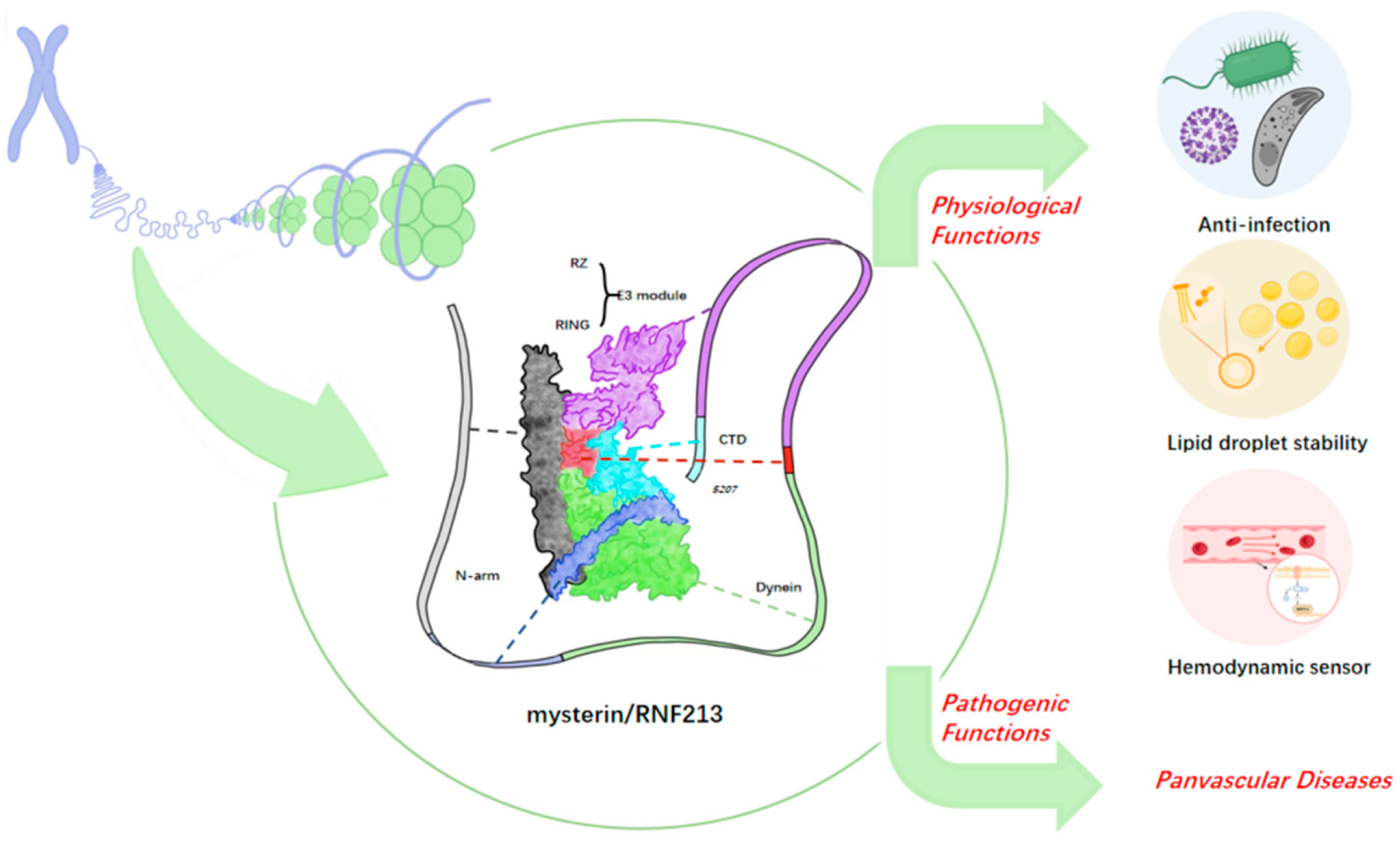
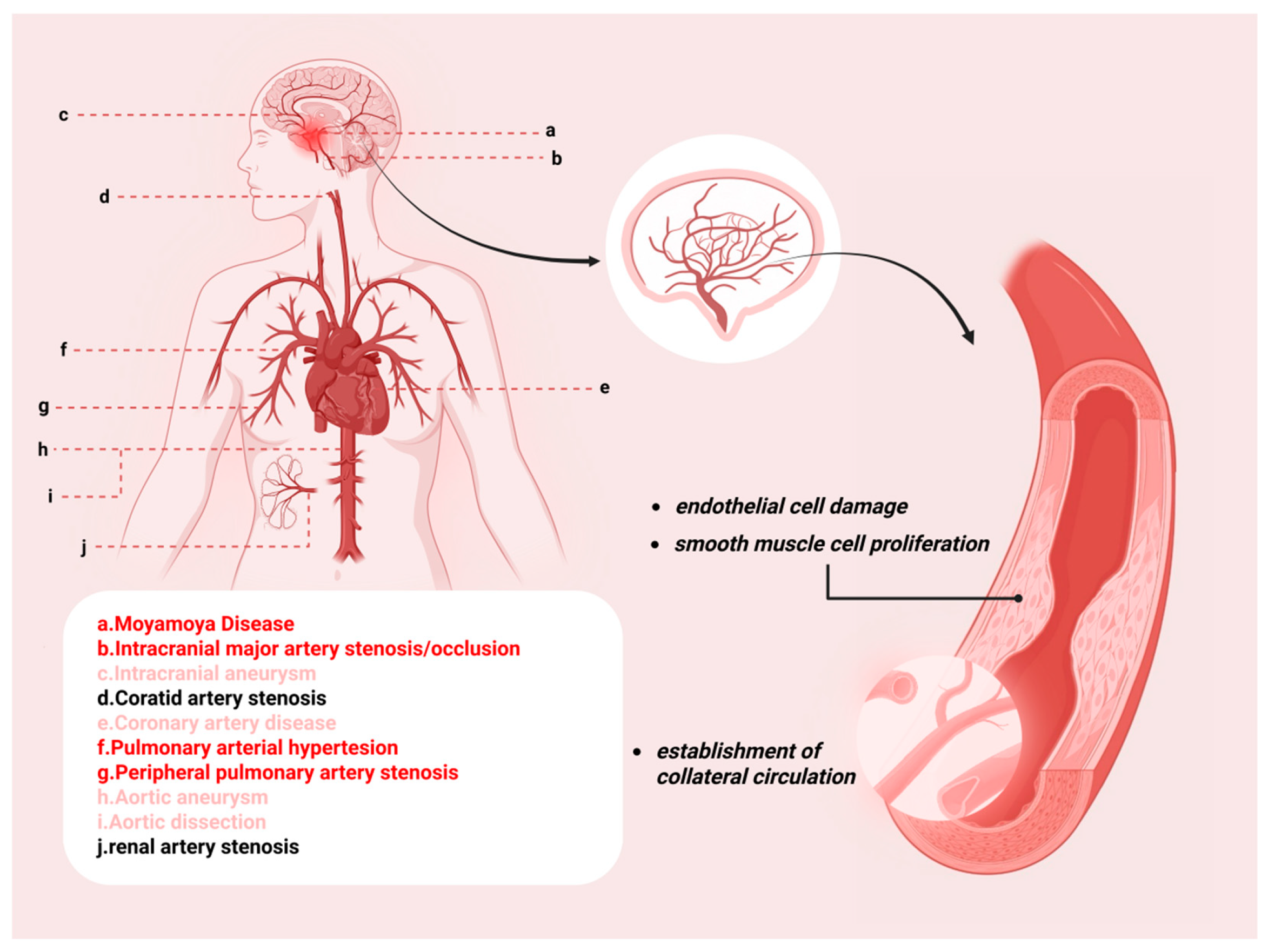
RNF213 in Panvascular Disease: A Molecular Hub Linking Genetic Susceptibility to Systemic Vasculopathy

Abstract
1. Introduction
1.1. RNF213 in Cerebrovascular Diseases
1.1.1. Moyamoya Disease
1.1.2. Intracranial Aneurysms
1.1.3. Intracranial Artery Dissection
1.1.4. Intracranial Large Artery Stenosis/Occlusion
1.2. Coronary Artery Disease
1.3. Pulmonary Artery Stenosis/Hypertension, Renal Artery Stenosis/Hypertension, and Other Peripheral Arterial Diseases
2. The Biological Functions and Mechanisms of RNF213
2.1. RNF213 Modulates the Progression of Vascular Diseases in Hypoxic Microenvironments
2.2. RNF213 Modulates Abnormal Vascular Development via Blood Flow Dynamics
- (1)
- RNF213 orchestrates mechanosensitive signaling in endothelial cells through coordinated regulation of membrane protein expression, including PECAM-1, VE-cadherin, and VEGFR2/VEGFR3 [145]. PECAM-1 colocalizes with Piezo1, and together they mediate wall shear stress-induced calcium influx and nitric oxide synthase activation [167]. Piezo channels convert mechanical stimuli into biochemical signals by regulating calcium homeostasis [168] and vascular morphogenesis [169].
- (2)
- (3)
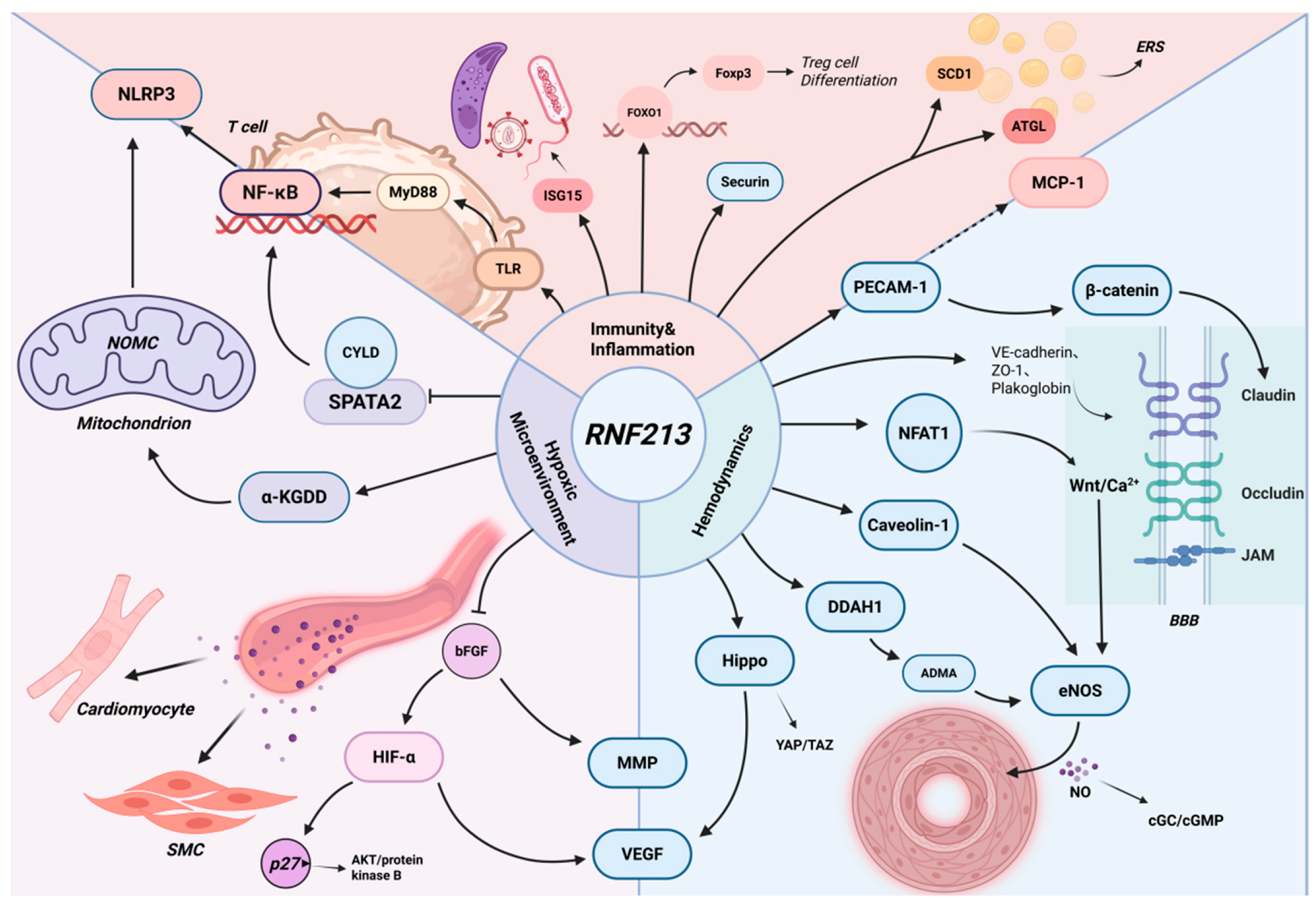
- Endothelial cells regulate the relaxation state of adjacent smooth muscle cells through nitric oxide (NO) [171,172], thereby modulating blood flow velocity and perfusion efficiency. As a key effector of vascular mechanobiology, NO is produced by endothelial cells in response to shear stress [173]. RNF213 may regulate eNOS activity by disrupting inhibitory caveolin-1-eNOS binding [174,175], promoting NFAT1 ubiquitination and degradation to suppress non-canonical Wnt/Ca2+ signaling [50,86], and enhancing DDAH1-mediated removal of ADMA-mediated eNOS inhibition [176,177] (Figure 3). Reduced caveolin-1 protein levels have been reported in MMD and in RNF213 variant carriers [178,179,180], and caveolin-1 dysregulation is also implicated in CAD [181,182] and PAH [110,183], supporting the importance of the RNF213/caveolin-1 axis in vascular pathology and angiogenesis [184,185]. Calcium/calmodulin can displace caveolin-1 from eNOS and thereby promote calcium-dependent eNOS activation [186]. NFAT1 also influences caveolin-1 expression and non-canonical Wnt/Ca2+ signaling, pathways involved in endothelial proliferation and vascular network formation [187,188]. RNF213 deficiency-induced NFAT1 nuclear translocation may reduce vascular regression and contribute to moyamoya vascular abnormalities [86]. NFAT1 overexpression has also been linked to MDM2 activation, p53 degradation, and hepatocellular carcinogenesis [189], indicating broader biological relevance.
2.3. RNF213 Participates in the Regulation of Vascular Inflammation and Immune Homeostasis
2.4. RNF213 Contributes to Panvascular Diseases via Its Antimicrobial Activity Against Bacterial, Viral, and Parasitic Infections
2.5. RNF213 Is Involved in Lipid and Carbohydrate Metabolism, Modulates Lipid Levels and Blood Glucose, and Influences the Progression of Vascular Diseases
2.6. RNF213 in Cell Cycle Regulation and Gut Microbiota Interactions
- (1)
- Downregulation of Cell Division-Related Clusters HL: Zhang et al. reported that knockdown of RNF213 (RNF213KD) results in the downregulation of gene clusters associated with cell division and proliferation, as revealed by clustering analysis [160].
- (2)
- (3)
- RNF213 has been implicated in increasing genomic instability during mitosis [301].
3. Concluding Remarks and Limitations
Author Contributions
Funding
Institutional Review Board Statement
Informed Consent Statement
Data Availability Statement
Conflicts of Interest
References
- Zhao, K.; Zeng, Z.; He, Y.; Zhao, R.; Niu, J.; Sun, H.; Li, S.; Dong, J.; Jing, Z.; Zhou, J. Recent advances in targeted therapy for inflammatory vascular diseases. J. Control. Release 2024, 372, 730–750. [Google Scholar] [CrossRef] [Scilit] [PubMed]
- Ahel, J.; Lehner, A.; Vogel, A.; Schleiffer, A.; Meinhart, A.; Haselbach, D.; Clausen, T. Moyamoya disease factor RNF213 is a giant E3 ligase with a dynein-like core and a distinct ubiquitin-transfer mechanism. eLife 2020, 9, e56185. [Google Scholar] [CrossRef] [Scilit] [PubMed]
- Zhou, X.; Yu, L.; Zhao, Y.; Ge, J. Panvascular medicine: An emerging discipline focusing on atherosclerotic diseases. Eur. Heart J. 2022, 43, 4528–4531. [Google Scholar] [CrossRef] [Scilit] [PubMed]
- Bang, O.Y.; Chung, J.W.; Kim, D.H.; Won, H.H.; Yeon, J.Y.; Ki, C.S.; Shin, H.J.; Kim, J.S.; Hong, S.C.; Kim, D.K.; et al. Moyamoya Disease and Spectrums of RNF213 Vasculopathy. Transl. Stroke Res. 2020, 11, 580–589. [Google Scholar] [PubMed]
- Hiraide, T.; Suzuki, H.; Momoi, M.; Shinya, Y.; Fukuda, K.; Kosaki, K.; Kataoka, M. RNF213-Associated Vascular Disease: A Concept Unifying Various Vasculopathies. Life 2022, 12, 555. [Google Scholar] [CrossRef] [Scilit] [PubMed]
- Hioki, H.; Miyashita, Y.; Miura, T.; Ebisawa, S.; Motoki, H.; Izawa, A.; Tomita, T.; Koyama, J.; Ikeda, U. Diagnostic value of peripheral fractional flow reserve in isolated iliac artery stenosis: A comparison with the post-exercise ankle-brachial index. J. Endovasc. Ther. 2014, 21, 625–632. [Google Scholar] [PubMed]
- Albayati, M.A.; Patel, A.; Modi, B.; Saha, P.; Karim, L.; Perera, D.; Smith, A.; Modarai, B. Intra-arterial Fractional Flow Reserve Measurements Provide an Objective Assessment of the Functional Significance of Peripheral Arterial Stenoses. Eur. J. Vasc. Endovasc. Surg. 2024, 67, 332–340. [Google Scholar] [CrossRef] [Scilit] [PubMed]
- Kobayashi, N.; Hirano, K.; Yamawaki, M.; Araki, M.; Sakai, T.; Takimura, H.; Sakamoto, Y.; Mori, S.; Tsutsumi, M.; Takama, T.; et al. Ability of Fractional Flow Reserve to Predict Restenosis After Superficial Femoral Artery Stenting. J. Endovasc. Ther. 2016, 23, 896–902. [Google Scholar] [CrossRef] [Scilit] [PubMed]
- Suzuki, J.; Takaku, A. Cerebrovascular “moyamoya” disease. Disease showing abnormal net-like vessels in base of brain. Arch. Neurol. 1969, 20, 288–299. [Google Scholar] [PubMed]
- Liu, W.; Hitomi, T.; Kobayashi, H.; Harada, K.H.; Koizumi, A. Distribution of moyamoya disease susceptibility polymorphism p.R4810K in RNF213 in East and Southeast Asian populations. Neurol. Med.-Chir. 2012, 52, 299–303. [Google Scholar]
- Ihara, M.; Yamamoto, Y.; Hattori, Y.; Liu, W.; Kobayashi, H.; Ishiyama, H.; Yoshimoto, T.; Miyawaki, S.; Clausen, T.; Bang, O.Y.; et al. Moyamoya disease: Diagnosis and interventions. Lancet Neurol. 2022, 21, 747–758. [Google Scholar] [CrossRef] [Scilit] [PubMed]
- Liu, W.; Morito, D.; Takashima, S.; Mineharu, Y.; Kobayashi, H.; Hitomi, T.; Hashikata, H.; Matsuura, N.; Yamazaki, S.; Toyoda, A.; et al. Identification of RNF213 as a susceptibility gene for moyamoya disease and its possible role in vascular development. PLoS ONE 2011, 6, e22542. [Google Scholar] [CrossRef] [Scilit] [PubMed]
- Kamada, F.; Aoki, Y.; Narisawa, A.; Abe, Y.; Komatsuzaki, S.; Kikuchi, A.; Kanno, J.; Niihori, T.; Ono, M.; Ishii, N.; et al. A genome-wide association study identifies RNF213 as the first Moyamoya disease gene. J. Hum. Genet. 2011, 56, 34–40. [Google Scholar] [PubMed]
- Wu, Z.; Jiang, H.; Zhang, L.; Xu, X.; Zhang, X.; Kang, Z.; Song, D.; Zhang, J.; Guan, M.; Gu, Y. Molecular analysis of RNF213 gene for moyamoya disease in the Chinese Han population. PLoS ONE 2012, 7, e48179. [Google Scholar] [CrossRef] [Scilit] [PubMed]
- Cecchi, A.C.; Guo, D.; Ren, Z.; Flynn, K.; Santos-Cortez, R.L.; Leal, S.M.; Wang, G.T.; Regalado, E.S.; Steinberg, G.K.; Shendure, J.; et al. RNF213 rare variants in an ethnically diverse population with Moyamoya disease. Stroke 2014, 45, 3200–3207. [Google Scholar] [CrossRef] [Scilit] [PubMed]
- Miyawaki, S.; Imai, H.; Takayanagi, S.; Mukasa, A.; Nakatomi, H.; Saito, N. Identification of a genetic variant common to moyamoya disease and intracranial major artery stenosis/occlusion. Stroke 2012, 43, 3371–3374. [Google Scholar] [CrossRef] [Scilit] [PubMed]
- Zhou, S.; Ambalavanan, A.; Rochefort, D.; Xie, P.; Bourassa, C.V.; Hince, P.; Dionne-Laporte, A.; Spiegelman, D.; Gan-Or, Z.; Mirarchi, C.; et al. RNF213 Is Associated with Intracranial Aneurysms in the French-Canadian Population. Am. J. Hum. Genet. 2016, 99, 1072–1085. [Google Scholar] [CrossRef] [Scilit] [PubMed]
- Momoi, M.; Katsumata, Y.; Hiraide, T.; Shinya, Y.; Goto, S.; Inami, T.; Kosaki, K.; Kataoka, M. Chronic Thromboembolic Pulmonary Hypertension as RNF213-Associated Vascular Disease. JACC Asia 2024, 4, 1017–1019. [Google Scholar] [CrossRef] [Scilit] [PubMed]
- Hiraide, T.; Kataoka, M.; Suzuki, H.; Aimi, Y.; Chiba, T.; Isobe, S.; Katsumata, Y.; Goto, S.; Kanekura, K.; Yamada, Y.; et al. Poor outcomes in carriers of the RNF213 variant (p.Arg4810Lys) with pulmonary arterial hypertension. J. Heart Lung Transplant. 2020, 39, 103–112. [Google Scholar] [PubMed]
- Kim, J.Y.; Cho, H. Renovascular hypertension and RNF213 p.R4810K variant in Korean children with Moyamoya disease. Clin. Nephrol. 2021, 96, 105–111. [Google Scholar] [PubMed]
- Kim, Y.J.; Lee, J.K.; Ahn, S.H.; Kim, B.J.; Kang, D.W.; Kim, J.S.; Kwon, S.U. Nonatheroscleotic Isolated Middle Cerebral Artery Disease May Be Early Manifestation of Moyamoya Disease. Stroke 2016, 47, 2229–2235. [Google Scholar] [CrossRef] [Scilit] [PubMed]
- Kim, J.S.; Lee, H.B.; Kwon, H.S. RNF213 Polymorphism in Intracranial Artery Dissection. J. Stroke 2018, 20, 404–406. [Google Scholar] [CrossRef] [Scilit] [PubMed]
- Morimoto, T.; Mineharu, Y.; Ono, K.; Nakatochi, M.; Ichihara, S.; Kabata, R.; Takagi, Y.; Cao, Y.; Zhao, L.; Kobayashi, H.; et al. Significant association of RNF213 p.R4810K, a moyamoya susceptibility variant, with coronary artery disease. PLoS ONE 2017, 12, e0175649. [Google Scholar] [PubMed]
- Shinya, Y.; Miyawaki, S.; Imai, H.; Hongo, H.; Ono, H.; Takenobu, A.; Nakatomi, H.; Teraoka, A.; Saito, N. Genetic Analysis of Ring Finger Protein 213 (RNF213) c.14576G>A in Intracranial Atherosclerosis of the Anterior and Posterior Circulations. J. Stroke Cerebrovasc. Dis. 2017, 26, 2638–2644. [Google Scholar] [PubMed]
- Yang, W.J.; Wong, K.S.; Chen, X.Y. Intracranial Atherosclerosis: From Microscopy to High-Resolution Magnetic Resonance Imaging. J. Stroke 2017, 19, 249–260. [Google Scholar] [CrossRef] [Scilit] [PubMed]
- Zervas, N.T.; Liszczak, T.M.; Mayberg, M.R.; Black, P.M. Cerebrospinal fluid may nourish cerebral vessels through pathways in the adventitia that may be analogous to systemic vasa vasorum. J. Neurosurg. 1982, 56, 475–481. [Google Scholar] [CrossRef] [Scilit] [PubMed]
- Kang, D.W.; Kim, D.Y.; Kim, J.; Baik, S.H.; Jung, C.; Singh, N.; Song, J.W.; Bae, H.J.; Kim, B.J. Emerging Concept of Intracranial Arterial Diseases: The Role of High Resolution Vessel Wall MRI. J. Stroke 2024, 26, 26–40. [Google Scholar] [CrossRef] [Scilit] [PubMed]
- Janda, P.H.; Bellew, J.G.; Veerappan, V. Moyamoya disease: Case report and literature review. J. Osteopath. Med. 2009, 109, 547–553. [Google Scholar]
- Asselman, C.; Hemelsoet, D.; Eggermont, D.; Dermaut, B.; Impens, F. Moyamoya disease emerging as an immune-related angiopathy. Trends Mol. Med. 2022, 28, 939–950. [Google Scholar] [CrossRef] [Scilit] [PubMed]
- Scott, R.M.; Smith, E.R. Moyamoya disease and moyamoya syndrome. N. Engl. J. Med. 2009, 360, 1226–1237. [Google Scholar] [CrossRef] [Scilit] [PubMed]
- Takagi, Y.; Kikuta, K.; Nozaki, K.; Fujimoto, M.; Hayashi, J.; Imamura, H.; Hashimoto, N. Expression of hypoxia-inducing factor-1 alpha and endoglin in intimal hyperplasia of the middle cerebral artery of patients with Moyamoya disease. Neurosurgery 2007, 60, 338–345, discussion 345. [Google Scholar] [PubMed]
- Takekawa, Y.; Umezawa, T.; Ueno, Y.; Sawada, T.; Kobayashi, M. Pathological and immunohistochemical findings of an autopsy case of adult moyamoya disease. Neuropathology 2004, 24, 236–242. [Google Scholar] [CrossRef] [Scilit] [PubMed]
- Abumiya, T.; Fujimura, M. Moyamoya Vasculopathy and Moyamoya-Related Systemic Vasculopathy: A Review with Histopathological and Genetic Viewpoints. Stroke 2024, 55, 1699–1706. [Google Scholar] [CrossRef] [Scilit] [PubMed]
- Liu, H.; Fukasawa, T.; Anno, T.; Takeuchi, M.; Shimazaki, S.; Yang, T.; Kawakami, K. Incidence, prevalence, and treatment of Moyamoya disease in Japan: A population-based descriptive study. J. Stroke Cerebrovasc. Dis. 2024, 33, 107770. [Google Scholar] [CrossRef] [Scilit] [PubMed]
- Sun, Y.; Zhou, G.; Feng, J.; Chen, L.; Liu, G.; Wang, J.; Wang, Q.; Yu, J.; Yang, X.; Yang, Z.; et al. Incidence and prevalence of moyamoya disease in urban China: A nationwide retrospective cohort study. Stroke Vasc. Neurol. 2021, 6, 615–623. [Google Scholar] [CrossRef] [Scilit] [PubMed]
- Ahn, I.M.; Park, D.H.; Hann, H.J.; Kim, K.H.; Kim, H.J.; Ahn, H.S. Incidence, prevalence, and survival of moyamoya disease in Korea: A nationwide, population-based study. Stroke 2014, 45, 1090–1095. [Google Scholar] [CrossRef] [Scilit] [PubMed]
- Uchino, K.; Johnston, S.C.; Becker, K.J.; Tirschwell, D.L. Moyamoya disease in Washington State and California. Neurology 2005, 65, 956–958. [Google Scholar] [CrossRef] [Scilit] [PubMed]
- Khan, N.; Yonekawa, Y. Moyamoya angiopathy in Europe. Acta Neurochir. Suppl. 2005, 94, 149–152. [Google Scholar] [PubMed]
- Ito, A.; Fujimura, M.; Niizuma, K.; Kanoke, A.; Sakata, H.; Morita-Fujimura, Y.; Kikuchi, A.; Kure, S.; Tominaga, T. Enhanced post-ischemic angiogenesis in mice lacking RNF213; a susceptibility gene for moyamoya disease. Brain Res. 2015, 1594, 310–320. [Google Scholar] [CrossRef] [Scilit] [PubMed]
- Kobayashi, H.; Matsuda, Y.; Hitomi, T.; Okuda, H.; Shioi, H.; Matsuda, T.; Imai, H.; Sone, M.; Taura, D.; Harada, K.H.; et al. Biochemical and Functional Characterization of RNF213 (Mysterin) R4810K, a Susceptibility Mutation of Moyamoya Disease, in Angiogenesis In Vitro and In Vivo. J. Am. Heart Assoc. 2015, 4, e002146. [Google Scholar] [CrossRef] [Scilit] [PubMed]
- Koizumi, A.; Kobayashi, H.; Hitomi, T.; Harada, K.H.; Habu, T.; Youssefian, S. A new horizon of moyamoya disease and associated health risks explored through RNF213. Environ. Health Prev. Med. 2016, 21, 55–70. [Google Scholar] [CrossRef] [Scilit] [PubMed]
- Wang, Y.; Yang, L.; Wang, X.; Zeng, F.; Zhang, K.; Zhang, Q.; Liu, M.; Liu, S.; Shang, M.; Li, Q.; et al. Meta-analysis of genotype and phenotype studies to confirm the predictive role of the RNF213 p.R4810K variant for moyamoya disease. Eur. J. Neurol. 2021, 28, 823–836. [Google Scholar] [PubMed]
- Miyatake, S.; Miyake, N.; Touho, H.; Nishimura-Tadaki, A.; Kondo, Y.; Okada, I.; Tsurusaki, Y.; Doi, H.; Sakai, H.; Saitsu, H.; et al. Homozygous c.14576G>A variant of RNF213 predicts early-onset and severe form of moyamoya disease. Neurology 2012, 78, 803–810. [Google Scholar] [PubMed]
- Miyatake, S.; Touho, H.; Miyake, N.; Ohba, C.; Doi, H.; Saitsu, H.; Taguri, M.; Morita, S.; Matsumoto, N. Sibling cases of moyamoya disease having homozygous and heterozygous c.14576G>A variant in RNF213 showed varying clinical course and severity. J. Hum. Genet. 2012, 57, 804–806. [Google Scholar] [CrossRef] [Scilit] [PubMed]
- Mertens, R.; Graupera, M.; Gerhardt, H.; Bersano, A.; Tournier-Lasserve, E.; Mensah, M.A.; Mundlos, S.; Vajkoczy, P. The Genetic Basis of Moyamoya Disease. Transl. Stroke Res. 2022, 13, 25–45. [Google Scholar] [PubMed]
- Tokairin, K.; Hamauchi, S.; Ito, M.; Kazumata, K.; Sugiyama, T.; Nakayama, N.; Kawabori, M.; Osanai, T.; Houkin, K. Vascular Smooth Muscle Cell Derived from IPS Cell of Moyamoya Disease—Comparative Characterization with Endothelial Cell Transcriptome. J. Stroke Cerebrovasc. Dis. 2020, 29, 105305. [Google Scholar] [CrossRef] [Scilit] [PubMed]
- Phi, J.H.; Suzuki, N.; Moon, Y.J.; Park, A.K.; Wang, K.C.; Lee, J.Y.; Choi, S.A.; Chong, S.; Shirane, R.; Kim, S.K. Chemokine Ligand 5 (CCL5) Derived from Endothelial Colony-Forming Cells (ECFCs) Mediates Recruitment of Smooth Muscle Progenitor Cells (SPCs) toward Critical Vascular Locations in Moyamoya Disease. PLoS ONE 2017, 12, e0169714. [Google Scholar] [CrossRef] [Scilit] [PubMed]
- Guo, D.C.; Papke, C.L.; Tran-Fadulu, V.; Regalado, E.S.; Avidan, N.; Johnson, R.J.; Kim, D.H.; Pannu, H.; Willing, M.C.; Sparks, E.; et al. Mutations in smooth muscle alpha-actin (ACTA2) cause coronary artery disease, stroke, and Moyamoya disease, along with thoracic aortic disease. Am. J. Hum. Genet. 2009, 84, 617–627. [Google Scholar] [CrossRef] [Scilit] [PubMed]
- Hervé, D.; Philippi, A.; Belbouab, R.; Zerah, M.; Chabrier, S.; Collardeau-Frachon, S.; Bergametti, F.; Essongue, A.; Berrou, E.; Krivosic, V.; et al. Loss of α1β1 soluble guanylate cyclase, the major nitric oxide receptor, leads to moyamoya and achalasia. Am. J. Hum. Genet. 2014, 94, 385–394. [Google Scholar] [CrossRef] [Scilit] [PubMed]
- Amal, H.; Gong, G.; Gjoneska, E.; Lewis, S.M.; Wishnok, J.S.; Tsai, L.H.; Tannenbaum, S.R. S-nitrosylation of E3 ubiquitin-protein ligase RNF213 alters non-canonical Wnt/Ca+2 signaling in the P301S mouse model of tauopathy. Transl. Psychiatry 2019, 9, 44. [Google Scholar] [CrossRef] [Scilit] [PubMed]
- Lattanzio, S.; Santilli, F.; Liani, R.; Vazzana, N.; Ueland, T.; Di Fulvio, P.; Formoso, G.; Consoli, A.; Aukrust, P.; Davì, G. Circulating dickkopf-1 in diabetes mellitus: Association with platelet activation and effects of improved metabolic control and low-dose aspirin. J. Am. Heart Assoc. 2014, 3, e001000. [Google Scholar] [PubMed]
- Dong, R.; Chen, W.; Feng, W.; Xia, C.; Hu, D.; Zhang, Y.; Yang, Y.; Wang, D.W.; Xu, X.; Tu, L. Exogenous Bradykinin Inhibits Tissue Factor Induction and Deep Vein Thrombosis via Activating the eNOS/Phosphoinositide 3-Kinase/Akt Signaling Pathway. Cell Physiol. Biochem. 2015, 37, 1592–1606. [Google Scholar] [CrossRef] [Scilit] [PubMed]
- Xu, S.; Chen, T.; Yu, J.; Wan, L.; Zhang, J.; Chen, J.; Wei, W.; Li, X. Insights into the regulatory role of epigenetics in moyamoya disease: Current advances and future prospectives. Mol. Ther. Nucleic Acids 2024, 35, 102281. [Google Scholar] [CrossRef] [Scilit] [PubMed]
- He, X.W.; Zhao, Y.; Shi, Y.H.; Zhao, R.; Liu, Y.S.; Hu, Y.; Zhuang, M.T.; Wu, Y.L.; Li, G.F.; Yin, J.W.; et al. DNA Methylation Analysis Identifies Differentially Methylated Sites Associated with Early-Onset Intracranial Atherosclerotic Stenosis. J. Atheroscler. Thromb. 2020, 27, 71–99. [Google Scholar] [CrossRef] [Scilit] [PubMed]
- Yoshimoto, T.; Yamagami, H.; Matsumaru, Y. Recent Advances in Stroke Genetics-Unraveling the Complexity of Cerebral Infarction: A Brief Review. Genes 2025, 16, 59. [Google Scholar] [CrossRef] [Scilit] [PubMed]
- Zhou, J.; Lei, Y.; Zhang, S.; Qin, R.; Liu, Y.; Yi, D. Identification of the potential role of S1PR1 in adult moyamoya disease based on multiple bioinformatics analysis and experimental verification. FASEB J. 2025, 39, e70461. [Google Scholar] [CrossRef] [Scilit] [PubMed]
- Mineharu, Y.; Miyamoto, S. RNF213 and GUCY1A3 in Moyamoya Disease: Key Regulators of Metabolism, Inflammation, and Vascular Stability. Front. Neurol. 2021, 12, 687088. [Google Scholar] [CrossRef] [Scilit] [PubMed]
- Schievink, W.I.; Schaid, D.J.; Rogers, H.M.; Piepgras, D.G.; Michels, V.V. On the inheritance of intracranial aneurysms. Stroke 1994, 25, 2028–2037. [Google Scholar] [CrossRef] [Scilit] [PubMed]
- Schievink, W.I. Genetics of intracranial aneurysms. Neurosurgery 1997, 40, 651–662, discussion 662–653. [Google Scholar] [CrossRef] [Scilit] [PubMed]
- Kissela, B.M.; Sauerbeck, L.; Woo, D.; Khoury, J.; Carrozzella, J.; Pancioli, A.; Jauch, E.; Moomaw, C.J.; Shukla, R.; Gebel, J.; et al. Subarachnoid hemorrhage: A preventable disease with a heritable component. Stroke 2002, 33, 1321–1326. [Google Scholar] [PubMed]
- Ronkainen, A.; Hernesniemi, J.; Puranen, M.; Niemitukia, L.; Vanninen, R.; Ryynänen, M.; Kuivaniemi, H.; Tromp, G. Familial intracranial aneurysms. Lancet 1997, 349, 380–384. [Google Scholar] [CrossRef] [Scilit] [PubMed]
- Liu, J.; Yan, J.; Li, Y.; Jiang, W. Progress in genetic research on familiar aneurysms. J. Cent. South Univ. Med. Sci. 2019, 44, 338–344. [Google Scholar]
- Imai, Y.; Sato, K.; Ishikawa, T.; Comerford, A.; David, T.; Yamaguchi, T. ATP transport in saccular cerebral aneurysms at arterial bends. Ann. Biomed. Eng. 2010, 38, 927–934. [Google Scholar] [PubMed]
- Fukushima, Y.; Miyawaki, S.; Inoue, T.; Shimizu, S.; Yoshikawa, G.; Imai, H.; Saito, N.; Tsutsumi, K. Repeated de novo aneurysm formation after anastomotic surgery: Potential risk of genetic variant RNF213 c.14576G>A. Surg. Neurol. Int. 2015, 6, 41. [Google Scholar] [CrossRef] [Scilit] [PubMed]
- Aoki, T.; Yoshitomi, M.; Yamamoto, M.; Hirohata, M.; Morioka, M. Ruptured de novo aneurysm arising at a site remote from the anastomosis 14 years after superficial temporal artery-middle cerebral artery bypass: A case report. Neurosurgery 2012, 71, E905–E909. [Google Scholar] [CrossRef] [Scilit] [PubMed]
- Kohno, K.; Ueda, T.; Kadota, O.; Sakaki, S. Subdural hemorrhage caused by de novo aneurysm complicating extracranial-intracranial bypass surgery: Case report. Neurosurgery 1996, 38, 1051–1055. [Google Scholar] [CrossRef] [Scilit] [PubMed]
- Lee, S.W.; Antiga, L.; Spence, J.D.; Steinman, D.A. Geometry of the carotid bifurcation predicts its exposure to disturbed flow. Stroke 2008, 39, 2341–2347. [Google Scholar] [CrossRef] [Scilit] [PubMed]
- Debette, S.; Compter, A.; Labeyrie, M.A.; Uyttenboogaart, M.; Metso, T.M.; Majersik, J.J.; Goeggel-Simonetti, B.; Engelter, S.T.; Pezzini, A.; Bijlenga, P.; et al. Epidemiology, pathophysiology, diagnosis, and management of intracranial artery dissection. Lancet Neurol. 2015, 14, 640–654. [Google Scholar] [CrossRef] [Scilit] [PubMed]
- Sikkema, T.; Uyttenboogaart, M.; Eshghi, O.; De Keyser, J.; Brouns, R.; van Dijk, J.M.; Luijckx, G.J. Intracranial artery dissection. Eur. J. Neurol. 2014, 21, 820–826. [Google Scholar] [CrossRef] [Scilit] [PubMed]
- Traenka, C.; Kloss, M.; Strom, T.; Lyrer, P.; Brandt, T.; Bonati, L.H.; Grond-Ginsbach, C.; Engelter, S. Rare genetic variants in patients with cervical artery dissection. Eur. Stroke J. 2019, 4, 355–362. [Google Scholar] [CrossRef] [Scilit] [PubMed]
- Shinya, Y.; Miyawaki, S.; Nakatomi, H.; Shin, M.; Teraoka, A.; Saito, N. Hemorrhagic Onset Intracranial Artery Dissection of Middle Cerebral Artery Followed by Progressive Arterial Stenosis with Genetic Variant RNF213 p.Arg4810Lys (rs112735431). World Neurosurg. 2020, 141, 192–195. [Google Scholar] [PubMed]
- Tashiro, R.; Fujimura, M.; Sakata, H.; Endo, H.; Tomata, Y.; Sato-Maeda, M.; Niizuma, K.; Tominaga, T. Genetic analysis of ring finger protein 213 (RNF213) c.14576G>A polymorphism in patients with vertebral artery dissection: A comparative study with moyamoya disease. Neurol. Res. 2019, 41, 811–816. [Google Scholar] [CrossRef] [Scilit] [PubMed]
- Mizutani, T.; Miki, Y.; Kojima, H.; Suzuki, H. Proposed classification of nonatherosclerotic cerebral fusiform and dissecting aneurysms. Neurosurgery 1999, 45, 253–259; discussion 259–260. [Google Scholar] [CrossRef] [Scilit]
- Mizutani, T. Natural course of intracranial arterial dissections. J. Neurosurg. 2011, 114, 1037–1044. [Google Scholar] [CrossRef] [Scilit] [PubMed]
- Mizutani, T.; Kojima, H.; Asamoto, S.; Miki, Y. Pathological mechanism and three-dimensional structure of cerebral dissecting aneurysms. J. Neurosurg. 2001, 94, 712–717. [Google Scholar] [CrossRef] [Scilit] [PubMed]
- Tawk, R.G.; Bendok, B.R.; Qureshi, A.I.; Getch, C.C.; Srinivasan, J.; Alberts, M.; Russell, E.J.; Batjer, H.H. Isolated dissections and dissecting aneurysms of the posterior inferior cerebellar artery: Topic and literature review. Neurosurg. Rev. 2003, 26, 180–187. [Google Scholar] [PubMed]
- Shlapakova, P.S.; Dobrynina, L.A.; Kalashnikova, L.A.; Gubanova, M.V.; Danilova, M.S.; Gnedovskaya, E.V.; Grigorenko, A.P.; Gusev, F.E.; Manakhov, A.D.; Rogaev, E.I. Peripheral Blood Gene Expression Profiling Reveals Molecular Pathways Associated with Cervical Artery Dissection. Int. J. Mol. Sci. 2024, 25, 5205. [Google Scholar] [CrossRef] [Scilit] [PubMed]
- Miyawaki, S.; Imai, H.; Shimizu, M.; Yagi, S.; Ono, H.; Mukasa, A.; Nakatomi, H.; Shimizu, T.; Saito, N. Genetic variant RNF213 c.14576G>A in various phenotypes of intracranial major artery stenosis/occlusion. Stroke 2013, 44, 2894–2897. [Google Scholar] [PubMed]
- Bang, O.Y.; Chung, J.W.; Cha, J.; Lee, M.J.; Yeon, J.Y.; Ki, C.S.; Jeon, P.; Kim, J.S.; Hong, S.C. A Polymorphism in RNF213 Is a Susceptibility Gene for Intracranial Atherosclerosis. PLoS ONE 2016, 11, e0156607. [Google Scholar] [CrossRef] [Scilit] [PubMed]
- Liao, X.; Deng, J.; Dai, W.; Zhang, T.; Yan, J. Rare variants of RNF213 and moyamoya/non-moyamoya intracranial artery stenosis/occlusion disease risk: A meta-analysis and systematic review. Environ. Health Prev. Med. 2017, 22, 75. [Google Scholar] [PubMed]
- Lin, J.; Sheng, W. RNF213 Variant Diversity Predisposes Distinct Populations to Dissimilar Cerebrovascular Diseases. Biomed. Res. Int. 2018, 2018, 6359174. [Google Scholar] [CrossRef] [Scilit] [PubMed]
- Hongo, H.; Miyawaki, S.; Imai, H.; Shimizu, M.; Yagi, S.; Mitsui, J.; Ishiura, H.; Yoshimura, J.; Doi, K.; Qu, W.; et al. Comprehensive investigation of RNF213 nonsynonymous variants associated with intracranial artery stenosis. Sci. Rep. 2020, 10, 11942. [Google Scholar] [CrossRef] [Scilit] [PubMed]
- Ozaki, K.; Tanaka, T. Molecular genetics of coronary artery disease. J. Hum. Genet. 2016, 61, 71–77. [Google Scholar] [PubMed]
- Ogura, M. PCSK9 inhibition in the management of familial hypercholesterolemia. J. Cardiol. 2018, 71, 1–7. [Google Scholar] [CrossRef] [Scilit] [PubMed]
- Hikino, K.; Koyama, S.; Ito, K.; Koike, Y.; Koido, M.; Matsumura, T.; Kurosawa, R.; Tomizuka, K.; Ito, S.; Liu, X.; et al. RNF213 Variants, Vasospastic Angina, and Risk of Fatal Myocardial Infarction. JAMA Cardiol. 2024, 9, 723–731. [Google Scholar] [CrossRef] [Scilit] [PubMed]
- Scholz, B.; Korn, C.; Wojtarowicz, J.; Mogler, C.; Augustin, I.; Boutros, M.; Niehrs, C.; Augustin, H.G. Endothelial RSPO3 Controls Vascular Stability and Pruning through Non-canonical WNT/Ca(2+)/NFAT Signaling. Dev. Cell 2016, 36, 79–93. [Google Scholar] [PubMed]
- Zamora, M.; Männer, J.; Ruiz-Lozano, P. Epicardium-derived progenitor cells require beta-catenin for coronary artery formation. Proc. Natl. Acad. Sci. USA 2007, 104, 18109–18114. [Google Scholar] [PubMed]
- Dumont, J.; Meroufel, D.; Bauters, C.; Hansmannel, F.; Bensemain, F.; Cottel, D.; Hamon, M.; Lambert, J.C.; Ducimetière, P.; Amouyel, P.; et al. Association of ornithine transcarbamylase gene polymorphisms with hypertension and coronary artery vasomotion. Am. J. Hypertens. 2009, 22, 993–1000. [Google Scholar] [CrossRef] [Scilit] [PubMed]
- Ford, T.J.; Corcoran, D.; Padmanabhan, S.; Aman, A.; Rocchiccioli, P.; Good, R.; McEntegart, M.; Maguire, J.J.; Watkins, S.; Eteiba, H.; et al. Genetic dysregulation of endothelin-1 is implicated in coronary microvascular dysfunction. Eur. Heart J. 2020, 41, 3239–3252. [Google Scholar] [CrossRef] [Scilit] [PubMed]
- Mizuno, Y.; Harada, E.; Morita, S.; Kinoshita, K.; Hayashida, M.; Shono, M.; Morikawa, Y.; Murohara, T.; Nakayama, M.; Yoshimura, M.; et al. East asian variant of aldehyde dehydrogenase 2 is associated with coronary spastic angina: Possible roles of reactive aldehydes and implications of alcohol flushing syndrome. Circulation 2015, 131, 1665–1673. [Google Scholar] [PubMed]
- Noda, K.; Hattori, Y.; Hori, M.; Harada-Shiba, M.; Ihara, M. A Case of Multiple Intracranial Major Artery Stenoses with Coexisting PCSK9 p.E32K and RNF213 p.R4810K Variants. Neurol. Genet. 2023, 9, e200099. [Google Scholar] [PubMed]
- Ikeda, E. Systemic vascular changes in spontaneous occlusion of the circle of Willis. Stroke 1991, 22, 1358–1362. [Google Scholar] [CrossRef] [Scilit] [PubMed]
- Nomura, S.; Aihara, Y.; Akagawa, H.; Chiba, K.; Yamaguchi, K.; Kawashima, A.; Okada, Y.; Kawamata, T. Can Moyamoya Disease Susceptibility Gene Affect Extracranial Systemic Artery Stenosis? J. Stroke Cerebrovasc. Dis. 2020, 29, 104532. [Google Scholar] [CrossRef] [Scilit] [PubMed]
- Pinard, A.; Fiander, M.D.J.; Cecchi, A.C.; Rideout, A.L.; Azouz, M.; Fraser, S.M.; McNeely, P.D.; Walling, S.; Novara, S.C.; Hurst, A.C.E.; et al. Association of De Novo RNF213 Variants with Childhood Onset Moyamoya Disease and Diffuse Occlusive Vasculopathy. Neurology 2021, 96, e1783–e1791. [Google Scholar] [CrossRef] [Scilit] [PubMed]
- Jee, T.K.; Yeon, J.Y.; Kim, S.M.; Bang, O.Y.; Kim, J.S.; Hong, S.C. Prospective Screening of Extracranial Systemic Arteriopathy in Young Adults with Moyamoya Disease. J. Am. Heart Assoc. 2020, 9, e016670. [Google Scholar] [CrossRef] [Scilit] [PubMed]
- Baek, J.W.; Jo, K.I.; Park, J.J.; Jeon, P.; Kim, K.H. Prevalence and clinical implications of renal artery stenosis in pediatric moyamoya disease. Eur. J. Paediatr. Neurol. 2016, 20, 20–24. [Google Scholar] [CrossRef] [Scilit] [PubMed]
- Liu, E.; Zhao, H.; Liu, C.; Tan, X.; Luo, C.; Yang, S. Research progress of moyamoya disease combined with renovascular hypertension. Front. Surg. 2022, 9, 969090. [Google Scholar] [CrossRef] [Scilit] [PubMed]
- Guey, S.; Tournier-Lasserve, E.; Hervé, D.; Kossorotoff, M. Moyamoya disease and syndromes: From genetics to clinical management. Appl. Clin. Genet. 2015, 8, 49–68. [Google Scholar] [CrossRef] [Scilit] [PubMed]
- Dorschel, K.B.; Wanebo, J.E. Genetic and Proteomic Contributions to the Pathophysiology of Moyamoya Angiopathy and Related Vascular Diseases. Appl. Clin. Genet. 2021, 14, 145–171. [Google Scholar] [CrossRef] [Scilit] [PubMed]
- Jansen, J.N.; Donker, A.J.; Luth, W.J.; Smit, L.M. Moyamoya disease associated with renovascular hypertension. Neuropediatrics 1990, 21, 44–47. [Google Scholar] [CrossRef] [Scilit] [PubMed]
- Kuwayama, F.; Hamasaki, Y.; Shinagawa, T.; Kubota, C.; Ichikawa, I.; Kato, Y.; Oi, S.; Shinohara, O. Moyamoya disease complicated with renal artery stenosis and nephrotic syndrome: Reversal of nephrotic syndrome after nephrectomy. J. Pediatr. 2001, 138, 418–420. [Google Scholar] [CrossRef] [Scilit] [PubMed]
- Akasaki, T.; Kagiyama, S.; Omae, T.; Ohya, Y.; Ibayashi, S.; Abe, I.; Fujishima, M. Asymptomatic moyamoya disease associated with coronary and renal artery stenoses—A case report. Jpn. Circ. J. 1998, 62, 136–138. [Google Scholar] [CrossRef] [Scilit] [PubMed]
- Chang, S.A.; Song, J.S.; Park, T.K.; Yang, J.H.; Kwon, W.C.; Kim, S.R.; Kim, S.M.; Cha, J.; Jang, S.Y.; Cho, Y.S.; et al. Nonsyndromic Peripheral Pulmonary Artery Stenosis Is Associated with Homozygosity of RNF213 p.Arg4810Lys Regardless of Co-occurrence of Moyamoya Disease. Chest 2018, 153, 404–413. [Google Scholar] [CrossRef] [Scilit] [PubMed]
- Fukushima, H.; Takenouchi, T.; Kosaki, K. Homozygosity for moyamoya disease risk allele leads to moyamoya disease with extracranial systemic and pulmonary vasculopathy. Am. J. Med. Genet. Part A 2016, 170, 2453–2456. [Google Scholar] [CrossRef] [Scilit] [PubMed]
- Suzuki, H.; Kataoka, M.; Hiraide, T.; Aimi, Y.; Yamada, Y.; Katsumata, Y.; Chiba, T.; Kanekura, K.; Isobe, S.; Sato, Y.; et al. Genomic Comparison with Supercentenarians Identifies RNF213 as a Risk Gene for Pulmonary Arterial Hypertension. Circ. Genom. Precis. Med. 2018, 11, e002317. [Google Scholar] [CrossRef] [Scilit] [PubMed]
- Kobayashi, H.; Kabata, R.; Kinoshita, H.; Morimoto, T.; Ono, K.; Takeda, M.; Choi, J.; Okuda, H.; Liu, W.; Harada, K.H.; et al. Rare variants in RNF213, a susceptibility gene for moyamoya disease, are found in patients with pulmonary hypertension and aggravate hypoxia-induced pulmonary hypertension in mice. Pulm. Circ. 2018, 8, 1–13. [Google Scholar] [CrossRef] [Scilit]
- Nickel, N.P.; Spiekerkoetter, E.; Gu, M.; Li, C.G.; Li, H.; Kaschwich, M.; Diebold, I.; Hennigs, J.K.; Kim, K.Y.; Miyagawa, K.; et al. Elafin Reverses Pulmonary Hypertension via Caveolin-1-Dependent Bone Morphogenetic Protein Signaling. Am. J. Respir. Crit. Care Med. 2015, 191, 1273–1286. [Google Scholar] [PubMed]
- Austin, E.D.; Ma, L.; LeDuc, C.; Berman Rosenzweig, E.; Borczuk, A.; Phillips, J.A., 3rd; Palomero, T.; Sumazin, P.; Kim, H.R.; Talati, M.H.; et al. Whole exome sequencing to identify a novel gene (caveolin-1) associated with human pulmonary arterial hypertension. Circ. Cardiovasc. Genet. 2012, 5, 336–343. [Google Scholar] [CrossRef] [Scilit] [PubMed]
- Zhao, Y.Y.; Liu, Y.; Stan, R.V.; Fan, L.; Gu, Y.; Dalton, N.; Chu, P.H.; Peterson, K.; Ross, J., Jr.; Chien, K.R. Defects in caveolin-1 cause dilated cardiomyopathy and pulmonary hypertension in knockout mice. Proc. Natl. Acad. Sci. USA 2002, 99, 11375–11380. [Google Scholar] [CrossRef] [Scilit] [PubMed]
- Bakhshi, F.R.; Mao, M.; Shajahan, A.N.; Piegeler, T.; Chen, Z.; Chernaya, O.; Sharma, T.; Elliott, W.M.; Szulcek, R.; Bogaard, H.J.; et al. Nitrosation-dependent caveolin 1 phosphorylation, ubiquitination, and degradation and its association with idiopathic pulmonary arterial hypertension. Pulm. Circ. 2013, 3, 816–830. [Google Scholar] [CrossRef] [Scilit] [PubMed]
- Döring, Y.; Pawig, L.; Weber, C.; Noels, H. The CXCL12/CXCR4 chemokine ligand/receptor axis in cardiovascular disease. Front. Physiol. 2014, 5, 212. [Google Scholar] [PubMed]
- Xu, J.; Li, X.; Zhou, S.; Wang, R.; Wu, M.; Tan, C.; Chen, J.; Wang, Z. Inhibition of CXCR4 ameliorates hypoxia-induced pulmonary arterial hypertension in rats. Am. J. Transl. Res. 2021, 13, 1458–1470. [Google Scholar] [PubMed]
- Yu, L.; Hales, C.A. Effect of chemokine receptor CXCR4 on hypoxia-induced pulmonary hypertension and vascular remodeling in rats. Respir. Res. 2011, 12, 21. [Google Scholar] [CrossRef] [Scilit] [PubMed]
- Bordenave, J.; Thuillet, R.; Tu, L.; Phan, C.; Cumont, A.; Marsol, C.; Huertas, A.; Savale, L.; Hibert, M.; Galzi, J.L.; et al. Neutralization of CXCL12 attenuates established pulmonary hypertension in rats. Cardiovasc. Res. 2020, 116, 686–697. [Google Scholar] [PubMed]
- Nitkin, C.R.; Xia, S.; Menden, H.; Yu, W.; Xiong, M.; Heruth, D.P.; Ye, S.Q.; Sampath, V. FOSL1 is a novel mediator of endotoxin/lipopolysaccharide-induced pulmonary angiogenic signaling. Sci. Rep. 2020, 10, 13143. [Google Scholar] [CrossRef] [Scilit] [PubMed]
- Strong, A.; O’Grady, G.; Shih, E.; Bishop, J.R.; Loomes, K.; Diamond, T.; Hartung, E.A.; Wong, W.; Cuddapah, S.; Cahill, A.M.; et al. A new syndrome of moyamoya disease, kidney dysplasia, aminotransferase elevation, and skin disease associated with de novo variants in RNF213. Am. J. Med. Genet. Part A 2021, 185, 2168–2174. [Google Scholar] [CrossRef] [Scilit] [PubMed]
- Henry, T.D.; Annex, B.H.; McKendall, G.R.; Azrin, M.A.; Lopez, J.J.; Giordano, F.J.; Shah, P.K.; Willerson, J.T.; Benza, R.L.; Berman, D.S.; et al. The VIVA trial: Vascular endothelial growth factor in Ischemia for Vascular Angiogenesis. Circulation 2003, 107, 1359–1365. [Google Scholar] [PubMed]
- Tsipis, C.P.; Sun, X.; Xu, K.; Lamanna, J.C. Hypoxia-induced angiogenesis and capillary density determination. Methods Mol. Biol. 2014, 1135, 69–80. [Google Scholar] [CrossRef] [Scilit] [PubMed]
- Bhardwaj, A.; Panepinto, M.C.; Ueberheide, B.; Neel, B.G. A mechanism for hypoxia-induced inflammatory cell death in cancer. Nature 2025, 637, 470–477. [Google Scholar] [PubMed]
- Yancopoulos, G.D.; Davis, S.; Gale, N.W.; Rudge, J.S.; Wiegand, S.J.; Holash, J. Vascular-specific growth factors and blood vessel formation. Nature 2000, 407, 242–248. [Google Scholar] [CrossRef] [Scilit] [PubMed]
- Sato, T.N.; Tozawa, Y.; Deutsch, U.; Wolburg-Buchholz, K.; Fujiwara, Y.; Gendron-Maguire, M.; Gridley, T.; Wolburg, H.; Risau, W.; Qin, Y. Distinct roles of the receptor tyrosine kinases Tie-1 and Tie-2 in blood vessel formation. Nature 1995, 376, 70–74. [Google Scholar] [CrossRef] [Scilit] [PubMed]
- Kovalenko, A.; Chable-Bessia, C.; Cantarella, G.; Israël, A.; Wallach, D.; Courtois, G. The tumour suppressor CYLD negatively regulates NF-kappaB signalling by deubiquitination. Nature 2003, 424, 801–805. [Google Scholar] [CrossRef] [Scilit] [PubMed]
- Banh, R.S.; Iorio, C.; Marcotte, R.; Xu, Y.; Cojocari, D.; Rahman, A.A.; Pawling, J.; Zhang, W.; Sinha, A.; Rose, C.M.; et al. PTP1B controls non-mitochondrial oxygen consumption by regulating RNF213 to promote tumour survival during hypoxia. Nat. Cell Biol. 2016, 18, 803–813. [Google Scholar] [PubMed]
- Fang, Y.; Tian, S.; Pan, Y.; Li, W.; Wang, Q.; Tang, Y.; Yu, T.; Wu, X.; Shi, Y.; Ma, P.; et al. Pyroptosis: A new frontier in cancer. Biomed. Pharmacother. 2020, 121, 109595. [Google Scholar] [CrossRef] [Scilit] [PubMed]
- Li, Z.; Liu, Y.; Li, X.; Yang, S.; Feng, S.; Li, G.; Jin, F.; Nie, S. Knockdown the moyamoya disease susceptibility gene, RNF213, upregulates the expression of basic fibroblast growth factor and matrix metalloproteinase-9 in bone marrow derived mesenchymal stem cells. Neurosurg. Rev. 2024, 47, 246. [Google Scholar] [CrossRef] [Scilit] [PubMed]
- Yoshimoto, T.; Houkin, K.; Takahashi, A.; Abe, H. Angiogenic factors in moyamoya disease. Stroke 1996, 27, 2160–2165. [Google Scholar] [CrossRef] [Scilit] [PubMed]
- Klagsbrun, M. Mediators of angiogenesis: The biological significance of basic fibroblast growth factor (bFGF)-heparin and heparan sulfate interactions. Semin. Cancer Biol. 1992, 3, 81–87. [Google Scholar] [PubMed]
- Blecharz-Lang, K.G.; Prinz, V.; Burek, M.; Frey, D.; Schenkel, T.; Krug, S.M.; Fromm, M.; Vajkoczy, P. Gelatinolytic activity of autocrine matrix metalloproteinase-9 leads to endothelial de-arrangement in Moyamoya disease. J. Cereb. Blood Flow. Metab. 2018, 38, 1940–1953. [Google Scholar] [CrossRef] [Scilit] [PubMed]
- Sengupta, N.; MacDonald, T.T. The role of matrix metalloproteinases in stromal/epithelial interactions in the gut. Physiology 2007, 22, 401–409. [Google Scholar] [CrossRef] [Scilit] [PubMed]
- Jaipersad, A.S.; Lip, G.Y.; Silverman, S.; Shantsila, E. The role of monocytes in angiogenesis and atherosclerosis. J. Am. Coll. Cardiol. 2014, 63, 1–11. [Google Scholar] [CrossRef] [Scilit] [PubMed]
- Hu, Y.; Zhao, Y.; Li, P.; Lu, H.; Li, H.; Ge, J. Hypoxia and panvascular diseases: Exploring the role of hypoxia-inducible factors in vascular smooth muscle cells under panvascular pathologies. Sci. Bull. 2023, 68, 1954–1974. [Google Scholar] [CrossRef] [Scilit]
- Yue, X.; Lin, X.; Yang, T.; Yang, X.; Yi, X.; Jiang, X.; Li, X.; Li, T.; Guo, J.; Dai, Y.; et al. Rnd3/RhoE Modulates Hypoxia-Inducible Factor 1α/Vascular Endothelial Growth Factor Signaling by Stabilizing Hypoxia-Inducible Factor 1α and Regulates Responsive Cardiac Angiogenesis. Hypertension 2016, 67, 597–605. [Google Scholar] [CrossRef] [Scilit] [PubMed]
- Tian, S.; Cai, Z.; Sen, P.; van Uden, D.; van de Kamp, E.; Thuillet, R.; Tu, L.; Guignabert, C.; Boomars, K.; Van der Heiden, K.; et al. Loss of lung microvascular endothelial Piezo2 expression impairs NO synthesis, induces EndMT, and is associated with pulmonary hypertension. Am. J. Physiol. Heart Circ. Physiol. 2022, 323, H958–H974. [Google Scholar] [CrossRef] [Scilit] [PubMed]
- Imanishi, M.; Tomita, S.; Ishizawa, K.; Kihira, Y.; Ueno, M.; Izawa-Ishizawa, Y.; Ikeda, Y.; Yamano, N.; Tsuchiya, K.; Tamaki, T. Smooth muscle cell-specific Hif-1α deficiency suppresses angiotensin II-induced vascular remodelling in mice. Cardiovasc. Res. 2014, 102, 460–468. [Google Scholar] [PubMed]
- Iwasaka, C.; Tanaka, K.; Abe, M.; Sato, Y. Ets-1 regulates angiogenesis by inducing the expression of urokinase-type plasminogen activator and matrix metalloproteinase-1 and the migration of vascular endothelial cells. J. Cell. Physiol. 1996, 169, 522–531. [Google Scholar] [CrossRef] [PubMed]
- Oda, N.; Abe, M.; Sato, Y. ETS-1 converts endothelial cells to the angiogenic phenotype by inducing the expression of matrix metalloproteinases and integrin beta3. J. Cell. Physiol. 1999, 178, 121–132. [Google Scholar] [PubMed]
- Kuwahara, F.; Kai, H.; Tokuda, K.; Shibata, R.; Kusaba, K.; Tahara, N.; Niiyama, H.; Nagata, T.; Imaizumi, T. Hypoxia-inducible factor-1alpha/vascular endothelial growth factor pathway for adventitial vasa vasorum formation in hypertensive rat aorta. Hypertension 2002, 39, 46–50. [Google Scholar] [PubMed]
- Björnheden, T.; Levin, M.; Evaldsson, M.; Wiklund, O. Evidence of hypoxic areas within the arterial wall in vivo. Arterioscler. Thromb. Vasc. Biol. 1999, 19, 870–876. [Google Scholar] [CrossRef] [Scilit] [PubMed]
- Melter, M.; Reinders, M.E.; Sho, M.; Pal, S.; Geehan, C.; Denton, M.D.; Mukhopadhyay, D.; Briscoe, D.M. Ligation of CD40 induces the expression of vascular endothelial growth factor by endothelial cells and monocytes and promotes angiogenesis in vivo. Blood 2000, 96, 3801–3808. [Google Scholar] [CrossRef] [Scilit] [PubMed]
- Kang, H.S.; Kim, J.H.; Phi, J.H.; Kim, Y.Y.; Kim, J.E.; Wang, K.C.; Cho, B.K.; Kim, S.K. Plasma matrix metalloproteinases, cytokines and angiogenic factors in moyamoya disease. J. Neurol. Neurosurg. Psychiatry 2010, 81, 673–678. [Google Scholar] [PubMed]
- Sawa, Y.; Sugimoto, Y.; Ueki, T.; Ishikawa, H.; Sato, A.; Nagato, T.; Yoshida, S. Effects of TNF-alpha on leukocyte adhesion molecule expressions in cultured human lymphatic endothelium. J. Histochem. Cytochem. 2007, 55, 721–733. [Google Scholar] [PubMed]
- Vestweber, D. How leukocytes cross the vascular endothelium. Nat. Rev. Immunol. 2015, 15, 692–704. [Google Scholar] [CrossRef] [Scilit] [PubMed]
- Menashi, S.; Lu, H.; Soria, C.; Legrand, Y. Endothelial cell proteases: Physiological role and regulation. Baillière’s Clin. Haematol. 1993, 6, 559–576. [Google Scholar] [CrossRef] [Scilit]
- O’Brien, K.D.; McDonald, T.O.; Chait, A.; Allen, M.D.; Alpers, C.E. Neovascular expression of E-selectin, intercellular adhesion molecule-1, and vascular cell adhesion molecule-1 in human atherosclerosis and their relation to intimal leukocyte content. Circulation 1996, 93, 672–682. [Google Scholar] [CrossRef] [Scilit] [PubMed]
- Roy, V.; Ross, J.P.; Pépin, R.; Cortez Ghio, S.; Brodeur, A.; Touzel Deschênes, L.; Le-Bel, G.; Phillips, D.E.; Milot, G.; Dion, P.A.; et al. Moyamoya Disease Susceptibility Gene RNF213 Regulates Endothelial Barrier Function. Stroke 2022, 53, 1263–1275. [Google Scholar] [CrossRef] [Scilit] [PubMed]
- Greene, C.; Hanley, N.; Campbell, M. Claudin-5: Gatekeeper of neurological function. Fluids Barriers CNS 2019, 16, 3. [Google Scholar] [CrossRef] [Scilit] [PubMed]
- Jang, A.S.; Concel, V.J.; Bein, K.; Brant, K.A.; Liu, S.; Pope-Varsalona, H.; Dopico, R.A., Jr.; Di, Y.P.; Knoell, D.L.; Barchowsky, A.; et al. Endothelial dysfunction and claudin 5 regulation during acrolein-induced lung injury. Am. J. Respir. Cell Mol. Biol. 2011, 44, 483–490. [Google Scholar] [CrossRef] [Scilit] [PubMed]
- Li, J.; Ananthapanyasut, W.; Yu, A.S. Claudins in renal physiology and disease. Pediatr. Nephrol. 2011, 26, 2133–2142. [Google Scholar] [CrossRef] [Scilit] [PubMed]
- Chen, Z.; Rubin, J.; Tzima, E. Role of PECAM-1 in arteriogenesis and specification of preexisting collaterals. Circ. Res. 2010, 107, 1355–1363. [Google Scholar] [CrossRef] [Scilit] [PubMed]
- Gao, Y.; Chen, Z.; Leung, S.W.; Vanhoutte, P.M. Hypoxic Vasospasm Mediated by cIMP: When Soluble Guanylyl Cyclase Turns Bad. J. Cardiovasc. Pharmacol. 2015, 65, 545–548. [Google Scholar] [PubMed]
- Abumiya, T.; Fujimura, M. The Pathogenetic Mechanism for Moyamoya Vasculopathy Including a Possible Trigger Effect of Increased Flow Velocity. JMA J. 2023, 6, 16–24. [Google Scholar] [CrossRef] [Scilit] [PubMed]
- Li, M.; Stenmark, K.R.; Shandas, R.; Tan, W. Effects of pathological flow on pulmonary artery endothelial production of vasoactive mediators and growth factors. J. Vasc. Res. 2009, 46, 561–571. [Google Scholar] [CrossRef] [Scilit] [PubMed]
- Nanjo, H.; Sho, E.; Komatsu, M.; Sho, M.; Zarins, C.K.; Masuda, H. Intermittent short-duration exposure to low wall shear stress induces intimal thickening in arteries exposed to chronic high shear stress. Exp. Mol. Pathol. 2006, 80, 38–45. [Google Scholar] [CrossRef] [Scilit] [PubMed]
- Pipp, F.; Boehm, S.; Cai, W.J.; Adili, F.; Ziegler, B.; Karanovic, G.; Ritter, R.; Balzer, J.; Scheler, C.; Schaper, W.; et al. Elevated fluid shear stress enhances postocclusive collateral artery growth and gene expression in the pig hind limb. Arterioscler. Thromb. Vasc. Biol. 2004, 24, 1664–1668. [Google Scholar] [CrossRef] [Scilit] [PubMed]
- Cooper, S.; Emmott, A.; McDonald, K.K.; Campeau, M.A.; Leask, R.L. Increased MMP activity in curved geometries disrupts the endothelial cell glycocalyx creating a proinflammatory environment. PLoS ONE 2018, 13, e0202526. [Google Scholar] [CrossRef] [Scilit] [PubMed]
- Belhassen, L.; Pelle, G.; Sediame, S.; Bachir, D.; Carville, C.; Bucherer, C.; Lacombe, C.; Galacteros, F.; Adnot, S. Endothelial dysfunction in patients with sickle cell disease is related to selective impairment of shear stress-mediated vasodilation. Blood 2001, 97, 1584–1589. [Google Scholar] [PubMed]
- Chien, S.; Li, S.; Shyy, Y.J. Effects of mechanical forces on signal transduction and gene expression in endothelial cells. Hypertension 1998, 31, 162–169. [Google Scholar] [CrossRef] [Scilit] [PubMed]
- Wang, C.; Baker, B.M.; Chen, C.S.; Schwartz, M.A. Endothelial cell sensing of flow direction. Arterioscler. Thromb. Vasc. Biol. 2013, 33, 2130–2136. [Google Scholar] [CrossRef] [Scilit] [PubMed]
- Sudhir, B.J.; Keelara, A.G.; Venkat, E.H.; Kazumata, K.; Sundararaman, A. The mechanobiological theory: A unifying hypothesis on the pathogenesis of moyamoya disease based on a systematic review. Neurosurg. Focus. 2021, 51, E6. [Google Scholar] [CrossRef] [Scilit] [PubMed]
- Zhang, L.; Rashad, S.; Zhou, Y.; Niizuma, K.; Tominaga, T. RNF213 loss of function reshapes vascular transcriptome and spliceosome leading to disrupted angiogenesis and aggravated vascular inflammatory responses. J. Cereb. Blood Flow. Metab. 2022, 42, 2107–2122. [Google Scholar] [CrossRef] [Scilit] [PubMed]
- Taddei, A.; Giampietro, C.; Conti, A.; Orsenigo, F.; Breviario, F.; Pirazzoli, V.; Potente, M.; Daly, C.; Dimmeler, S.; Dejana, E. Endothelial adherens junctions control tight junctions by VE-cadherin-mediated upregulation of claudin-5. Nat. Cell Biol. 2008, 10, 923–934. [Google Scholar] [CrossRef] [Scilit] [PubMed]
- Gavard, J.; Gutkind, J.S. VE-cadherin and claudin-5: It takes two to tango. Nat. Cell Biol. 2008, 10, 883–885. [Google Scholar] [CrossRef] [Scilit] [PubMed]
- Wolburg, H.; Lippoldt, A. Tight junctions of the blood-brain barrier: Development, composition and regulation. Vasc. Pharmacol. 2002, 38, 323–337. [Google Scholar] [CrossRef] [Scilit]
- Narducci, A.; Yasuyuki, K.; Onken, J.; Blecharz, K.; Vajkoczy, P. In vivo demonstration of blood-brain barrier impairment in Moyamoya disease. Acta Neurochir. 2019, 161, 371–378. [Google Scholar] [CrossRef] [Scilit] [PubMed]
- Shi, S.M.; Suh, R.J.; Shon, D.J.; Garcia, F.J.; Buff, J.K.; Atkins, M.; Li, L.; Lu, N.; Sun, B.; Luo, J.; et al. Glycocalyx dysregulation impairs blood-brain barrier in ageing and disease. Nature 2025, 639, 985–994. [Google Scholar] [PubMed]
- Cruz Hernández, J.C.; Bracko, O.; Kersbergen, C.J.; Muse, V.; Haft-Javaherian, M.; Berg, M.; Park, L.; Vinarcsik, L.K.; Ivasyk, I.; Rivera, D.A.; et al. Neutrophil adhesion in brain capillaries reduces cortical blood flow and impairs memory function in Alzheimer’s disease mouse models. Nat. Neurosci. 2019, 22, 413–420. [Google Scholar] [CrossRef] [Scilit] [PubMed]
- Albarrán-Juárez, J.; Iring, A.; Wang, S.; Joseph, S.; Grimm, M.; Strilic, B.; Wettschureck, N.; Althoff, T.F.; Offermanns, S. Piezo1 and G(q)/G(11) promote endothelial inflammation depending on flow pattern and integrin activation. J. Exp. Med. 2018, 215, 2655–2672. [Google Scholar] [PubMed]
- Coste, B.; Mathur, J.; Schmidt, M.; Earley, T.J.; Ranade, S.; Petrus, M.J.; Dubin, A.E.; Patapoutian, A. Piezo1 and Piezo2 are essential components of distinct mechanically activated cation channels. Science 2010, 330, 55–60. [Google Scholar] [CrossRef] [Scilit] [PubMed]
- Ranade, S.S.; Qiu, Z.; Woo, S.H.; Hur, S.S.; Murthy, S.E.; Cahalan, S.M.; Xu, J.; Mathur, J.; Bandell, M.; Coste, B.; et al. Piezo1, a mechanically activated ion channel, is required for vascular development in mice. Proc. Natl. Acad. Sci. USA 2014, 111, 10347–10352. [Google Scholar] [CrossRef] [Scilit] [PubMed]
- Ye, F.; Niu, X.; Liang, F.; Dai, Y.; Liang, J.; Li, J.; Wu, X.; Zheng, H.; Qi, T.; Sheng, W. RNF213 loss-of-function promotes pathological angiogenesis in moyamoya disease via the Hippo pathway. Brain 2023, 146, 4674–4689. [Google Scholar] [PubMed]
- Waldman, S.A.; Murad, F. Biochemical mechanisms underlying vascular smooth muscle relaxation: The guanylate cyclase-cyclic GMP system. J. Cardiovasc. Pharmacol. 1988, 12, S115–S118. [Google Scholar] [PubMed]
- Davies, P.F.; Olesen, S.P.; Clapham, D.E.; Morrel, E.M.; Schoen, F.J. Endothelial communication. State of the art lecture. Hypertension 1988, 11, 563–572. [Google Scholar] [CrossRef] [Scilit] [PubMed]
- Bharath, L.P.; Cho, J.M.; Park, S.K.; Ruan, T.; Li, Y.; Mueller, R.; Bean, T.; Reese, V.; Richardson, R.S.; Cai, J.; et al. Endothelial Cell Autophagy Maintains Shear Stress-Induced Nitric Oxide Generation via Glycolysis-Dependent Purinergic Signaling to Endothelial Nitric Oxide Synthase. Arterioscler. Thromb. Vasc. Biol. 2017, 37, 1646–1656. [Google Scholar] [PubMed]
- Bucci, M.; Gratton, J.P.; Rudic, R.D.; Acevedo, L.; Roviezzo, F.; Cirino, G.; Sessa, W.C. In vivo delivery of the caveolin-1 scaffolding domain inhibits nitric oxide synthesis and reduces inflammation. Nat. Med. 2000, 6, 1362–1367. [Google Scholar] [CrossRef] [Scilit] [PubMed]
- Zhao, Y.Y.; Zhao, Y.D.; Mirza, M.K.; Huang, J.H.; Potula, H.H.; Vogel, S.M.; Brovkovych, V.; Yuan, J.X.; Wharton, J.; Malik, A.B. Persistent eNOS activation secondary to caveolin-1 deficiency induces pulmonary hypertension in mice and humans through PKG nitration. J. Clin. Investig. 2009, 119, 2009–2018. [Google Scholar] [CrossRef] [Scilit] [PubMed]
- Sasaki, A.; Doi, S.; Mizutani, S.; Azuma, H. Roles of accumulated endogenous nitric oxide synthase inhibitors, enhanced arginase activity, and attenuated nitric oxide synthase activity in endothelial cells for pulmonary hypertension in rats. Am. J. Physiol. Lung Cell Mol. Physiol. 2007, 292, L1480–L1487. [Google Scholar] [CrossRef] [Scilit] [PubMed]
- Martina, L.; Asselman, C.; Thery, F.; Boucher, K.; Delhaye, L.; Maia, T.M.; Dermaut, B.; Eyckerman, S.; Impens, F. Proteome Profiling of RNF213 Depleted Cells Reveals Nitric Oxide Regulator DDAH1 Antilisterial Activity. Front. Cell Infect. Microbiol. 2021, 11, 735416. [Google Scholar] [CrossRef] [Scilit] [PubMed]
- Bang, O.Y.; Chung, J.W.; Kim, S.J.; Oh, M.J.; Kim, S.Y.; Cho, Y.H.; Cha, J.; Yeon, J.Y.; Kim, K.H.; Kim, G.M.; et al. Caveolin-1, Ring finger protein 213, and endothelial function in Moyamoya disease. Int. J. Stroke 2016, 11, 999–1008. [Google Scholar] [CrossRef] [Scilit] [PubMed]
- Chung, J.W.; Kim, D.H.; Oh, M.J.; Cho, Y.H.; Kim, E.H.; Moon, G.J.; Ki, C.S.; Cha, J.; Kim, K.H.; Jeon, P.; et al. Cav-1 (Caveolin-1) and Arterial Remodeling in Adult Moyamoya Disease. Stroke 2018, 49, 2597–2604. [Google Scholar] [CrossRef] [Scilit] [PubMed]
- Fujimura, M.; Sonobe, S.; Nishijima, Y.; Niizuma, K.; Sakata, H.; Kure, S.; Tominaga, T. Genetics and Biomarkers of Moyamoya Disease: Significance of RNF213 as a Susceptibility Gene. J. Stroke 2014, 16, 65–72. [Google Scholar] [PubMed]
- Chen, S.; Wang, X.; Wang, J.; Zhao, Y.; Wang, D.; Tan, C.; Fa, J.; Zhang, R.; Wang, F.; Xu, C.; et al. Genomic variant in CAV1 increases susceptibility to coronary artery disease and myocardial infarction. Atherosclerosis 2016, 246, 148–156. [Google Scholar] [CrossRef] [Scilit] [PubMed]
- Cao, X.; Ye, Z.; Jin, M.; Yan, S.; Song, X.; Huang, R. Downregulated caveolin-1 expression serves a potential role in coronary artery spasm by inducing nitric oxide production in vitro. Exp. Ther. Med. 2018, 16, 3567–3573. [Google Scholar] [PubMed]
- Patel, H.H.; Zhang, S.; Murray, F.; Suda, R.Y.; Head, B.P.; Yokoyama, U.; Swaney, J.S.; Niesman, I.R.; Schermuly, R.T.; Pullamsetti, S.S.; et al. Increased smooth muscle cell expression of caveolin-1 and caveolae contribute to the pathophysiology of idiopathic pulmonary arterial hypertension. FASEB J. 2007, 21, 2970–2979. [Google Scholar] [PubMed]
- Frank, P.G.; Woodman, S.E.; Park, D.S.; Lisanti, M.P. Caveolin, caveolae, and endothelial cell function. Arterioscler. Thromb. Vasc. Biol. 2003, 23, 1161–1168. [Google Scholar] [CrossRef] [Scilit] [PubMed]
- Liu, J.; Wang, X.B.; Park, D.S.; Lisanti, M.P. Caveolin-1 expression enhances endothelial capillary tubule formation. J. Biol. Chem. 2002, 277, 10661–10668. [Google Scholar] [CrossRef] [Scilit] [PubMed]
- Michel, J.B.; Feron, O.; Sacks, D.; Michel, T. Reciprocal regulation of endothelial nitric-oxide synthase by Ca2+-calmodulin and caveolin. J. Biol. Chem. 1997, 272, 15583–15586. [Google Scholar] [PubMed]
- Cheng, C.W.; Yeh, J.C.; Fan, T.P.; Smith, S.K.; Charnock-Jones, D.S. Wnt5a-mediated non-canonical Wnt signalling regulates human endothelial cell proliferation and migration. Biochem. Biophys. Res. Commun. 2008, 365, 285–290. [Google Scholar] [PubMed]
- Descamps, B.; Sewduth, R.; Ferreira Tojais, N.; Jaspard, B.; Reynaud, A.; Sohet, F.; Lacolley, P.; Allières, C.; Lamazière, J.M.; Moreau, C.; et al. Frizzled 4 regulates arterial network organization through noncanonical Wnt/planar cell polarity signaling. Circ. Res. 2012, 110, 47–58. [Google Scholar] [CrossRef] [Scilit] [PubMed]
- Li, X.; Xu, W.; Kang, W.; Wong, S.H.; Wang, M.; Zhou, Y.; Fang, X.; Zhang, X.; Yang, H.; Wong, C.H.; et al. Genomic analysis of liver cancer unveils novel driver genes and distinct prognostic features. Theranostics 2018, 8, 1740–1751. [Google Scholar] [CrossRef] [Scilit] [PubMed]
- Gao, Q.; Ni, P.; Wang, Y.; Huo, P.; Zhang, X.; Wang, S.; Xiao, F.; Li, Y.; Feng, W.; Yuan, J.; et al. DDAH1 promotes neurogenesis and neural repair in cerebral ischemia. Acta Pharm. Sin. B 2024, 14, 2097–2118. [Google Scholar] [CrossRef] [Scilit] [PubMed]
- Feng, F.; Luo, K.; Yuan, X.; Lan, T.; Wang, S.; Xu, X.; Lu, Z. Aerobic Exercise Protects against Cardiotoxin-Induced Skeletal Muscle Injury in a DDAH1-Dependent Manner. Antioxidants 2024, 13, 1069. [Google Scholar] [CrossRef] [Scilit] [PubMed]
- Fu, Q.; Gao, Q.; Jiao, S.; Da, F.; Guo, J.; Liu, Y.; Liu, J. Adipose-derived stem cells ameliorate radiation-induced lung injury by activating the DDAH1/ADMA/eNOS signaling pathway. Regen. Ther. 2024, 27, 398–407. [Google Scholar] [PubMed]
- Zhang, Y.; Janssens, S.P.; Wingler, K.; Schmidt, H.H.; Moens, A.L. Modulating endothelial nitric oxide synthase: A new cardiovascular therapeutic strategy. Am. J. Physiol. Heart Circ. Physiol. 2011, 301, H634–H646. [Google Scholar] [CrossRef] [Scilit] [PubMed]
- Mónica, F.Z.; Bian, K.; Murad, F. The Endothelium-Dependent Nitric Oxide-cGMP Pathway. Adv. Pharmacol. 2016, 77, 1–27. [Google Scholar] [CrossRef] [Scilit] [PubMed]
- Mikkelsen, R.B.; Wardman, P. Biological chemistry of reactive oxygen and nitrogen and radiation-induced signal transduction mechanisms. Oncogene 2003, 22, 5734–5754. [Google Scholar] [CrossRef] [Scilit] [PubMed]
- Lee, J.H.; Yang, E.S.; Park, J.W. Inactivation of NADP+-dependent isocitrate dehydrogenase by peroxynitrite. Implications for cytotoxicity and alcohol-induced liver injury. J. Biol. Chem. 2003, 278, 51360–51371. [Google Scholar] [CrossRef] [Scilit] [PubMed]
- Fehsel, K.; Jalowy, A.; Qi, S.; Burkart, V.; Hartmann, B.; Kolb, H. Islet cell DNA is a target of inflammatory attack by nitric oxide. Diabetes 1993, 42, 496–500. [Google Scholar] [CrossRef] [Scilit] [PubMed]
- Kröncke, K.D.; Kolb-Bachofen, V.; Berschick, B.; Burkart, V.; Kolb, H. Activated macrophages kill pancreatic syngeneic islet cells via arginine-dependent nitric oxide generation. Biochem. Biophys. Res. Commun. 1991, 175, 752–758. [Google Scholar] [CrossRef] [Scilit] [PubMed]
- Murohara, T.; Asahara, T.; Silver, M.; Bauters, C.; Masuda, H.; Kalka, C.; Kearney, M.; Chen, D.; Symes, J.F.; Fishman, M.C.; et al. Nitric oxide synthase modulates angiogenesis in response to tissue ischemia. J. Clin. Investig. 1998, 101, 2567–2578. [Google Scholar] [CrossRef] [Scilit] [PubMed]
- Dimmeler, S.; Zeiher, A.M. Nitric oxide-an endothelial cell survival factor. Cell Death Differ. 1999, 6, 964–968. [Google Scholar] [CrossRef] [Scilit] [PubMed]
- Furchgott, R.F.; Zawadzki, J.V. The obligatory role of endothelial cells in the relaxation of arterial smooth muscle by acetylcholine. Nature 1980, 288, 373–376. [Google Scholar] [CrossRef] [Scilit] [PubMed]
- Waldron, G.J.; Ding, H.; Lovren, F.; Kubes, P.; Triggle, C.R. Acetylcholine-induced relaxation of peripheral arteries isolated from mice lacking endothelial nitric oxide synthase. Br. J. Pharmacol. 1999, 128, 653–658. [Google Scholar] [CrossRef] [Scilit] [PubMed]
- Tejero, J.; Shiva, S.; Gladwin, M.T. Sources of Vascular Nitric Oxide and Reactive Oxygen Species and Their Regulation. Physiol. Rev. 2019, 99, 311–379. [Google Scholar] [CrossRef] [Scilit] [PubMed]
- Numata, G.; Takimoto, E. Cyclic GMP and PKG Signaling in Heart Failure. Front. Pharmacol. 2022, 13, 792798. [Google Scholar] [CrossRef] [Scilit] [PubMed]
- Kraehling, J.R.; Sessa, W.C. Contemporary Approaches to Modulating the Nitric Oxide-cGMP Pathway in Cardiovascular Disease. Circ. Res. 2017, 120, 1174–1182. [Google Scholar] [CrossRef] [Scilit] [PubMed]
- Andrabi, S.M.; Sharma, N.S.; Karan, A.; Shahriar, S.M.S.; Cordon, B.; Ma, B.; Xie, J. Nitric Oxide: Physiological Functions, Delivery, and Biomedical Applications. Adv. Sci. 2023, 10, e2303259. [Google Scholar] [CrossRef] [Scilit]
- Zhang, Q.; Liu, Y.; Yu, L.; Duan, R.; Ma, Y.; Ge, P.; Zhang, D.; Zhang, Y.; Wang, R.; Wang, S.; et al. The Association of the RNF213 p.R4810K Polymorphism with Quasi-Moyamoya Disease and a Review of the Pertinent Literature. World Neurosurg. 2017, 99, 701–708.e1. [Google Scholar] [CrossRef] [Scilit] [PubMed]
- Li, D.; Yang, W.; Xian, P.; Liu, P.; Bao, X.; Zong, R.; Duan, L. Coexistence of moyamoya and Graves’ diseases: The clinical characteristics and treatment effects of 21 Chinese patients. Clin. Neurol. Neurosurg. 2013, 115, 1647–1652. [Google Scholar] [CrossRef] [Scilit] [PubMed]
- Bower, R.S.; Mallory, G.W.; Nwojo, M.; Kudva, Y.C.; Flemming, K.D.; Meyer, F.B. Moyamoya disease in a primarily white, midwestern US population: Increased prevalence of autoimmune disease. Stroke 2013, 44, 1997–1999. [Google Scholar] [CrossRef] [Scilit] [PubMed]
- Li, H.; Zhang, Z.S.; Dong, Z.N.; Ma, M.J.; Yang, W.Z.; Han, C.; Du, M.M.; Liu, Y.X.; Yang, H.; Liu, W.; et al. Increased thyroid function and elevated thyroid autoantibodies in pediatric patients with moyamoya disease: A case-control study. Stroke 2011, 42, 1138–1139. [Google Scholar] [PubMed]
- Kim, S.J.; Heo, K.G.; Shin, H.Y.; Bang, O.Y.; Kim, G.M.; Chung, C.S.; Kim, K.H.; Jeon, P.; Kim, J.S.; Hong, S.C.; et al. Association of thyroid autoantibodies with moyamoya-type cerebrovascular disease: A prospective study. Stroke 2010, 41, 173–176. [Google Scholar] [CrossRef] [Scilit] [PubMed]
- Wang, R.; Xu, Y.; Lv, R.; Chen, J. Systemic lupus erythematosus associated with Moyamoya syndrome: A case report and literature review. Lupus 2013, 22, 629–633. [Google Scholar] [CrossRef] [Scilit] [PubMed]
- Singh, R.; Bauman, M.M.J.; Seas, A.; Harrison, D.J.; Pennington, Z.; Brown, N.J.; Gendreau, J.; Rahmani, R.; Ellens, N.; Catapano, J.; et al. Association of moyamoya vasculopathy with autoimmune disease: A systematic review and pooled analysis. Neurosurg. Rev. 2023, 46, 220. [Google Scholar] [CrossRef] [Scilit] [PubMed]
- Fujimura, M.; Fujimura, T.; Kakizaki, A.; Sato-Maeda, M.; Niizuma, K.; Tomata, Y.; Aiba, S.; Tominaga, T. Increased serum production of soluble CD163 and CXCL5 in patients with moyamoya disease: Involvement of intrinsic immune reaction in its pathogenesis. Brain Res. 2018, 1679, 39–44. [Google Scholar] [CrossRef] [Scilit] [PubMed]
- Mak, A.; Chan, J.K.Y. Endothelial function and endothelial progenitor cells in systemic lupus erythematosus. Nat. Rev. Rheumatol. 2022, 18, 286–300. [Google Scholar] [CrossRef] [Scilit] [PubMed]
- Corban, M.T.; Duarte-Garcia, A.; McBane, R.D.; Matteson, E.L.; Lerman, L.O.; Lerman, A. Antiphospholipid Syndrome: Role of Vascular Endothelial Cells and Implications for Risk Stratification and Targeted Therapeutics. J. Am. Coll. Cardiol. 2017, 69, 2317–2330. [Google Scholar] [CrossRef] [Scilit] [PubMed]
- Rao, M.; Zhang, H.; Liu, Q.; Zhang, S.; Hu, L.; Deng, F. Clinical and experimental pathology of Moyamoya disease. Chin. Med. J. 2003, 116, 1845–1849. [Google Scholar] [PubMed]
- Hosoda, Y.; Ikeda, E.; Hirose, S. Histopathological studies on spontaneous occlusion of the circle of Willis (cerebrovascular moyamoya disease). Clin. Neurol. Neurosurg. 1997, 99, S203–S208. [Google Scholar] [CrossRef] [Scilit] [PubMed]
- Masuda, J.; Ogata, J.; Yutani, C. Smooth muscle cell proliferation and localization of macrophages and T cells in the occlusive intracranial major arteries in moyamoya disease. Stroke 1993, 24, 1960–1967. [Google Scholar] [CrossRef] [Scilit] [PubMed]
- Liu, E.; Liu, C.; Jin, L.; Zhou, H.; Tan, X.; Zhang, G.; Tao, W.; Gao, X.; Zhao, H.; Luo, C.; et al. Clinical value of the systemic immune-inflammation index in moyamoya disease. Front. Neurol. 2023, 14, 1123951. [Google Scholar] [CrossRef] [Scilit] [PubMed]
- Ge, P.; Tao, C.; Wang, W.; He, Q.; Liu, C.; Zheng, Z.; Mou, S.; Zhang, B.; Liu, X.; Zhang, Q.; et al. Circulating immune cell landscape and T-cell abnormalities in patients with moyamoya disease. Clin. Transl. Med. 2024, 14, e1647. [Google Scholar] [CrossRef] [Scilit] [PubMed]
- Liu, C.; Mou, S.; Zhang, B.; Pang, Y.; Chan, L.; Li, J.; He, Q.; Zheng, Z.; Zhao, Z.; Sun, W.; et al. Innate Immune Cell Profiling in Peripheral Blood Mononuclear Cells of Patients with Moyamoya Disease. Inflammation 2024, 48, 2444–2457. [Google Scholar] [CrossRef] [Scilit] [PubMed]
- Tashiro, R.; Niizuma, K.; Kasamatsu, J.; Okuyama, Y.; Rashad, S.; Kikuchi, A.; Fujimura, M.; Kure, S.; Ishii, N.; Tominaga, T. Dysregulation of Rnf 213 gene contributes to T cell response via antigen uptake, processing, and presentation. J. Cell. Physiol. 2021, 236, 7554–7564. [Google Scholar] [CrossRef] [Scilit] [PubMed]
- Dixon, K.B.; Davies, S.S.; Kirabo, A. Dendritic cells and isolevuglandins in immunity, inflammation, and hypertension. Am. J. Physiol. Heart Circ. Physiol. 2017, 312, H368–H374. [Google Scholar] [CrossRef] [Scilit] [PubMed]
- Yan, S.; Kotschenreuther, K.; Deng, S.; Kofler, D.M. Regulatory T cells in rheumatoid arthritis: Functions, development, regulation, and therapeutic potential. Cell Mol. Life Sci. 2022, 79, 533. [Google Scholar] [CrossRef] [Scilit] [PubMed]
- Wang, J.; Zhao, X.; Wan, Y.Y. Intricacies of TGF-β signaling in Treg and Th17 cell biology. Cell Mol. Immunol. 2023, 20, 1002–1022. [Google Scholar] [CrossRef] [Scilit] [PubMed]
- Kitagawa, Y.; Sakaguchi, S. Molecular control of regulatory T cell development and function. Curr. Opin. Immunol. 2017, 49, 64–70. [Google Scholar] [CrossRef] [Scilit] [PubMed]
- Yang, X.; Zhu, X.; Sheng, J.; Fu, Y.; Nie, D.; You, X.; Chen, Y.; Yang, X.; Ling, Q.; Zhang, H.; et al. RNF213 promotes Treg cell differentiation by facilitating K63-linked ubiquitination and nuclear translocation of FOXO1. Nat. Commun. 2024, 15, 5961. [Google Scholar] [CrossRef] [Scilit] [PubMed]
- Hori, S.; Nomura, T.; Sakaguchi, S. Control of regulatory T cell development by the transcription factor Foxp3. Science 2003, 299, 1057–1061. [Google Scholar] [CrossRef] [Scilit] [PubMed]
- Lužnik, Z.; Anchouche, S.; Dana, R.; Yin, J. Regulatory T Cells in Angiogenesis. J. Immunol. 2020, 205, 2557–2565. [Google Scholar] [CrossRef] [Scilit] [PubMed]
- Wang, S.; Jiang, Q.; Liu, Y.; Zhang, X.; Huang, Y.; Zhang, H. The Role of Immune Cells in Moyamoya Disease. Brain Sci. 2025, 15, 137. [Google Scholar] [CrossRef] [Scilit] [PubMed]
- Lin, R.; Xie, Z.; Zhang, J.; Xu, H.; Su, H.; Tan, X.; Tian, D.; Su, M. Clinical and immunopathological features of Moyamoya disease. PLoS ONE 2012, 7, e36386. [Google Scholar] [CrossRef] [Scilit] [PubMed]
- He, S.; Zhou, Z.; Cheng, M.Y.; Hao, X.; Chiang, T.; Wang, Y.; Zhang, J.; Wang, X.; Ye, X.; Wang, R.; et al. Advances in moyamoya disease: Pathogenesis, diagnosis, and therapeutic interventions. MedComm 2025, 6, e70054. [Google Scholar] [CrossRef] [Scilit] [PubMed]
- Muller, W.A. Getting leukocytes to the site of inflammation. Vet. Pathol. 2013, 50, 7–22. [Google Scholar] [CrossRef] [Scilit] [PubMed]
- Rahman, A.; Fazal, F. Hug tightly and say goodbye: Role of endothelial ICAM-1 in leukocyte transmigration. Antioxid. Redox Signal 2009, 11, 823–839. [Google Scholar] [CrossRef] [Scilit] [PubMed]
- Langer, H.F.; Chavakis, T. Leukocyte-endothelial interactions in inflammation. J. Cell Mol. Med. 2009, 13, 1211–1220. [Google Scholar] [CrossRef] [Scilit] [PubMed]
- Peng, W.; Xie, Y.; Liu, Y.; Xu, J.; Yuan, F.; Li, C.; Qin, T.; Lu, H.; Duan, C.; Hu, J. Targeted delivery of CD163(+) macrophage-derived small extracellular vesicles via RGD peptides promote vascular regeneration and stabilization after spinal cord injury. J. Control. Release 2023, 361, 750–765. [Google Scholar] [CrossRef] [Scilit] [PubMed]
- Guo, L.; Akahori, H.; Harari, E.; Smith, S.L.; Polavarapu, R.; Karmali, V.; Otsuka, F.; Gannon, R.L.; Braumann, R.E.; Dickinson, M.H.; et al. CD163+ macrophages promote angiogenesis and vascular permeability accompanied by inflammation in atherosclerosis. J. Clin. Investig. 2018, 128, 1106–1124. [Google Scholar] [CrossRef] [Scilit] [PubMed]
- Lee, C.Y.; Wu, S.W.; Yang, J.J.; Chen, W.Y.; Chen, C.J.; Chen, H.H.; Lee, Y.C.; Su, C.H.; Kuan, Y.H. Vascular endothelial dysfunction induced by 3-bromofluoranthene via MAPK-mediated-NFκB pro-inflammatory pathway and intracellular ROS generation. Arch. Toxicol. 2024, 98, 2247–2259. [Google Scholar] [CrossRef] [Scilit] [PubMed]
- Ohkubo, K.; Sakai, Y.; Inoue, H.; Akamine, S.; Ishizaki, Y.; Matsushita, Y.; Sanefuji, M.; Torisu, H.; Ihara, K.; Sardiello, M.; et al. Moyamoya disease susceptibility gene RNF213 links inflammatory and angiogenic signals in endothelial cells. Sci. Rep. 2015, 5, 13191. [Google Scholar] [CrossRef] [Scilit] [PubMed]
- Takeda, M.; Tezuka, T.; Kim, M.; Choi, J.; Oichi, Y.; Kobayashi, H.; Harada, K.H.; Mizushima, T.; Taketani, S.; Koizumi, A.; et al. Moyamoya disease patient mutations in the RING domain of RNF213 reduce its ubiquitin ligase activity and enhance NFκB activation and apoptosis in an AAA+ domain-dependent manner. Biochem. Biophys. Res. Commun. 2020, 525, 668–674. [Google Scholar] [CrossRef] [Scilit] [PubMed]
- Shin, H.S.; Park, G.H.; Choi, E.S.; Park, S.Y.; Kim, D.S.; Chang, J.; Hong, J.M. RNF213 variant and autophagic impairment: A pivotal link to endothelial dysfunction in moyamoya disease. J. Cereb. Blood Flow. Metab. 2024, 44, 1801–1815. [Google Scholar] [CrossRef] [Scilit] [PubMed]
- Piccolis, M.; Bond, L.M.; Kampmann, M.; Pulimeno, P.; Chitraju, C.; Jayson, C.B.K.; Vaites, L.P.; Boland, S.; Lai, Z.W.; Gabriel, K.R.; et al. Probing the Global Cellular Responses to Lipotoxicity Caused by Saturated Fatty Acids. Mol. Cell 2019, 74, 32–44.e38. [Google Scholar] [CrossRef] [Scilit] [PubMed]
- Sugihara, M.; Morito, D.; Ainuki, S.; Hirano, Y.; Ogino, K.; Kitamura, A.; Hirata, H.; Nagata, K. The AAA+ ATPase/ubiquitin ligase mysterin stabilizes cytoplasmic lipid droplets. J. Cell Biol. 2019, 218, 949–960. [Google Scholar] [CrossRef] [Scilit] [PubMed]
- Forbes, H.J.; Williamson, E.; Benjamin, L.; Breuer, J.; Brown, M.M.; Langan, S.M.; Minassian, C.; Smeeth, L.; Thomas, S.L.; Warren-Gash, C. Association of herpesviruses and stroke: Systematic review and meta-analysis. PLoS ONE 2018, 13, e0206163. [Google Scholar] [CrossRef] [Scilit] [PubMed]
- Helmuth, I.G.; Mølbak, K.; Uldall, P.V.; Poulsen, A. Post-varicella Arterial Ischemic Stroke in Denmark 2010 to 2016. Pediatr. Neurol. 2018, 80, 42–50. [Google Scholar] [CrossRef] [Scilit] [PubMed]
- Kutlesa, M.; Tesović, G.; Knezović, I.; Mise, B.; Visković, K.; Barisić, N. Ischemic stroke associated with adenoviral infection in a 4-year-old boy. Wien. Klin. Wochenschr. 2009, 121, 776–779. [Google Scholar] [CrossRef] [Scilit] [PubMed]
- Piccolo, B.; Barsacchi, M.; Greco, F.; Cerasti, D.; Ormitti, F.; Pisani, F. Transient posterior cerebral arteriopathy: An unusual case enterovirus-related. Brain Dev. 2019, 41, 214–216. [Google Scholar] [CrossRef] [Scilit] [PubMed]
- Bell, M.L.; Buchhalter, J.R. Influenza A-associated stroke in a 4-year-old male. Pediatr. Neurol. 2004, 31, 56–58. [Google Scholar] [CrossRef] [Scilit] [PubMed]
- Kerr, L.; Filloux, F.M. Cerebral infarction as a remote complication of childhood Haemophilus influenzae meningitis. West. J. Med. 1992, 157, 179–182. [Google Scholar] [PubMed]
- Palacio, S.; Hart, R.G.; Vollmer, D.G.; Kagan-Hallet, K. Late-developing cerebral arteropathy after pyogenic meningitis. Arch. Neurol. 2003, 60, 431–433. [Google Scholar] [CrossRef] [Scilit] [PubMed]
- Czartoski, T.; Hallam, D.; Lacy, J.M.; Chun, M.R.; Becker, K. Postinfectious vasculopathy with evolution to moyamoya syndrome. J. Neurol. Neurosurg. Psychiatry 2005, 76, 256–259. [Google Scholar] [CrossRef] [Scilit] [PubMed]
- Khan, F.Y.; Kamal, H.; Musa, R.; Hayati, A. Moyamoya syndrome in a known case of pulmonary tuberculosis. J. Neurosci. Rural. Pract. 2010, 1, 105–108. [Google Scholar] [CrossRef] [Scilit] [PubMed]
- Yamada, H.; Deguchi, K.; Tanigawara, T.; Takenaka, K.; Nishimura, Y.; Shinoda, J.; Hattori, T.; Andoh, T.; Sakai, N. The relationship between moyamoya disease and bacterial infection. Clin. Neurol. Neurosurg. 1997, 99, S221–S224. [Google Scholar] [CrossRef] [Scilit] [PubMed]
- Matsushima, Y.; Qian, L.; Aoyagi, M. Comparison of moyamoya disease in Japan and moyamoya disease (or syndrome) in the People’s Republic of China. Clin. Neurol. Neurosurg. 1997, 99, S19–S22. [Google Scholar] [CrossRef] [Scilit] [PubMed]
- Houzelstein, D.; Simon-Chazottes, D.; Batista, L.; Tokuda, S.; Langa Vives, F.; Flamand, M.; Montagutelli, X.; Panthier, J.J. The ring finger protein 213 gene (Rnf213) contributes to Rift Valley fever resistance in mice. Mamm. Genome 2021, 32, 30–37. [Google Scholar] [CrossRef] [Scilit] [PubMed]
- Thery, F.; Martina, L.; Asselman, C.; Zhang, Y.; Vessely, M.; Repo, H.; Sedeyn, K.; Moschonas, G.D.; Bredow, C.; Teo, Q.W.; et al. Ring finger protein 213 assembles into a sensor for ISGylated proteins with antimicrobial activity. Nat. Commun. 2021, 12, 5772. [Google Scholar] [CrossRef] [Scilit] [PubMed]
- Yuan, W.; Krug, R.M. Influenza B virus NS1 protein inhibits conjugation of the interferon (IFN)-induced ubiquitin-like ISG15 protein. EMBO J. 2001, 20, 362–371. [Google Scholar] [CrossRef] [Scilit] [PubMed]
- Farrell, P.J.; Broeze, R.J.; Lengyel, P. Accumulation of an mRNA and protein in interferon-treated Ehrlich ascites tumour cells. Nature 1979, 279, 523–525. [Google Scholar] [CrossRef] [Scilit] [PubMed]
- Werneke, S.W.; Schilte, C.; Rohatgi, A.; Monte, K.J.; Michault, A.; Arenzana-Seisdedos, F.; Vanlandingham, D.L.; Higgs, S.; Fontanet, A.; Albert, M.L.; et al. ISG15 is critical in the control of Chikungunya virus infection independent of UbE1L mediated conjugation. PLoS Pathog. 2011, 7, e1002322. [Google Scholar] [PubMed]
- Radoshevich, L.; Impens, F.; Ribet, D.; Quereda, J.J.; Nam Tham, T.; Nahori, M.A.; Bierne, H.; Dussurget, O.; Pizarro-Cerdá, J.; Knobeloch, K.P.; et al. ISG15 counteracts Listeria monocytogenes infection. eLife 2015, 4, e06848. [Google Scholar] [CrossRef] [Scilit] [PubMed]
- González-Sanz, R.; Mata, M.; Bermejo-Martín, J.; Álvarez, A.; Cortijo, J.; Melero, J.A.; Martínez, I. ISG15 Is Upregulated in Respiratory Syncytial Virus Infection and Reduces Virus Growth through Protein ISGylation. J. Virol. 2016, 90, 3428–3438. [Google Scholar] [CrossRef] [Scilit] [PubMed]
- Loeb, K.R.; Haas, A.L. The interferon-inducible 15-kDa ubiquitin homolog conjugates to intracellular proteins. J. Biol. Chem. 1992, 267, 7806–7813. [Google Scholar] [PubMed]
- Hershko, A.; Ciechanover, A. The ubiquitin system. Annu. Rev. Biochem. 1998, 67, 425–479. [Google Scholar] [CrossRef] [Scilit] [PubMed]
- Zhang, Y.; Thery, F.; Wu, N.C.; Luhmann, E.K.; Dussurget, O.; Foecke, M.; Bredow, C.; Jiménez-Fernández, D.; Leandro, K.; Beling, A.; et al. The in vivo ISGylome links ISG15 to metabolic pathways and autophagy upon Listeria monocytogenes infection. Nat. Commun. 2019, 10, 5383. [Google Scholar] [CrossRef] [Scilit] [PubMed]
- Tian, H.; Yu, K.; He, L.; Xu, H.; Han, C.; Zhang, X.; Wang, X.; Zhang, X.; Zhang, L.; Gao, G.; et al. RNF213 modulates γ-herpesvirus infection and reactivation via targeting the viral Replication and Transcription Activator. Proc. Natl. Acad. Sci. USA 2023, 120, e2218825120. [Google Scholar] [CrossRef] [Scilit] [PubMed]
- Egorov, A.I.; Converse, R.R.; Griffin, S.M.; Styles, J.N.; Sams, E.; Hudgens, E.; Wade, T.J. Latent Toxoplasma gondii infections are associated with elevated biomarkers of inflammation and vascular injury. BMC Infect. Dis. 2021, 21, 188. [Google Scholar] [CrossRef] [Scilit] [PubMed]
- Haefliger, E.; Müller, O. Branch artery occlusion due to focal necrotizing retinitis—Probably caused by toxoplasmosis (author’s transl). Klin. Monbl. Augenheilkd. 1980, 176, 613–618. [Google Scholar] [PubMed]
- Hernandez, D.; Walsh, S.; Saavedra Sanchez, L.; Dickinson, M.S.; Coers, J. Interferon-Inducible E3 Ligase RNF213 Facilitates Host-Protective Linear and K63-Linked Ubiquitylation of Toxoplasma gondii Parasitophorous Vacuoles. mBio 2022, 13, e0188822. [Google Scholar] [CrossRef] [Scilit] [PubMed]
- Gao, S.; Gao, L.; Yuan, D.; Lin, X.; van der Veen, S. Gonococcal OMV-delivered PorB induces epithelial cell mitophagy. Nat. Commun. 2024, 15, 1669. [Google Scholar] [PubMed]
- Wang, Y.; Hollingsworth, L.R.; Sangaré, L.O.; Paredes-Santos, T.C.; Krishnamurthy, S.; Penn, B.H.; Wu, H.; Saeij, J.P.J. Host E3 ubiquitin ligase ITCH mediates Toxoplasma gondii effector GRA35-triggered NLRP1 inflammasome activation and cell-autonomous immunity. mBio 2024, 15, e0330223. [Google Scholar] [PubMed]
- Otten, E.G.; Werner, E.; Crespillo-Casado, A.; Boyle, K.B.; Dharamdasani, V.; Pathe, C.; Santhanam, B.; Randow, F. Ubiquitylation of lipopolysaccharide by RNF213 during bacterial infection. Nature 2021, 594, 111–116. [Google Scholar] [CrossRef] [Scilit] [PubMed]
- Rahnefeld, A.; Klingel, K.; Schuermann, A.; Diny, N.L.; Althof, N.; Lindner, A.; Bleienheuft, P.; Savvatis, K.; Respondek, D.; Opitz, E.; et al. Ubiquitin-like protein ISG15 (interferon-stimulated gene of 15 kDa) in host defense against heart failure in a mouse model of virus-induced cardiomyopathy. Circulation 2014, 130, 1589–1600. [Google Scholar] [PubMed]
- Zeng, C.; Ge, P.; Yin, Z.; Lu, J.; Yu, X.; Li, J.; Zhai, Y.; Liu, C.; He, Q.; Liu, W.; et al. RNF213 Variant and Infectious Burden Associated with Intracranial Artery Stenosis in Moyamoya Disease. J. Am. Heart Assoc. 2025, 14, e036830. [Google Scholar] [CrossRef] [Scilit] [PubMed]
- Kanoke, A.; Fujimura, M.; Niizuma, K.; Ito, A.; Sakata, H.; Sato-Maeda, M.; Morita-Fujimura, Y.; Kure, S.; Tominaga, T. Temporal profile of the vascular anatomy evaluated by 9.4-tesla magnetic resonance angiography and histological analysis in mice with the R4859K mutation of RNF213, the susceptibility gene for moyamoya disease. Brain Res. 2015, 1624, 497–505. [Google Scholar] [CrossRef] [Scilit] [PubMed]
- Sonobe, S.; Fujimura, M.; Niizuma, K.; Nishijima, Y.; Ito, A.; Shimizu, H.; Kikuchi, A.; Arai-Ichinoi, N.; Kure, S.; Tominaga, T. Temporal profile of the vascular anatomy evaluated by 9.4-T magnetic resonance angiography and histopathological analysis in mice lacking RNF213: A susceptibility gene for moyamoya disease. Brain Res. 2014, 1552, 64–71. [Google Scholar] [CrossRef] [Scilit] [PubMed]
- Dei Cas, M.; Carrozzini, T.; Pollaci, G.; Potenza, A.; Nava, S.; Canavero, I.; Tinelli, F.; Gorla, G.; Vetrano, I.G.; Acerbi, F.; et al. Plasma Lipid Profiling Contributes to Untangle the Complexity of Moyamoya Arteriopathy. Int. J. Mol. Sci. 2021, 22, 13410. [Google Scholar] [CrossRef] [Scilit] [PubMed]
- Pollaci, G.; Gorla, G.; Potenza, A.; Carrozzini, T.; Canavero, I.; Bersano, A.; Gatti, L. Novel Multifaceted Roles for RNF213 Protein. Int. J. Mol. Sci. 2022, 23, 4492. [Google Scholar] [CrossRef] [Scilit] [PubMed]
- Hill, J.M.; Zalos, G.; Halcox, J.P.; Schenke, W.H.; Waclawiw, M.A.; Quyyumi, A.A.; Finkel, T. Circulating endothelial progenitor cells, vascular function, and cardiovascular risk. N. Engl. J. Med. 2003, 348, 593–600. [Google Scholar] [CrossRef] [Scilit] [PubMed]
- Tinelli, F.; Nava, S.; Arioli, F.; Bedini, G.; Scelzo, E.; Lisini, D.; Faragò, G.; Gioppo, A.; Ciceri, E.F.; Acerbi, F.; et al. Vascular Remodeling in Moyamoya Angiopathy: From Peripheral Blood Mononuclear Cells to Endothelial Cells. Int. J. Mol. Sci. 2020, 21, 5763. [Google Scholar] [CrossRef] [Scilit] [PubMed]
- Wang, D.D.; Hu, F.B. Dietary Fat and Risk of Cardiovascular Disease: Recent Controversies and Advances. Annu. Rev. Nutr. 2017, 37, 423–446. [Google Scholar] [CrossRef] [Scilit] [PubMed]
- Lemaitre, R.N.; King, I.B. Very long-chain saturated fatty acids and diabetes and cardiovascular disease. Curr. Opin. Lipidol. 2022, 33, 76–82. [Google Scholar] [PubMed]
- Peters, L.; Kuebler, W.M.; Simmons, S. Sphingolipids in Atherosclerosis: Chimeras in Structure and Function. Int. J. Mol. Sci. 2022, 23, 11948. [Google Scholar] [CrossRef] [Scilit] [PubMed]
- Listenberger, L.L.; Han, X.; Lewis, S.E.; Cases, S.; Farese, R.V., Jr.; Ory, D.S.; Schaffer, J.E. Triglyceride accumulation protects against fatty acid-induced lipotoxicity. Proc. Natl. Acad. Sci. USA 2003, 100, 3077–3082. [Google Scholar] [CrossRef] [Scilit] [PubMed]
- Paumen, M.B.; Ishida, Y.; Muramatsu, M.; Yamamoto, M.; Honjo, T. Inhibition of carnitine palmitoyltransferase I augments sphingolipid synthesis and palmitate-induced apoptosis. J. Biol. Chem. 1997, 272, 3324–3329. [Google Scholar] [CrossRef] [Scilit] [PubMed]
- Borradaile, N.M.; Han, X.; Harp, J.D.; Gale, S.E.; Ory, D.S.; Schaffer, J.E. Disruption of endoplasmic reticulum structure and integrity in lipotoxic cell death. J. Lipid Res. 2006, 47, 2726–2737. [Google Scholar] [CrossRef] [Scilit] [PubMed]
- Wei, Y.; Wang, D.; Topczewski, F.; Pagliassotti, M.J. Saturated fatty acids induce endoplasmic reticulum stress and apoptosis independently of ceramide in liver cells. Am. J. Physiol. Endocrinol. Metab. 2006, 291, E275–E281. [Google Scholar] [CrossRef] [Scilit] [PubMed]
- Anderson, E.K.; Hill, A.A.; Hasty, A.H. Stearic acid accumulation in macrophages induces toll-like receptor 4/2-independent inflammation leading to endoplasmic reticulum stress-mediated apoptosis. Arterioscler. Thromb. Vasc. Biol. 2012, 32, 1687–1695. [Google Scholar] [CrossRef] [Scilit] [PubMed]
- Turpin, S.M.; Lancaster, G.I.; Darby, I.; Febbraio, M.A.; Watt, M.J. Apoptosis in skeletal muscle myotubes is induced by ceramides and is positively related to insulin resistance. Am. J. Physiol. Endocrinol. Metab. 2006, 291, E1341–E1350. [Google Scholar] [CrossRef] [Scilit] [PubMed]
- Gao, D.; Nong, S.; Huang, X.; Lu, Y.; Zhao, H.; Lin, Y.; Man, Y.; Wang, S.; Yang, J.; Li, J. The effects of palmitate on hepatic insulin resistance are mediated by NADPH Oxidase 3-derived reactive oxygen species through JNK and p38MAPK pathways. J. Biol. Chem. 2010, 285, 29965–29973. [Google Scholar] [CrossRef] [Scilit] [PubMed]
- Michel, C.I.; Holley, C.L.; Scruggs, B.S.; Sidhu, R.; Brookheart, R.T.; Listenberger, L.L.; Behlke, M.A.; Ory, D.S.; Schaffer, J.E. Small nucleolar RNAs U32a, U33, and U35a are critical mediators of metabolic stress. Cell Metab. 2011, 14, 33–44. [Google Scholar] [CrossRef] [Scilit] [PubMed]
- Sarkar, P.; Thirumurugan, K. New insights into TNFα/PTP1B and PPARγ pathway through RNF213—A link between inflammation, obesity, insulin resistance, and Moyamoya disease. Gene 2021, 771, 145340. [Google Scholar] [CrossRef] [Scilit] [PubMed]
- Song, D.D.; Chen, Y.; Li, Z.Y.; Guan, Y.F.; Zou, D.J.; Miao, C.Y. Protein tyrosine phosphatase 1B inhibits adipocyte differentiation and mediates TNFα action in obesity. Biochim. Biophys. Acta 2013, 1831, 1368–1376. [Google Scholar] [CrossRef] [Scilit] [PubMed]
- Howangyin, K.Y.; Silvestre, J.S. Diabetes mellitus and ischemic diseases: Molecular mechanisms of vascular repair dysfunction. Arterioscler. Thromb. Vasc. Biol. 2014, 34, 1126–1135. [Google Scholar] [CrossRef] [Scilit] [PubMed]
- De Bock, K.; Georgiadou, M.; Schoors, S.; Kuchnio, A.; Wong, B.W.; Cantelmo, A.R.; Quaegebeur, A.; Ghesquière, B.; Cauwenberghs, S.; Eelen, G.; et al. Role of PFKFB3-driven glycolysis in vessel sprouting. Cell 2013, 154, 651–663. [Google Scholar] [CrossRef] [Scilit] [PubMed]
- Schoors, S.; De Bock, K.; Cantelmo, A.R.; Georgiadou, M.; Ghesquière, B.; Cauwenberghs, S.; Kuchnio, A.; Wong, B.W.; Quaegebeur, A.; Goveia, J.; et al. Partial and transient reduction of glycolysis by PFKFB3 blockade reduces pathological angiogenesis. Cell Metab. 2014, 19, 37–48. [Google Scholar] [CrossRef] [Scilit] [PubMed]
- Kobayashi, H.; Yamazaki, S.; Takashima, S.; Liu, W.; Okuda, H.; Yan, J.; Fujii, Y.; Hitomi, T.; Harada, K.H.; Habu, T.; et al. Ablation of Rnf213 retards progression of diabetes in the Akita mouse. Biochem. Biophys. Res. Commun. 2013, 432, 519–525. [Google Scholar] [CrossRef] [Scilit] [PubMed]
- Lorenzo, M.; Fernández-Veledo, S.; Vila-Bedmar, R.; Garcia-Guerra, L.; De Alvaro, C.; Nieto-Vazquez, I. Insulin resistance induced by tumor necrosis factor-alpha in myocytes and brown adipocytes. J. Anim. Sci. 2008, 86, E94–E104. [Google Scholar] [CrossRef] [Scilit] [PubMed]
- Moreno-Mateos, M.A.; Espina; Á, G.; Torres, B.; Gámez del Estal, M.M.; Romero-Franco, A.; Ríos, R.M.; Pintor-Toro, J.A. PTTG1/securin modulates microtubule nucleation and cell migration. Mol. Biol. Cell 2011, 22, 4302–4311. [Google Scholar] [CrossRef] [Scilit] [PubMed]
- Hitomi, T.; Habu, T.; Kobayashi, H.; Okuda, H.; Harada, K.H.; Osafune, K.; Taura, D.; Sone, M.; Asaka, I.; Ameku, T.; et al. Downregulation of Securin by the variant RNF213 R4810K (rs112735431, G>A) reduces angiogenic activity of induced pluripotent stem cell-derived vascular endothelial cells from moyamoya patients. Biochem. Biophys. Res. Commun. 2013, 438, 13–19. [Google Scholar] [CrossRef] [Scilit] [PubMed]
- Hitomi, T.; Habu, T.; Kobayashi, H.; Okuda, H.; Harada, K.H.; Osafune, K.; Taura, D.; Sone, M.; Asaka, I.; Ameku, T.; et al. The moyamoya disease susceptibility variant RNF213 R4810K (rs112735431) induces genomic instability by mitotic abnormality. Biochem. Biophys. Res. Commun. 2013, 439, 419–426. [Google Scholar] [CrossRef] [Scilit] [PubMed]
- Zhang, Q.; Zhang, L.; Chen, C.; Li, P.; Lu, B. The gut microbiota-artery axis: A bridge between dietary lipids and atherosclerosis? Prog. Lipid Res. 2023, 89, 101209. [Google Scholar] [CrossRef] [Scilit] [PubMed]
- Mineharu, Y.; Nakamura, Y.; Sato, N.; Kamata, T.; Oichi, Y.; Fujitani, T.; Funaki, T.; Okuno, Y.; Miyamoto, S.; Koizumi, A.; et al. Increased abundance of Ruminococcus gnavus in gut microbiota is associated with moyamoya disease and non-moyamoya intracranial large artery disease. Sci. Rep. 2022, 12, 20244. [Google Scholar] [CrossRef] [Scilit] [PubMed]
- Tomofuji, Y.; Maeda, Y.; Oguro-Igashira, E.; Kishikawa, T.; Yamamoto, K.; Sonehara, K.; Motooka, D.; Matsumoto, Y.; Matsuoka, H.; Yoshimura, M.; et al. Metagenome-wide association study revealed disease-specific landscape of the gut microbiome of systemic lupus erythematosus in Japanese. Ann. Rheum. Dis. 2021, 80, 1575–1583. [Google Scholar] [CrossRef] [Scilit] [PubMed]
- Esposito, S.; Polinori, I.; Rigante, D. The Gut Microbiota-Host Partnership as a Potential Driver of Kawasaki Syndrome. Front. Pediatr. 2019, 7, 124. [Google Scholar] [CrossRef] [Scilit] [PubMed]
- Henke, M.T.; Kenny, D.J.; Cassilly, C.D.; Vlamakis, H.; Xavier, R.J.; Clardy, J. Ruminococcus gnavus, a member of the human gut microbiome associated with Crohn’s disease, produces an inflammatory polysaccharide. Proc. Natl. Acad. Sci. USA 2019, 116, 12672–12677. [Google Scholar] [CrossRef] [Scilit] [PubMed]
- Yeung, W.T.E.; Mizuta, I.; Watanabe-Hosomi, A.; Yokote, A.; Koizumi, T.; Mukai, M.; Kinoshita, M.; Ohara, T.; Mizuno, T. RNF213-related susceptibility of Japanese CADASIL patients to intracranial arterial stenosis. J. Hum. Genet. 2018, 63, 687–690. [Google Scholar] [CrossRef] [Scilit] [PubMed]
- Kiando, S.R.; Barlassina, C.; Cusi, D.; Galan, P.; Lathrop, M.; Plouin, P.F.; Jeunemaitre, X.; Bouatia-Naji, N. Exome sequencing in seven families and gene-based association studies indicate genetic heterogeneity and suggest possible candidates for fibromuscular dysplasia. J. Hypertens. 2015, 33, 1802–1810, discussion 1810. [Google Scholar] [CrossRef] [Scilit] [PubMed]
- Chong, P.F.; Ogata, R.; Kobayashi, H.; Koizumi, A.; Kira, R. Early onset of moyamoya syndrome in a Down syndrome patient with the genetic variant RNF213 p.R4810K. Brain Dev. 2015, 37, 822–824. [Google Scholar] [CrossRef] [Scilit] [PubMed]
- Yamamoto, K.; Kaku, Y.; Koga, H. Case Report: RNF213 variant and choroidal anastomosis as potential risk factors for early stroke in moyamoya syndrome associated with Down syndrome. Front. Pediatr. 2023, 11, 1289554. [Google Scholar] [CrossRef] [Scilit] [PubMed]
- Phi, J.H.; Choi, J.W.; Seong, M.W.; Kim, T.; Moon, Y.J.; Lee, J.; Koh, E.J.; Ryu, S.K.; Kang, T.H.; Bang, J.S.; et al. Association between moyamoya syndrome and the RNF213 c.14576G>A variant in patients with neurofibromatosis Type 1. J. Neurosurg. Pediatr. 2016, 17, 717–722. [Google Scholar] [PubMed]



| Type £ | Variant | Author and Year § | Sample Size Case (M) ££/ Control (M) | OR/P | Country/Area | Disease | Domain | Study Type |
|---|---|---|---|---|---|---|---|---|
| 1 | c.14427G>A (p.R4810K) | (Liu et al. 2011) [12] | 161 (145)/384 (10) 38 (30)/228 (6) 52 (12)/150 (2) | 338.9/1.0 × 10−100 135.6/1.0 × 10−26 14.7/1 × 10−4 | Japan Korea China | MMD | other | Heritage Research |
| 1 | c.14576G>A (p.R4859K) | (Kamada et al. 2011) [13] | 63 (46)/429 (6) | 190.8/1.2 × 10−43 | Japan | MMD | other | Heritage Research |
| 1 | (p.D4013N) | (Liu et al. 2011) [12] | 8 (1)/120 (0) | / | Czech | MMD | RING finger | Heritage Research |
| 1 | (p.A4399T) | (Wu et al. 2012) [14] | 170 (28)/507 (45) | 0.008 | China | MMD (bleeding phenotype) | other | Case–Control Study |
| 2 | c.12343_12345delAAA (p.K4115del) | (Cecchi et al. 2014) [15] | / | / | America | MMD | other | Heritage Research |
| 2 | c.1587_1589delCGC (p.A529del) | (Cecchi et al. 2014) [15] | / | / | America | MMD | other | Heritage Research |
| 1 | c.14576G>A (p.R4859K) | (Miyawaki et al. 2012) [16] | 41 (9)/25 (0) | 12.9/0.01 | Japan | ICASO | other | Case–Control Study |
| 1 | c.7312C>T (p.R2438C) | (Zhou et al. 2016) [17] | / | / | French-Canada | IA | AAA | Heritage Research |
| 1 | c.8476G>A (p.A2826T) | (Zhou et al. 2016) [17] | / | / | French-Canada | IA | AAA | Heritage Research |
| 1 | c.14427G>A (p.R4810K) | (Momoi et al. 2024) [18] | 140 (9) | / | Japan | CTEPH | other | Case–Control Study |
| 1 | c.14427G>A (p.R4810K) | (Hiraide et al. 2020) [19] | 139 (11) | / | Japan | PAH | other | Case–Control Study |
| 1 | c.14427G>A (p.R4810K) | (Kim and Cho 2021) [20] | 32 (30) | 8.3(RR) | Korea | RVH | other | Case–Control Study |
| 1 | c.14427G>A (p.R4810K) | (Kim et al. 2016) [21] | 15 (4)/16 (4) | 0.045 | Korea | MCAD | other | Case–Control Study |
| 2 | c.1214_1216delGAG | (Zhou et al. 2016) [17] | / | / | French-Canada | IA | Treacle | Heritage Research |
| 2 | c.11415delC | (Zhou et al. 2016) [17] | / | / | French-Canada | IA | other | Heritage Research |
| 1 | c.14427G>A (p.R4810K) | (Kim, Lee and Kwon 2018) [22] | 24 (8)/24 (1) | 14.247/0.018 | Korea | CAD | other | Case–Control Study |
| 1 | c.14427G>A (p.R4810K) | (Morimoto et al. 2017) [23] | 956/716 | 2.9/0.005 | Japan | Coronary artery disease | other | Case–Control Study |
| 1 | c.14427G>A (p.R4810K) | (Shinya et al. 2017) [24] | 43 (10)/100 (2) | 14.8/2.6 × 10−5 | Japan | Anterior ICAS | other | Case–Control Study |
Disclaimer/Publisher’s Note: The statements, opinions and data contained in all publications are solely those of the individual author(s) and contributor(s) and not of MDPI and/or the editor(s). MDPI and/or the editor(s) disclaim responsibility for any injury to people or property resulting from any ideas, methods, instructions or products referred to in the content. |
© 2026 by the authors. Licensee MDPI, Basel, Switzerland. This article is an open access article distributed under the terms and conditions of the Creative Commons Attribution (CC BY) license.
Share and Cite
Qiu, Z.; Kuang, G.; Ji, H.; Feng, X.; Wu, K.; Sun, H.; Liu, Y. RNF213 in Panvascular Disease: A Molecular Hub Linking Genetic Susceptibility to Systemic Vasculopathy. Biomedicines 2026, 14, 1748. https://doi.org/10.3390/biomedicines14081748
Qiu Z, Kuang G, Ji H, Feng X, Wu K, Sun H, Liu Y. RNF213 in Panvascular Disease: A Molecular Hub Linking Genetic Susceptibility to Systemic Vasculopathy. Biomedicines. 2026; 14(8):1748. https://doi.org/10.3390/biomedicines14081748
Chicago/Turabian StyleQiu, Zhenghaonan, Guicheng Kuang, Hang Ji, Xinyao Feng, Kunhao Wu, Haogeng Sun, and Yi Liu. 2026. "RNF213 in Panvascular Disease: A Molecular Hub Linking Genetic Susceptibility to Systemic Vasculopathy" Biomedicines 14, no. 8: 1748. https://doi.org/10.3390/biomedicines14081748
APA StyleQiu, Z., Kuang, G., Ji, H., Feng, X., Wu, K., Sun, H., & Liu, Y. (2026). RNF213 in Panvascular Disease: A Molecular Hub Linking Genetic Susceptibility to Systemic Vasculopathy. Biomedicines, 14(8), 1748. https://doi.org/10.3390/biomedicines14081748