Koumine’s Therapeutic Impact on Hepatocellular Carcinoma: A Combined Network Pharmacology and Experimental Study
Abstract
1. Introduction
2. Materials and Methods
2.1. Koumine
2.2. Cell Culture and Reagents
2.3. Cell Proliferation Experiment
2.4. Cell Scratch Healing Experiment
2.5. Colony Formation Assay
2.6. Transwell Assay
2.7. Flow Cytometry
2.8. Immunoblot Analysis
2.9. Cellular Thermal Shift Assay (CETSA)
2.10. Animal Experiment
2.11. Histopathology and Immunohistochemistry
2.12. Pharmacokinetic Analysis
2.13. Protein Extraction and Analysis by LC–MS/MS
2.14. Network Pharmacology for Target Pathways of Koumine in Anti-HCC [16]
2.15. Protein–Protein Interaction (PPI) Network Topological Analysis
2.16. Functional Enrichment Analysis
2.17. Construction of Compound-Target-Pathway-Disease Network
2.18. Compound-Target Molecular Docking
2.19. Statistic Analysis
3. Results
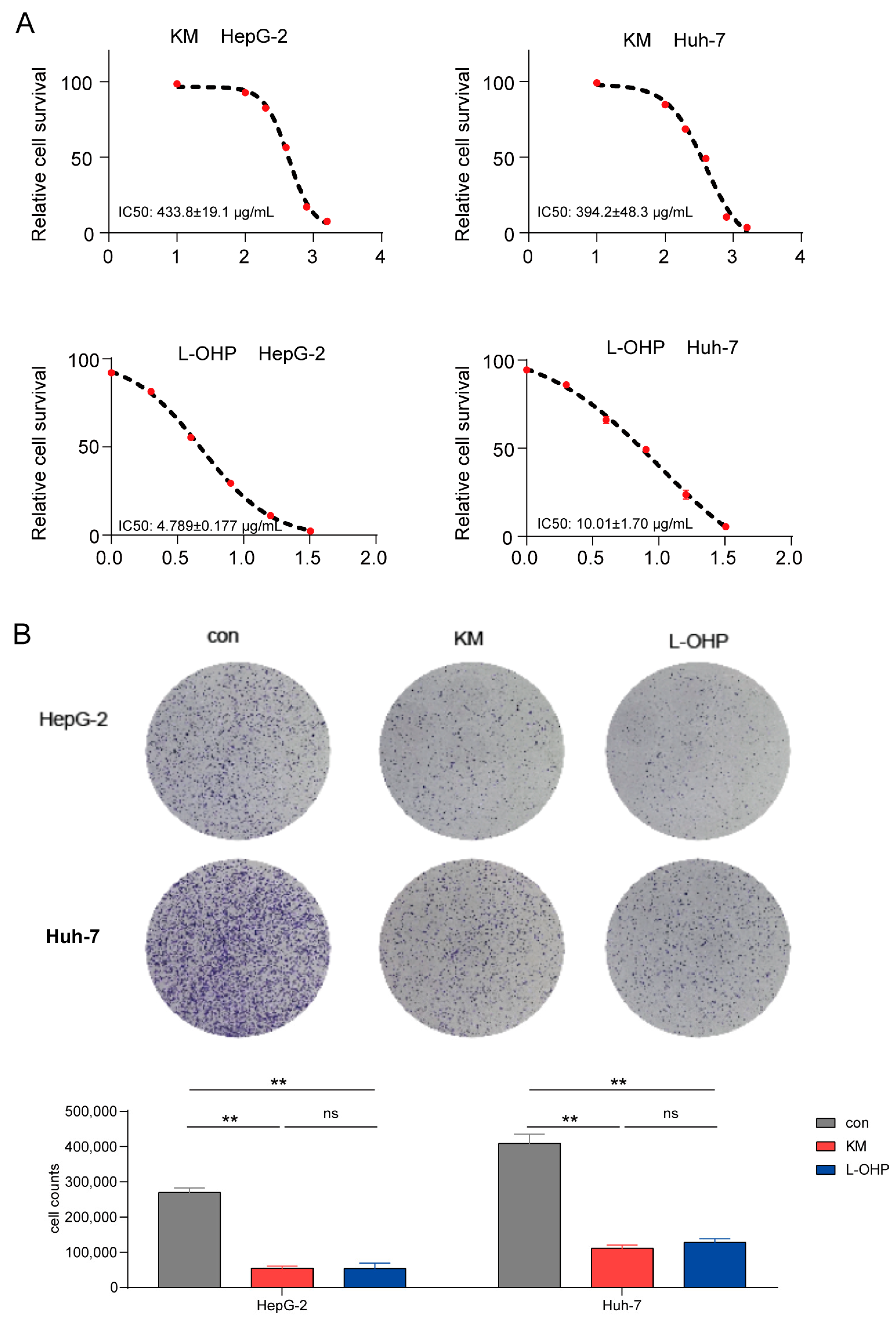
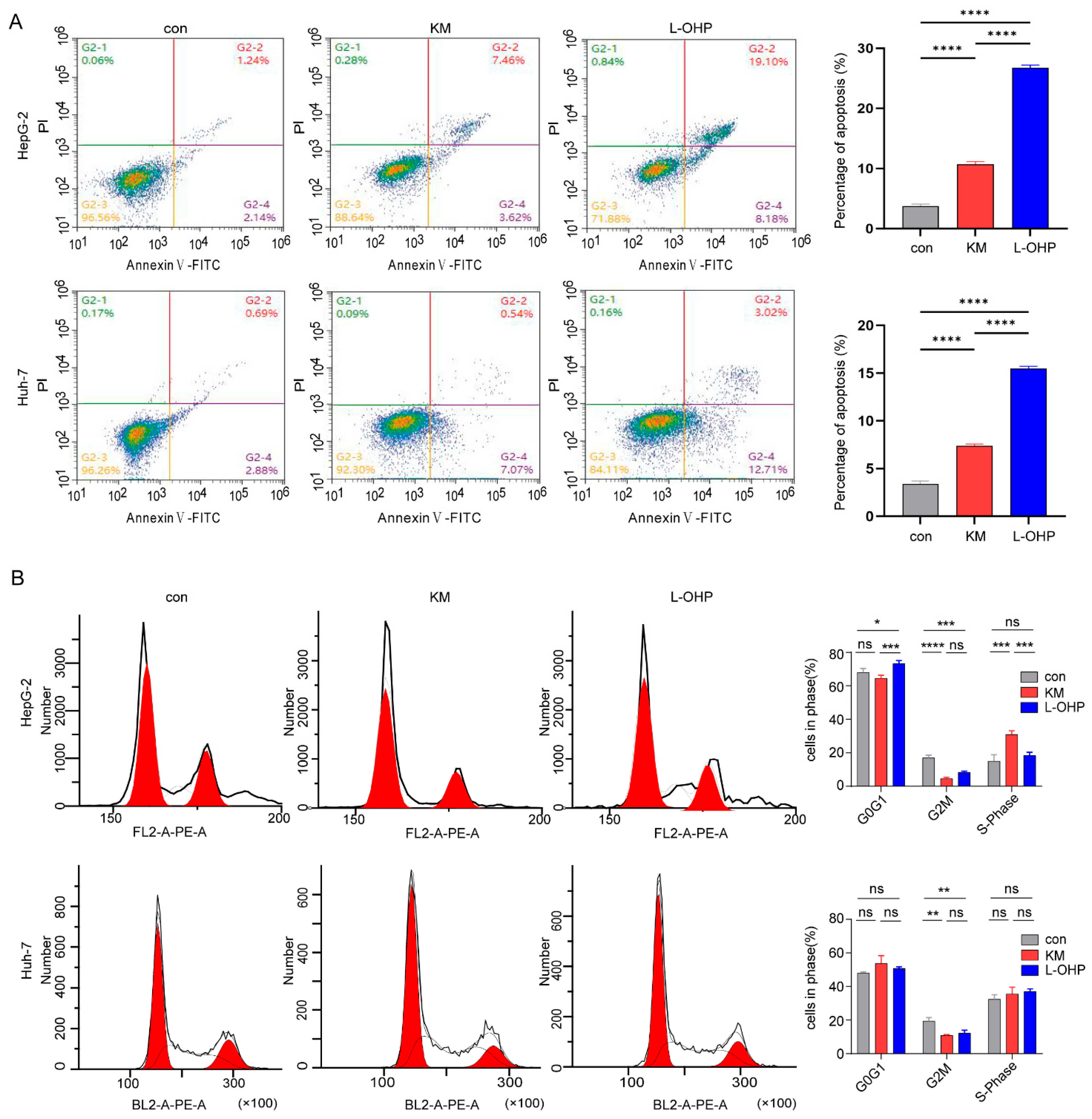
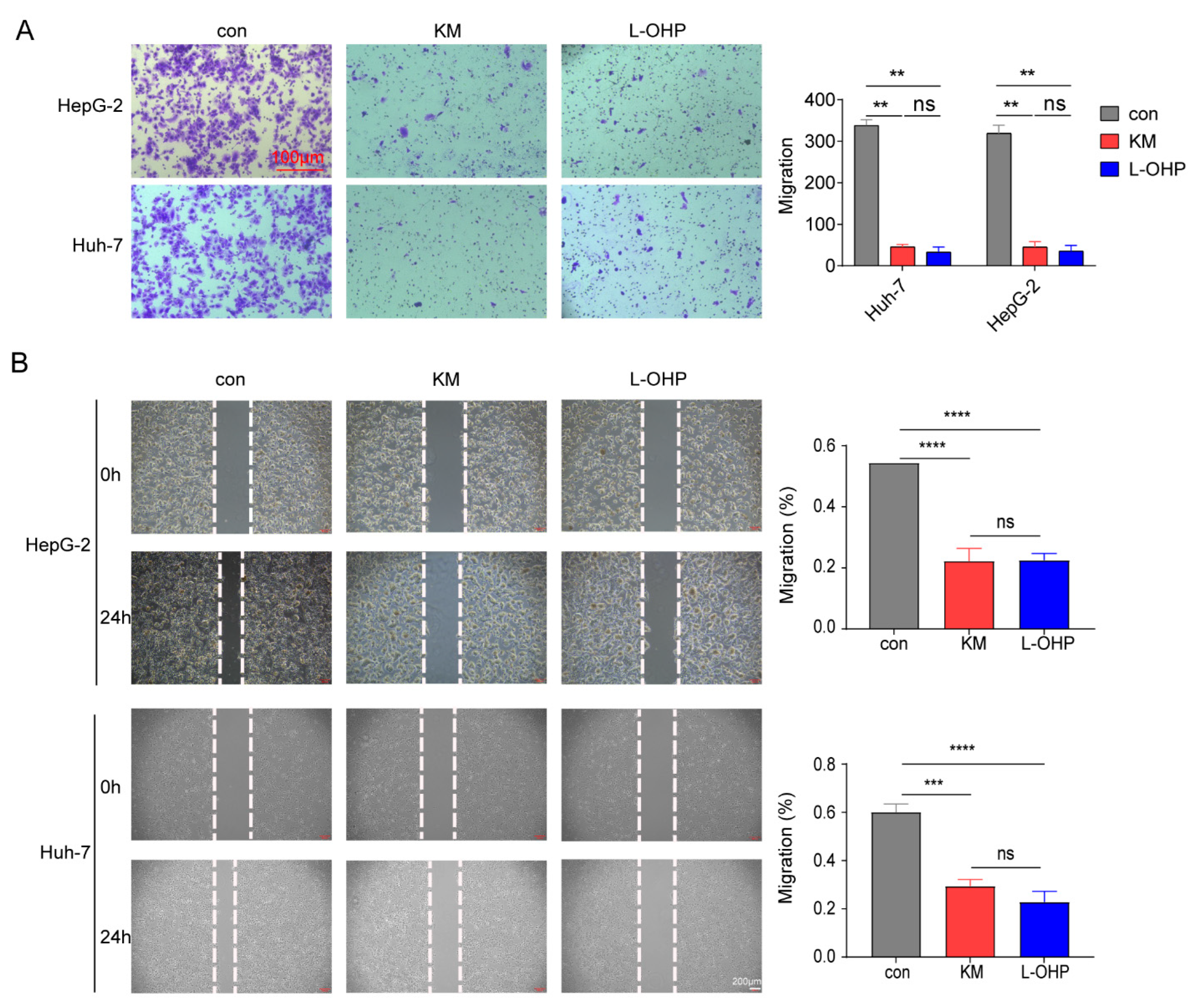
3.1. In Vitro Experiments Confirm the Anti-Tumor Effect of Koumine
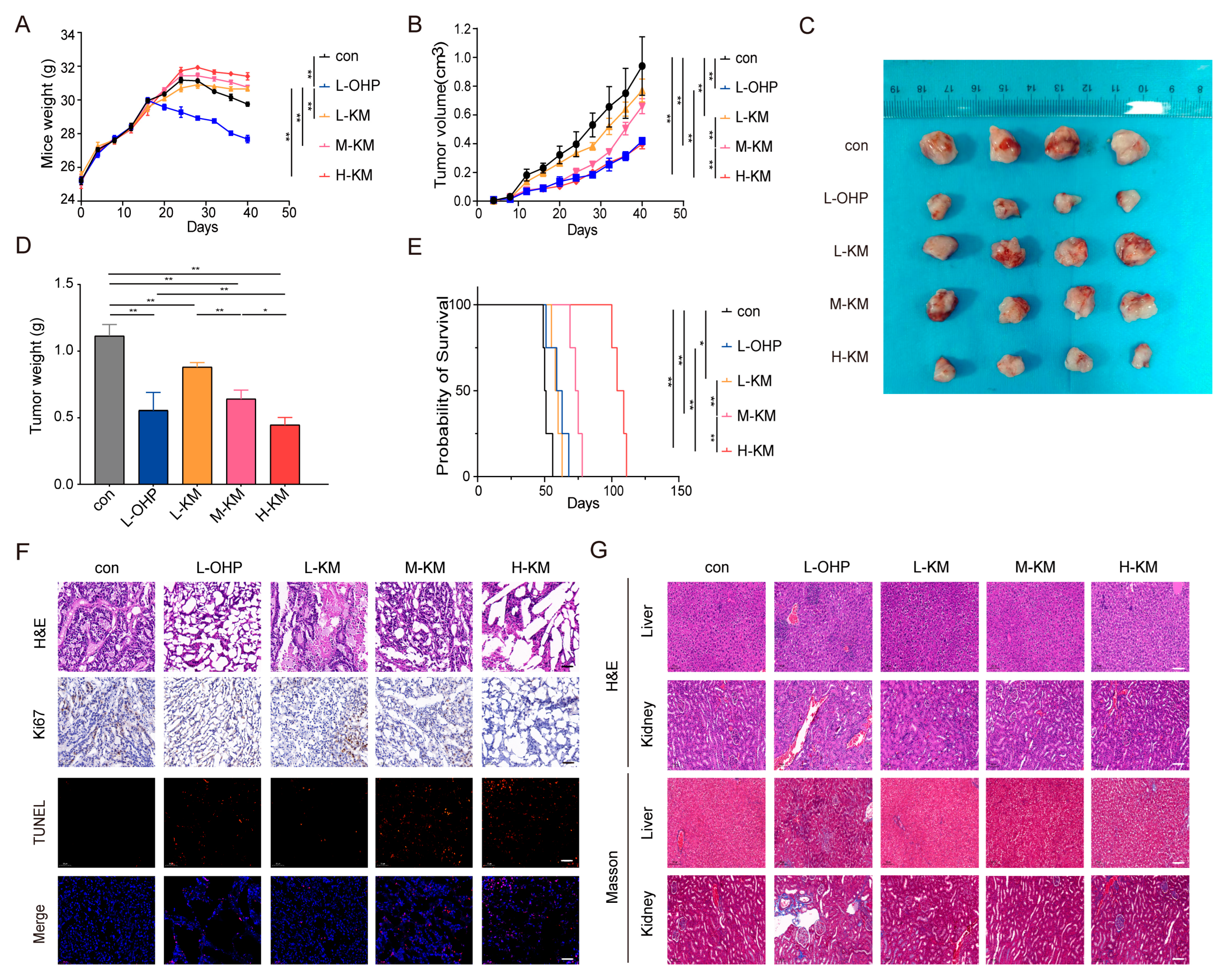
3.2. In Vivo Experiments Confirm the Anti-Tumor Effect of Koumine
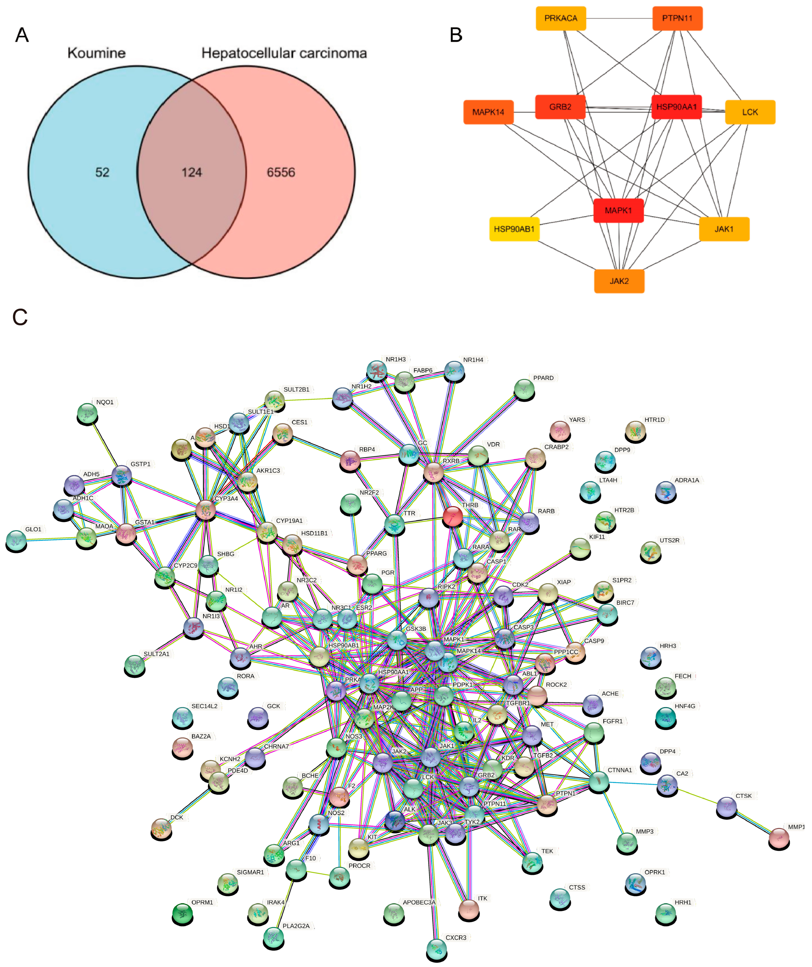
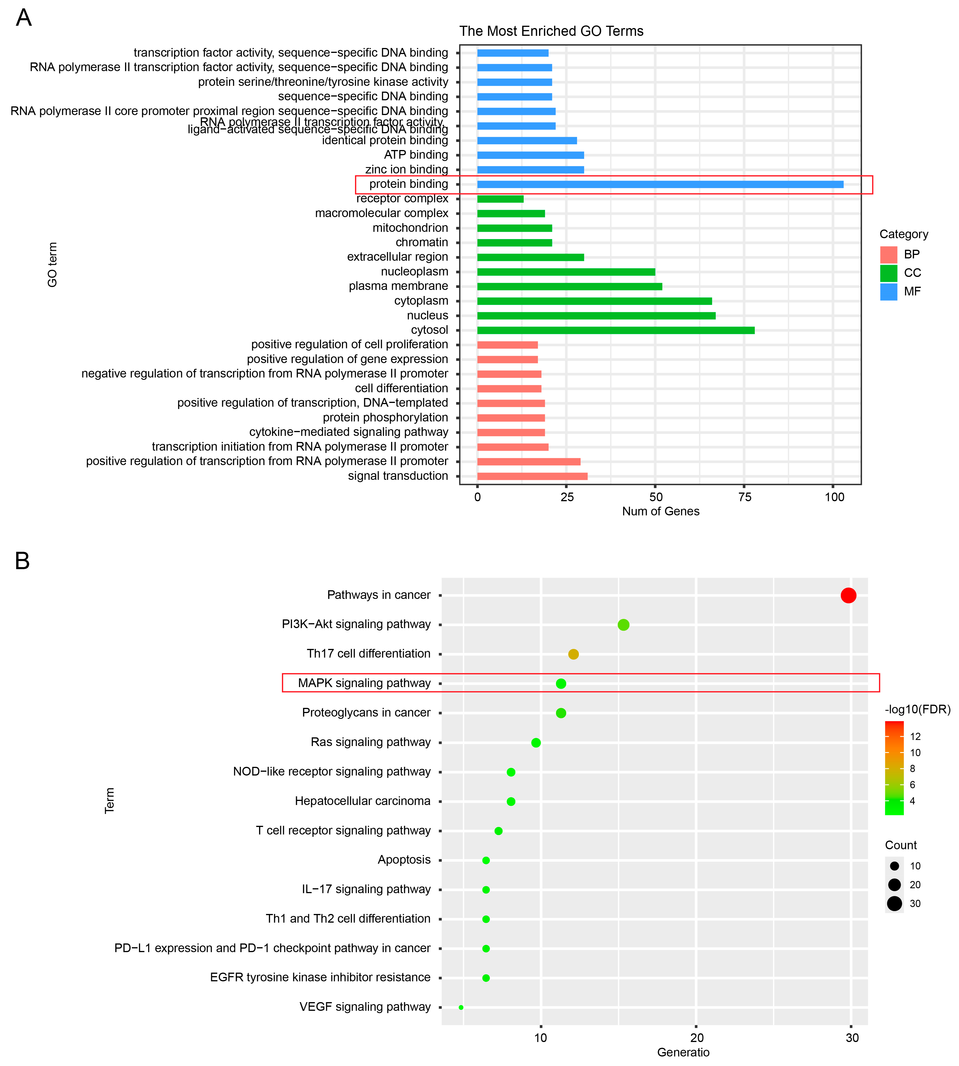
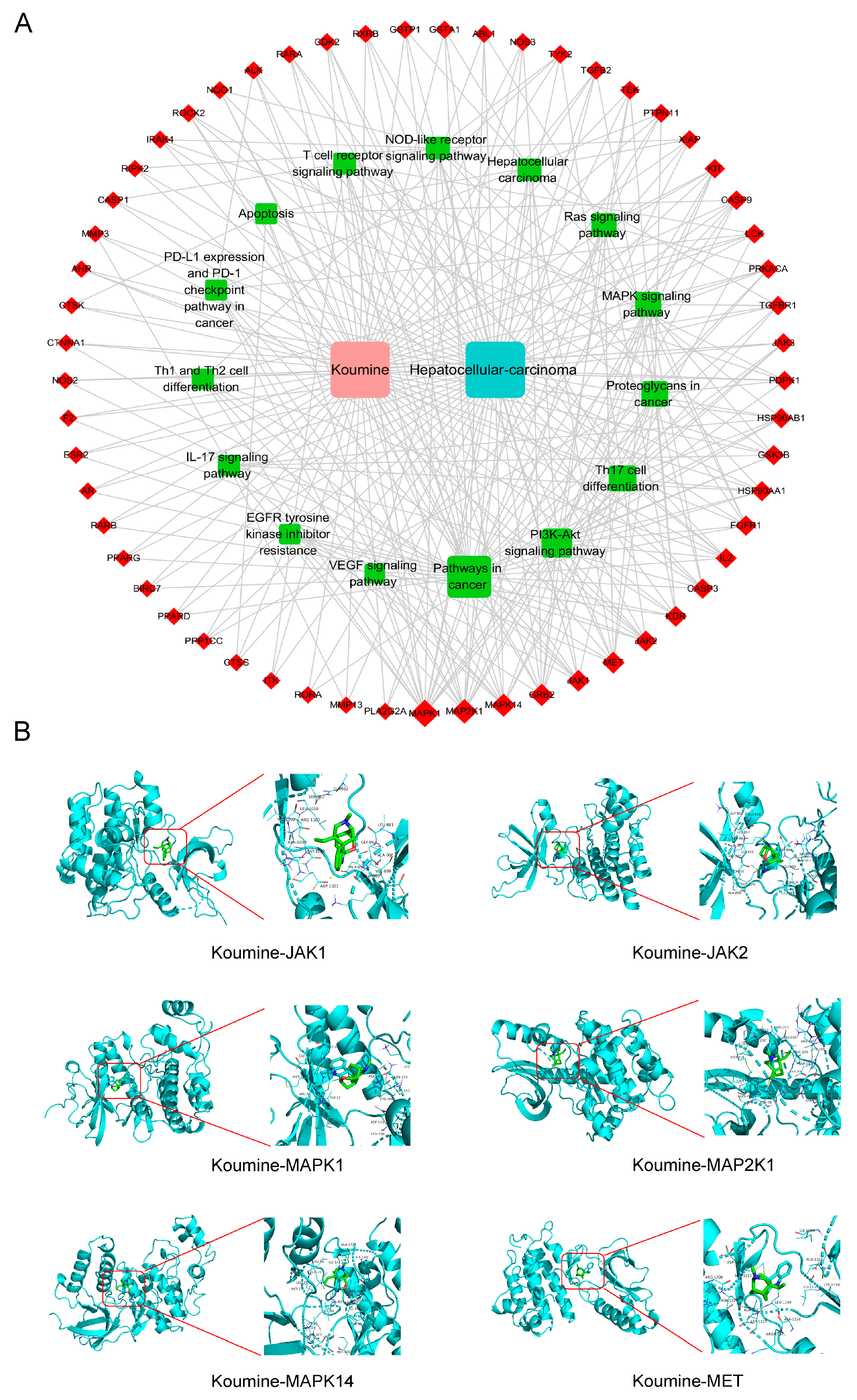
3.3. Selection of Compound-Disease-Related Targets via Network Pharmacology
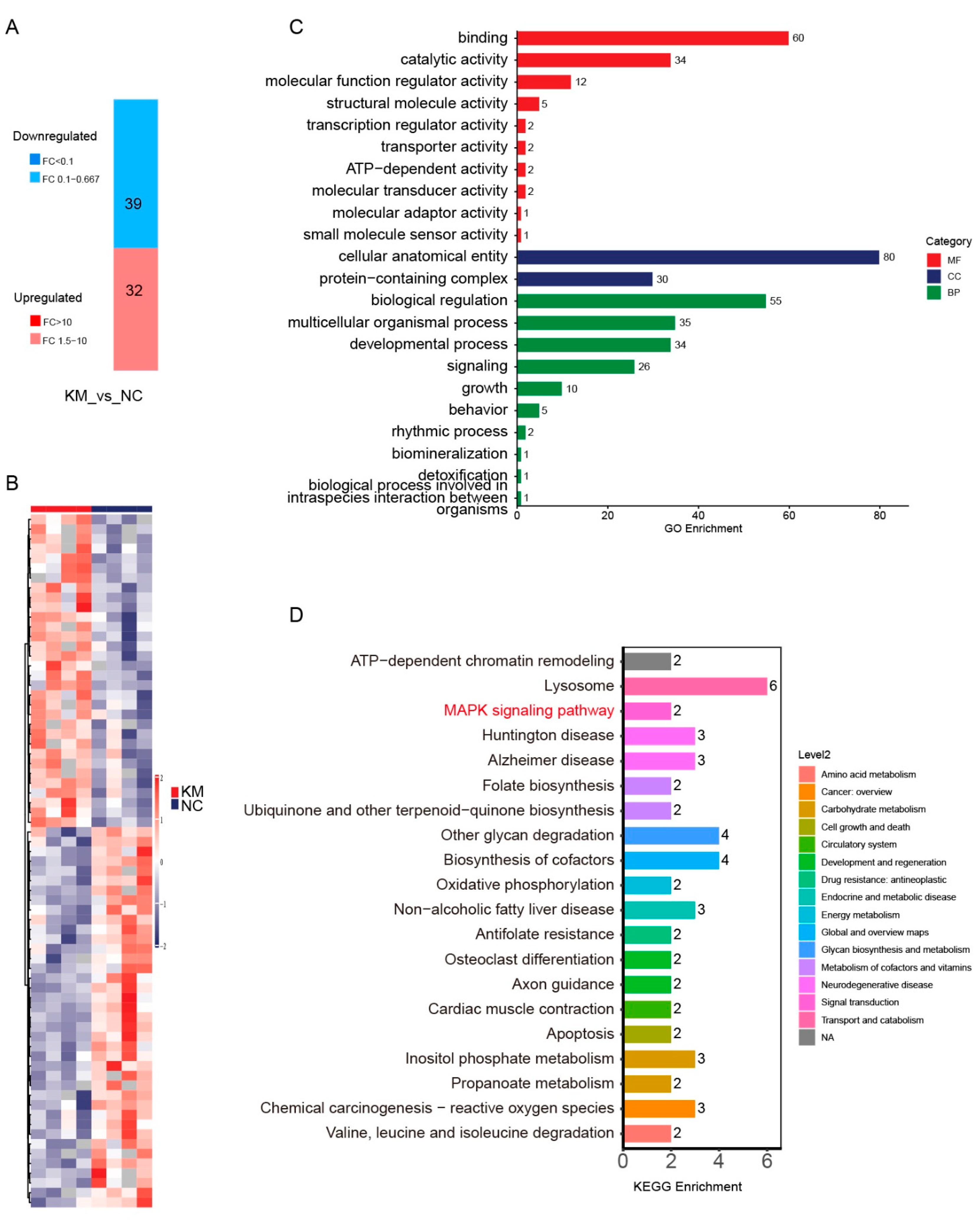
3.4. Selection of Compound-Disease-Related Targets via Proteomics
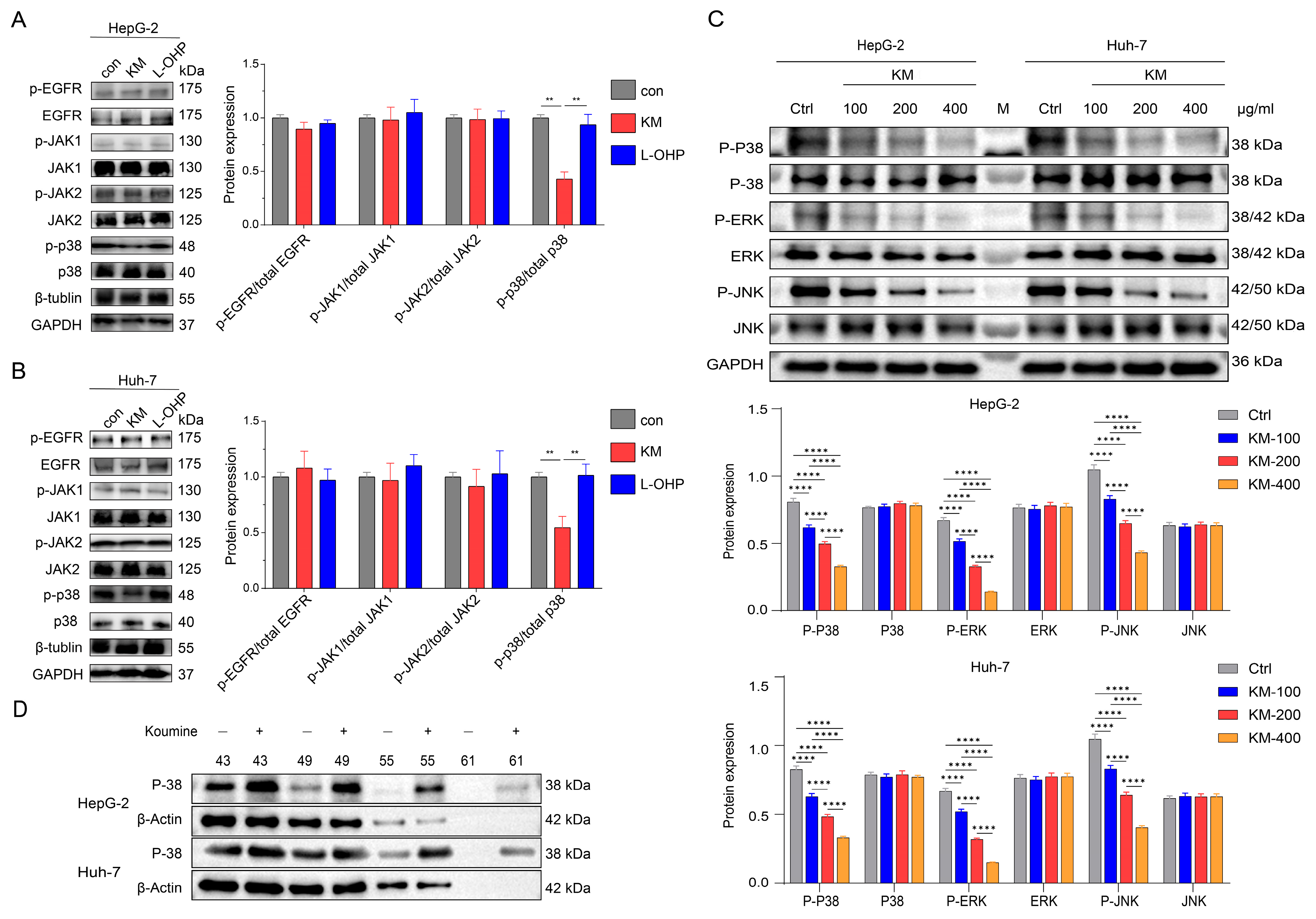
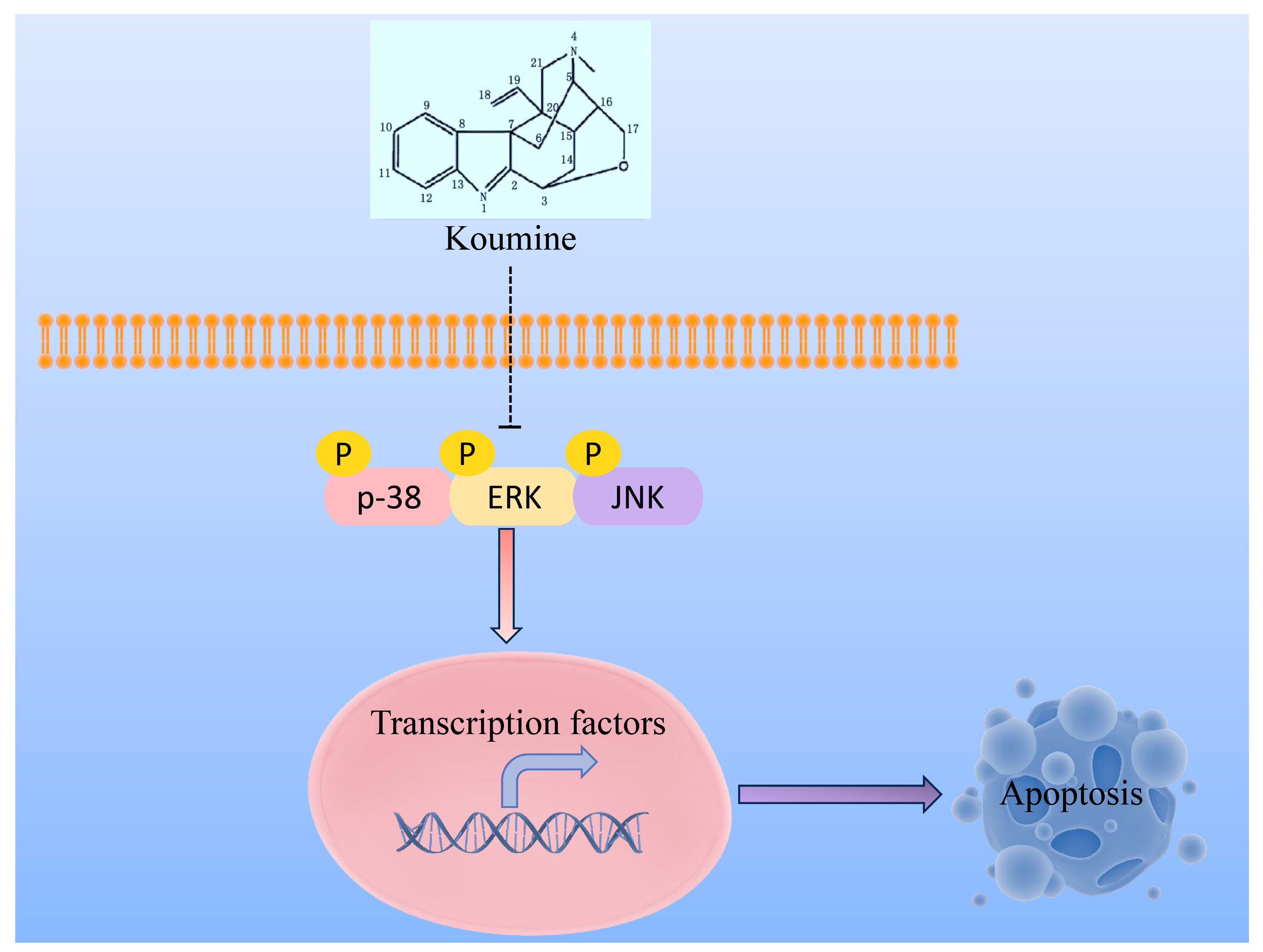
3.5. Impact of Koumine on P38 Pathway in HCC Cells
4. Discussion
5. Conclusions
Supplementary Materials
Author Contributions
Funding
Institutional Review Board Statement
Informed Consent Statement
Data Availability Statement
Conflicts of Interest
Abbreviations
References
- Zhu, R.X.; Seto, W.K.; Lai, C.L.; Yuen, M.F. Epidemiology of Hepatocellular Carcinoma in the Asia-Pacific Region. Gut Liver 2016, 10, 332–339. [Google Scholar] [CrossRef] [Scilit] [PubMed]
- Sung, H.; Ferlay, J.; Siegel, R.L.; Laversanne, M.; Soerjomataram, I.; Jemal, A.; Bray, F. Global Cancer Statistics 2020: GLOBOCAN Estimates of Incidence and Mortality Worldwide for 36 Cancers in 185 Countries. CA Cancer J. Clin. 2021, 71, 209–249. [Google Scholar] [CrossRef] [Scilit]
- Bray, F.; Ferlay, J.; Soerjomataram, I.; Siegel, R.L.; Torre, L.A.; Jemal, A. Global cancer statistics 2018: GLOBOCAN estimates of incidence and mortality worldwide for 36 cancers in 185 countries. CA Cancer J. Clin. 2018, 68, 394–424. [Google Scholar] [CrossRef] [Scilit]
- Yang, Y.M.; Kim, S.Y.; Seki, E. Inflammation and Liver Cancer: Molecular Mechanisms and Therapeutic Targets. Semin. Liver Dis. 2019, 39, 26–42. [Google Scholar] [CrossRef] [Scilit] [PubMed]
- Jin, H.; Shi, Y.; Lv, Y.; Yuan, S.; Ramirez, C.F.; Lieftink, C.; Wang, L.; Wang, S.; Wang, C.; Dias, M.H.; et al. EGFR activation limits the response of liver cancer to lenvatinib. Nature 2021, 595, 730–734. [Google Scholar] [CrossRef] [Scilit] [PubMed]
- Kudo, M. Targeted and immune therapies for hepatocellular carcinoma: Predictions for 2019 and beyond. World J. Gastroenterol. 2019, 25, 789–807. [Google Scholar] [CrossRef] [Scilit]
- Viveiros, P.; Riaz, A.; Lewandowski, R.J.; Mahalingam, D. Current State of Liver-Directed Therapies and Combinatory Approaches with Systemic Therapy in Hepatocellular Carcinoma (HCC). Cancers 2019, 11, 1085. [Google Scholar] [CrossRef] [Scilit]
- Zhu, K.; Huang, J.; Lai, L.; Huang, W.; Cai, M.; Zhou, J.; Guo, Y.; Chen, J. Medium or Large Hepatocellular Carcinoma: Sorafenib Combined with Transarterial Chemoembolization and Radiofrequency Ablation. Radiology 2018, 288, 300–307. [Google Scholar] [CrossRef] [Scilit]
- Nishijima, T.F.; Shachar, S.S.; Nyrop, K.A.; Muss, H.B. Safety and Tolerability of PD-1/PD-L1 Inhibitors Compared with Chemotherapy in Patients with Advanced Cancer: A Meta-Analysis. Oncologist 2017, 22, 470–479. [Google Scholar] [CrossRef] [Scilit]
- Sun, M.X.; Cui, Y.; Li, Y.; Meng, W.Q.; Xu, Q.Q.; Zhao, J.; Lu, J.C.; Xiao, K. Indole alkaloids from Gelsemium elegans. Phytochemistry 2019, 162, 232–240. [Google Scholar] [CrossRef] [Scilit]
- Lin, H.; Qiu, H.; Cheng, Y.; Liu, M.; Chen, M.; Que, Y.; Que, W. Gelsemium elegans Benth: Chemical Components, Pharmacological Effects, and Toxicity Mechanisms. Molecules 2021, 26, 7145. [Google Scholar] [CrossRef] [Scilit] [PubMed]
- Gao, M.Y.; Shen, W.Z.; Wu, Y.H.; Cao, C.S.; Zhang, D.M.; Gao, J.H. Study on anti-proliferation activity and the mechanisms of alkaloid monomers from Gelsemium elegans on HepG2 cell in vitro. J. Chin. Med. Mater. 2012, 35, 438–442. [Google Scholar]
- Wang, L.; Xu, H.L.; Liang, J.W.; Ding, Y.Y.; Meng, F.H. An Integrated Network, RNA Sequencing, and Experiment Pharmacology Approach Reveals the Active Component, Potential Target, and Mechanism of Gelsemium elegans in the Treatment of Colorectal Cancer. Front. Oncol. 2020, 10, 616628. [Google Scholar] [CrossRef] [Scilit]
- Xu, Y.K.; Yang, S.P.; Liao, S.G.; Zhang, H.; Lin, L.P.; Ding, J.; Yue, J.M. Alkaloids from Gelsemium elegans. J. Nat. Prod. 2006, 69, 1347–1350. [Google Scholar] [CrossRef] [Scilit]
- Kitajima, M.; Nakamura, T.; Kogure, N.; Ogawa, M.; Mitsuno, Y.; Ono, K.; Yano, S.; Aimi, N.; Takayama, H. Isolation of gelsedine-type indole alkaloids from Gelsemium elegans and evaluation of the cytotoxic activity of gelsemium alkaloids for A431 epidermoid carcinoma cells. J. Nat. Prod. 2006, 69, 715–718. [Google Scholar] [CrossRef] [Scilit]
- Zhou, J.; Sun, H.; Wang, Z.; Cong, W.; Zeng, M.; Zhou, W.; Bie, P.; Liu, L.; Wen, T.; Kuang, M.; et al. Guidelines for the Diagnosis and Treatment of Primary Liver Cancer (2022 Edition). Liver Cancer 2023, 12, 405–444. [Google Scholar] [CrossRef] [Scilit]
- Nogales, C.; Mamdouh, Z.M.; List, M.; Kiel, C.; Casas, A.I.; Schmidt, H.H.H.W. Network pharmacology: Curing causal mechanisms instead of treating symptoms. Trends Pharmacol. Sci. 2022, 43, 136–150. [Google Scholar] [CrossRef] [Scilit]
- Yang, H.Y.; Liu, M.L.; Luo, P.; Yao, X.S.; Zhou, H. Network pharmacology provides a systematic approach to understanding the treatment of ischemic heart diseases with traditional Chinese medicine. Phytomedicine 2022, 104, 154268. [Google Scholar] [CrossRef] [Scilit]
- Chen, W.; Li, Y.; Zhang, C.; Zhou, H.; Ma, J.; Vaishnani, D.K.; Zeng, B.; Yu, J.; Mao, H.; Zheng, J. Multi-Omics and Experimental Validation Reveal Anti-HCC Mechanisms of Tibetan Liuwei Muxiang Pill and Quercetin. Pharmaceuticals 2025, 18, 900. [Google Scholar] [CrossRef] [Scilit] [PubMed]
- Gao, Y.; Chen, W.; Zarogoulidis, P.; Yallapu, M.M.; Nikolic, M.V.; Vaishnani, D.K.; Zheng, J.; Nedeljkovic, N.; Ye, K.; Guo, Y.; et al. Multiomics and experimental validation reveal theophylline’s mechanism targeting IL1A/ACTB/TLR4 and identify synergistic drugs in hepatocellular carcinoma. J. Pharmacol. Exp. Ther. 2026, 393, 103836. [Google Scholar] [CrossRef] [Scilit] [PubMed]
- Huang, R.Z.; Jin, L.; Wang, C.G.; Xu, X.J.; Du, Y.; Liao, N.; Liao, Z.X.; Wang, H.S. A pentacyclic triterpene derivative possessing polyhydroxyl ring A suppresses growth of HeLa cells by reactive oxygen species-dependent NF-κB pathway. Eur. J. Pharmacol. 2018, 838, 157–169. [Google Scholar] [CrossRef] [Scilit]
- Yuan, Z.; Liang, Z.; Yi, J.; Chen, X.; Li, R.; Wu, J.; Sun, Z. Koumine Promotes ROS Production to Suppress Hepatocellular Carcinoma Cell Proliferation Via NF-κB and ERK/p38 MAPK Signaling. Biomolecules 2019, 9, 559. [Google Scholar] [CrossRef] [Scilit]
- Lee, T.H.; Tai, D.I.; Cheng, C.J.; Sun, C.S.; Lin, C.Y.; Sheu, M.J.; Lee, W.P.; Peng, C.Y.; Wang, A.H.; Tsai, S.L. Enhanced nuclear factor-kappa B-associated Wnt-1 expression in hepatitis B- and C-related hepatocarcinogenesis: Identification by functional proteomics. J. Biomed. Sci. 2006, 13, 27–39. [Google Scholar] [CrossRef] [Scilit]
- Guo, Y.J.; Pan, W.W.; Liu, S.B.; Shen, Z.F.; Xu, Y.; Hu, L.L. ERK/MAPK signalling pathway and tumorigenesis. Exp. Ther. Med. 2020, 19, 1997–2007. [Google Scholar] [CrossRef] [Scilit] [PubMed]
- Huang, S.S.; Chen, W.; Vaishnani, D.K.; Huang, L.J.; Li, J.Z.; Huang, S.R.; Li, Y.Z.; Xie, Q.P. Leucine-rich repeat-containing protein 19 suppresses colorectal cancer by targeting cyclin-dependent kinase 6/E2F1 and remodeling the immune microenvironment. World J. Gastroenterol. 2025, 31, 107893. [Google Scholar] [CrossRef] [Scilit]
- Qin, S.; Cheng, Y.; Liang, J.; Shen, L.; Bai, Y.; Li, J.; Fan, J.; Liang, L.; Zhang, Y.; Wu, G.; et al. Efficacy and safety of the FOLFOX4 regimen versus doxorubicin in Chinese patients with advanced hepatocellular carcinoma: A subgroup analysis of the EACH study. Oncologist 2014, 19, 1169–1178. [Google Scholar] [CrossRef] [Scilit] [PubMed]
- Wilhelm, S.; Carter, C.; Lynch, M.; Lowinger, T.; Dumas, J.; Smith, R.A.; Schwartz, B.; Simantov, R.; Kelley, S. Discovery and development of sorafenib: A multikinase inhibitor for treating cancer. Nat. Rev. Drug Discov. 2006, 5, 835–844. [Google Scholar] [CrossRef] [Scilit]
- Gueritte-Voegelein, F.; Guenard, D.; Dubois, J.; Wahl, A.; Potier, P. Chemical and biological studies on Taxol (Paclitaxel) and Taxotere (Docetaxel), new antineoplastic agents. J. Pharm. Belg. 1994, 49, 193–205. [Google Scholar]
- Ma, N.; Zhang, Z.; Liao, F.; Jiang, T.; Tu, Y. The birth of artemisinin. Pharmacol. Ther. 2020, 216, 107658. [Google Scholar] [CrossRef] [Scilit] [PubMed]
- Siqueira-Neto, J.L.; Wicht, K.J.; Chibale, K.; Burrows, J.N.; Fidock, D.A.; Winzeler, E.A. Antimalarial drug discovery: Progress and approaches. Nat. Rev. Drug Discov. 2023, 22, 807–826. [Google Scholar] [CrossRef] [Scilit]
- Robinson, J.G. Simvastatin: Present and future perspectives. Expert Opin. Pharmacother. 2007, 8, 2159. [Google Scholar] [CrossRef] [Scilit]
- Ikeda, M.; Morizane, C.; Ueno, M.; Okusaka, T.; Ishii, H.; Furuse, J. Chemotherapy for hepatocellular carcinoma: Current status and future perspectives. Jpn. J. Clin. Oncol. 2018, 48, 103–114. [Google Scholar] [CrossRef] [Scilit]
- Chang, W.T.; Bow, Y.D.; Fu, P.J.; Li, C.Y.; Wu, C.Y.; Chang, Y.H.; Teng, Y.N.; Li, R.N.; Lu, M.C.; Liu, Y.C.; et al. A Marine Terpenoid, Heteronemin, Induces Both the Apoptosis and Ferroptosis of Hepatocellular Carcinoma Cells and Involves the ROS and MAPK Pathways. Oxidative Med. Cell. Longev. 2021, 2021, 7689045. [Google Scholar] [CrossRef] [Scilit]
- Wagner, E.F.; Nebreda, A.R. Signal integration by JNK and p38 MAPK pathways in cancer development. Nat. Rev. Cancer 2009, 9, 537–549. [Google Scholar] [CrossRef] [Scilit]
- Huynh, H.; Nguyen, T.T.; Chow, K.H.; Tan, P.H.; Soo, K.C.; Tran, E. Over-expression of the mitogen-activated protein kinase (MAPK) kinase (MEK)-MAPK in hepatocellular carcinoma: Its role in tumor progression and apoptosis. BMC Gastroenterol. 2003, 3, 19. [Google Scholar] [CrossRef] [Scilit]
- Ma, J.; Chen, W.; Vaishnani, D.K.; Wang, C.; Xue, S.; Yang, Q.; Tong, Y.; Lei, N.; Zhao, Z.; Ying, F. Curcumin Analog J7 Attenuates Liver Fibrosis and Metabolic Dysregulation in a Rat Model of Type 2 Diabetes via Modulation of TGF-β/Smad and NF-κB/BCL-2/BAX Pathways. Drug Des. Dev. Ther. 2025, 19, 2411–2432. [Google Scholar] [CrossRef] [Scilit]
- Ma, Y.; Lin, Y.; Wang, C.; Lv, Y.; Chen, W. YY1 as a mediator to enhance the resistance of KRAS mutant colorectal cancer cells to cetuximab. J. Genet. 2025, 104, 3. [Google Scholar] [CrossRef] [Scilit]



| Parameter Name | Parameter Value |
|---|---|
| Compound | Koumine |
| PubChem CID | 102004413 |
| Cas | 1358-76-5 |
| Structural Formula | C20H22N2O |
| Molecular Weight (g/mol) | 306.4 |
| Canonical SMILES | CN1CC2(C3CC4C5=NC6=CC=CC=C6C52CC1C3CO4)C=C |
| Constitutional Formula |  |
| Parameter Name | Parameter Value |
|---|---|
| Instrument | Waters-HPLC |
| Column | C18, 4.6 × 250 nm, 5 μm |
| Detection Mode: | 260 nm |
| Column Temperature | 35 °C |
| Flow Rate | 1.0 mL/min |
| Sample dissolution Mobile Phase Gradientelution | 60% methanol in water A: Acetonitrile B: 0.1% Triethylamine phosphate in water A: 10%–30%, 0–15 min |
| ChemoPprofile |  |
| Target | PDB: ID | Ligand | Affinity (KJ/mol) |
|---|---|---|---|
| MAPK1 | 5ik4 | NA | −7.83 ± 0.02 |
| MAP2K1 | 7b94 | LYS′97 | −6.97 ± 0.01 |
| MAPK14 | 6sfi | NA | −7.23 ± 1.57 |
| JAK1 | 4ehz | ASP′1021 | −7.56 ± 0.01 |
| JAK2 | 4d1s | NA | −8.53 ± 0.02 |
| MET | 3dkg | NA | −6.38 ± 0.00 |
Disclaimer/Publisher’s Note: The statements, opinions and data contained in all publications are solely those of the individual author(s) and contributor(s) and not of MDPI and/or the editor(s). MDPI and/or the editor(s) disclaim responsibility for any injury to people or property resulting from any ideas, methods, instructions or products referred to in the content. |
© 2026 by the authors. Licensee MDPI, Basel, Switzerland. This article is an open access article distributed under the terms and conditions of the Creative Commons Attribution (CC BY) license.
Share and Cite
Lin, H.; Tang, Y.; Shi, L.; Zhu, S.; Yan, W.; Chen, W.; Que, W. Koumine’s Therapeutic Impact on Hepatocellular Carcinoma: A Combined Network Pharmacology and Experimental Study. Biomedicines 2026, 14, 1250. https://doi.org/10.3390/biomedicines14061250
Lin H, Tang Y, Shi L, Zhu S, Yan W, Chen W, Que W. Koumine’s Therapeutic Impact on Hepatocellular Carcinoma: A Combined Network Pharmacology and Experimental Study. Biomedicines. 2026; 14(6):1250. https://doi.org/10.3390/biomedicines14061250
Chicago/Turabian StyleLin, Hailing, Yuli Tang, Lingfei Shi, Shengjie Zhu, Wenqiang Yan, Weihong Chen, and Wancai Que. 2026. "Koumine’s Therapeutic Impact on Hepatocellular Carcinoma: A Combined Network Pharmacology and Experimental Study" Biomedicines 14, no. 6: 1250. https://doi.org/10.3390/biomedicines14061250
APA StyleLin, H., Tang, Y., Shi, L., Zhu, S., Yan, W., Chen, W., & Que, W. (2026). Koumine’s Therapeutic Impact on Hepatocellular Carcinoma: A Combined Network Pharmacology and Experimental Study. Biomedicines, 14(6), 1250. https://doi.org/10.3390/biomedicines14061250