


Abstract
Background/Objectives: Intramedullary tumors are uncommon spinal cord lesions that account for a small proportion of central nervous system neoplasms but are associated with a high risk of neurological morbidity. Accurate preoperative characterization is essential because therapeutic strategies, surgical planning, and functional prognosis depend strongly on tumor biology and growth behavior within the confined spinal cord environment. This study aims to characterize the radiological phenotype of intramedullary tumors and to identify imaging patterns that may assist in lesion characterization and diagnostic stratification. Methods: A retrospective analysis of preoperative MRI findings in patients with histopathologically confirmed intramedullary tumors was performed. Preoperative MRI examinations were systematically analyzed to describe imaging features according to tumor histology using conventional sequences (T1-weighted, T2-weighted, and contrast-enhanced imaging). Results: Distinct radiological phenotypes were observed across a wide spectrum of lesions. Glial tumors, including subependymoma, ependymoma, pilocytic astrocytoma, diffuse midline glioma H3K27M, glioblastoma, high-grade astrocytoma with piloid features, ganglioglioma, and diffuse leptomeningeal glioneural tumors, demonstrated variable combinations of cord expansion, margin definition, enhancement patterns, and tract involvement, reflecting differences between expansile and infiltrative growth. Secondary tumors such as metastases frequently exhibited aggressive imaging features, including extensive edema and intense or heterogeneous enhancement. Vascular lesions, including hemangioblastoma and cavernoma, showed characteristic vascular signatures, such as nodular enhancement with flow voids or susceptibility-related signal changes. Developmental lesions, such as epidermoid cysts, neurenteric cysts, and lipoma, displayed distinctive signal characteristics, especially on diffusion and T1, that aided differentiation from neoplastic processes. Conclusions: In conclusion, the structured radiological interpretation functions proposed herein are not only useful for diagnostic purposes, but could also be useful for risk stratification and therapeutic guidance.
1. Introduction
Intramedullary tumors represent a heterogeneous group of lesions that originate within the spinal cord parenchyma, accounting for approximately 2–4% of all central nervous system (CNS) neoplasms and 30–47% of intradural tumors in adults [1]. Despite their rarity, these lesions are of major clinical importance because of the high risk of neurological sequelae and the challenges inherent in their diagnosis and surgical management. The compact anatomy of the spinal cord makes accurate preoperative characterization essential [2,3], as treatment strategies depend heavily on the underlying histological and molecular nature of the tumor.
Magnetic resonance imaging (MRI) is the cornerstone of spinal cord tumor evaluation. In recent years, advances in MRI technology have improved the radiological approach to spinal cord tumors [4,5]. It offers unparalleled soft-tissue contrast and the ability to define tumor morphology, cord expansion, cystic components, and patterns of enhancement [5]. MRI not only serves diagnostic purposes but also plays a crucial role in surgical planning [6], assessment of resectability [7,8], and postoperative follow-up. Moreover, multiparametric imaging may provide new insights into the tumor microstructure of the spinal cord [9,10,11], potentially improving lesion characterization beyond conventional morphology.
In parallel, the 2021 World Health Organization (WHO) classification of CNS tumors introduced integrated molecular diagnostics incorporating genetic and epigenetic alterations into histopathological definitions [12,13]. This paradigm shift has established a biologically driven framework in which imaging is increasingly expected to mirror tumor behavior rather than merely depict anatomy. However, in the spinal cord, radiological knowledge originates from small, heterogeneous series, and rare tumor entities remain poorly described or are extrapolated from intracranial counterparts despite distinct spinal microenvironmental constraints.
The aim of this review is therefore to provide a comprehensive and structured overview of the neuroimaging features of intramedullary tumors, organized according to the main histological entities encountered in clinical practice (Table 1). To achieve this, we retrospectively reviewed preoperative spinal cord MRIs in patients with histopathologically confirmed intramedullary tumors in order to systematically illustrate and describe their imaging patterns. This approach seeks to support clinicians in the differential diagnosis of intramedullary lesions and to promote a radiological framework aligned with the current WHO classification paradigm.
Table 1.
Pathological classification of intramedullary lesions according to the 2021 WHO classification system.
2. Methodology and MRI Protocol
All procedures performed in this study involving human participants were approved by the Ethics Committee (IRB 00011687–2026/26) on 26 April 2026 and were conducted in accordance with the 1964 Declaration of Helsinki and its later amendments or comparable ethical standards.
All cases of intramedullary tumor included in this retrospective study had histopathological confirmation following surgical resection or biopsy, in accordance with the current WHO classification criteria. This study was not designed as a cohort-based analysis with consecutive patient inclusion, but rather as an illustrative review based on representative, histopathologically confirmed cases selected to reflect typical imaging patters of each tumor entity. Only patients with available preoperative MRI examinations of sufficient quality were included. For each tumor entity, a single representative case was selected to illustrate its characteristic imaging features. MRI studies were reviewed to assess conventional imaging features, including lesion location, longitudinal extent, signal characteristics, and contrast enhancement patterns, with additional evaluation of advanced imaging techniques when available. MRI examinations were interpreted by a senior radiologist and systematically reviewed by a neuroradiologist with more than 30 years of experience.
Given the complex microanatomy and the small cross-sectional area of the spinal cord, high-resolution imaging protocols are indispensable for accurate lesion characterization. The principal objectives of MRI evaluation are to determine the lesion’s longitudinal and axial extent, its relationship with the spinal cord parenchyma, its internal architecture, and its enhancement pattern.
2.1. Technical Considerations
High-field MRI scanners (3 Tesla) are recommended for improved signal-to-noise ratio and spatial resolution. Thin-slice acquisitions (≤2 mm) are preferred to minimize partial volume effects. Sagittal imaging is optimal for assessing the craniocaudal extent, whereas axial imaging provides essential details about tumor localization within the cord, infiltration, and peritumoral changes. The use of dedicated spine coils or multi-channel phased-array coils enhances image quality.
A standard MRI examination should include sagittal and axial T1-weighted, T2-weighted, as well as post-gadolinium T1-weighted imaging with a 3–5-min delay post-injection to allow optimal contrast uptake. Coronal MRI sequences have limited utility in the evaluation of strictly intramedullary spinal cord tumors since the cranio-caudal extent of lesions is optimally assessed on sagittal images, while axial sequences provide detailed information regarding lesion symmetry, eccentricity, and relationship to spinal cord structures (Table 2).
Table 2.
MRI protocol.
2.2. Sagittal View
The sagittal view is the most informative for assessing the craniocaudal extent of intramedullary tumors and their relationship to the spinal cord. Because intramedullary tumors typically expand the spinal cord rather than displace it, careful evaluation of the degree of swelling and signal extension beyond the visible mass is essential. Lesions are conventionally categorized as follows: (1) focal (<2 vertebral segments; e.g., hemangioblastomas, metastases…), (2) intermediate (2–3 segments; e.g., typical of low-grade astrocytomas or gangliogliomas), and (3) longitudinally extensive (>3 segments, e.g., diffuse astrocytomas, H3K27-altered gliomas).
2.3. Axial View
Axial images provide a cross-sectional understanding of tumor location relative to the central canal, gray matter, and white matter tracts, and are keys for evaluating tract infiltration. Central lesions are usually represented by ependymomas, which originate from the ependymal lining and symmetrically expand the cord. Eccentric lesions are characteristic of astrocytomas and gangliogliomas, which arise from glial cells within the hemicord, producing asymmetric expansion. Subpial or exophytic lesions, including hemangioblastomas, cavernomas, and metastases, are located near the cord surface and may exhibit pial-vessel involvement or exophytic components extending into the subarachnoid space.
2.4. Enhancement Patterns: Morphological and Diagnostic Implications
Gadolinium-enhanced MRI is crucial for evaluating blood–spinal cord barrier integrity and remains one of the most powerful differentiating tools among intramedullary lesions. The pattern, intensity, and distribution of enhancement offer valuable insights. Homogeneous, intense enhancement throughout the lesion is typical of ependymomas and lymphomas. According to the type of ependymoma (cystic contingent), enhancement may be more heterogeneous due to an intra-tumoral cystic component, whereas lymphomas enhance homogeneously despite low perfusion due to high cellularity. Heterogeneous or ring-like enhancement is characteristic of astrocytomas, particularly high-grade variants and glioblastomas, which are caused by necrosis and heterogeneous vascular permeability. Nodular enhancement, frequently associated with pial-flow voids or feeding vessels, is characteristic of hemangioblastomas. Minimal or absent enhancement may be seen in some low-grade tumors, including pilocytic astrocytomas and oligodendrogliomas. The absence of enhancement should not exclude neoplasia, as it may simply reflect an intact blood–spinal cord barrier.
3. Radiological Features of Intramedullary Tumors
3.1. Primary CNS Tumors
3.1.1. Spinal Ependymal Tumors
Subependymoma (Figure 1A)
Spinal subependymomas most commonly involve the cervical and upper thoracic spinal cord and are typically located in a central or slightly eccentric intramedullary position, often in close relationship to the central canal [14]. They present as well-circumscribed, non-infiltrative masses. On MRI, they are generally iso- to hypointense on T1-weighted sequences and hyperintense on T2-weighted images, often with a remarkably homogeneous signals [15]. Peritumoral edema is minimal or absent, reflecting the non-invasive nature of the lesion. A marked fusiform spinal cord enlargement with abrupt margins on sagittal MRI creates the characteristic “bamboo leaf sign,” reflecting subpial tumor growth [16]. Gadolinium enhancement is absent or minimal in most cases. When present, enhancement is usually faint. The lack of intense or homogeneous enhancement is a key feature differentiating subependymomas from classic ependymomas [14,15].
Figure 1.
(A) Subependymoma: (a) Sagittal T2-weighted image showing an extensive well-defined hypersignal-T2 mass within the cord; (b) Sagittal T1-contrast-enhanced image showing the absence of gadolinium enhancement; (c) Sagittal pictorial representation; (d,f) Axial T2-weighted image showing the central tumor; (e,g) Axial pictorial representation. (B) Ependymoma: (a) Sagittal T2-weighted image showing an extensive well-defined mass with multiple intumoral cysts and polar cyst; (b) Sagittal T1-contrast-enhanced image showing the gadolinium enhancement of the tumor associated with the polar cyst; (c) Sagittal pictorial representation; (d) Axial T2-weighted image showing a central tumor with an intratumoral cyst; (e) Axial T1-gadolinium enhanced image showing gadolinium enhancement of the tumor without enhancement of the cysts; (f) Axial pictorial representation.
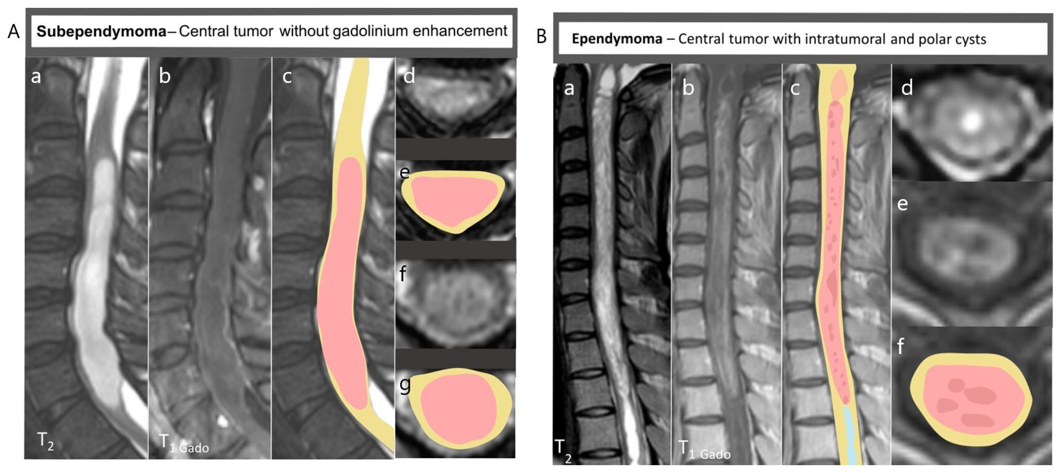
Spinal Ependymoma (Figure 1B)
Ependymomas are the most frequent intramedullary tumor in adults, accounting for approximately 60% of adult cases and 30% of those in children. Arising from the ependymal lining of the central canal, these tumors are typically centrally located, symmetrical, well circumscribed, and occur most frequently in the cervical or cervicothoracic region [17]. They often span several vertebral segments and may be associated with both polar cysts and syringomyelia, the latter resulting from altered cerebrospinal fluid (CSF) dynamics. Ependymomas are typically iso- to hypointense on T1, with a sharp demarcation from the surrounding spinal cord [18]. A highly characteristic feature is the hemosiderin “cap sign”, a T2/SWI hypointense rim at one or both poles of the tumor caused by chronic microhemorrhage (Table 2). Internal signal heterogeneity may arise from hemorrhage or cystic components (which were present in more than 60% of ependymomas) [18,19]. Contrast administration usually produces intense enhancement, which may extend into the walls of associated polar cysts [20]. Advanced imaging often shows displacement rather than infiltration of white matter tracts on diffusion tensor imaging (DTI) and tractography [9,21] (Table 3).
Table 3.
Radiological comparison of ependymoma and astrocytoma.
3.1.2. Spinal Astrocytoma
Pilocytic Astrocytoma (Figure 2A)
Pilocytic astrocytomas are the most common intramedullary tumors in children, accounting for 60–70% of pediatric cases, but are rarer in adulthood. Pilocytic astrocytomas most commonly arise in the cervical spinal cord. They are typically eccentric or mildly asymmetric within the spinal cord, and normally well circumscribed, in contrast to diffuse astrocytoma [14]. The longitudinal extent is generally moderate. On MRI, they appear iso- to hypointense on T1- and T2-weighted imaging. Signal homogeneity is common, although mild heterogeneity may be observed due to microcystic changes. Contrast enhancement is variable, but often mild to moderate with patchy, nodular, or faint enhancement [22]. On DTI, they characteristically show reduced fractional anisotropy, with infiltration and disruption of white matter tracts on tractography [23,24]. Perfusion imaging typically demonstrates low to moderately increased relative blood volume compared with high-grade gliomas, reflecting limited neoangiogenesis consistent with their low-grade biological behavior.
Figure 2.
(A) Pilocytic astrocytoma: (a) Sagittal T2-weighted image showing eccentric and irregular intramedullary hyposignal; (b) Sagittal T1-contrast-enhanced image showing a patchy enhancement; (c) Sagittal pictorial representation; (d,e) axial T1-contrast-enhanced images showing irregular and patchy enhancement; (f) Axial pictorial representation. (B) Diffuse midline glioma H3K27M: (a) Sagittal T2-weighted image showing extensive midline intramedullary mass; (b) Sagittal T1-contrast-enhanced image showing the absence of gadolinium enhancement; (c) Sagittal pictorial representation; (d) Axial T2-weighted image showing a diffuse and eccentric intramedullary mass; (e) Axial pictorial representation. (C) Glioblastoma: (a) Sagittal T2-weighted image showing a diffuse hypersignal associated to intramedullary edema; (b) Sagittal T1-contrast-enhanced image showing a high enhancement with necrosis; (c) Sagittal pictorial representation; (d) Axial T2-weighted image; (e) Axial T1-contrast-enhanced image showing eccentric lesion; (f) Axial pictorial representation. (D) High-grade astrocytoma with piloid features: (a) Sagittal T2-weighted image showing fusiform hyperT2 intramedullary mass with extensive edema; (b) Sagittal T1-contrast-enhanced image showing a high enhancement (c) Sagittal pictorial representation; (d) Axial T2-weighted image; (e) Axial T1-contrast-enhanced image; (f) Axial pictorial representation.
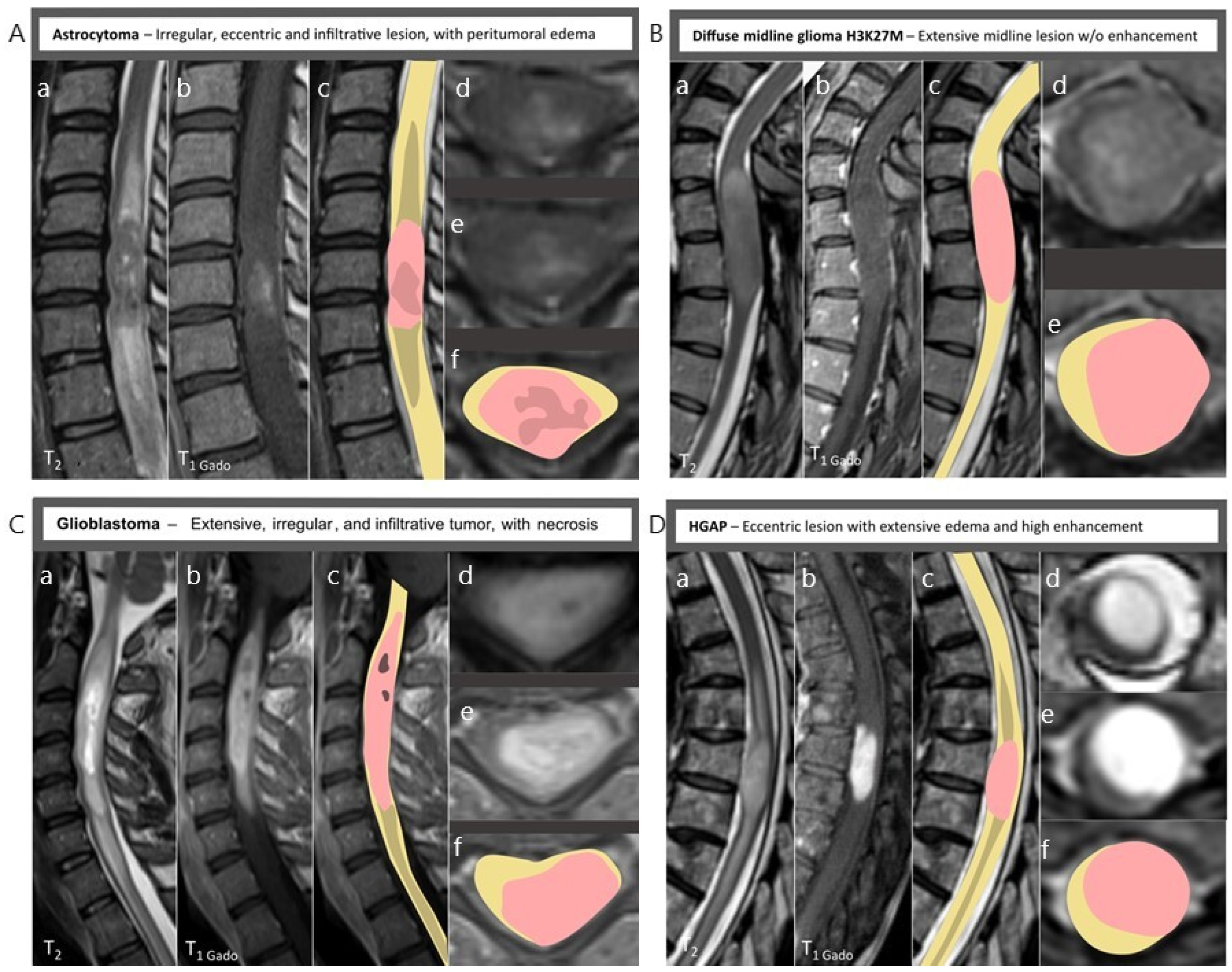
Diffuse Midline Glioma H3K27M (Figure 2B)
Spinal cord diffuse midline glioma, H3K27-altered, most commonly involves the cervical or cervicothoracic levels. These tumors typically present as longitudinally extensive intramedullary lesions with poorly defined margins and marked cord expansion. On MRI, they are usually iso- to hypointense on T1-weighted sequences and heterogeneously hyperintense on T2-weighted images, often extending over multiple vertebral segments. In cases of H3K27-altered diffuse midline gliomas, the enhancement may be surprisingly subtle despite aggressive histology, underscoring the need for integration with advanced imaging markers [25]. Peritumoral edema is common.
Glioblastoma (Figure 2C)
Primary spinal cord glioblastoma is exceedingly rare (<1% of all glioblastomas), representing the most aggressive end of the spinal glial tumor spectrum [26]. Glioblastomas can arise anywhere along the spinal cord. They typically present as poorly defined, infiltrative masses causing marked cord expansion with indistinct margins. Longitudinal involvement over multiple vertebral levels is common, and extension into the cervicomedullary junction may occur. Unlike ependymomas, glioblastomas do not produce polar cysts, and syrinx formation is uncommon. On MRI, they typically display a heterogeneous signal on both T1- and T2-weighted sequences, reflecting the coexistence of hypercellular tumor tissue, necrosis, microhemorrhage, and infiltrated normal parenchyma [26]. Contrast administration usually reveals irregular ring-like or patchy enhancement accompanied by substantial perilesional edema [27].
High-Grade Astrocytoma with Piloid Features (HGAP) (Figure 2D)
HGAP is a recently characterized infiltrative astrocytic tumor defined by distinct DNA methylation profiling and recurrent MAPK pathway alterations. Spinal HGAP remains rare and may involve the cervical or cervicothoracic cord. On MRI, these tumors typically present as intramedullary expansile lesions with heterogeneous signal characteristics, appearing iso- to hypointense on T1-weighted images and heterogeneously hyperintense on T2-weighted sequences [28]. Unlike classic pilocytic astrocytomas, HGAP frequently demonstrates irregular or heterogeneous contrast enhancement, sometimes with nodular or ring-like components, and may show areas of necrosis or microhemorrhage [28,29]. The margins are often partially infiltrative rather than sharply circumscribed, and longitudinal extension over multiple vertebral segments can occur.
3.1.3. Spinal Glioneuronal Tumor
Ganglioglioma (Figure 3A)
Gangliogliomas are low-grade glioneuronal tumors composed of dysplastic ganglion cells mixed with neoplastic glial elements, typically presenting in young adults [30]. Their radiological appearance reflects their mixed cellular composition and slow growth. They typically arise within the thoracic spinal cord and tend to be eccentrically located with well-defined margins. The lesions usually involve one to three vertebral segments, and although they can be expansive, they are less infiltrative than astrocytoma. On MRI, they appear hypointense on T1-weighted images and hyperintense on T2-weighted sequences, but the signal may be heterogeneous due to the presence of microcysts, calcifications, or mixed glioneuronal components. Gangliogliomas usually cause localized cord expansion but are not associated with extensive peritumoral edema, supporting a low-grade process. Their pattern of enhancement is variable, most often mild or patchy rather than intense.
Figure 3.
(A) Ganglioma: (a) Sagittal T2-weighted image showing asymmetric intramedullary lesion with intratumoral cysts; (b) Sagittal T1-contrast-enhanced image showing a mild enhancement; (c) Sagittal pictorial representation; (d) Axial T2-weighted image; (e) Axial T1-contrast-enhanced image; (f) Axial pictorial representation. (B) Diffuse leptomeningeal glioneural tumor: (a) Sagittal T2-weighted image heterogeneous isosignal lesion with hyposignal nodule, associated with syrinx and edema; (b) Sagittal T1-contrast-enhanced image showing nodular and subpial enhancement; (c) Sagittal pictorial representation; (d) Axial T2-weighted image; (e) Axial T1-contrast-enhanced image showing a high enhancement of the nodule; (f) Axial pictorial representation.
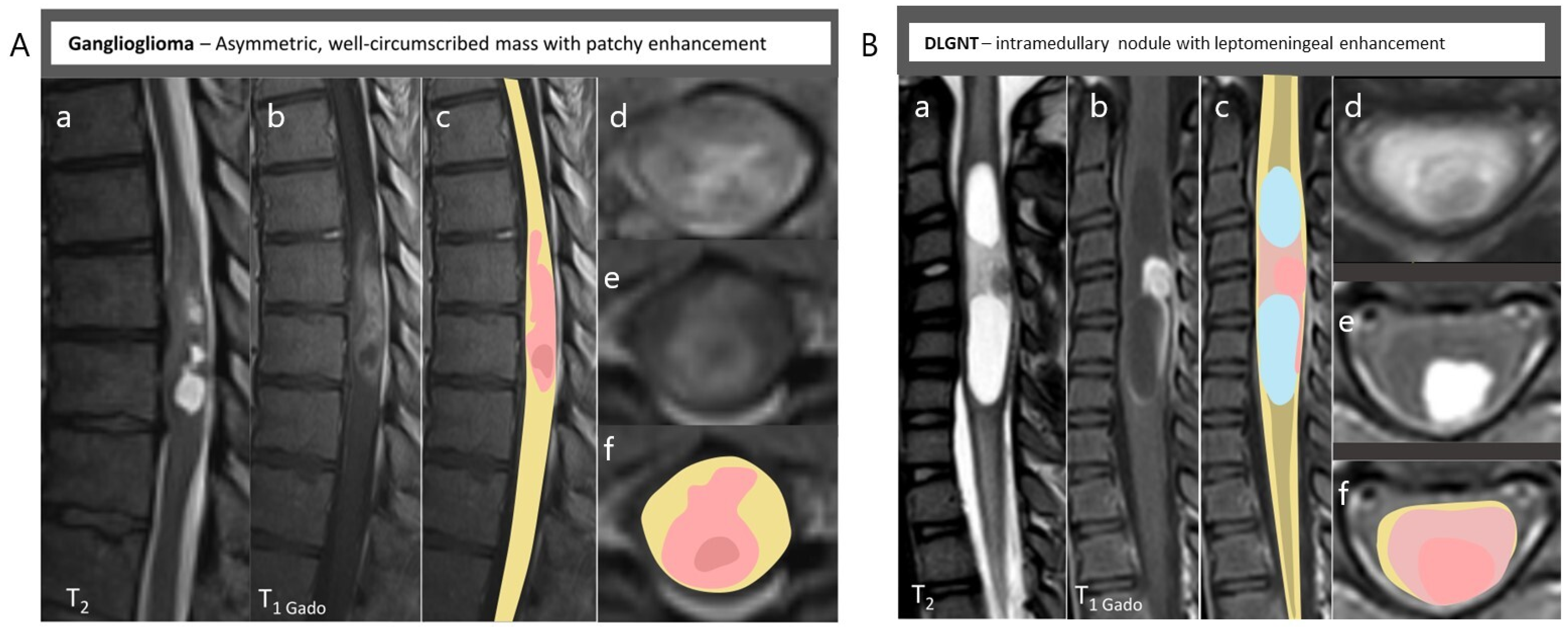
Diffuse Leptomeningeal Glioneural Tumors (DLGNTs) (Figure 3B)
DLGNT is a rare glioneuronal neoplasm characterized by diffuse leptomeningeal dissemination. Although primarily affecting children and young adults, spinal involvement is common and may dominate the clinical presentation. On MRI, DLGNT typically manifests as diffuse or nodular leptomeningeal enhancement along the spinal cord, extending over multiple vertebral levels [31]. The underlying spinal cord parenchyma may appear normal or show mild T2 hyperintensity without a discrete intramedullary mass, although secondary cord edema or focal intramedullary nodules can be observed. T1-weighted images are usually unremarkable aside from leptomeningeal thickening, while T2-weighted sequences may demonstrate subtle subpial signal abnormalities [31].
3.1.4. Spinal Hemangioblastoma (Figure 4A)
Hemangioblastomas are benign, highly vascular tumors representing about 3–8% of intramedullary tumors. They may occur sporadically or in the context of von Hippel–Lindau disease. Hemangioblastomas have a predilection for a subpial or exophytic location. They typically present as small, well-circumscribed nodules. On MRI, they appear iso- to hypointense on T1- and T2-weighted sequences, frequently accompanied by a peritumoral syrinx that may extend over several vertebral levels [32]. The classic “cyst-with-nodule” configuration is highly suggestive, with the enhancing mural nodule corresponding to the solid, vascular component of the tumor [32,33]. After contrast administration, they characteristically show intense nodular enhancement, and feeding or draining vessels are commonly visualized as serpiginous flow voids on T2 or SWI sequences. Spinal angiography allows the delineation of arterial feeders and venous drainage, confirming the diagnosis and aiding operative strategy [32].
Figure 4.
(A) Hemangioblastoma: (a) Sagittal T2-weighted image showing a hyposignal nodule with draining vessels associated to syrinx; (b) Sagittal T1-contrast-enhanced image showing an intense enhancement; (c) Sagittal pictorial representation; (d) Axial T1-contrast-enhanced image showing a nodular tumor with hyposignal draining vessels at the ventral and dorsal poles of the tumor; (e) Axial pictorial representation. (B) Solitary fibrous tumor: (a) Sagittal T2-weighted image showing a fusiform intramedullary tumor with extensive edema; (b) Sagittal T1-contrast-enhanced image showing intense enhancement; (c) Sagittal pictorial representation; (d) Axial T1-contrast-enhanced image; (e) Axial pictorial representation. (C) Lymphoma: (a) Sagittal T2-weighted image showing intramedullary hyposignal; (b) Sagittal T1-contrast-enhanced image showing homogeneous enhancement with blurred edges; (c) Sagittal pictorial representation; (d) Axial T2-weighted image; (e) Axial T1-contrast-enhanced image; (f) Axial pictorial representation.
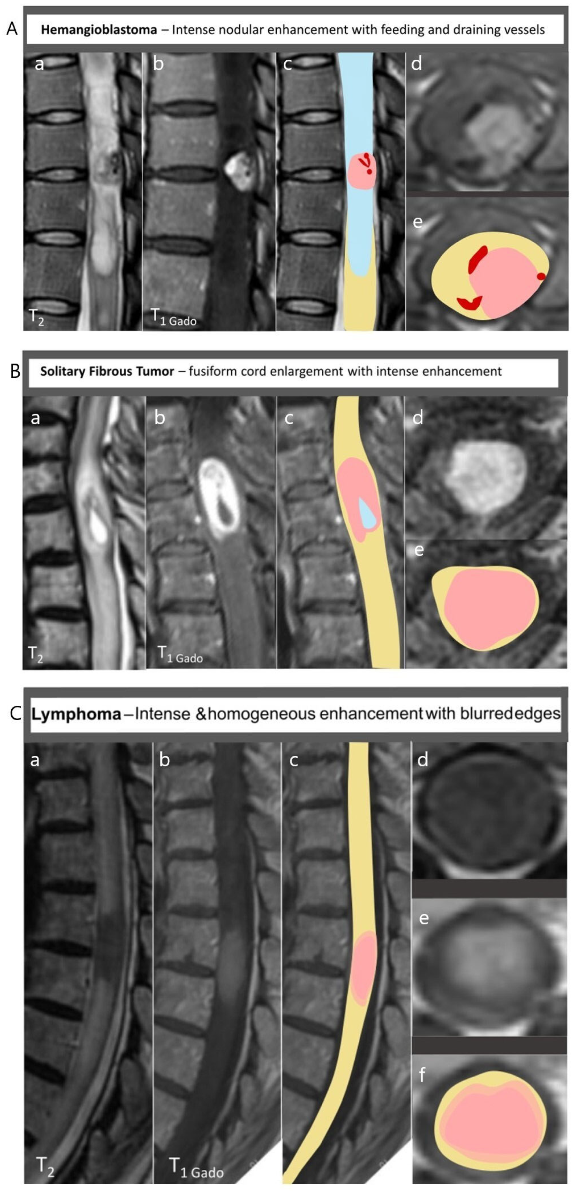
3.1.5. Solitary Fibrous Tumor (Figure 4B)
Hemangiopericytomas, now classified within the solitary fibrous tumor (SFT) spectrum, are rare but clinically important due to their recurrence potential. SFTs can be intradural extramedullary, intra-extramedullary, or intramedullary only [34,35]. Intramedullary SFTs usually appear as well-demarcated, expansile masses causing fusiform cord enlargement. On MRI, the solid component is iso- to hypointense on T1-weighted images and commonly hypointense on T2-weighted sequences, a feature attributed to their dense collagen content [36]. Areas of intermediate or slightly hyperintense T2 signal may appear when cellularity increases. After contrast administration, they demonstrate intense and homogeneous enhancement with sharp margins, and present without associated polar cysts, helping differentiate them from ependymomas [34,36].
3.1.6. Lymphoma (Figure 4C)
Primary intramedullary lymphoma is uncommon (most often arising in the context of primary CNS lymphoma or systemic lymphoproliferative disease), but should be included in the differential diagnosis of enhancing intramedullary lesions. Intramedullary lymphomas typically present as diffuse or fusiform cord enlargement rather than a discrete mass [33,37]. On MRI, lymphoma typically appears iso- to hypointense on T1-weighted sequences and hypointense on T2-weighted images, often accompanied by diffuse swelling of the spinal cord [33,37]. Following gadolinium administration, it demonstrates intense and homogeneous enhancement [37], a feature that reflects its dense cellular architecture rather than true hypervascularity. Moreover, tumor contours may be blurred after gadolinium injection. Diffusion-weighted imaging (DWI) usually shows marked diffusion restriction with low apparent diffusion coefficient (ADC) values, consistent with the high cellularity characteristic of lymphoma. Perfusion imaging shows low rCBV despite marked enhancement, differentiating it from hypervascular gliomas.
3.2. Metastases
3.2.1. Secondary CNS Tumors: Metastases (Figure 5A)
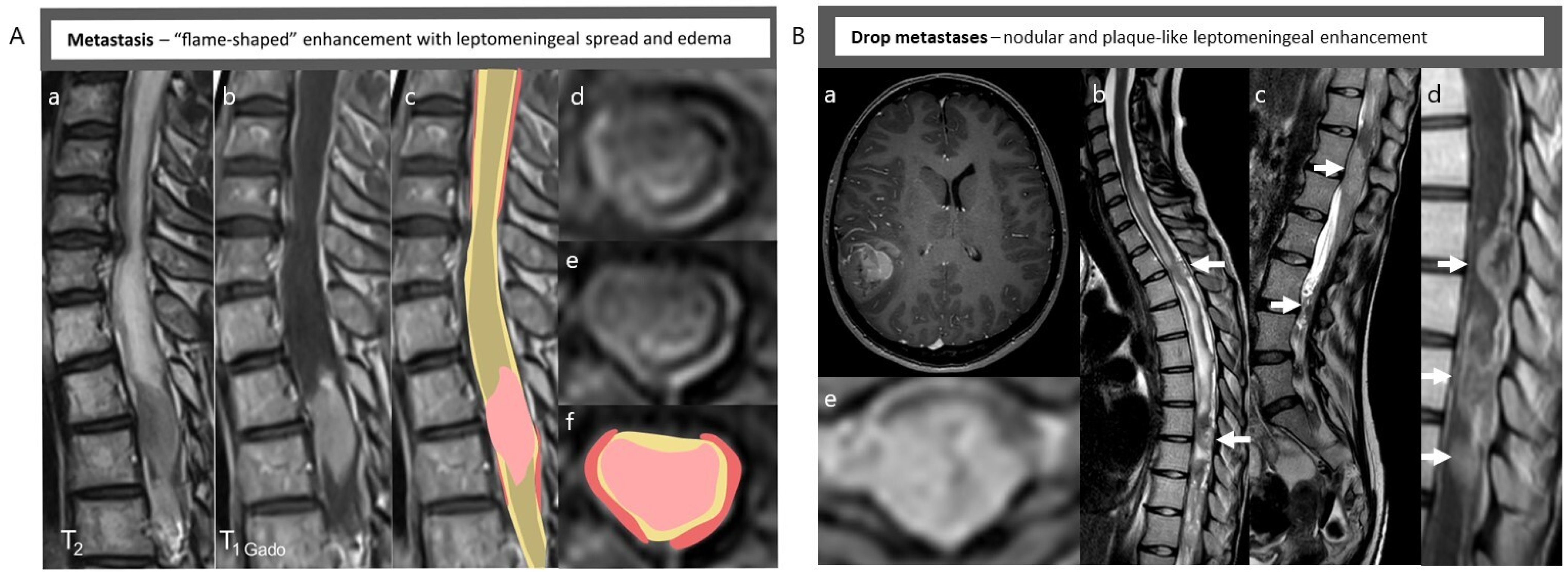
Intramedullary metastases are a very rare intramedullary entity, most frequently from lung, breast, renal cell carcinoma or melanoma primaries [38,39]. They frequently involve the cervical or lower thoracic spinal cord, consistent with the regional vascular supply and higher blood flow in these segments. Metastases often involve shorter cord segments than diffuse gliomas, but multiple lesions may be present. On MRI, intramedullary metastases are often eccentric or subpial in location [39]. They appear iso- to hypointense on T1-weighted images while hyperintense on T2-weighted sequences is usually observed due to extensive peritumoral vasogenic edema [39]. Gadolinium enhancement is typically intense, nodular, reflecting breakdown of the blood–spinal cord barrier [39]. After contrast injection, the “flame sign” is characteristic (corresponding to a leptomeningeal involvement), reflecting dissemination along the subarachnoid space.
Figure 5.
(A) Metastases: (a) Sagittal T2-weighted image showing a hyposignal intramedullary mass with extensive edema; (b) Sagittal T1-contrast-enhanced image showing an intense enhancement with leptomeningeal dissemination; (c) Sagittal pictorial representation; (d,e) Axial T1-contrast-enhanced image; (f) Axial pictorial representation. (B) Drop metastases: (a) Axial T1-contrast-enhanced image showing a posterior temporal glioblastoma; (b,c) Sagittal T2-weighted images of the entire spinal cord showing multiple (arrows) extraxial and intraxial nodule, with extensive intramedullary edema; (d) Sagittal T1-contrast-enhanced image; (e) Axial T1-contrast-enhanced image showing extra/intra axial tumor.
3.2.2. Drop Metastases (Figure 5B)
In addition, secondary spinal involvement may also occur through cerebrospinal fluid dissemination, commonly referred to as “drop metastases.” These lesions result from tumor cell spread along the neuraxis from an intracranial primary tumor, most classically medulloblastoma, intracranial ependymoma [40], or high-grade gliomas [41]. Spinal drop metastases typically present as nodular or plaque-like leptomeningeal enhancement along the spinal cord or cauda equina rather than as true parenchymal intramedullary masses [42]. Although less common, intramedullary implantation has been described in this context. Recognition of this dissemination pathway is essential, as it significantly alters staging, prognosis, and therapeutic strategy, necessitating complete craniospinal imaging.
3.3. Non-Neoplastic Intramedullary Lesions
3.3.1. Cavernoma (Figure 6A)
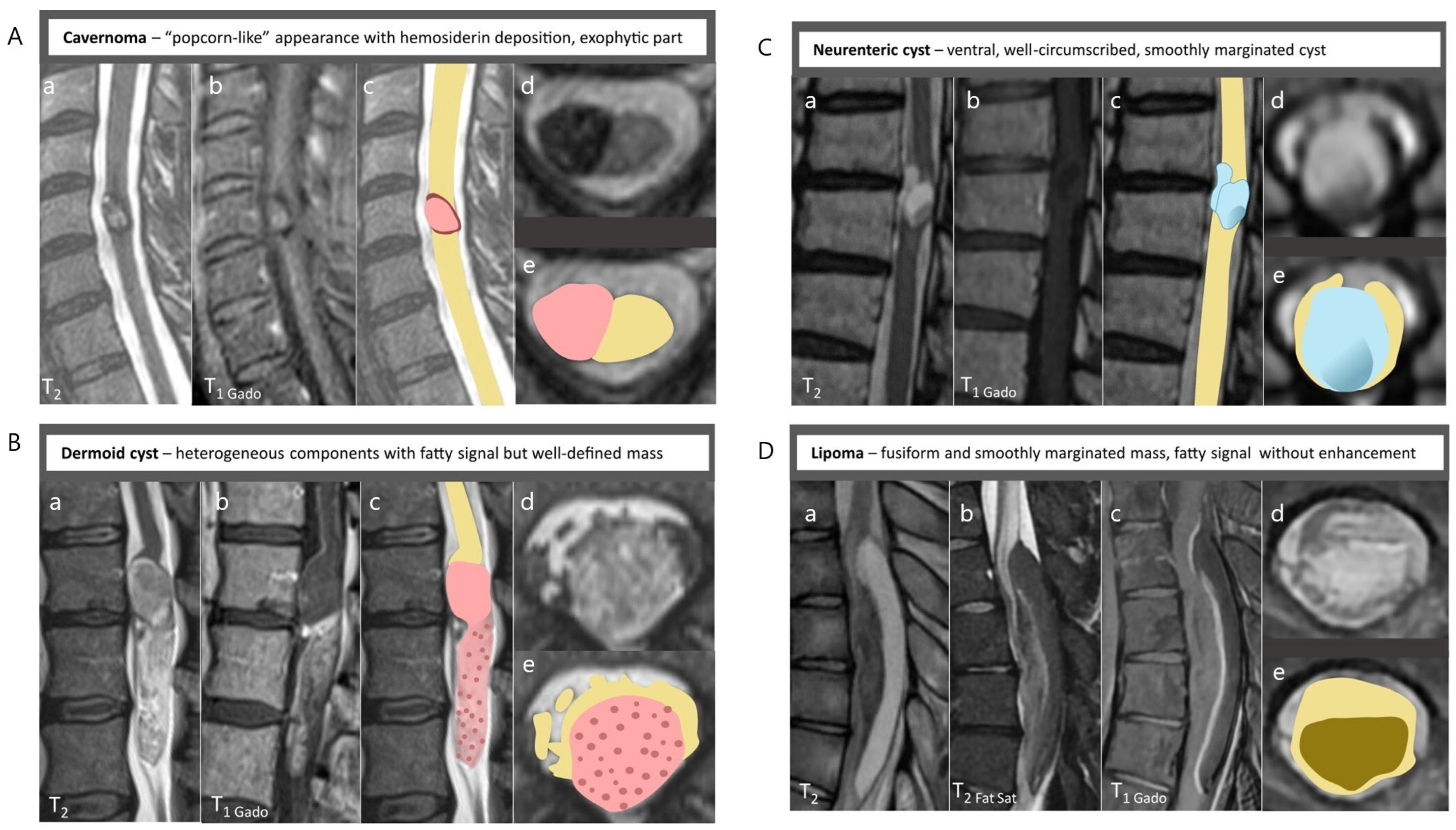
Cavernous malformations are composed of dilated capillary channels without intervening neural tissue. Although they are non-neoplastic lesions, they are an important diagnostic consideration in the differential diagnosis of intramedullary tumors due to their mass effect and hemorrhagic potential. Intramedullary cavernomas can occur at any spinal level. They typically present as well-defined, compact lesions, and are eccentric and/or exophytic [43,44]. On MRI, they typically show a mixture of hyperintense and hypointense signal on both T1- and T2-weighted sequences, producing the classic “popcorn” or “mulberry”-like appearance that reflects blood products of varying ages [43,45]. On susceptibility-weighted imaging, they are surrounded by a prominent hypointense rim caused by hemosiderin deposition. Contrast enhancement is generally absent, helping distinguish them from true neoplastic lesions [43,45].
Figure 6.
(A) Cavernoma: (a) Sagittal T2-weighted image showing “popcorn-like” appearance hemosiderin deposition; (b) Sagittal T1-contrast-enhanced image showing the absence of gadolinium enhancement; (c) Sagittal pictorial representation; (d) Axial T2-weighted image showing exophytic part of the cavernoma within the cord; (e) Axial pictorial representation. (B) Dermoid cyst: (a) Sagittal T2-weighted image showing a well-defined mass within the conus medullaris; (b) Sagittal T1-contrast-enhanced image showing the absence of gadolinium enhancement; (c) Sagittal pictorial representation; (d) Axial T2-weighted image showing the heterogeneous component of the tumor; (e) Axial pictorial representation. (C) Neurenteric cyst: (a) Sagittal T2-weighted image showing a well-circumscribed bilobed cyst; (b) Sagittal T1-contrast-enhanced image showing the absence of gadolinium enhancement; (c) Sagittal pictorial representation; (d) Axial T2-weighted image showing an intramedullary cyst opening the spinal cord ventrally, with CSF flux inside; (e) Axial pictorial representation. (D) Lipoma: (a) Sagittal T2-weighted image showing fusiform hyper-T2 mass within the dorsal columns; (b) Sagittal T2 FatSat-weighted image showing a hyposignal (c) Sagittal T1-contrast-enhanced image showing the absence of gadolinium enhancement; (d) Axial T2-weighted image showing the intramedullary counterpart of the lipoma at the cranial pole; (e) Axial pictorial representation.
3.3.2. Dermoid and Epidermoid Cysts (Figure 6B)
Dermoid and epidermoid cysts of the spinal cord are rare congenital lesions arising from ectodermal inclusions during neural tube closure. They are most frequently encountered in the lumbosacral region and in association with spinal dysraphism [46]. When intramedullary, they present as well-defined, slowly expanding lesions that may cause focal cord enlargement. Dermoid cysts tend to be more heterogeneous due to their complex contents, whereas epidermoid cysts are typically more uniform. Dermoid cysts, which contain fat, hair, and sebaceous material, often appear hyperintense on T1-weighted sequences and heterogeneously hyperintense on T2 [46,47], with signal characteristics influenced by lipid-rich components. Epidermoid cysts, composed of keratin debris, usually demonstrate T1 hypointensity or isointensity and T2 hyperintensity, often resembling CSF but with slightly higher signal intensity [47,48]. Neither dermoid nor epidermoid cysts typically exhibit contrast enhancement. Thin peripheral enhancement may occasionally be observed in dermoid cysts if an inflammatory reaction is present, but the cyst contents themselves do not enhance.
3.3.3. Neurenteric Cysts (Figure 6C)
Neurenteric cysts are benign congenital lesions resulting from abnormal notochord–foregut separation during embryologic development [49]. Although they are rare, when intramedullary, they can mimic a spinal cord tumor. On MRI, they are typically located ventral to the spinal cord [49], most commonly in the cervical or upper thoracic region. Morphologically, these cysts are well circumscribed and non-expansile, usually causing only mild compression of the adjacent spinal cord without infiltrative features [48,49]. Their signal characteristics reflect their proteinaceous or mucinous content, appearing hyperintense on both T1- and T2-weighted sequences [48,49], and they do not enhance after gadolinium administration, reflecting the absence of a vascularized solid component, consistent with their benign epithelial nature. Diffusion imaging shows no restriction, confirming a cystic rather than solid nature. In rare cases, however, susceptibility-weighted imaging (SWI) has demonstrated a calcified cystic component within an intramedullary neurenteric cyst, appearing as marked susceptibility signal drop-out [50].
3.3.4. Lipomas (Figure 6D)
Intramedullary lipomas are rare congenital lesions, and can be associated with dysraphic anomalies such as spinal dysraphism or diastematomyelia. They result from abnormal inclusion of mesenchymal tissue during neural tube closure rather than representing true neoplasms. Intramedullary lipomas typically occur in the thoracic cord and often present as fusiform, smoothly marginated masses expanding the spinal cord [51,52]. On MRI, they show a characteristic fat-equivalent signal pattern: the lesion is hyperintense on T1-weighted sequences and hyperintense on T2, mirroring the signal of subcutaneous fat [51,52]. Fat-suppressed sequences or STIR imaging produce a marked signal drop, confirming the fatty nature of the lesion [51,52]. Contrast enhancement is absent, although thin septations may occasionally display minimal enhancement.
4. Advances in Imaging Biomarkers
The growing availability of multiparametric MRI techniques has expanded the diagnostic scope of spinal cord imaging beyond morphology, allowing the characterization of tumor microstructure, vascularity, and metabolism.
4.1. DWI
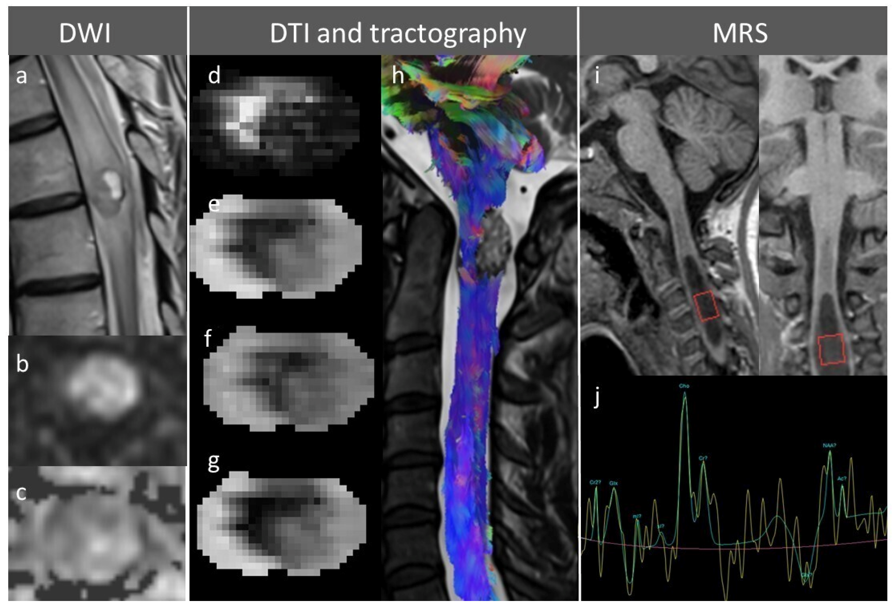
DWI provides a rapid, quantitative assessment of tumor cellularity through measurement of the ADC [53]. Restricted diffusion (low ADC) is caused by decreased free motion of water molecules and characterizes hypercellular tumors (such as lymphoma) [53] or high protein content (such as in epidermoid cysts) [54]; facilitated diffusion (high ADC) is observed in low-grade astrocytomas (Figure 7).
Figure 7.
(a) Sagittal T2-weighted image showing intramedullary tumor with extensive edema; (b) Axial diffusion weighted-image of the intramedullary tumor presented in panel (a) showing hypersignal; (c) Apparent diffusion coefficient map of the intramedullary tumor presented in panel (a) showing hypersignal; (d) Fractional anisotropy (FA) map of cervical spinal cord with cavernoma showing decreased FA (i.e., microarchitectural disorganization); (e) Mean diffusivity (MD) map of cervical spinal cord with cavernoma showing increase MD; (f) Axial diffusivity map of cervical spinal cord with cavernoma; (g) Radial diffusivity map of cervical spinal cord with cavernoma; (h) Spinal cord tractography showing displacement of spinal white matter due to cervical cavernoma; (i) Sagittal and coronal views showing region of interest placement for spectroscopy in case of cervical diffuse leptomeningeal glioneural tumor; (j) Spectroscopy of diffuse leptomeningeal glioneural tumor showing a high pic of choline and creatine, demonstrating increased membrane turnover and tumor proliferation.
4.2. DTI and Tractography
DTI quantifies the anisotropy of water diffusion within white-matter tracts, providing insights into the organization and integrity of spinal cord fibers [55]. It is particularly useful for distinguishing displacing from infiltrating lesions: ependymomas tend to displace spinal tracts, leading to sharply demarcated margins on color-coded fractional anisotropy (FA) maps [6,21], whereas pilocytic astrocytomas and high-grade gliomas typically infiltrate surrounding fibers, resulting in decreased FA and elevated mean diffusivity due to the disruption of tract coherence (often associated with a decrease in axial diffusivity corresponding to axonal loss and an increase in radial diffusivity corresponding to the demyelination process) [23]. Clinically, DTI-based tractography assists neurosurgeons in differentiating spinal tracts from one another and providing preoperative planning that defines the relationship of each tract of interest to the intramedullary tumor, finally establishing predictive factors for tumor resectability [6,7,56] (Figure 7).
4.3. Magnetic Resonance Spectroscopy (MRS)
MRS provides metabolic fingerprints by quantifying resonances from choline (Cho, biomarker of cellular turn over), N-acetylaspartate (NAA, biomarker of neural viability), creatine (Cr, biomarker of energy metabolism), lactate (biomarker of anaerobic metabolism), and lipids, which can help to determine tumor grade, biological activity, and response to therapy [57]. Semi-quantitative analysis using ratios of various metabolites is used to better predict the grade. Intramedullary ependymomas show strongly reduced NAA/Cr levels, whereas Cho/Cr and myoInosotol/Cr are moderately increased [10]. In contrast, high-grade intramedullary tumors tend to present a decreased NAA and Cr, with a high peak of myo-inositol (a biomarker of astrocyte proliferation) [58]. These metabolic patterns complement morphological findings and may provide indirect information on tumor aggressiveness and biological behavior. However, technical limitations—including voxel size constraints, partial volume effects, low signal-to-noise ratio, and susceptibility to motion—currently restrict the routine use of spinal cord MRS, confining its role mainly to specialized centers and research settings [57] (Figure 7).
4.4. Perfusion-Weighted Imaging (PWI)
PWI, using dynamic susceptibility contrast or dynamic contrast-enhanced techniques, allows the quantitative assessment of tumor vascularity through parameters such as relative blood volume (rCBV). In intramedullary tumors, PWI enables vascular phenotyping, which complements conventional contrast-enhanced MRI. High-grade gliomas [11,59] and glioblastomas [60] consistently demonstrate markedly elevated rCBV, correlating with dense capillary networks and microvascular proliferation. However, clinical adoption remains limited by technical challenges related to the small cord diameter, motion and CSF-pulsation artifacts, and the lack of standardized acquisition and post-processing protocols [59].
5. Discussion
Intramedullary tumors represent a rare but clinically critical subset of CNS neoplasms [1,61]. Despite their histological diversity, a structured analysis of imaging features—integrating morphology, signal characteristics, and enhancement patterns—can substantially narrow differential diagnosis and guide optimal management. Today, conventional MRI remains the cornerstone of evaluation, providing essential information on lesion localization, longitudinal extent, and cord morphology [5,62]. Nevertheless, advanced MRI techniques could revolutionize diagnostic accuracy by defining radiological biomarkers specific to each histological entity.
This pictorial review provides a structured overview of MRI features across a broad spectrum of histologically confirmed intramedullary lesions. Across the entities illustrated in this study—ranging from glial tumors (subependymoma, ependymoma, pilocytic astrocytoma, high-grade astrocytoma, glioblastoma, oligodendroglioma, ganglioglioma) to secondary neoplasms (metastases, lymphoma), vascular lesions (hemangioblastoma, cavernoma), mesenchymal tumors (solitary fibrous tumor), and developmental lesions (epidermoid cyst, neurenteric cyst, lipoma)—distinct imaging phenotypes emerge. These phenotypes are shaped by fundamental biological traits: expansile versus infiltrative growth, cellular density, vascular proliferation, cyst formation, hemorrhagic propensity, and preservation or disruption of the blood–spinal cord barrier. This concept aligns with the broader evolution of neuro-oncology toward biologically informed classification systems [12] and supports the idea that radiological assessment, when structured and systematic, may contribute to a more mechanistic understanding of spinal cord disease.
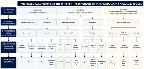
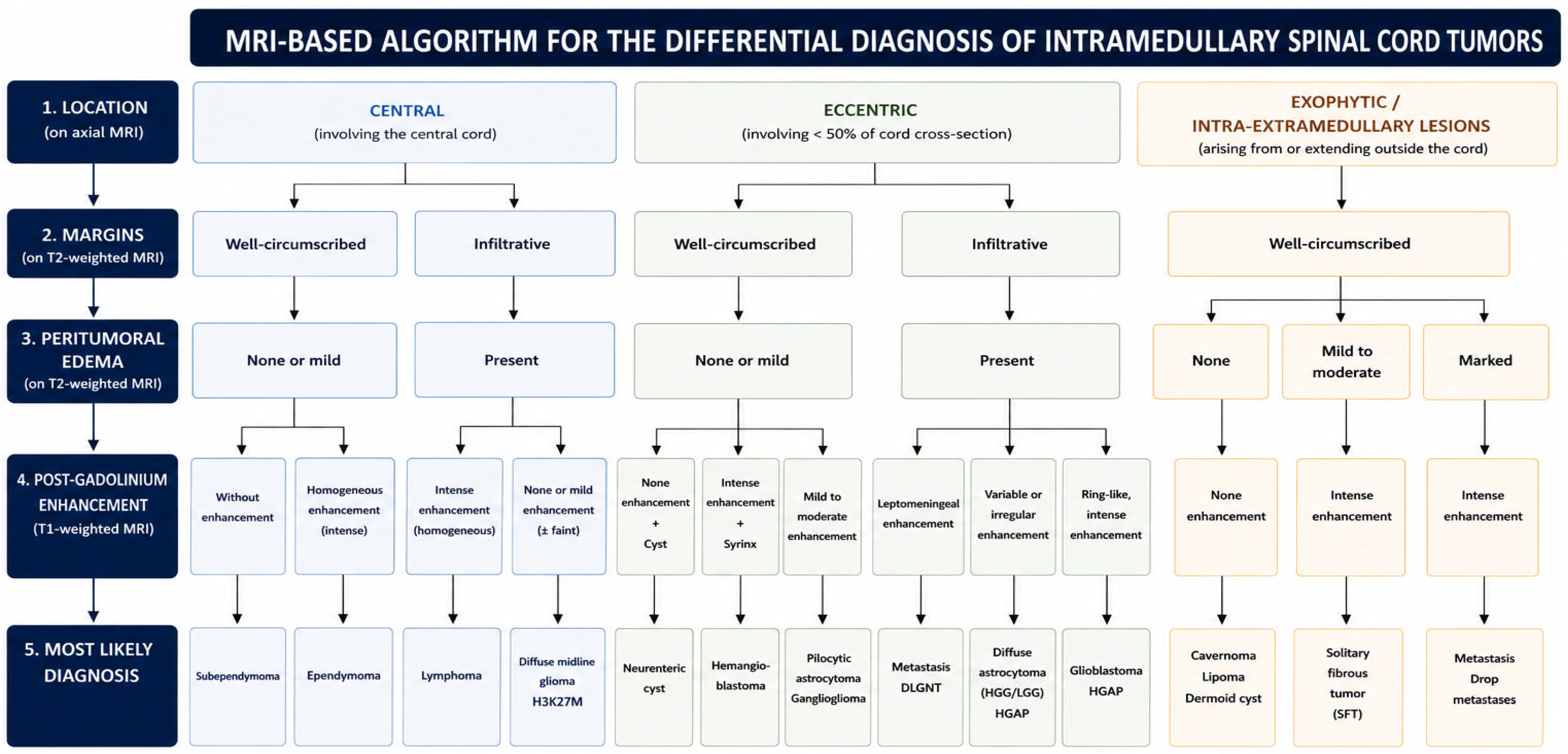
The imaging features described in our study are consistent with prior reports emphasizing the importance of lesion location, morphology, and enhancement patterns in narrowing the differential diagnosis [14,17,20,25,32]. Beyond confirming previously described imaging patterns, the present work adopts a didactic, pattern-based approach designed to reflect real-world diagnostic reasoning. In daily clinical practice, radiological interpretation relies less on isolated imaging signs than on a stepwise integration of key features, including lesion location, margin definition, enhancement pattern, and the presence of peritumoral edema or associated findings (Figure 8).
Figure 8.
MRI-based algorithm for the differential diagnosis of intramedullary spinal cord tumors.
The clinical value of this framework lies in its ability to inform decision-making in a context where biopsy or extensive resection carries significant neurological risk [63]. In intramedullary pathology, surgical strategy is closely tied to anticipated tumor behavior. Lesions with imaging features suggesting expansile growth (e.g., ependymoma, hemangioblastoma) are often approached with an intent of maximal safe resection [63,64,65], whereas imaging patterns consistent with infiltrative gliomas or lymphoma may favor limited resection, biopsy, or primarily oncological treatment. Therefore, structured radiological interpretation functions not only as a diagnostic exercise but also as a form of risk stratification and therapeutic guidance.
5.1. Future Directions
The future of intramedullary tumor imaging lies in moving beyond qualitative morphologic assessment toward quantitative, biologically informative imaging biomarkers. While conventional MRI provides essential structural information, it remains predominantly descriptive and insufficient to fully capture the microstructural, vascular, and metabolic heterogeneity that defines tumor behavior. A multiparametric approach, integrating advanced MRI techniques, will offer a pathway toward radio-genomics or radio-molecular correlations.
As the 2021 WHO classification [12] increasingly integrates genomic and epigenetic features into tumor taxonomy, imaging must evolve toward a radio-molecular paradigm, where MRI serves as a proxy for underlying molecular identity. The emerging field of radio-genomics will seek to establish robust correlations between quantitative imaging metrics and genomic alterations, enabling early and non-invasive molecular profiling. Such signatures could support growth pattern, or biological aggressiveness, thereby contributing to risk stratification and personalized treatment planning, aligning diagnostic radiology with the molecular era of neuro-oncology.
Ultra-high-field MRI (7T) further expands this perspective. Its improved signal-to-noise ratio and spatial resolution may enhance visualization of gray–white matter interfaces [66], microvascular architecture (due to enhancement of susceptibility-based contrast), and subtle infiltrative changes, potentially refining margin delineation and tract involvement. In this perspective, 7T MRI does not merely represent a higher-resolution version of conventional imaging but rather a shift toward microstructural and vascular sensitivity that may better capture the biological footprint of intramedullary tumors. Its integration with multiparametric analysis and computational modeling could contribute to establishing imaging-derived biomarkers that more closely reflect tumor infiltration and microenvironmental alterations.
5.2. Strengths and Limitations
A key strength of this study is the use of histopathologically confirmed cases as a reference standard, ensuring that the illustrated imaging patterns are grounded in definitive diagnoses according to current WHO classification criteria. The structured, entity-based organization reflects real-world clinical reasoning and encompasses both common and rare intramedullary lesions, thereby providing a didactic and practical framework. However, several limitations should be acknowledged. First, the retrospective design introduces inherent selection bias, since MRI acquisition protocols were not fully standardized and have evolved over time. Second, this is a single-center study, which may limit the generalizability of the findings. In addition, for each tumor entity, only a single representative case was selected; therefore, the spectrum of imaging variability within a given histological subtype is not fully captured, and atypical presentations may not be reflected. Moreover, the relatively limited sample size for rare tumor subtypes restricts the generalizability of specific imaging patterns. While advanced MRI techniques, including DTI, perfusion imaging, and MRS, have demonstrated potential in improving lesion characterization and providing insights into tumor microstructure and vascularity, their clinical use in spinal cord imaging remains limited by technical constraints, susceptibility artifacts, and the lack of standardized acquisition [67,68]. Furthermore, the analysis is based exclusively on MRI, without integration of other imaging modalities. Finally, significant overlap persists between the imaging features of different tumor entities, and radiological phenotypes, while highly informative, cannot replace histopathological and molecular diagnosis, which remain the gold standard.
6. Conclusions
Intramedullary tumors represent a diagnostically and therapeutically challenging group of spinal cord lesions in which clinical decision-making must balance oncological control with preservation of neurological function. This review highlights that a structured MRI approach can help differentiate major tumor entities and guide preoperative strategy.
Beyond conventional morphological assessment, emerging multiparametric and ultra-high-field MRI techniques provide additional insights into tumor microstructure, vascularity, and metabolism. Although currently limited by technical and standardization constraints, these approaches hold promise for the development of non-invasive imaging biomarkers and for strengthening radiological–molecular correlations. In this evolving paradigm, MRI is expected to play an increasingly central role not only in diagnosis but also in treatment planning and radiogenomic characterization of intramedullary spinal cord tumors.
Author Contributions
Conceptualization, C.D. and F.C.; methodology, C.D. and D.M.; validation, C.D., D.M., F.D., P.M., and F.C.; formal analysis, C.D., F.D., P.M., and F.C.; writing—original draft preparation, C.D.; writing—review and editing, C.D., D.M., F.D., P.M., and F.C.; visualization C.D.; supervision, F.C.; project administration C.D. All authors have read and agreed to the published version of the manuscript.
Funding
This research received no external funding.
Institutional Review Board Statement
The study was conducted in accordance with the Declaration of Helsinki and approved by the Ethics Committee of Collège de neurochirurgie (protocol code IRB 00011687–2026/26; date of approval: 26 April 2026). The official administrative approval document was formally issued at a later date due to institutional processing timelines.
Informed Consent Statement
Informed consent was obtained from all subjects involved in the study.
Data Availability Statement
The data presented in this study are available on request from the corresponding author due to ethical restrictions.
Conflicts of Interest
The authors declare no conflicts of interest.
References
- Schellinger, K.A.; Propp, J.M.; Villano, J.L.; McCarthy, B.J. Descriptive Epidemiology of Primary Spinal Cord Tumors. J. Neuro-Oncol. 2008, 87, 173–179. [Google Scholar] [CrossRef] [Scilit] [PubMed]
- Bican, O.; Minagar, A.; Pruitt, A.A. The Spinal Cord: A Review of Functional Neuroanatomy. Neurol. Clin. 2013, 31, 1–18. [Google Scholar] [CrossRef] [Scilit] [PubMed]
- Diaz, E.; Morales, H. Spinal Cord Anatomy and Clinical Syndromes. Semin. Ultrasound CT MRI 2016, 37, 360–371. [Google Scholar] [CrossRef] [Scilit]
- Lemay, A.; Gros, C.; Zhuo, Z.; Zhang, J.; Duan, Y.; Cohen-Adad, J.; Liu, Y. Automatic Multiclass Intramedullary Spinal Cord Tumor Segmentation on MRI with Deep Learning. Neuroimage Clin. 2021, 31, 102766. [Google Scholar] [CrossRef] [Scilit]
- Zimmerman, R.A.; Bilaniuk, L.T. Imaging of Tumors of the Spinal Canal and Cord. Radiol. Clin. N. Am. 1988, 26, 965–1007. [Google Scholar] [CrossRef] [Scilit] [PubMed]
- Dauleac, C.; Jacquesson, T.; Frindel, C.; André-Obadia, N.; Ducray, F.; Mertens, P.; Cotton, F. Value of Spinal Cord Diffusion Imaging and Tractography in Providing Predictive Factors for Tumor Resection in Patients with Intramedullary Tumors: A Pilot Study. Cancers 2024, 16, 2834. [Google Scholar] [CrossRef] [Scilit]
- Setzer, M.; Murtagh, R.D.; Murtagh, F.R.; Eleraky, M.; Jain, S.; Marquardt, G.; Seifert, V.; Vrionis, F.D. Diffusion Tensor Imaging Tractography in Patients with Intramedullary Tumors: Comparison with Intraoperative Findings and Value for Prediction of Tumor Resectability. J. Neurosurg. Spine 2010, 13, 371–380. [Google Scholar] [CrossRef] [Scilit]
- Czernicki, T.; Maj, E.; Podgórska, A.; Kunert, P.; Prokopienko, M.; Nowak, A.; Cieszanowski, A.; Marchel, A. Diffusion Tensor Tractography of Pyramidal Tracts in Patients with Brainstem and Intramedullary Spinal Cord Tumors: Relationship with Motor Deficits and Intraoperative MEP Changes. J. Magn. Reson. Imaging 2017, 46, 715–723. [Google Scholar] [CrossRef] [Scilit]
- Dauleac, C.; Frindel, C.; Pélissou-Guyotat, I.; Nicolas, C.; Yeh, F.-C.; Fernandez-Miranda, J.; Cotton, F.; Jacquesson, T. Full Cervical Cord Tractography: A New Method for Clinical Use. Front. Neuroanat. 2022, 16, 993464. [Google Scholar] [CrossRef] [Scilit]
- Hock, A.; Alexander, F.; Boesiger, P.; Henning, A.; Kollias, S.S. Spinal Cord MR Spectroscopy in Neoplastic Lesions. In Proceedings of the Annual Meeting of the International Society of Magnetic Resonance in Medicine, Melbourne, Australia, 5–11 May 2012. [Google Scholar]
- Liu, X.; Tian, W.; Kolar, B.; Hu, R.; Huang, Y.; Huang, J.; Ekholm, S. Advanced MR Diffusion Tensor Imaging and Perfusion Weighted Imaging of Intramedullary Tumors and Tumor like Lesions in the Cervicomedullary Junction Region and the Cervical Spinal Cord. J. Neuro-Oncol. 2014, 116, 559–566. [Google Scholar] [CrossRef] [Scilit]
- Louis, D.N.; Perry, A.; Wesseling, P.; Brat, D.J.; Cree, I.A.; Figarella-Branger, D.; Hawkins, C.; Ng, H.K.; Pfister, S.M.; Reifenberger, G.; et al. The 2021 WHO Classification of Tumors of the Central Nervous System: A Summary. Neuro-Oncology 2021, 23, 1231–1251. [Google Scholar] [CrossRef] [Scilit]
- Esparragosa Vazquez, I.; Ducray, F. The Role of Radiotherapy, Chemotherapy, and Targeted Therapies in Adult Intramedullary Spinal Cord Tumors. Cancers 2024, 16, 2781. [Google Scholar] [CrossRef] [Scilit] [PubMed]
- Shih, R.Y.; Koeller, K.K. Intramedullary Masses of the Spinal Cord: Radiologic-Pathologic Correlation. Radiographics 2020, 40, 1125–1145. [Google Scholar] [CrossRef] [Scilit]
- Zhou, S.; Xiong, J.; Pan, J.; Zhang, T.; Geng, D.; Zhang, J. Neuroradiological Features of Cervical and Cervicothoracic Intraspinal Subependymomas: A Study of Five Cases. Clin. Radiol. 2016, 71, 499.e9–499.e15. [Google Scholar] [CrossRef] [Scilit]
- Toi, H.; Ogawa, Y.; Kinoshita, K.; Hirai, S.; Takai, H.; Hara, K.; Matsushita, N.; Matsubara, S.; Uno, M. Bamboo Leaf Sign as a Sensitive Magnetic Resonance Imaging Finding in Spinal Subependymoma: Case Report and Literature Review. Case Rep. Neurol. Med. 2016, 2016, 9108641. [Google Scholar] [CrossRef] [Scilit] [PubMed]
- Sun, B.; Wang, C.; Wang, J.; Liu, A. MRI Features of Intramedullary Spinal Cord Ependymomas. J. Neuroimaging 2003, 13, 346–351. [Google Scholar] [CrossRef]
- Tomek, M.; Jayajothi, A.; Brandner, S.; Jaunmuktane, Z.; Lee, C.H.; Davagnanam, I. Imaging Features of Spinal Tanycytic Ependymoma. Neuroradiol. J. 2016, 29, 61–65. [Google Scholar] [CrossRef] [Scilit]
- Dauleac, C.; Messerer, R.; Obadia-Andre, N.; Afathi, M.; Barrey, C.Y. Cysts Associated with Intramedullary Ependymomas of the Spinal Cord: Clinical, MRI and Oncological Features. J. Neuro-Oncol. 2019, 19, 385–391. [Google Scholar] [CrossRef] [Scilit] [PubMed]
- Miyazawa, N.; Hida, K.; Iwasaki, Y.; Koyanagi, I.; Abe, H. MRI at 1.5 T of Intramedullary Ependymoma and Classification of Pattern of Contrast Enhancement. Neuroradiology 2000, 42, 828–832. [Google Scholar] [CrossRef] [Scilit]
- Dauleac, C.; Boulogne, S.; Cotton, F.; Mertens, P. Spinal Cord Tractography and Neuromonitoring-Based Surgical Strategy for Intramedullary Ependymoma. Neurosurg. Focus. Video 2023, 9, V4. [Google Scholar] [CrossRef] [Scilit]
- She, D.-J.; Lu, Y.-P.; Xiong, J.; Geng, D.-Y.; Yin, B. MR Imaging Features of Spinal Pilocytic Astrocytoma. BMC Med. Imaging 2019, 19, 5. [Google Scholar] [CrossRef] [Scilit]
- Ducreux, D.; Lepeintre, J.-F.; Fillard, P.; Loureiro, C.; Tadié, M.; Lasjaunias, P. MR Diffusion Tensor Imaging and Fiber Tracking in 5 Spinal Cord Astrocytomas. AJNR Am. J. Neuroradiol. 2006, 27, 214–216. [Google Scholar] [PubMed]
- Landi, A.; Palmarini, V.; D’Elia, A.; Marotta, N.; Salvati, M.; Santoro, A.; Delfini, R. Magnetic Resonance Diffusion Tensor Imaging and Fiber-Tracking Diffusion Tensor Tractography in the Management of Spinal Astrocytomas. World J. Clin. Cases 2016, 4, 1–4. [Google Scholar] [CrossRef] [Scilit]
- Cheng, L.; Wang, L.; Yao, Q.; Ma, L.; Duan, W.; Guan, J.; Zhang, C.; Wang, K.; Liu, Z.; Wang, X.; et al. Clinicoradiological Characteristics of Primary Spinal Cord H3 K27M-Mutant Diffuse Midline Glioma. J. Neurosurg. Spine 2022, 36, 303–314. [Google Scholar] [CrossRef] [Scilit] [PubMed]
- Ferrante, P.; Mora, J.A.; Salazar, L.; Sáez, E.M.; Auger, C.; Rovira, À. MR Imaging Findings in Primary Spinal Cord Glioblastoma. Radiol. Case Rep. 2021, 16, 72–77. [Google Scholar] [CrossRef] [Scilit] [PubMed]
- Cerron-Vela, C.R.; Gonçalves, F.G.; Tierradentro-García, L.O.; Viaene, A.N.; Lerebo, W.; Andronikou, S. A Comprehensive Evaluation of Imaging Features in Pediatric Spinal Gliomas and Their Value in Predicting Tumor Grade and Histology. Neuroradiology 2024, 66, 1311–1324. [Google Scholar] [CrossRef] [Scilit]
- Soni, N.; Agarwal, A.; Ajmera, P.; Mehta, P.; Gupta, V.; Vibhute, M.; Gubbiotti, M.; Mark, I.T.; Messina, S.A.; Mohan, S.; et al. High-Grade Astrocytoma with Piloid Features: A Dual Institutional Review of Imaging Findings of a Novel Entity. AJNR Am. J. Neuroradiol. 2024, 45, 468–474. [Google Scholar] [CrossRef] [Scilit]
- Bender, K.; Perez, E.; Chirica, M.; Onken, J.; Kahn, J.; Brenner, W.; Ehret, F.; Euskirchen, P.; Koch, A.; Capper, D.; et al. High-Grade Astrocytoma with Piloid Features (HGAP): The Charité Experience with a New Central Nervous System Tumor Entity. J. Neuro-Oncol. 2021, 153, 109–120. [Google Scholar] [CrossRef] [Scilit]
- Vlachos, N.; Lampros, M.G.; Zigouris, A.; Voulgaris, S.; Alexiou, G.A. Anaplastic Gangliogliomas of the Spinal Cord: A Scoping Review of the Literature. Neurosurg. Rev. 2022, 45, 295–304. [Google Scholar] [CrossRef] [Scilit]
- Manoharan, N.; Ajuyah, P.; Senapati, A.; Wong, M.; Mullins, A.; Rodriguez, M.; Doyle, H.; McCowage, G.; Lau, L.M.S.; Ekert, P.G.; et al. Diffuse Leptomeningeal Glioneuronal Tumour (DLGNT) in Children: The Emerging Role of Genomic Analysis. Acta Neuropathol. Commun. 2021, 9, 147. [Google Scholar] [CrossRef] [Scilit]
- Chu, B.C.; Terae, S.; Hida, K.; Furukawa, M.; Abe, S.; Miyasaka, K. MR Findings in Spinal Hemangioblastoma: Correlation with Symptoms and with Angiographic and Surgical Findings. AJNR Am. J. Neuroradiol. 2001, 22, 206–217. [Google Scholar]
- Koeller, K.K.; Rosenblum, R.S.; Morrison, A.L. Neoplasms of the Spinal Cord and Filum Terminale: Radiologic-Pathologic Correlation. Radiographics 2000, 20, 1721–1749. [Google Scholar] [CrossRef] [Scilit] [PubMed]
- Liu, H.; Yang, A.; Chen, N.; Yang, J.; Qiu, X.; Zhang, J. Hemangiopericytomas in the Spine: Clinical Features, Classification, Treatment, and Long-Term Follow-up in 26 Patients. Neurosurgery 2013, 72, 16–24; discussion 24. [Google Scholar] [CrossRef] [Scilit] [PubMed]
- Dauleac, C.; Vasiljevic, A.; Berhouma, M. How to Differentiate Spinal Cord Hemangiopericytoma from Common Spinal Cord Tumor? Neurochirurgie 2020, 66, 53–55. [Google Scholar] [CrossRef] [Scilit]
- Mariniello, G.; Napoli, M.; Russo, C.; Briganti, F.; Giamundo, A.; Maiuri, F.; Del Basso De Caro, M.L. MRI Features of Spinal Solitary Fibrous Tumors. A Report of Two Cases and Literature Review. Neuroradiol. J. 2012, 25, 610–616. [Google Scholar] [CrossRef] [Scilit]
- Flanagan, E.P.; O’Neill, B.P.; Porter, A.B.; Lanzino, G.; Haberman, T.M.; Keegan, B.M. Primary Intramedullary Spinal Cord Lymphoma. Neurology 2011, 77, 784–791. [Google Scholar] [CrossRef] [Scilit] [PubMed]
- Sung, W.-S.; Sung, M.-J.; Chan, J.H.; Manion, B.; Song, J.; Dubey, A.; Erasmus, A.; Hunn, A. Intramedullary Spinal Cord Metastases: A 20-Year Institutional Experience with a Comprehensive Literature Review. World Neurosurg. 2013, 79, 576–584. [Google Scholar] [CrossRef] [Scilit]
- Rykken, J.B.; Diehn, F.E.; Hunt, C.H.; Schwartz, K.M.; Eckel, L.J.; Wood, C.P.; Kaufmann, T.J.; Lingineni, R.K.; Carter, R.E.; Wald, J.T. Intramedullary Spinal Cord Metastases: MRI and Relevant Clinical Features from a 13-Year Institutional Case Series. AJNR Am. J. Neuroradiol. 2013, 34, 2043–2049. [Google Scholar] [CrossRef] [Scilit]
- Javed, Z.; Sadiq, R.; Fatima, H.; Shamim, M.S. Intramedullary Spinal Cord Metastasis in Spinal Ependymomas. J. Pak. Med. Assoc. 2025, 75, 509–511. [Google Scholar] [CrossRef] [Scilit]
- Lawton, C.D.; Nagasawa, D.T.; Yang, I.; Fessler, R.G.; Smith, Z.A. Leptomeningeal Spinal Metastases from Glioblastoma Multiforme: Treatment and Management of an Uncommon Manifestation of Disease. J. Neurosurg. Spine 2012, 17, 438–448. [Google Scholar] [CrossRef] [Scilit]
- Yeamans, C.L.; Gutierrez-Quintana, R.; Haley, A.; Lamm, C.G. Magnetic Resonance Imaging and Clinical Findings Associated with Choroid Plexus Spinal Cord “Drop” Metastases. J. Am. Anim. Hosp. Assoc. 2017, 53, 265–269. [Google Scholar] [CrossRef] [Scilit]
- Weinzierl, M.R.; Krings, T.; Korinth, M.C.; Reinges, M.H.T.; Gilsbach, J.M. MRI and Intraoperative Findings in Cavernous Haemangiomas of the Spinal Cord. Neuroradiology 2004, 46, 65–71. [Google Scholar] [CrossRef] [Scilit]
- Dauleac, C.; Frindel, C.; Cotton, F.; Pelissou-Guyotat, I. Tractography-Based Surgical Strategy for Cavernoma of the Conus Medullaris: Case Illustration. J. Neurosurg. Spine 2019, 32, 611–612. [Google Scholar] [CrossRef] [Scilit] [PubMed]
- Panda, A.; Diehn, F.E.; Kim, D.K.; Bydon, M.; Goyal, A.; Benson, J.C.; Carr, C.M.; Rinaldo, L.; Flemming, K.D.; Lanzino, G. Spinal Cord Cavernous Malformations: MRI Commonly Shows Adjacent Intramedullary Hemorrhage. J. Neuroimaging 2020, 30, 690–696. [Google Scholar] [CrossRef] [Scilit] [PubMed]
- Zhang, Y.; Cheng, J.-L.; Zhang, L.; Bai, J.; Wang, J.; Li, H.-L.; Yang, Y.-J. Magnetic Resonance Imaging of Ruptured Spinal Dermoid Tumors with Spread of Fatty Droplets in the Central Spinal Canal and/or Spinal Subarachnoidal Space. J. Neuroimaging 2013, 23, 71–74. [Google Scholar] [CrossRef] [Scilit]
- Ogden, A.T.; Khandji, A.G.; McCormick, P.C.; Kaiser, M.G. Intramedullary Inclusion Cysts of the Cervicothoracic Junction. Report of Two Cases in Adults and Review of the Literature. J. Neurosurg. Spine 2007, 7, 236–242. [Google Scholar] [CrossRef] [Scilit] [PubMed]
- Evans, A.; Stoodley, N.; Halpin, S. Magnetic Resonance Imaging of Intraspinal Cystic Lesions: A Pictorial Review. Curr. Probl. Diagn. Radiol. 2002, 31, 79–94. [Google Scholar] [CrossRef]
- Brooks, B.S.; Duvall, E.R.; el Gammal, T.; Garcia, J.H.; Gupta, K.L.; Kapila, A. Neuroimaging Features of Neurenteric Cysts: Analysis of Nine Cases and Review of the Literature. AJNR Am. J. Neuroradiol. 1993, 14, 735–746. [Google Scholar]
- Ziu, M.; Vibhute, P.; Vecil, G.G.; Henry, J. Isolated Spinal Neurenteric Cyst Presenting as Intramedullary Calcified Cystic Mass on Imaging Studies: Case Report and Review of Literature. Neuroradiology 2010, 52, 119–123. [Google Scholar] [CrossRef] [Scilit]
- Ben Ali, H.; Batarfi, L.; Aldraihem, M.O.; Bashir, S. Unusual Presentation of a Giant Thoracic Spinal Cord Lipoma. Spinal Cord Ser. Cases 2022, 8, 35. [Google Scholar] [CrossRef] [Scilit]
- Kabir, S.M.R.; Thompson, D.; Rezajooi, K.; Casey, A.T.H. Non-Dysraphic Intradural Spinal Cord Lipoma: Case Series, Literature Review and Guidelines for Management. Acta Neurochir. 2010, 152, 1139–1144. [Google Scholar] [CrossRef] [Scilit]
- Yildiz, S.; Schecht, M.; Aggarwal, A.; Nael, K.; Doshi, A.; Pawha, P.S. Diffusion Weighted Imaging in Spine Tumors. Neuroimaging Clin. N. Am. 2023, 33, 459–475. [Google Scholar] [CrossRef] [Scilit]
- Celik, A.O.; Baris, M.M.; Demirtas, H.; Umul, A. Unusual Case of Spinal Epidermoid Cyst and a Concomitant Spinal Arachnoid Cyst. BMJ Case Rep. 2016, 2016, bcr2015214002. [Google Scholar] [CrossRef] [Scilit]
- Maier, S.E.; Mamata, H. Diffusion Tensor Imaging of the Spinal Cord. Ann. N. Y. Acad. Sci. 2005, 1064, 50–60. [Google Scholar] [CrossRef] [Scilit]
- Granata, F.; Racchiusa, S.; Mormina, E.; Barresi, V.; Garufi, G.; Grasso, G.; Salpietro, F.M.; Longo, M.; Alafaci, C. Presurgical Role of MRI Tractography in a Case of Extensive Cervicothoracic Spinal Ependymoma. Surg. Neurol. Int. 2017, 8, 56. [Google Scholar] [CrossRef] [Scilit]
- Wyss, P.O.; Hock, A.; Kollias, S. The Application of Human Spinal Cord Magnetic Resonance Spectroscopy to Clinical Studies: A Review. Semin. Ultrasound CT MRI 2017, 38, 153–162. [Google Scholar] [CrossRef] [Scilit]
- Henning, A.; Schär, M.; Kollias, S.S.; Boesiger, P.; Dydak, U. Quantitative Magnetic Resonance Spectroscopy in the Entire Human Cervical Spinal Cord and beyond at 3T. Magn. Reson. Med. 2008, 59, 1250–1258. [Google Scholar] [CrossRef] [Scilit]
- Liu, X.; Tian, W.; Chen, H.; LoStracco, T.A.; Zhang, J.; Li, M.Y.; Germin, B.; Wang, H.Z. Advanced Neuroimaging in the Evaluation of Spinal Cord Tumors and Tumor Mimics: Diffusion Tensor and Perfusion-Weighted Imaging. Semin. Ultrasound CT MRI 2017, 38, 163–175. [Google Scholar] [CrossRef] [Scilit]
- Liu, X.; Germin, B.I.; Ekholm, S. A Case of Cervical Spinal Cord Glioblastoma Diagnosed with MR Diffusion Tensor and Perfusion Imaging. J. Neuroimaging 2011, 21, 292–296. [Google Scholar] [CrossRef] [Scilit]
- Duong, L.M.; McCarthy, B.J.; McLendon, R.E.; Dolecek, T.A.; Kruchko, C.; Douglas, L.L.; Ajani, U.A. Descriptive Epidemiology of Malignant and Nonmalignant Primary Spinal Cord, Spinal Meninges, and Cauda Equina Tumors, United States, 2004–2007. Cancer 2012, 118, 4220–4227. [Google Scholar] [CrossRef] [Scilit]
- Shah, L.M.; Salzman, K.L. Conventional and Advanced Imaging of Spinal Cord Tumors. Neuroimaging Clin. N. Am. 2023, 33, 389–406. [Google Scholar] [CrossRef] [Scilit] [PubMed]
- Hussain, I.; Parker, W.E.; Barzilai, O.; Bilsky, M.H. Surgical Management of Intramedullary Spinal Cord Tumors. Neurosurg. Clin. N. Am. 2020, 31, 237–249. [Google Scholar] [CrossRef] [Scilit]
- Aghakhani, N.; David, P.; Parker, F.; Lacroix, C.; Benoudiba, F.; Tadie, M. Intramedullary Spinal Ependymomas: Analysis of a Consecutive Series of 82 Adult Cases with Particular Attention to Patients with No Preoperative Neurological Deficit. Neurosurgery 2008, 62, 1279–1285; discussion 1285–1286. [Google Scholar] [CrossRef] [Scilit]
- Klekamp, J. Spinal Ependymomas. Part 1: Intramedullary Ependymomas. Neurosurg. Focus 2015, 39, E6. [Google Scholar] [CrossRef] [Scilit]
- Barry, R.L.; Vannesjo, S.J.; By, S.; Gore, J.C.; Smith, S.A. Spinal Cord MRI at 7T. Neuroimage 2018, 168, 437–451. [Google Scholar] [CrossRef] [Scilit] [PubMed]
- Hendrix, P.; Griessenauer, C.J.; Cohen-Adad, J.; Rajasekaran, S.; Cauley, K.A.; Shoja, M.M.; Pezeshk, P.; Tubbs, R.S. Spinal Diffusion Tensor Imaging: A Comprehensive Review with Emphasis on Spinal Cord Anatomy and Clinical Applications. Clin. Anat. 2015, 28, 88–95. [Google Scholar] [CrossRef] [Scilit] [PubMed]
- Cohen-Adad, J. Functional Magnetic Resonance Imaging of the Spinal Cord: Current Status and Future Developments. Semin. Ultrasound CT MRI 2017, 38, 176–186. [Google Scholar] [CrossRef] [Scilit]

Disclaimer/Publisher’s Note: The statements, opinions and data contained in all publications are solely those of the individual author(s) and contributor(s) and not of MDPI and/or the editor(s). MDPI and/or the editor(s) disclaim responsibility for any injury to people or property resulting from any ideas, methods, instructions or products referred to in the content. |
© 2026 by the authors. Licensee MDPI, Basel, Switzerland. This article is an open access article distributed under the terms and conditions of the Creative Commons Attribution (CC BY) license.