Article

Ultrasound-Assisted Wound Debridement for Diabetic Foot Ulcers: A Systematic Review and Meta-Analysis of Randomized Controlled Trials

1
Department of Nursing, Shanghai 6th Peoples Hospital, Shanghai Jiao Tong University, Shanghai 200240, China
2
Department of Anesthesiology, Shanghai 6th Peoples Hospital, Shanghai Jiao Tong University, Shanghai 200240, China
3
Department of Orthopedic Surgery, Shanghai 6th Peoples Hospital, Shanghai Jiao Tong University, Shanghai 200240, China
*
Authors to whom correspondence should be addressed.
These authors contributed equally to this work.
Biomedicines 2026, 14(4), 846; https://doi.org/10.3390/biomedicines14040846
Submission received: 21 February 2026 / Revised: 22 March 2026 / Accepted: 31 March 2026 / Published: 8 April 2026
(This article belongs to the Special Issue Diabetes: Comorbidities, Therapeutics and Insights (3rd Edition))

Abstract

Background: Diabetic foot ulcers (DFUs) represent a severe and costly complication of diabetes mellitus. This meta-analysis aims to compare the efficacy of ultrasound-assisted wound debridement (UAWD) and conventional debridement in promoting wound healing in patients with DFUs. Methods: A systematic literature search was conducted using PubMed, EMBASE, BIOSIS, Web of Science, and the Cochrane Library from inception to 31 October 2025. Randomized controlled trials (RCTs) that compared UAWD with a placebo or standard wound care in patients with DFUs were included. Primary outcomes were the healing rate, time to complete healing, and reduction in wound area. Results were expressed as odds ratios (ORs) or mean differences (MDs) with 95% confidence intervals (CIs). This study was registered on the PROSPERO platform (CRD420251229633). Results: Ten RCTs that involved 386 patients were included. The meta-analysis showed that the treatment group had a significantly higher complete wound healing rate compared with the control group (OR: 2.92; 95% CI: 1.82 to 4.70; p = 0.75; I2 = 0%). The rate of wound area reduction was also significantly greater in the treatment group (MD: 21.29%; 95% CI: 3.03 to 39.56; p = 0.003; I2 = 75%). Furthermore, the time to complete healing was significantly shorter in the treatment group (MD: −4.84 weeks; 95% CI: −8.65 to −1.03; I2 = 61%, p = 0.05). Conclusions: UAWD appears to be more effective than conventional debridement alone in improving healing rates and accelerating wound closure in diabetic foot ulcers. However, safety data were inadequately reported across most included studies, with adverse events poorly characterized. Future large-scale RCTs should prioritize rigorous adverse event reporting to establish both the efficacy and safety profile of this intervention.

1. Introduction

Diabetic foot ulcer (DFU) represents one of the most severe and costly complications of diabetes mellitus [1]. During their lifetime, 19–34% of individuals with diabetes may develop a DFU, with a substantial proportion requiring amputation of the affected limb [2,3]. Therefore, there is an urgent need for innovative and effective wound management strategies.
The current treatment guidelines for DFU recommend debridement, infection management, revascularization, and decompression to promote healing [4]. A critical element of wound management is debridement, which involves the removal of non-viable tissue from the wound bed to facilitate its replacement with healthy tissue and accelerate healing [5]. Debridement may be performed using mechanical, sharp/surgical, autolytic, enzymatic, or biological methods [6,7]. Among these, sharp or surgical debridement is recommended by the International Working Group on the Diabetic Foot as the preferred approach, given its rapidity, cost-effectiveness, and widespread availability for wound bed preparation [6].
Recently, ultrasound-assisted wound debridement (UAWD) has emerged as an innovative therapeutic modality with significant potential to transform conventional DFU management [8]. A case series by Campitiello et al. demonstrated that the application of an ultrasonic debridement system reduces debridement time, preserves viable tissue safely, and is associated with low complication rates in DFU surgery [9]. By utilizing ultrasonic energy, UAWD enables precise removal of necrotic tissue, disruption of biofilms, and reduction of microbial load within ulcer lesions, thereby potentially accelerating the healing process [10,11]. Furthermore, the acoustic energy delivered via ultrasonic waves is proposed to stimulate essential wound healing processes, including enhanced angiogenesis and accelerated formation of granulation tissue [12]. However, despite a compelling theoretical rationale and promising outcomes reported in preliminary studies, the scientific community remains cautious. Current evidence supporting the clinical efficacy of UAWD in DFU management remains fragmented and inconsistent.
Previous systematic reviews on ultrasound-assisted wound debridement for diabetic foot ulcers have been limited by incomplete search strategies. To address these gaps, the present study offers several methodological advancements: (1) restricting inclusion to RCTs, (2) limiting inclusion to English-language publications, and (3) providing an updated literature search. By addressing these limitations, we aim to provide a more robust and clinically interpretable synthesis of the evidence.
This study therefore aimed to synthesize available evidence from RCTs to comprehensively compare the efficacy and safety of UAWD versus conventional debridement in DFU management.

2. Methods

2.1. Search Strategy

This systematic review was conducted in accordance with the Preferred Reporting Items for Systematic Reviews and Meta-Analyses (PRISMA) guidelines. It was registered on the PROSPERO platform (CRD420251229633).
A comprehensive literature search was performed across multiple electronic databases, including PubMed, EMBASE, BIOSIS, Web of Science, and the Cochrane Library. The study was carried out from inception to 31 October 2025, with only English-language papers being considered. The search terms were as follows: (‘ultrasonic debridement’ or ‘ultrasound debridement’ or ‘sonic debridement’) AND (‘diabetic foot’, ‘diabetic wound’, or ‘DFU’). The detailed search strategies are described in Supplementary Materials File S1. In addition to the electronic database search, the reference lists of all identified articles were manually screened to capture any potentially eligible studies not retrieved through the initial search. Grey literature and clinical trial registries were also searched.

2.2. Inclusion and Exclusion Criteria

2.2.1. Inclusion Criteria

The inclusion criteria were as follows: (1) type of studies: only RCTs evaluating the efficacy of UAWD for DFUs were included; (2) type of participants: patients were adults diagnosed with DFU, regardless of age, gender, or diabetes type; (3) type of intervention: patients in the treatment group underwent UAWD and those in the control group were treated with a placebo or standard wound care; (4) outcomes: wound healing rate, time to complete healing, percentage reduction in wound area, and adverse events. Standard wound care was defined as the routine clinical practice at the study institution, which could include surgical debridement, mechanical debridement, dressing changes, offloading, and infection control.
The exclusion criteria included: (1) reviews and case reports; (2) the data was incomplete.

2.2.2. Data Extraction

Two investigators independently extracted the following data from each included study: first author, publication year, study design, country, sample size, treatment details (UAWD and control), treatment time, and outcomes. Any disagreements in the data extraction process were adjudicated by a third reviewer. Wound healing was defined as complete epithelialization of the DFUs. The healing rate was calculated (No. of healed patients/No. of patients per group × 100). Time to healing was defined as the time from the day of ulcer inclusion in the study to wound healing. The percentage of wound area reduction was calculated by comparing the wound area at week zero to the wound area at the end of treatment.

2.2.3. Quality Assessment

The methodological quality of the included studies was evaluated with the Cochrane Risk of Bias Tool for Randomized Controlled Trials (RCTs). This assessment was conducted independently by two authors across seven domains: random sequence generation, allocation concealment, blinding of participants and personnel, blinding of outcome assessment, incomplete outcome data, selective reporting, and other bias. The quality of evidence was graded through the Grading of Recommendations Assessment Development and Evaluations (GRADE) approach. The GRADE system summarizes the evidence quality on a 4-point scale (very low to high) [13].

2.3. Statistical Analysis

All statistical analyses were performed using Review Manager 5.4.1 software and Stata 14.1 software. For continuous outcomes, the effect sizes were expressed as mean differences (MDs) with 95% confidence intervals (95% CIs). For dichotomous data, odds ratios (ORs) with 95% CIs were calculated. Multi-arm trials were handled using the control group splitting method (Cochrane Handbook) to avoid unit-of-analysis errors.
Given the anticipated clinical heterogeneity across studies (e.g., differences in ultrasound devices, treatment protocols, and follow-up durations), the random-effects model was used as the default approach for all meta-analyses to account for both within- and between-study variability. Statistical heterogeneity was assessed using the I2 statistic; however, model selection was not solely based on I2 thresholds but rather on clinical and methodological considerations. Sensitivity analyses were conducted to assess the robustness of the findings. Specifically, we compared the pooled effect sizes calculated using risk ratios (RRs) versus odds ratios (ORs). Additionally, we compared the results obtained from random-effects models with those from fixed-effect models. The influence of individual studies on the overall effect was evaluated using the leave-one-out method. Publication bias was assessed using funnel plots, Begg’s test, and Egger’s test. A two-sided p-value of less than 0.05 was considered statistically significant for all analyses.

3. Results

3.1. Study Selection and Characteristics

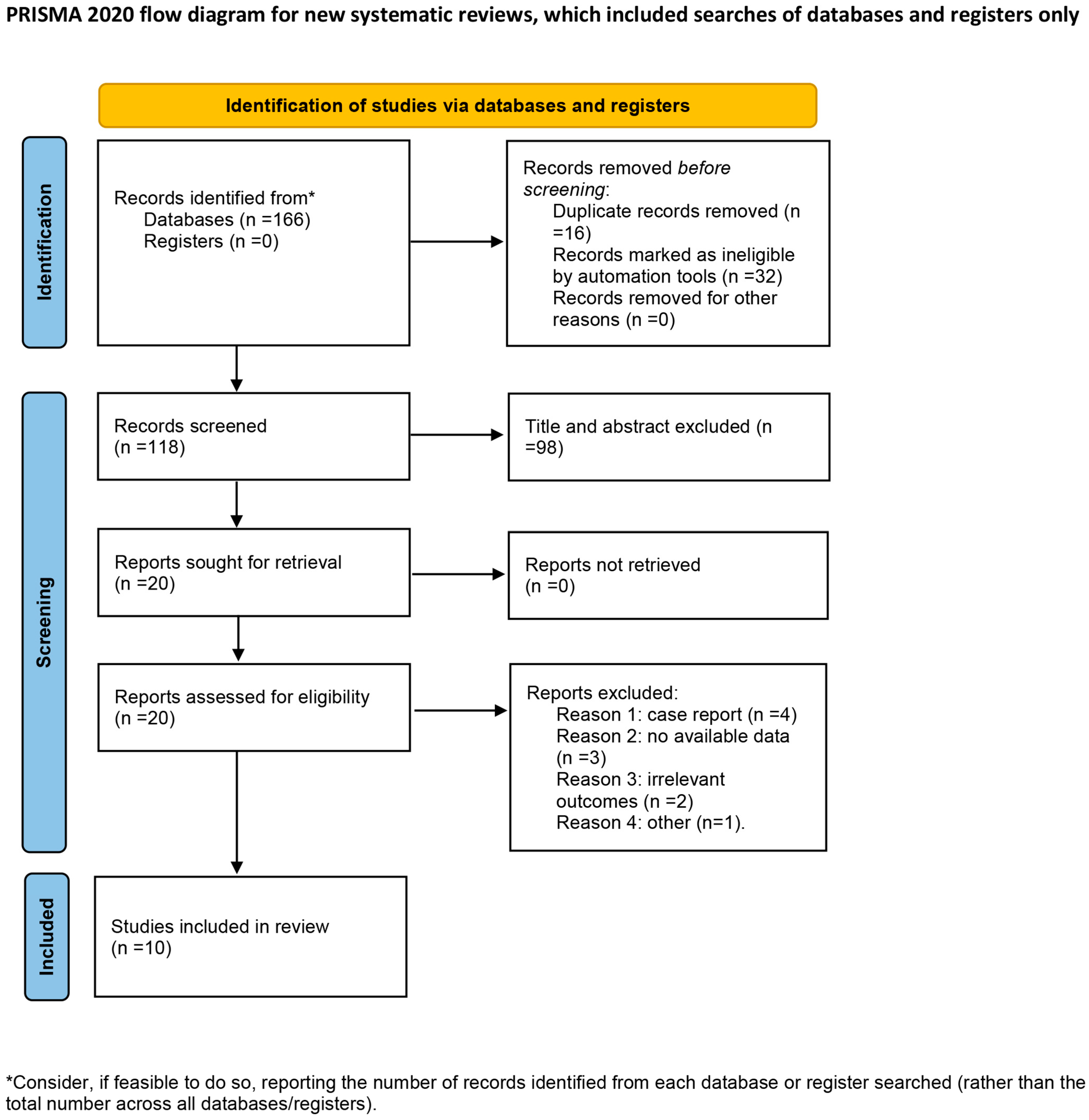
The initial literature search yielded 166 records. Following the removal of duplicates and the screening of titles and abstracts, 20 full-text articles were assessed for eligibility (Figure 1). Ten randomized controlled trials (RCTs), which enrolled a total of 386 participants, met the inclusion criteria [11,14,15,16,17,18,19,20,21,22]. The details of the included RCTs are summarized in Table 1. The studies were conducted in nine countries: the USA (n = 3), Spain (n = 2), Egypt, Australia, Iran, Ireland, and India (n = 1 each). The sample sizes ranged from 8 to 70 participants, with a mean age that spanned 51.8 to 75 years. The intervention characteristics varied across studies: the ultrasound frequency used ranged from 25 kHz to 3 MHz, the treatment duration lasted from four weeks to six months, and the session frequency of UAWD ranged from three times per week to biweekly.
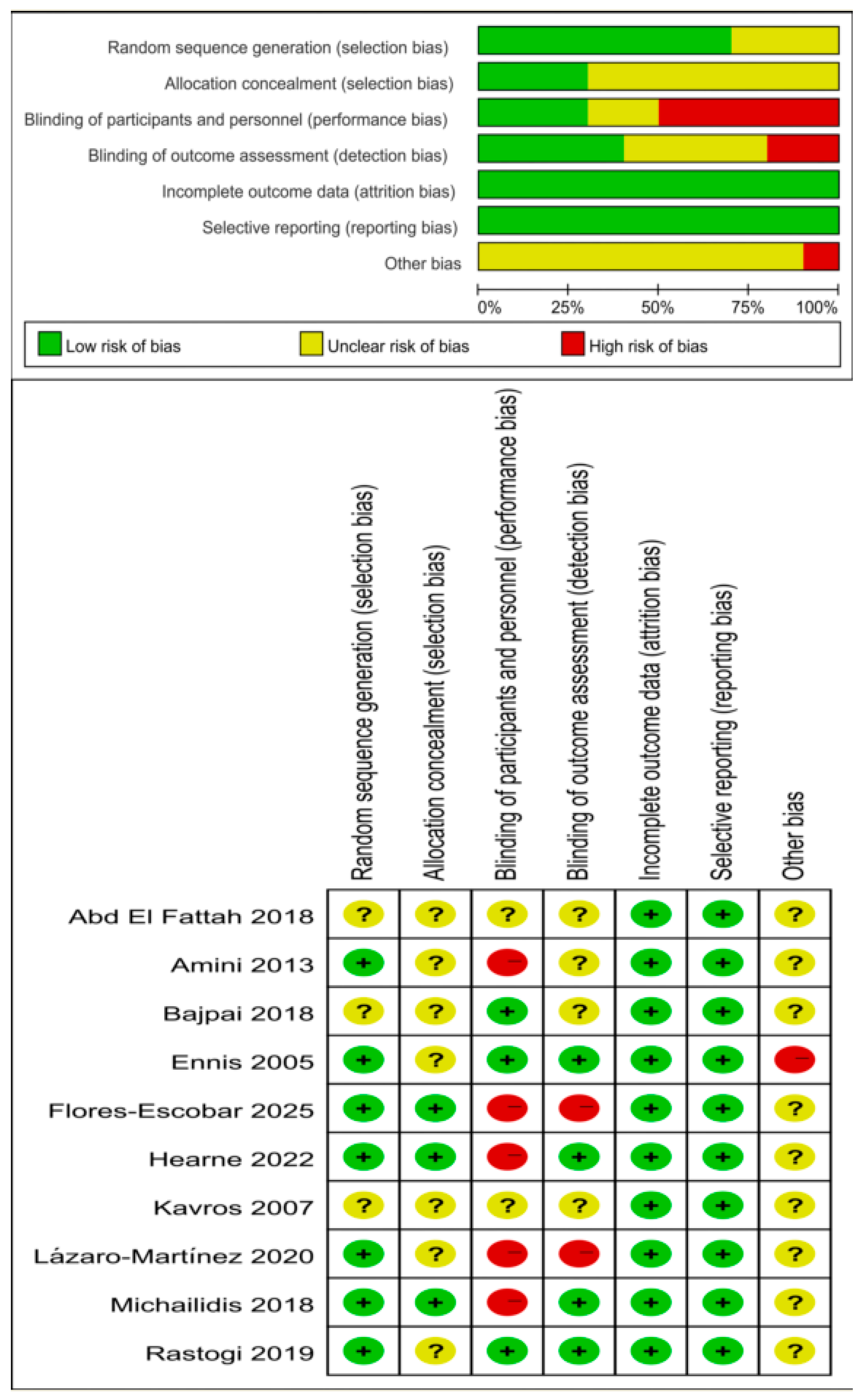
The risk of bias assessment of the ten RCTs included is summarized in Figure 2. According to the GRADE system, the overall certainty of evidence for the outcomes of time to healing and wound area reduction percentage was rated as ‘very low’, owing to methodological limitations regarding risk of bias, small sample sizes, substantial heterogeneity, and potential publication bias. In contrast, the certainty of evidence for healing rate was rate as ‘moderate’. A summary of these findings is presented in Table 2.

3.2. Meta-Analysis Findings

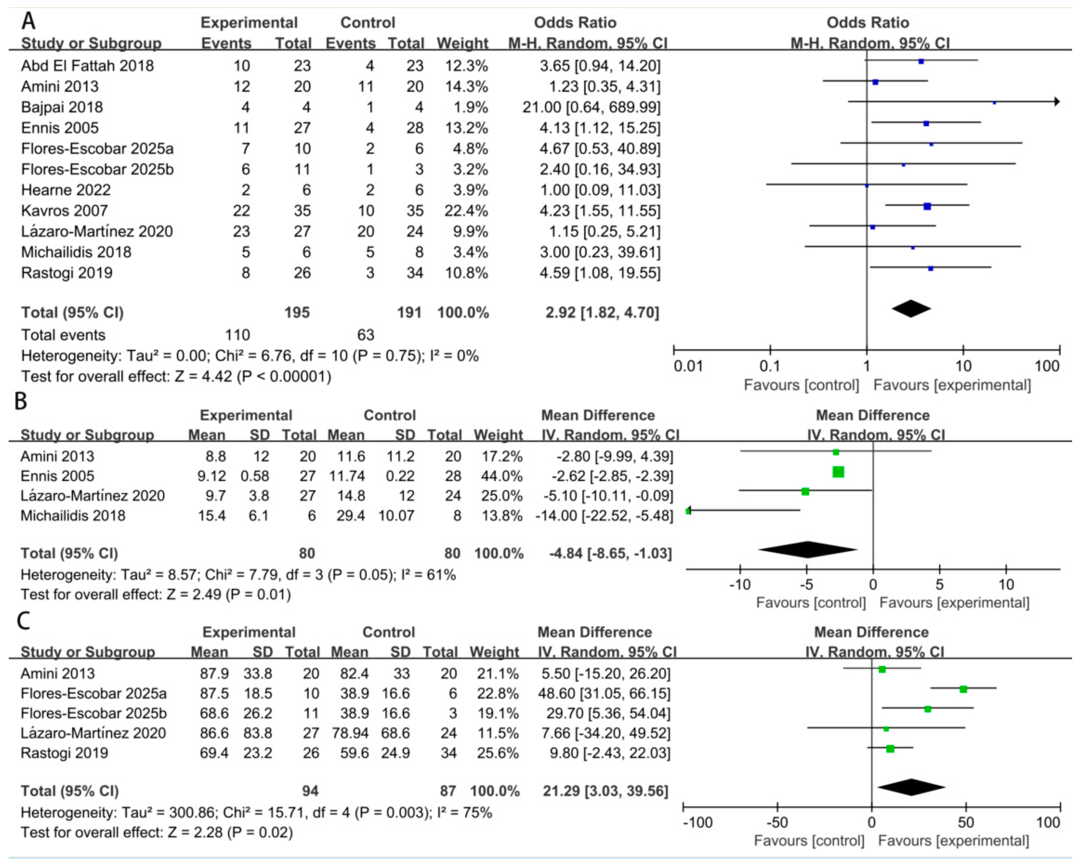
Complete wound healing: Ten studies reported on the complete healing rate. The pooled analysis demonstrated a significant higher wound healing rate when the treatment group was compared with the control group (OR: 2.92; 95% CI: 1.82 to 4.70; p = 0.75; I2 = 0%) (Figure 3A). The comparison of fixed-effect models and random-effects analyses showed no substantive differences in the direction or significance of the findings. Sensitivity analysis using risk ratios yielded consistent findings (RR: 1.67, 95% CI: 1.16 to 2.41; p = 0.01; I2 = 57%), confirming the robustness of the results (Table 3).
Time to complete healing: Four studies reported on time to healing. The pooled result indicated that healing was achieved significantly faster in the treatment group by an average of 4.84 weeks (MD: −4.84 weeks; 95% CI: −8.65 to −1.03; p = 0.05; I2 = 61%) (Figure 3B).
Wound area reduction percentage: Four studies reported quantitative data on the wound area reduction percentage. The meta-analysis showed a significantly greater reduction in wound size in the treatment group (MD: 21.29%; 95% CI: 3.03 to 39.56; p = 0.003; I2 = 75%) (Figure 3C). Given the variability in measurement time points (ranging from 4 weeks to 6 months) and baseline wound sizes across studies, we performed an additional meta-analysis using standardized mean differences (SMDs) to account for differences in measurement scales. The overall effect remained consistent with the primary analysis using raw mean differences (SMD: 0.58; 95% CI: 0.01 to 1.16; p = 0.02; I2 = 65%).

3.3. Adverse Events

Of the ten studies, six did not provide any safety data, while two only stated that ‘no serious adverse events occurred’ without further details. Ennis et al. documented pain in four cases—three in the control group and one in the ultrasound group—as well as two cases of wound infection in the ultrasound group. Michailidis et al. reported that three cases developed mild soft tissue infections.

3.4. Sensitivity Analysis

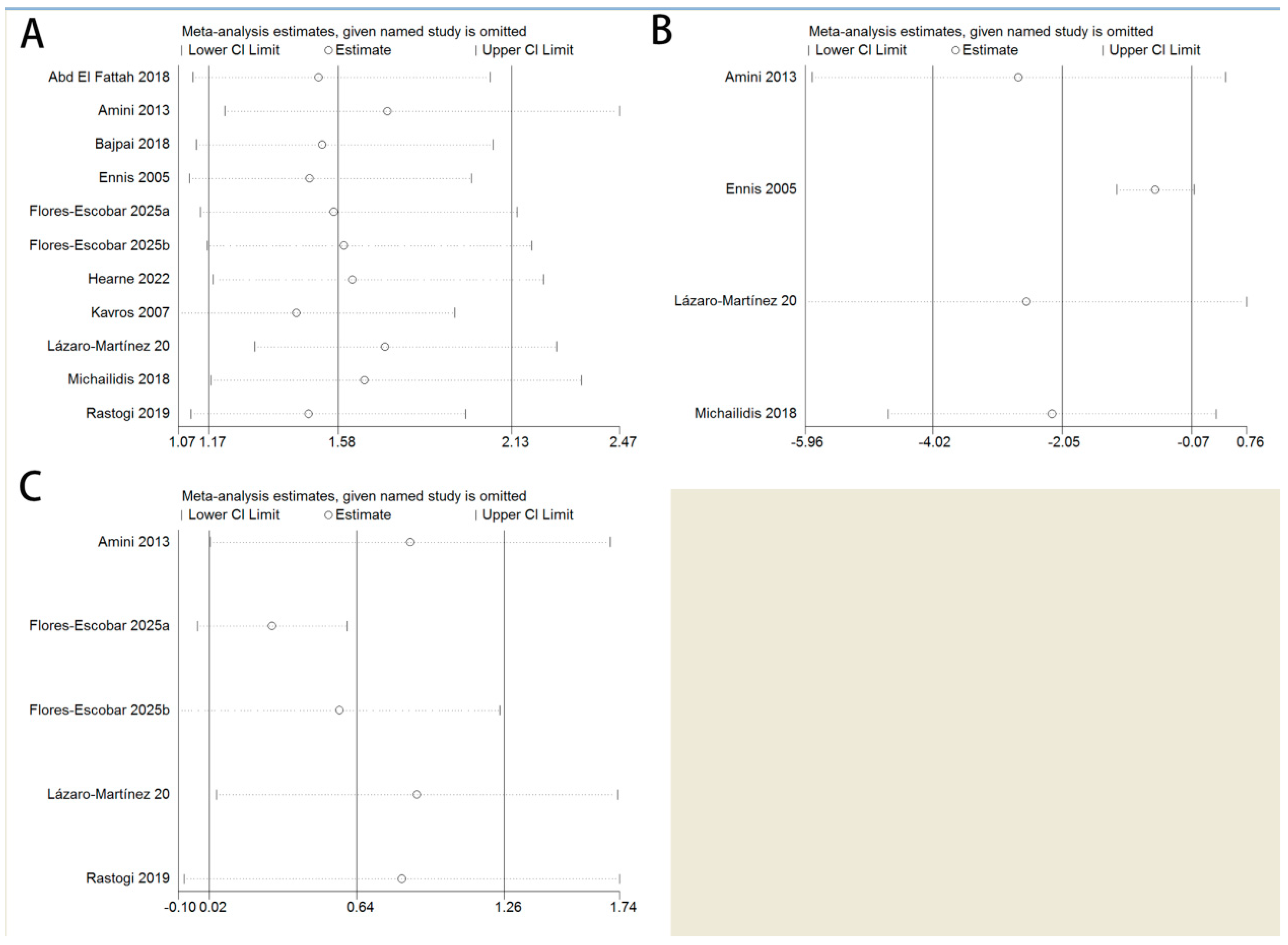
We further performed sensitivity analyses to detect whether one trial significantly influenced the outcomes or greatly contributed to the heterogeneity. The leave-one-out analysis confirmed that no single study unduly influenced the overall effect sizes (Figure 4).

3.5. Publication Bias

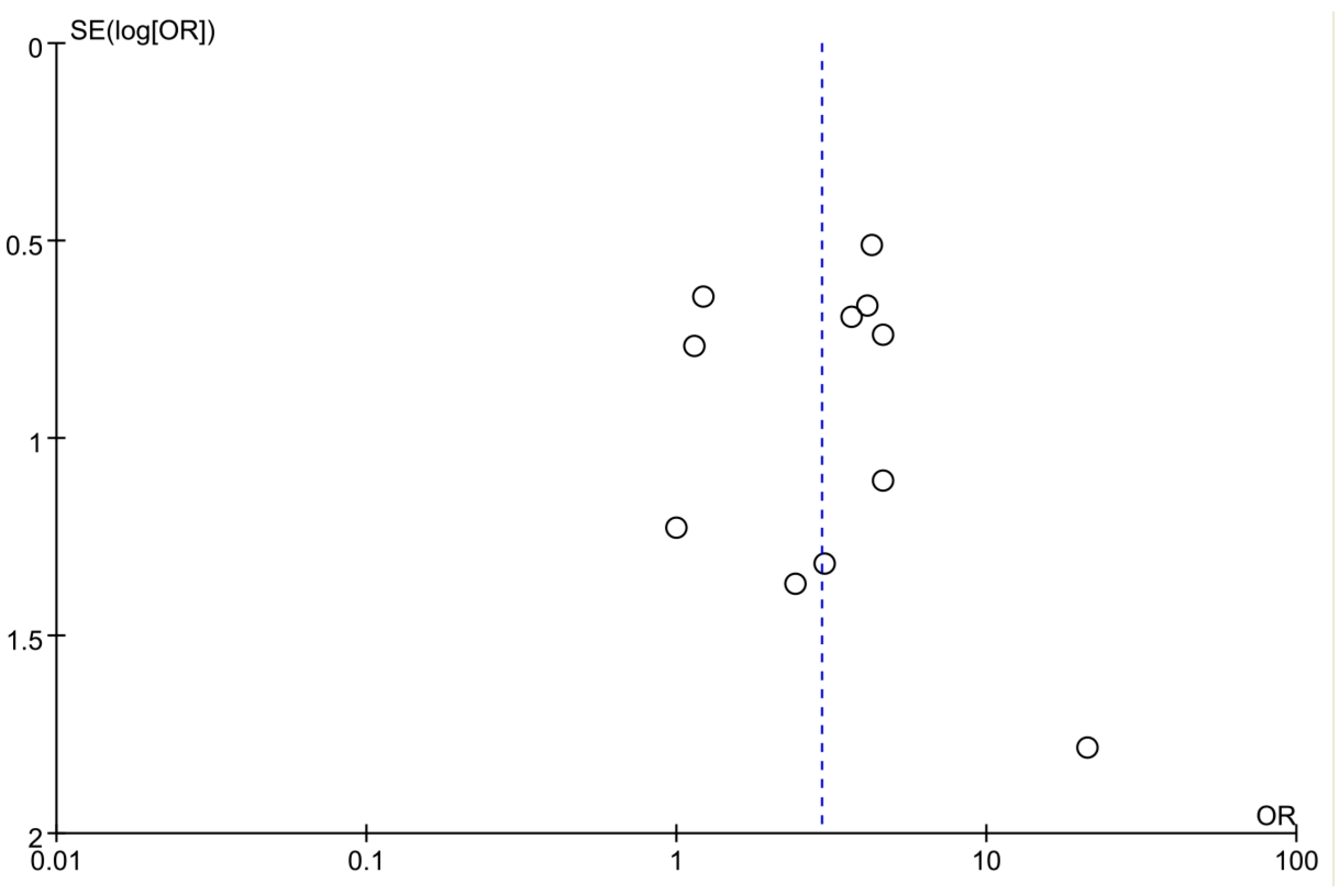
Begg’s test and Egger’s test were applied to evaluate the publication bias of healing rate in this study. The funnel plot in the meta-analysis showed no evidence of publication bias in relation to risk of ulcer healing rate (Figure 5). It was also proved by Egger’s test (p = 0.149) and Begg’s test (p = 1.00). Publication bias was not assessed for the outcomes of time to healing and wound area reduction percentage due to the insufficient number of studies available for each meta-analysis.

4. Discussion

In this study, we aimed to conduct a systematic review of the current literature to evaluate the effectiveness of UAWD in DFU management. This meta-analysis provides robust evidence that ultrasound-assisted debridement is superior to conventional debridement methods for treating DFU. The results consistently favored UAWD across all primary efficacy endpoints: a higher proportion of wounds achieved complete closure, a greater percentage reduction in wound size was observed, and healing occurred more rapidly.
The therapeutic benefits of UAWD are likely multifactorial. While its efficacy in removing biofilm and non-viable tissue is well-established, the key differentiator may be its biostimulatory effect [23]. Low-frequency ultrasound is thought to enhance healing at a cellular level by promoting fibroblast proliferation, increasing angiogenesis, and modulating inflammatory cytokines, thereby addressing the underlying pathophysiology of chronic diabetic wounds [24]. Microstreaming refers to the unidirectional flow of interstitial fluids induced by ultrasonic vibrations, which alters cell membrane permeability and modulates second messenger activity. These effects enhance protein synthesis, stimulate mast cell degranulation, and promote the production of growth factors, thereby activating fibroblasts and inducing neoangiogenesis at the wound site [25]. In addition, sharp or surgical debridement necessitates a high level of clinical proficiency, especially when underlying structures such as bone, joint tissue, or ligaments are exposed, given the inherent risk of iatrogenic tissue injury [5]. As a result, UAWD can be a useful alternative to traditional debridement techniques when sharp/surgical debridement is contraindicated, such as in individuals with bad vascular health.
A previous meta-analysis by Flores-Escobar evaluated the effect of UAWD in patients with DFU [26]. The author concluded that UAWD showed a higher healing rate, a greater percentage of wound area reduction, and similar healing times when compared with the control, although no statistically significant differences were observed between groups. Another meta-analysis analyzed the efficacy and safety of UAWD on DFU [27]. In the eleven included articles, five were written in Chinese. There might be a significant bias with low methodological quality. In this study, ten RCTs with 386 patients were included according to the inclusion criteria. The observed outcomes—specifically, an OR of 2.92 for healing rate, a mean reduction of 4.84 weeks in time to wound healing, and a 21.29% greater wound area reduction in the UAWD group—collectively underscore both the clinical relevance of UAWD and its potential to substantially improve the standard of care for patients with DFUs.
The risk of bias assessment revealed substantial methodological concerns across the included studies. Allocation concealment was unclear in seven studies (70%), while blinding of participants and personnel was rated as unclear or at high risk in seven studies (70%), and blinding of outcome assessment was rated as unclear or at high risk in six studies (60%). Inadequate allocation concealment increases the risk of selection bias, potentially leading to systematic baseline differences between groups that favor the intervention. Similarly, lack of blinding in outcome assessment—particularly relevant for wound healing outcomes where subjective judgment is involved—can inflate effect estimates.
These methodological limitations collectively explain the moderate to very low certainty of evidence according to the GRADE framework. The absence of blinding introduces a high risk of performance bias, especially for subjective outcomes such as time to healing, where treatment effects may be overestimated. Additionally, imprecision is a major concern: small sample sizes resulted in wide confidence intervals for the pooled estimates of the time to healing and wound area reduction percentage, directly leading to very low GRADE ratings for these outcomes. Given that most included studies had an unclear or high risk of bias in key domains, the observed treatment effects are likely overestimates of the true effect. Therefore, while our findings suggest that ultrasound-assisted debridement may be effective, the magnitude of benefit remains uncertain, and the true effect may be more modest than reported. Future RCTs should prioritize rigorous methodological quality, including adequate allocation concealment and blinded outcome assessment, to provide more reliable estimates of treatment efficacy.
The significant heterogeneity in treatment parameters across the included studies—such as ultrasound frequency (ranging from 25 kHz to 3 MHz) and treatment frequency (ranging from three times per week to biweekly)—precludes the recommendation of a single, optimized protocol. For example, Flores-Escobar et al. compared weekly versus biweekly UAWD in patients with DFU and found no significant difference in healing rates between the two regimens [14]. Furthermore, an expert panel recommended that UAWD may be performed sequentially, potentially at each dressing change if necessary [25]. This variability in treatment delivery represents a considerable confounding factor that may affect the outcome comparability. Another important limitation is the heterogeneity in outcome definitions across included studies. For the wound area reduction, measurement time points varied from 4 to 24 weeks. For complete healing, definitions of ‘healed’ were generally consistent (100% epithelialization), but the maximum follow-up durations varied from 6 to 24 weeks. Studies with a shorter follow-up may underestimate the proportion of patients that achieved complete healing. Translating these findings into clinical practice faces several challenges, including the need for specialized equipment and trained operators, the absence of standardized protocols due to inconsistent parameters, and the lack of economic data for cost-effectiveness evaluation. Consequently, future guidelines should aim to establish standardized treatment parameters based on robust efficacy and safety evidence.
This systematic review has several limitations. First, the analysis included only ten RCTs with a total of 386 patients. A small number of included RCTs with limited sample sizes and varying follow-up durations across these studies limit the statistical power and generalizability of the findings. Small trials are known to exaggerate treatment effects and increase the risk of publication bias. Although our sensitivity analyses suggested that no single study unduly influenced the pooled estimate, the possibility of small-study effects cannot be entirely excluded. Second, the time to complete healing was pooled using mean differences rather than hazard ratios due to insufficient reporting in the primary studies. This approach does not account for censoring and may underestimate or overestimate treatment effects. Third, significant heterogeneity was observed for the outcomes of time to healing (I2 = 61%) and wound area reduction percentage (I2 = 75%), which limits the certainty and generalizability of their clinical interpretation. Formal subgroup analyses to investigate sources of heterogeneity were not feasible due to the limited number of studies; however, a leave-one-out sensitivity analysis confirmed the directional robustness of the pooled estimates. Fourth, critical reporting gaps constrain the interpretation: inadequate reporting of patient-centered outcomes (e.g., quality of life, pain, and cost-effectiveness) precluded a comprehensive assessment of their broader clinical implications, while sparse data limit the evaluation of long-term outcomes (e.g., recurrence, amputation). Fifth, the safety profile of the intervention could not be adequately characterized due to the incomplete reporting of adverse events in the included trials, which precludes a comprehensive risk–benefit assessment. Sixth, although we conducted a comprehensive search across multiple databases, restricting the search to English-language publications may have introduced language bias, potentially excluding relevant studies published in other languages. Additionally, the variability in control interventions reflects real-world clinical practice but also limits the precision of our effect estimates. Future studies should adopt standardized protocols for control groups or clearly document all components of standard care to facilitate comparisons across trials. Finally, publication bias assessment was constrained by the limited number of studies. While a funnel plot inspection and Egger’s test did not show significant asymmetry, these methods have low power with fewer than 10 studies and should be interpreted cautiously. To address the limitations identified and build upon our findings, future research should prioritize the following: (1) large, pragmatic RCTs with adequate sample size; (2) standardized intervention protocols that clearly define and justify ultrasound parameters, enabling reproducibility and pooled analysis; and (3) adoption of a core outcome set for DFU trials, mandating consistent reporting of healing metrics, patient-reported outcomes (e.g., pain, quality of life), adverse events, and long-term endpoints like ulcer recurrence.

5. Conclusions

This meta-analysis demonstrates that UAWD is a highly effective and safe therapeutic modality that significantly improves healing outcomes for diabetic foot ulcers compared with conventional debridement alone. It could be considered as a potential adjunctive option within standardized wound care pathways. However, these findings are tempered by several important limitations. The certainty of evidence was low to very low for most outcomes, substantial heterogeneity was observed across studies, and adverse event reporting was incomplete. Without robust safety data, the true risk–benefit profile of this intervention cannot be adequately assessed, and widespread clinical adoption cannot yet be recommended. Further high-quality, adequately powered randomized controlled trials with standardized outcome reporting are needed to confirm these findings.

Supplementary Materials

The following supporting information can be downloaded from https://www.mdpi.com/article/10.3390/biomedicines14040846/s1, File S1: Search strategy.

Author Contributions

Conceptualization, J.D.; Investigation, S.M.; Methodology, S.M.; Writing—original draft, J.D. and S.M.; Writing—review & editing, J.D. and H.C. All authors have read and agreed to the published version of the manuscript.

Funding

This research received no external funding.

Institutional Review Board Statement

Not applicable.

Informed Consent Statement

Not applicable.

Data Availability Statement

The original contributions presented in this study are included in the article/Supplementary Material. Further inquiries can be directed to the corresponding authors.

Conflicts of Interest

The authors declare that they have no competing interests.

References

  1. Chen, H.; Zhou, Y.; Dai, J. Association of inflammation and nutrition-based indicators and diabetic foot ulcers: A cross-sectional study and a retrospective study. Front. Endocrinol. 2025, 16, 1654831. [Google Scholar] [CrossRef]
  2. Armstrong, D.G.; Boulton, A.J.M.; Bus, S.A. Diabetic Foot Ulcers and Their Recurrence. N. Engl. J. Med. 2017, 376, 2367–2375. [Google Scholar] [CrossRef] [PubMed]
  3. Qi, X.; Li, Y.; Xiang, Y.; Chen, Y.; Shi, Y.; Ge, X.; Zeng, B.; Shen, J. Hyperthermia-enhanced immunoregulation hydrogel for oxygenation and ROS neutralization in diabetic foot ulcers. Cell Biomater. 2025, 1, 100020. [Google Scholar] [CrossRef]
  4. Alexiadou, K.; Doupis, J. Management of diabetic foot ulcers. Diabetes Ther. 2012, 3, 4. [Google Scholar] [CrossRef]
  5. Rayman, G.; Vas, P.; Dhatariya, K.; Driver, V.; Hartemann, A.; Londahl, M.; Piaggesi, A.; Apelqvist, J.; Attinger, C.; Game, F. Guidelines on use of interventions to enhance healing of chronic foot ulcers in diabetes (IWGDF 2019 update). Diabetes Metab. Res Rev 2020, 36, e3283. [Google Scholar] [CrossRef]
  6. Hingorani, A.; LaMuraglia, G.M.; Henke, P.; Meissner, M.H.; Loretz, L.; Zinszer, K.M.; Driver, V.R.; Frykberg, R.; Carman, T.L.; Marston, W.; et al. The management of diabetic foot: A clinical practice guideline by the Society for Vascular Surgery in collaboration with the American Podiatric Medical Association and the Society for Vascular Medicine. J. Vasc. Surg. 2016, 63, 3s–21s. [Google Scholar] [CrossRef] [PubMed]
  7. Xiang, Y.; Pan, Z.; Qi, X.; Ge, X.; Xiang, J.; Xu, H.; Cai, E.; Lan, Y.; Chen, X.; Li, Y.; et al. A cuttlefish ink nanoparticle-reinforced biopolymer hydrogel with robust adhesive and immunomodulatory features for treating oral ulcers in diabetes. Bioact. Mater. 2024, 39, 562–581. [Google Scholar] [CrossRef]
  8. Yang, R.; Chen, G.; Pan, Q.Y.; Yao, Y.; Li, Y.F.; Chen, H.T.; Lei, C.J.; Liang, X. Evaluating the effectiveness of ultrasound-assisted wound debridement in managing diabetic foot ulcers: A systematic review and meta-analysis. World J. Diabetes 2025, 16, 97077. [Google Scholar] [CrossRef]
  9. Campitiello, F.; Mancone, M.; Corte, A.D.; Guerniero, R.; Canonico, S. An evaluation of an ultrasonic debridement system in patients with diabetic foot ulcers: A case series. J. Wound Care 2018, 27, 222–228. [Google Scholar] [CrossRef]
  10. Dayya, D.; O’Neill, O.J.; Huedo-Medina, T.B.; Habib, N.; Moore, J.; Iyer, K. Debridement of Diabetic Foot Ulcers. Adv. Wound Care 2022, 11, 666–686. [Google Scholar] [CrossRef] [PubMed]
  11. Hearne, C.L.J.; Patton, D.; Moore, Z.E.; Wilson, P.; Gillen, C.; O’Connor, T. Effectiveness of combined modulated ultrasound and electric current stimulation to treat diabetic foot ulcers. J. Wound Care 2022, 31, 12–20. [Google Scholar] [CrossRef]
  12. Chhabra, J.; Chopra, H.; Pahwa, R.; Raina, N.; Wadhwa, K.; Saini, S.; Negi, P.; Gupta, M.; Singh, I.; Dureja, H.; et al. Potential of nanoemulsions for accelerated wound healing: Innovative strategies. Int. J. Surg. 2023, 109, 2365–2377. [Google Scholar] [CrossRef]
  13. Guyatt, G.; Oxman, A.D.; Akl, E.A.; Kunz, R.; Vist, G.; Brozek, J.; Norris, S.; Falck-Ytter, Y.; Glasziou, P.; DeBeer, H.; et al. GRADE guidelines: 1. Introduction-GRADE evidence profiles and summary of findings tables. J. Clin. Epidemiol. 2011, 64, 383–394. [Google Scholar] [CrossRef]
  14. Flores-Escobar, S.; García-Álvarez, Y.; Álvaro-Afonso, F.J.; López-Moral, M.; García-Madrid, M.; Lázaro-Martínez, J.L. Clinical Effects of Weekly and Biweekly Low-Frequency Ultrasound Debridement Versus Standard of Wound Care in Patients with Diabetic Foot Ulcers: A Pilot Randomized Clinical Trial. Int. J. Low. Extrem. Wounds 2025, accepted. [Google Scholar] [CrossRef]
  15. Bajpai, A.; Nadkarni, S.; Neidrauer, M.; Weingarten, M.S.; Lewin, P.A.; Spiller, K.L. Effects of Non-thermal, Non-cavitational Ultrasound Exposure on Human Diabetic Ulcer Healing and Inflammatory Gene Expression in a Pilot Study. Ultrasound Med. Biol. 2018, 44, 2043–2049. [Google Scholar] [CrossRef] [PubMed]
  16. Ennis, W.J.; Foremann, P.; Mozen, N.; Massey, J.; Conner-Kerr, T.; Meneses, P. Ultrasound therapy for recalcitrant diabetic foot ulcers: Results of a randomized, double-blind, controlled, multicenter study. Ostomy Wound Manag. 2005, 51, 24–39. [Google Scholar]
  17. Kavros, S.J.; Miller, J.L.; Hanna, S.W. Treatment of ischemic wounds with noncontact, low-frequency ultrasound: The Mayo clinic experience, 2004–2006. Adv. Ski. Wound Care 2007, 20, 221–226. [Google Scholar] [CrossRef]
  18. Lázaro-Martínez, J.L.; Álvaro-Afonso, F.J.; Sevillano-Fernández, D.; García-Álvarez, Y.; Sanz-Corbalan, I.; García-Morales, E. Cellular Proliferation, Dermal Repair, and Microbiological Effectiveness of Ultrasound-Assisted Wound Debridement (UAW) Versus Standard Wound Treatment in Complicated Diabetic Foot Ulcers (DFU): An Open-Label Randomized Controlled Trial. J. Clin. Med. 2020, 9, 4032. [Google Scholar] [CrossRef]
  19. Michailidis, L.; Bergin, S.M.; Haines, T.P.; Williams, C.M. Healing rates in diabetes-related foot ulcers using low frequency ultrasonic debridement versus non-surgical sharps debridement: A randomised controlled trial. BMC Res. Notes 2018, 11, 732. [Google Scholar] [CrossRef] [PubMed]
  20. Rastogi, A.; Bhansali, A.; Ramachandran, S. Efficacy and Safety of Low-Frequency, Noncontact Airborne Ultrasound Therapy (Glybetac) For Neuropathic Diabetic Foot Ulcers: A Randomized, Double-Blind, Sham-Control Study. Int. J. Low. Extrem. Wounds 2019, 18, 81–88. [Google Scholar] [CrossRef] [PubMed]
  21. Amini, S.; ShojaeeFard, A.; Annabestani, Z.; Hammami, M.R.; Shaiganmehr, Z.; Larijani, B.; Mohseni, S.; Afshani, H.R.; Rad, M.A.; Mohajeri-Tehrani, M.R. Low-frequency ultrasound debridement in patients with diabetic foot ulcers and osteomyelitis. Wounds A Compend. Clin. Res. Pract. 2013, 25, 193–198. [Google Scholar]
  22. Fattah, A.A.E.; Shaaban, M.; Gawish, H.; Mashad, N.E.; Dawood, A.E.D. Effect of ultrasound-assisted debridement on wound healing and infection outcomes in diabetic foot. Menoufia Med. J. 2018, 31, 462. [Google Scholar]
  23. Kataoka, Y.; Kunimitsu, M.; Nakagami, G.; Koudounas, S.; Weller, C.D.; Sanada, H. Effectiveness of ultrasonic debridement on reduction of bacteria and biofilm in patients with chronic wounds: A scoping review. Int. Wound J. 2021, 18, 176–186. [Google Scholar] [CrossRef]
  24. Maan, Z.N.; Januszyk, M.; Rennert, R.C.; Duscher, D.; Rodrigues, M.; Fujiwara, T.; Ho, N.; Whitmore, A.; Hu, M.S.; Longaker, M.T.; et al. Noncontact, low-frequency ultrasound therapy enhances neovascularization and wound healing in diabetic mice. Plast. Reconstr. Surg. 2014, 134, 402e–411e. [Google Scholar] [CrossRef]
  25. Swanson, T.; Lázaro-Martínez, J.L.; Braumann, C.; Kirchhoff, J.B.; Gächter, B.; van Acker, K. Ultrasonic-assisted wound debridement: Report from a closed panel meeting. J. Wound Care 2020, 29, 128–135. [Google Scholar] [CrossRef] [PubMed]
  26. Flores-Escobar, S.; Álvaro-Afonso, F.J.; García-Álvarez, Y.; López-Moral, M.; Lázaro-Martínez, J.L.; García-Morales, E. Ultrasound-Assisted Wound (UAW) Debridement in the Treatment of Diabetic Foot Ulcer: A Systematic Review and Meta-Analysis. J. Clin. Med. 2022, 11, 1911. [Google Scholar] [CrossRef] [PubMed]
  27. Liu, E.; Hu, X.; Zhang, W.; Xiao, W.; Shen, Y.; Luo, Y.; Zheng, Z.; Zhou, P.; He, Y.; Que, H. Efficacy and safety of ultrasound-assisted wound debridement in the treatment of diabetic foot ulcers: A systematic review and meta-analysis of 11 randomized controlled trials. Front. Endocrinol. 2024, 15, 1393251. [Google Scholar] [CrossRef] [PubMed]
Figure 1. Flow diagram for study selection.
Biomedicines 14 00846 g001
Figure 2. Quality evaluation of the included studies [14,15,16,17,18,19,20,21,22].
Biomedicines 14 00846 g002
Figure 3. Meta-analysis of healing rate (A), time to complete healing (B) and wound area reduction percentage (C) [14,15,16,17,18,19,20,21,22].
Biomedicines 14 00846 g003
Figure 4. Sensitivity analysis of the meta-analysis using leave-one-out method: (A) healing rate, (B) time to complete healing, and (C) wound area reduction percentage [14,15,16,17,18,19,20,21,22].
Biomedicines 14 00846 g004
Figure 5. Publication bias analysis of the meta-analysis. Circles = individual studies. Blue dashed vertical line = pooled effect estimate.
Biomedicines 14 00846 g005
Table 1. The characteristics of the included studies.
CountryStudy DesignUlcer GradeNo.InterventionMean Age (Years)Ultrasound Frequency OutcomeAdverse EventTreatment Time
TG CG TG CG
Abd El Fattah 2018 [22]EgyptRCTNR2323SC + UAWDSCNRNR1NR12 weeks
Amini 2013 [21]IranRCTWagner Classification: 32020SC + UAWDSC55.2NR1, 2, 3NR6 months
Bajpai 2018 [15]USARCTNR44SC + UAWD (weekly)SC57.625 kHz1NR12 weeks
Ennis 2005 [16]USARCTWagner Classifications: 1, 22728SC + UAWD (weekly)SCNR40 kHz1, 2Pain: 4, Infection: 212 weeks
Flores-Escobar 2025 [14]SpainRCTTexas Classifications: 1, 2109SC + UAWD (weekly)SC70.225 kHz1, 3NR12 weeks
11 SC + UAWD (biweekly)
Hearne 2022 [11]IrelandRCTNR65SC + UAWD (twice a week)SC71.61.0–3.0 MHz 108 weeks
Kavros 2007 [17]USARCTNR3535SC + UAWD (3 times a week)SC7540 kHz1NR12 weeks
Lázaro-Martínez 2020 [18]SpainRCTTexas Classifications: 1, 22724SC + UAWD (weekly)SC61.225 kHz1, 2, 3NR6 weeks
Michailidis 2018 [19] AustraliaRCTTexas Classifications: 1, 268SC + UAWD (weekly)SCNRNR1, 2Infection: 36 weeks
Rastogi 2019 [20]IndiaRCTWagner Classifications: 2, 32634SC + UAWD (daily for the initial 6 days, followed by twice a week)SC51.826 kHz to 60 kHz1, 3NR4 weeks
TG: treatment group; CG: control group; SC: standard of care treatment; NR: not reported; 1: healing rate, 2: time to complete healing, 3: wound area reduction percentage.
Table 2. Grading of Recommendations Assessment, Development and Evaluation (GRADE) assessment of overall certainty.
OutcomesNo. of StudiesRisk of BiasInconsistencyIndirectnessImprecisionPublication BiasQuality of Evidence
Healing rate10SeriousNot seriousNot seriousNot seriousNot serious⨁⨁⨁◯
Moderate
Time to healing4SeriousSeriousNot seriousSeriousSerious⨁◯◯◯
Very Low
Wound area reduction percentage4SeriousSeriousNot seriousSeriousSerious⨁◯◯◯
Very Low
The GRADE system rates evidence as Moderate (⨁⨁⨁◯), or Very low (⨁◯◯◯), where each ⨁ represents one point and ◯ represents zero points.
Table 3. Sensitive analysis for outcome of healing rate.
ORRR
Effect Size95% CIpI2Effect Size95% CIpI2
Fixed-effect models2.941.85–4.680.7501.641.32–2.040.0157%
Random-effects models2.921.82–4.700.7501.671.16–2.410.0157%
Disclaimer/Publisher’s Note: The statements, opinions and data contained in all publications are solely those of the individual author(s) and contributor(s) and not of MDPI and/or the editor(s). MDPI and/or the editor(s) disclaim responsibility for any injury to people or property resulting from any ideas, methods, instructions or products referred to in the content.