Management Strategies for Congestive Heart Failure in Chronic Kidney Disease: Narrative Review
Abstract
1. Introduction
- Reduced Ejection Fraction (HFrEF) is defined by a left ventricular ejection fraction (LVEF) of 40% or less.
- Mildly reduced or improved Ejection Fraction (HFmrEF or HFimpEF) is characterized by a LVEF between 41% and 49%.
- Preserved Ejection Fraction (HFpEF) is defined by a LVEF of 50% or greater.
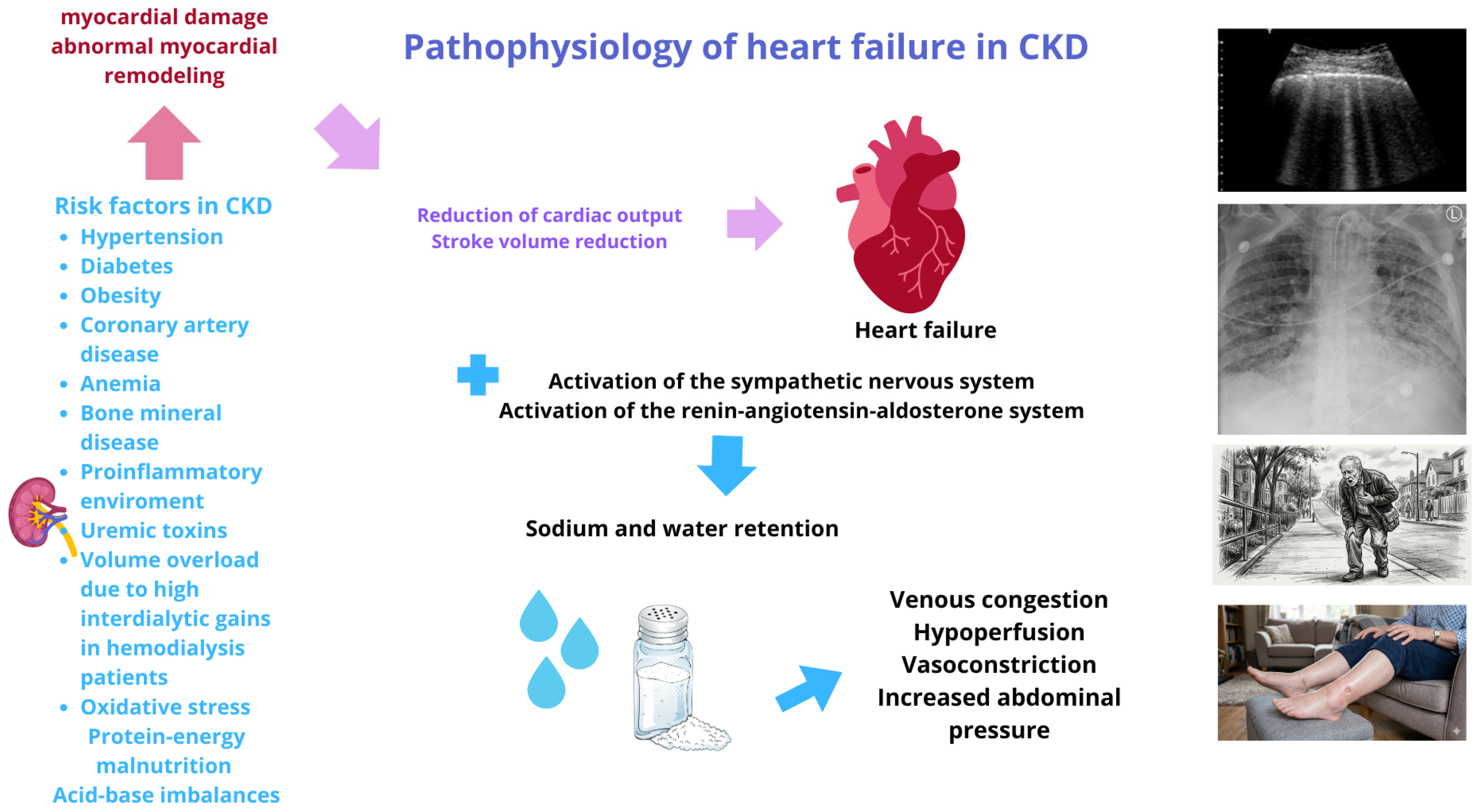
2. Pathophysiology of Chronic Kidney Disease Associated with Heart Failure
3. Diagnosis of Heart Failure in Chronic Kidney Disease
4. Diagnosis and Biomarkers of Heart Failure in CKD
4.1. Framingham Clinical Diagnostic Criteria for Heart Failure
4.2. Troponins
4.3. Galectin
4.4. ST2
4.5. GDF-15
4.6. CA 125
4.7. Imaging
5. Heart Failure Treatment
6. Treatment of Heart Failure in Chronic Kidney Disease
6.1. Beta-Blockers
6.2. Angiotensin Receptor-Neprilysin Inhibitors (ARNI)
6.3. ACE and ARB Inhibitors
6.4. Steroidal and Non-Steroidal Mineralocorticoid Receptor Antagonists
6.5. Glucagon-like Peptide-1 (GLP-1) Agonist and Cardiovascular Disease
6.6. Other Medications
7. Strategies for Monitoring Fluid Overload in Patients with Chronic Kidney Disease on Renal Replacement Therapy
7.1. Bioimpedance
7.2. Ultrasound
8. Future Perspectives
9. Conclusions
Author Contributions
Funding
Institutional Review Board Statement
Informed Consent Statement
Data Availability Statement
Conflicts of Interest
References
- Savarese, G.; Becher, P.M.; Lund, L.H.; Seferovic, P.; Rosano, G.M.C.; Coats, A.J.S. Global burden of heart failure: A comprehensive and updated review of epidemiology. Cardiovasc. Res. 2023, 118, 3272–3287, Erratum in Cardiovasc. Res. 2023, 119, 1453. https://doi.org/10.1093/cvr/cvac013. [Google Scholar] [CrossRef] [Scilit]
- Arrigo, M.; Jessup, M.; Mullens, W.; Reza, N.; Shah, A.M.; Sliwa, K.; Mebazaa, A. Acute heart failure. Nat. Rev. Dis. Primers 2020, 6, 16. [Google Scholar] [CrossRef] [Scilit] [PubMed]
- Murphy, S.P.; Ibrahim, N.E.; Januzzi, J.L., Jr. Heart Failure With Reduced Ejection Fraction: A Review. JAMA 2020, 324, 488–504, Erratum in JAMA 2020, 324, 2107. https://doi.org/10.1001/jama.2020.10262. [Google Scholar] [CrossRef] [Scilit]
- Ponikowski, P.; Voors, A.A.; Anker, S.D.; Bueno, H.; Cleland, J.G.F.; Coats, A.J.S.; Falk, V.; González-Juanatey, J.R.; Harjola, V.-P.; Jankowska, E.A.; et al. 2016 ESC Guidelines for the diagnosis and treatment of acute and chronic heart failure: The Task Force for the diagnosis and treatment of acute and chronic heart failure of the European Society of Cardiology (ESC)Developed with the special contribution of the Heart Failure Association (HFA) of the ESC. Eur. Heart J. 2016, 37, 2129–2200, Erratum in Eur. Heart J. 2018, 39, 860. [Google Scholar] [CrossRef] [Scilit]
- Yancy, C.W.; Jessup, M.; Bozkurt, B.; Butler, J.; Casey, D.E., Jr.; Drazner, M.H.; Fonarow, G.C.; Geraci, S.A.; Horwich, T.; Januzzi, J.L.; et al. 2013 ACCF/AHAguideline for the management of heart failure: A report of the American College of Cardiology Foundation/American Heart Association Task Force on Practice Guidelines. J. Am. Coll. Cardiol. 2013, 62, e147–e239. [Google Scholar] [CrossRef] [Scilit]
- Bozkurt, B.; Coats, A.J.S.; Tsutsui, H.; Abdelhamid, C.M.; Adamopoulos, S.; Albert, N.; Anker, S.D.; Atherton, J.; Böhm, M.; Butler, J.; et al. Universal definition and classification of heart failure: A report of the Heart Failure Society of America, Heart Failure Association of the European Society of Cardiology, Japanese Heart Failure Society and Writing Committee of the Universal Definition of Heart Failure: Endorsed by the Canadian Heart Failure Society, Heart Failure Association of India, Cardiac Society of Australia and New Zealand, and Chinese Heart Failure Association. Eur. J. Heart Fail. 2021, 23, 352–380. [Google Scholar] [CrossRef] [Scilit]
- Li, Y.; Shi, Y.; Zhu, B.; Chen, Y.; Shen, B.; Zhao, S.; Song, N.; Fang, Y.; Ding, X. Association of chronic kidney disease and cardiovascular disease risk with all-cause mortality: An interaction, joint and mediation analysis in Chinese adults. BMC Public Health 2025, 25, 1685. [Google Scholar] [CrossRef] [Scilit] [PubMed]
- Damman, K.; Valente, M.A.; Voors, A.A.; O’Connor, C.M.; van Veldhuisen, D.J.; Hillege, H.L. Renal impairment, worsening renal function, and outcome in patients with heart failure: An updated meta-analysis. Eur. Heart J. 2014, 35, 455–469. [Google Scholar] [CrossRef] [Scilit]
- Kottgen, A.; Russell, S.D.; Loehr, L.R.; Crainiceanu, C.M.; Rosamond, W.D.; Chang, P.P.; Chambless, L.E.; Coresh, J. Reduced kidney function as a risk factor for incident heart failure: The atherosclerosis risk in communities (ARIC) study. J. Am. Soc. Nephrol. 2007, 18, 1307–1315. [Google Scholar] [CrossRef] [Scilit]
- Amsalem, Y.; Garty, M.; Schwartz, R.; Sandach, A.; Behar, S.; Caspi, A.; Gottlieb, S.; Ezra, D.; Lewis, B.S.; Leor, J. Prevalence and significance of unrecognized renal insufficiency in patients with heart failure. Eur. Heart J. 2008, 29, 1029–1036. [Google Scholar] [CrossRef] [Scilit] [PubMed]
- Chen, T.H.; Wo, H.T.; Chang, P.C.; Wang, C.C.; Wen, M.S.; Chou, C.C. A meta-analysis of mortality in end-stage renal disease patients receiving implantable cardioverter defibrillators (ICDs). PLoS ONE 2014, 9, e99418. [Google Scholar] [CrossRef] [Scilit]
- McCallum, W.; Sarnak, M.J. Cardiorenal Syndrome in the Hospital. Clin. J. Am. Soc. Nephrol. 2023, 18, 933–945. [Google Scholar] [CrossRef] [Scilit]
- London, G.M. Left ventricular alterations and end-stage renal disease. Nephrol. Dial. Transplant. 2002, 17, 29–36. [Google Scholar] [CrossRef] [Scilit]
- Schunk, S.J.; Zimmermann, P. Cardiovascular Risk and Its Presentation in Chronic Kidney Disease. J. Clin. Med. 2025, 14, 4567. [Google Scholar] [CrossRef] [Scilit]
- Nardi, E.; Mulè, G.; Giammanco, A.; Mattina, A.; Geraci, G.; Nardi, C.; Averna, M. Left ventricular hypertrophy in chronic kidney disease: A diagnostic criteria comparison. Nutr. Metab. Cardiovasc. Dis. 2021, 31, 137–144. [Google Scholar] [CrossRef] [Scilit]
- Maqbool, S.; Shafiq, S.; Ali, S.; Rehman, M.E.U.; Malik, J.; Lee, K.Y. Left Ventricular Hypertrophy (LVH) and Left Ventricular Geometric Patterns in Patients with Chronic Kidney Disease (CKD) Stage 2–5 With Preserved Ejection Fraction (EF): A Systematic Review to Explore CKD Stage-wise LVH Patterns. Curr. Probl. Cardiol. 2023, 48, 101590. [Google Scholar] [CrossRef] [Scilit]
- Foley, R.N.; Curtis, B.M.; Randell, E.W.; Parfrey, P.S. Left ventricular hypertrophy in new hemodialysis patients without symptomatic cardiac disease. Clin. J. Am. Soc. Nephrol. 2010, 5, 805–813. [Google Scholar] [CrossRef] [Scilit] [PubMed]
- Wang, A.Y.; Fang, F.; Chan, J.; Wen, Y.Y.; Qing, S.; Chan, I.H.; Lo, G.; Lai, K.N.; Lo, W.K.; Lam, C.W.; et al. Effect of paricalcitol on left ventricular mass and function in CKD—The OPERA trial. J. Am. Soc. Nephrol. 2014, 25, 175–186. [Google Scholar] [CrossRef] [Scilit] [PubMed]
- Moe, S.M.; Thadhani, R. What have we learned about chronic kidney disease-mineral bone disorder from the EVOLVE and PRIMO trials? Curr. Opin. Nephrol. Hypertens. 2013, 22, 651–655. [Google Scholar] [CrossRef] [Scilit]
- Aoki, J.; Ikari, Y.; Nakajima, H.; Mori, M.; Sugimoto, T.; Hatori, M.; Tanimoto, S.; Amiya, E.; Hara, K. Clinical and pathologic characteristics of dilated cardiomyopathy in hemodialysis patients. Kidney Int. 2005, 67, 333–340. [Google Scholar] [CrossRef] [Scilit] [PubMed]
- Narayanan, G.; Halim, A.; Hu, A.; Avin, K.G.; Lu, T.; Zehnder, D.; Hato, T.; Chen, N.X.; Moe, S.M.; Lim, K. Molecular Phenotyping and Mechanisms of Myocardial Fibrosis in Advanced Chronic Kidney Disease. Kidney 2023, 4, 1562–1579. [Google Scholar] [CrossRef] [Scilit]
- Romero-González, G.; González, A.; López, B.; Ravassa, S.; Díez, J. Heart failure in chronic kidney disease: The emerging role of myocardial fibrosis. Nephrol. Dial. Transplant. 2022, 37, 817–824. [Google Scholar] [CrossRef] [Scilit]
- Echefu, G.; Stowe, I.; Burka, S.; Basu-Ray, I.; Kumbala, D. Pathophysiological concepts and screening of cardiovascular disease in dialysis patients. Front. Nephrol. 2023, 3, 1198560. [Google Scholar] [CrossRef] [Scilit] [PubMed]
- Saleh, M.A.; El Kilany, W.M.; Keddis, V.W.; El Said, T.W. Effect of high flow arteriovenous fistula on cardiac function in hemodialysis patients. Egypt. Heart J. 2018, 70, 337–341. [Google Scholar] [CrossRef] [Scilit] [PubMed]
- Hutcheson, J.D.; Goettsch, C. Cardiovascular Calcification Heterogeneity in Chronic Kidney Disease. Circ. Res. 2023, 132, 993–1012. [Google Scholar] [CrossRef] [Scilit] [PubMed]
- Ksiazek, S.H.; Hu, L.; Andò, S.; Pirklbauer, M.; Säemann, M.D.; Ruotolo, C.; Zaza, G.; La Manna, G.; De Nicola, L.; Mayer, G.; et al. Renin-Angiotensin-Aldosterone System: From History to Practice of a Secular Topic. Int. J. Mol. Sci. 2024, 25, 4035. [Google Scholar] [CrossRef] [Scilit]
- Shimada, T.; Kakitani, M.; Yamazaki, Y.; Hasegawa, H.; Takeuchi, Y.; Fujita, T.; Fukumoto, S.; Tomizuka, K.; Yamashita, T. Targeted ablation of Fgf23 demonstrates an essential physiological role of FGF23 in phosphate and vitamin D metabolism. J. Clin. Investig. 2004, 113, 561–568. [Google Scholar] [CrossRef]
- Ho, B.B.; Bergwitz, C. FGF23 signalling and physiology. J. Mol. Endocrinol. 2021, 66, R23–R32. [Google Scholar] [CrossRef] [Scilit]
- Vogt, J.; Föller, M. Regulation of αKlotho. Cell. Physiol. Biochem. 2025, 59, 511–524. [Google Scholar] [CrossRef] [Scilit] [PubMed]
- Silva, A.P.; Mendes, F.; Carias, E.; Gonçalves, R.B.; Fragoso, A.; Dias, C.; Tavares, N.; Café, H.M.; Santos, N.; Rato, F.; et al. Plasmatic Klotho and FGF23 Levels as Biomarkers of CKD-Associated Cardiac Disease in Type 2 Diabetic Patients. Int. J. Mol. Sci. 2019, 20, 1536. [Google Scholar] [CrossRef] [Scilit] [PubMed]
- Leidner, A.S.; Cai, X.; Zelnick, L.R.; Lee, J.; Bansal, N.; Pasch, A.; Kansal, M.; Chen, J.; Anderson, A.H.; Sondheimer, J.H.; et al. Fibroblast Growth Factor 23 and Risk of Heart Failure Subtype: The CRIC (Chronic Renal Insufficiency Cohort) Study. Kidney Med. 2023, 5, 100723. [Google Scholar] [CrossRef] [Scilit] [PubMed]
- Fuchs, M.A.A.; Burke, E.J.; Latic, N.; Murray, S.L.; Li, H.; Sparks, M.A.; Abraham, D.; Zhang, H.; Rosenberg, P.; Saleem, U.; et al. Fibroblast growth factor 23 and fibroblast growth factor receptor 4 promote cardiac metabolic remodeling in chronic kidney disease. Kidney Int. 2025, 107, 852–868. [Google Scholar] [CrossRef] [Scilit]
- Chawla, L.S.; Herzog, C.A.; Costanzo, M.R.; Tumlin, J.; Kellum, J.A.; McCullough, P.A.; Ronco, C.; ADQI XI Workgroup. Proposal for a functional classification system of heart failure in patients with end-stage renal disease: Proceedings of the acute dialysis quality initiative (ADQI) XIworkgroup. J. Am. Coll. Cardiol. 2014, 63, 1246–1252. [Google Scholar] [CrossRef] [Scilit] [PubMed]
- Shams, P.; Malik, A.; Chhabra, L. Heart Failure (Congestive Heart Failure). In StatPearls; StatPearls Publishing: Treasure Island, FL, USA, 2025. [Google Scholar]
- King, M.; Kingery, J.; Casey, B. Diagnosis and evaluation of heart failure. Am. Fam. Physician 2012, 85, 1161–1168. [Google Scholar] [PubMed]
- Goetze, J.P.; Bruneau, B.G.; Ramos, H.R.; Ogawa, T.; de Bold, M.K.; de Bold, A.J. Cardiac natriuretic peptides. Nat. Rev. Cardiol. 2020, 17, 698–717. [Google Scholar] [CrossRef] [Scilit]
- Cheng, Y.J.; Yao, F.J.; Liu, L.J.; Tang, K.; Lin, X.X.; Li, W.J.; Zhang, J.; Wu, S.H. B-type natriuretic peptide and prognosis of end-stage renal disease: A meta-analysis. PLoS ONE 2013, 8, e79302. [Google Scholar] [CrossRef] [Scilit]
- Paniagua, R.; Ventura, M.D.; Avila-Díaz, M.; Hinojosa-Heredia, H.; Méndez-Durán, A.; Cueto-Manzano, A.; Cisneros, A.; Ramos, A.; Madonia-Juseino, C.; Belio-Caro, F.; et al. NT-proBNP, fluid volume overload and dialysis modality are independent predictors of mortality in ESRD patients. Nephrol. Dial. Transplant. 2010, 25, 551–557. [Google Scholar] [CrossRef] [Scilit]
- Neuen, B.L.; Vaduganathan, M.; Claggett, B.L.; Beldhuis, I.; Myhre, P.; Desai, A.S.; Skali, H.; Mc Causland, F.R.; McGrath, M.; Anand, I.; et al. Natriuretic Peptides, Kidney Function, and Clinical Outcomes in Heart Failure with Preserved Ejection Fraction. JACC Heart Fail. 2025, 13, 28–39. [Google Scholar] [CrossRef] [Scilit]
- Masson, S.; Latini, R.; Anand, I.S.; Vago, T.; Angelici, L.; Barlera, S.; Missov, E.D.; Clerico, A.; Tognoni, G.; Cohn, J.N.; et al. Direct comparison of B-type natriuretic peptide (BNP) and amino-terminal proBNP in a large population of patients with chronic and symptomatic heart failure: The Valsartan Heart Failure (Val-HeFT) data. Clin. Chem. 2006, 52, 1528–1538. [Google Scholar] [CrossRef] [Scilit]
- McCullough, P.A.; Kuncheria, J.; Mathur, V.S. Diagnostic and therapeutic utility of B-type natriuretic peptide in patients with renal insufficiency and decompensated heart failure. Rev. Cardiovasc. Med. 2004, 5, 16–25. [Google Scholar]
- Horii, M.; Matsumoto, T.; Uemura, S.; Sugawara, Y.; Takitsume, A.; Ueda, T.; Nakagawa, H.; Nishida, T.; Soeda, T.; Okayama, S.; et al. Prognostic value of B-type natriuretic peptide and its amino-terminal proBNP fragment for cardiovascular events with stratification by renal function. J. Cardiol. 2013, 61, 410–416. [Google Scholar] [CrossRef] [Scilit]
- Fujii, H.; Goto, S. Comparative analysis of plasma BNP and NT-proBNP levels, and NT-proBNP/BNP ratio in patients with chronic kidney disease. Hypertens. Res. 2025, 48, 2303–2314. [Google Scholar] [CrossRef] [Scilit] [PubMed]
- Dubin, R.F.; Li, Y.; He, J.; Jaar, B.G.; Kallem, R.; Lash, J.P.; Makos, G.; Rosas, S.E.; Soliman, E.Z.; Townsend, R.R.; et al. Predictors of high sensitivity cardiac troponin T in chronic kidney disease patients: A cross-sectional study in the chronic renal insufficiency cohort (CRIC). BMC Nephrol. 2013, 14, 229. [Google Scholar] [CrossRef] [Scilit] [PubMed]
- Gregg, L.P.; Adams-Huet, B.; Li, X.; Colbert, G.; Jain, N.; de Lemos, J.A.; Hedayati, S.S. Effect Modification of Chronic Kidney Disease on the Association of Circulating and Imaging Cardiac Biomarkers with Outcomes. J. Am. Heart Assoc. 2017, 6, e005235. [Google Scholar] [CrossRef] [Scilit]
- Rehm, M.; Rothenbacher, D.; Iacoviello, L.; Costanzo, S.; Tunstall-Pedoe, H.; Fitton, C.A.; Söderberg, S.; Hultdin, J.; Salomaa, V.; Jousilahti, P.; et al. Chronic kidney disease and risk of atrial fibrillation and heart failure in general population-based cohorts: The BiomarCaRE project. ESC Heart Fail. 2022, 9, 57–65. [Google Scholar] [CrossRef] [Scilit]
- Geladari, E.V.; Vallianou, N.G.; Evangelopoulos, A.; Koufopoulos, P.; Panagopoulos, F.; Margellou, E.; Dalamaga, M.; Sevastianos, V.; Geladari, C.V. Cardiac Troponin Levels in Patients with Chronic Kidney Disease: “Markers of High Risk or Just Noise’’? Diagnostics 2024, 14, 2316. [Google Scholar] [CrossRef] [Scilit] [PubMed]
- Wu, C.; Lv, Z.; Li, X.; Zhou, X.; Mao, W.; Zhu, M. Galectin-3 in Predicting Mortality of Heart Failure: A Systematic Review and Meta-Analysis. Heart Surg. Forum 2021, 24, E327–E332. [Google Scholar] [CrossRef] [Scilit]
- Shi, Y.; Dong, G.; Liu, J.; Shuang, X.; Liu, C.; Yang, C.; Qing, W.; Qiao, W. Clinical Implications of Plasma Galectin-3 in Heart Failure With Preserved Ejection Fraction: A Meta-Analysis. Front. Cardiovasc. Med. 2022, 9, 854501. [Google Scholar] [CrossRef] [Scilit]
- Baccouche, B.M.; Mahmoud, M.A.; Nief, C.; Patel, K.; Natterson-Horowitz, B. Galectin-3 is Associated with Heart Failure Incidence: A Meta-Analysis. Curr. Cardiol. Rev. 2023, 19, e171122211004. [Google Scholar] [CrossRef] [Scilit]
- Min, S.H.; Kim, I.; An, J.N.; Lee, H.S.; Kim, S.G.; Kim, J.K. Galectin-3 as a prognostic biomarker in haemodialysis patients with preserved or mildly reduced ejection fraction. Clin. Kidney J. 2025, 18, sfaf306. [Google Scholar] [CrossRef] [Scilit]
- Heleniak, Z.; Bohdan, M.; Gruchała, M.; Dębska-Ślizień, A. Heart failure biomarkers in hemodialysis patients. Cardiol. J. 2024, 31, 628–636. [Google Scholar] [CrossRef] [Scilit]
- Najjar, E.; Faxén, U.L.; Hage, C.; Donal, E.; Daubert, J.C.; Linde, C.; Lund, L.H. ST2 in heart failure with preserved and reduced ejection fraction. Scand. Cardiovasc. J. 2019, 53, 21–27. [Google Scholar] [CrossRef] [Scilit]
- Obokata, M.; Sunaga, H.; Ishida, H.; Ito, K.; Ogawa, T.; Ando, Y.; Kurabayashi, M.; Negishi, K. Independent and incremental prognostic value of novel cardiac biomarkers in chronic hemodialysis patients. Am. Heart J. 2016, 179, 29–41. [Google Scholar] [CrossRef] [Scilit] [PubMed]
- Zhang, Z.; Shen, B.; Cao, X.; Liu, Z.; Chen, X.; Nie, Y.; Yu, J.; Zou, J.; Ding, X. Increased Soluble Suppression of Tumorigenicity 2 Level Predicts All-Cause and Cardiovascular Mortality in Maintenance Hemodialysis Patients: A Prospective Cohort Study. Blood Purif. 2017, 43, 37–45. [Google Scholar] [CrossRef] [Scilit]
- Rochette, L.; Zeller, M.; Cottin, Y.; Vergely, C. Insights Into Mechanisms of GDF15 and Receptor GFRAL: Therapeutic Targets. Trends Endocrinol. Metab. 2020, 31, 939–951. [Google Scholar] [CrossRef] [Scilit]
- Benes, J.; Kotrc, M.; Wohlfahrt, P.; Conrad, M.J.; Franekova, J.; Jabor, A.; Lupinek, P.; Kautzner, J.; Melenovsky, V.; Jarolim, P. The Role of GDF-15 in Heart Failure Patients With Chronic Kidney Disease. Can. J. Cardiol. 2019, 35, 462–470. [Google Scholar] [CrossRef] [Scilit]
- Nikitiuk, B.E.; Rydzewska-Rosołowska, A.; Kakareko, K.; Głowińska, I.; Hryszko, T. On Whether Ca-125 Is the Answer for Diagnosing Overhydration, Particularly in End-Stage Kidney Disease Patients-A Systematic Review. Int. J. Mol. Sci. 2024, 25, 2192. [Google Scholar] [CrossRef] [Scilit]
- Yilmaz, H.; Gürel, O.M.; Celik, H.T.; Sahiner, E.; Yildirim, M.E.; Bilgiç, M.A.; Bavbek, N.; Akcay, A. CA 125 levels and left ventricular function in patients with end-stage renal disease on maintenance hemodialysis. Ren. Fail. 2014, 36, 210–216. [Google Scholar] [CrossRef] [Scilit]
- Nishikimi, T.; Nakagawa, Y. Potential pitfalls when interpreting plasma BNP levels in heart failure practice. J. Cardiol. 2021, 78, 269–274. [Google Scholar] [CrossRef] [Scilit] [PubMed]
- Januzzi, J.L., Jr.; McCarthy, C.P. Cardiac Troponin and the True False Positive. JACC Case Rep. 2020, 2, 461–463. [Google Scholar] [CrossRef] [Scilit] [PubMed]
- Netala, V.R.; Hou, T.; Wang, Y.; Zhang, Z.; Teertam, S.K. Cardiovascular Biomarkers: Tools for Precision Diagnosis and Prognosis. Int. J. Mol. Sci. 2025, 26, 3218. [Google Scholar] [CrossRef] [Scilit]
- Syn, G.; Lee, Y.Q.; Lim, Z.Y.; Chan, G.C. Galectin-3: Action and clinical utility in chronic kidney disease. Int. Urol. Nephrol. 2024, 56, 3535–3543. [Google Scholar] [CrossRef] [Scilit]
- Chou, Y.-L.; Chen, H.-L.; Hsu, B.-G.; Yang, C.-Y.; Chen, C.-H.; Lee, Y.-C.; Tsai, I.-L.; Sung, C.-C.; Wu, C.-C.; Yang, S.-R.; et al. Galectin-3 contributes to pathogenesis of IgA nephropathy. Kidney Int. 2024, 106, 658–670. [Google Scholar] [CrossRef] [Scilit] [PubMed]
- Sabapathy, V.; Price, A.; Cheru, N.T.; Venkatadri, R.; Dogan, M.; Costlow, G.; Mohammad, S.; Sharma, R. ST2 + T-Regulatory Cells in Renal Inflammation and Fibrosis after Ischemic Kidney Injury. J. Am. Soc. Nephrol. 2025, 36, 73–86. [Google Scholar] [CrossRef] [Scilit]
- Matsuzaka, Y.; Iyoda, M. The IL-33/ST2/ILC2 pathway in kidney disease: Balancing inflammation, fibrosis, and repair. Am. J. Physiol. Cell Physiol. 2025, 329, C718–C725. [Google Scholar] [CrossRef] [Scilit]
- Filippini, D.M.; Romaniello, D.; Carosi, F.; Fabbri, L.; Carlini, A.; Giusti, R.; Di Maio, M.; Alfieri, S.; Lauriola, M.; Pantaleo, M.A.; et al. The Multifaceted Role of Growth Differentiation Factor 15 (GDF15): A Narrative Review from Cancer Cachexia to Target Therapy. Biomedicines 2025, 13, 1931. [Google Scholar] [CrossRef] [Scilit] [PubMed]
- Howe, T.; Sokolovsky, N.; Sayasneh, A.; Omar, K.; Tahmasebi, F. Raised CA125—What we actually know. Obstet. Gynaecol. 2021, 23, 21–27. [Google Scholar] [CrossRef] [Scilit]
- Palumbo, P.; Cannizzaro, E.; Palumbo, M.M.; Di Cesare, A.; Bruno, F.; Acanfora, C.; Arceri, A.; Evangelista, L.; Arrigoni, F.; Grassi, F.; et al. Heart Failure and Cardiomyopathies: CT and MR from Basics to Advanced Imaging. Diagnostics 2022, 12, 2298. [Google Scholar] [CrossRef] [Scilit]
- Rudnick, M.R.; Wahba, I.M.; Leonberg-Yoo, A.K.; Miskulin, D.; Litt, H.I. Risks and Options With Gadolinium-Based Contrast Agents in Patients With CKD: A Review. Am. J. Kidney Dis. 2021, 77, 517–528. [Google Scholar] [CrossRef] [Scilit]
- Beghini, A.; Sammartino, A.M.; Papp, Z.; von Haehling, S.; Biegus, J.; Ponikowski, P.; Adamo, M.; Falco, L.; Lombardi, C.M.; Pagnesi, M.; et al. 2024 update in heart failure. ESC Heart Fail. 2025, 12, 8–42. [Google Scholar] [CrossRef] [Scilit]
- Solomon, S.D.; McMurray, J.J.V.; Anand, I.S.; Ge, J.; Lam, C.S.P.; Maggioni, A.P.; Martinez, F.; Packer, M.; Pfeffer, M.A.; Pieske, B.; et al. Angiotensin–Neprilysin Inhibition in Heart Failure with Preserved Ejection Fraction. N. Engl. J. Med. 2019, 381, 1609–1620. [Google Scholar] [CrossRef] [Scilit]
- Packer, M.; McMurray, J.J.; Desai, A.S.; Gong, J.; Lefkowitz, M.P.; Rizkala, A.R.; Rouleau, J.L.; Shi, V.C.; Solomon, S.D.; Swedberg, K.; et al. Angiotensin receptor neprilysin inhibition compared with enalapril on the risk of clinical progression in surviving patients with heart failure. Circulation 2015, 131, 54–61. [Google Scholar] [CrossRef] [Scilit]
- Santos-Gallego, C.G.; Vargas-Delgado, A.P.; Requena-Ibanez, J.A.; Garcia-Ropero, A.; Mancini, D.; Pinney, S.; Macaluso, F.; Sartori, S.; Roque, M.; Sabatel-Perez, F.; et al. Randomized Trial of Empagliflozin in Nondiabetic Patients with Heart Failure and Reduced Ejection Fraction. J. Am. Coll. Cardiol. 2021, 77, 243–255. [Google Scholar] [CrossRef] [Scilit] [PubMed]
- Fitchett, D.; Inzucchi, S.E.; Cannon, C.P.; McGuire, D.K.; Scirica, B.M.; Johansen, O.E.; Sambevski, S.; Kaspers, S.; Pfarr, E.; George, J.T.; et al. Empagliflozin Reduced Mortality and Hospitalization for Heart Failure Across the Spectrum of Cardiovascular Risk in the EMPA-REG OUTCOME Trial. Circulation 2019, 139, 1384–1395. [Google Scholar] [CrossRef] [Scilit] [PubMed]
- Jhund, P.S.; McMurray, J.J. The neprilysin pathway in heart failure: A review and guide on the use of sacubitril/valsartan. Heart 2016, 102, 1342–1347. [Google Scholar] [CrossRef] [Scilit]
- Sible, A.M.; Nawarskas, J.J.; Alajajian, D.; Anderson, J.R. Sacubitril/Valsartan: A Novel Cardiovascular Combination Agent. Cardiol. Rev. 2016, 24, 41–47. [Google Scholar] [CrossRef] [Scilit] [PubMed]
- Kaddoura, R.; Madurasinghe, V.; Chapra, A.; Abushanab, D.; Al-Badriyeh, D.; Patel, A. Beta-blocker therapy in heart failure with preserved ejection fraction (B-HFpEF): A systematic review and meta-analysis. Curr. Probl. Cardiol. 2024, 49, 102376. [Google Scholar] [CrossRef] [Scilit]
- Poole-Wilson, P.A.; Swedberg, K.; Cleland, J.G.; Di Lenarda, A.; Hanrath, P.; Komajda, M.; Lubsen, J.; Lutiger, B.; Metra, M.; Remme, W.J.; et al. Comparison of carvedilol and metoprolol on clinical outcomes in patients with chronic heart failure in the Carvedilol Or Metoprolol European Trial (COMET): Randomised controlled trial. Lancet 2003, 362, 7–13. [Google Scholar] [CrossRef] [Scilit]
- CIBIS-II Investigators and Committees. The Cardiac Insufficiency Bisoprolol Study II (CIBIS-II): A randomised trial. Lancet 1999, 353, 9–13. [CrossRef] [Scilit]
- MERIT-HF Study Group. Effect of metoprolol CR/XL in chronic heart failure: Metoprolol CR/XL Randomised Intervention Trial in Congestive Heart Failure (MERIT-HF). Lancet 1999, 353, 2001–2007. [Google Scholar] [CrossRef] [Scilit]
- Packer, M.; Fowler, M.B.; Roecker, E.B.; Coats, A.J.; Katus, H.A.; Krum, H.; Mohacsi, P.; Rouleau, J.L.; Tendera, M.; Staiger, C.; et al. Effect of carvedilol on the morbidity of patients with severe chronic heart failure: Results of the carvedilol prospective randomized cumulative survival (COPERNICUS) study. Circulation 2002, 106, 2194–2199. [Google Scholar] [CrossRef] [Scilit]
- Flather, M.D.; Shibata, M.C.; Coats, A.J.; Van Veldhuisen, D.J.; Parkhomenko, A.; Borbola, J.; Cohen-Solal, A.; Dumitrascu, D.; Ferrari, R.; Lechat, P.; et al. Randomized trial to determine the effect of nebivolol on mortality cardiovascular hospital admission in elderly patients with heart failure (SENIORS). Eur. Heart J. 2005, 26, 215–225. [Google Scholar] [CrossRef] [Scilit] [PubMed]
- Wali, R.K.; Iyengar, M.; Beck, G.J.; Chartyan, D.M.; Chonchol, M.; Lukas, M.A.; Cooper, C.; Himmelfarb, J.; Weir, M.R.; Berl, T.; et al. Efficacy and safety of carvedilol in treatment of heart failure with chronic kidney disease: A meta-analysis of randomized trials. Circ. Heart Fail. 2011, 4, 18–26. [Google Scholar] [CrossRef] [Scilit]
- Cice, G.; Ferrara, L.; Di Benedetto, A.; Russo, P.E.; Marinelli, G.; Pavese, F.; Iacono, A. Dilated cardiomyopathy in dialysis patients--beneficial effects of carvedilol: A double-blind, placebo-controlled trial. J. Am. Coll. Cardiol. 2001, 37, 407–411. [Google Scholar] [CrossRef] [Scilit]
- Cice, G.; Ferrara, L.; D’Andrea, A.; D’Isa, S.; Di Benedetto, A.; Cittadini, A.; Russo, P.E.; Golino, P.; Calabrò, R. Carvedilol increases two-year survivalin dialysis patients with dilated cardiomyopathy: A prospective, placebo-controlled trial. J. Am. Coll. Cardiol. 2003, 41, 1438–1444. [Google Scholar] [CrossRef] [Scilit] [PubMed]
- Jin, J.; Guo, X.; Yu, Q. Effects of Beta-Blockers on Cardiovascular Events and Mortality in Dialysis Patients: A Systematic Review and Meta-Analysis. Blood Purif. 2019, 48, 51–59. [Google Scholar] [CrossRef] [Scilit]
- Ayalasomayajula, S.; Langenickel, T.; Pal, P.; Boggarapu, S.; Sunkara, G. Erratum to: Clinical Pharmacokinetics of Sacubitril/Valsartan (LCZ696): A Novel Angiotensin Receptor-Neprilysin Inhibitor. Clin. Pharmacokinet. 2018, 57, 105–123. [Google Scholar] [CrossRef] [Scilit]
- Gu, J.; Noe, A.; Chandra, P.; Al-Fayoumi, S.; Ligueros-Saylan, M.; Sarangapani, R.; Maahs, S.; Ksander, G.; Rigel, D.F.; Jeng, A.Y.; et al. Pharmacokinetics and pharmacodynamics of LCZ696, a novel dual-acting angiotensin receptor-neprilysin inhibitor (ARNi). J. Clin. Pharmacol. 2010, 50, 401–414. [Google Scholar] [CrossRef] [Scilit]
- von Lueder, T.G.; Sangaralingham, S.J.; Wang, B.H.; Kompa, A.R.; Atar, D.; Burnett, J.C., Jr.; Krum, H. Renin-angiotensin blockade combined with natriuretic peptide system augmentation: Novel therapeutic concepts to combat heart failure. Circ. Heart Fail. 2013, 6, 594–605. [Google Scholar] [CrossRef] [Scilit] [PubMed]
- Wang, L.; Cheng, L.; Chen, H.; Wei, F.; Jiang, A. Use of angiotensin receptor neprilysin inhibitor in patients on maintenance hemodialysis with reduced cardiac ejection fraction real-world experience from a single center. Iran. J. Kidney Dis. 2021, 15, 288–299. [Google Scholar] [CrossRef] [Scilit]
- Lee, S.; Oh, J.; Kim, H.; Ha, J.; Chun, K.H.; Lee, C.J.; Park, S.; Lee, S.H.; Kang, S.M. Sacubitril/valsartan in patients with heart failure with reduced ejection fraction with end-stage of renal disease. ESC Heart Fail. 2020, 7, 1125–1129. [Google Scholar] [CrossRef] [Scilit]
- Jiang, L.; Ran, J.; Zhu, Y.; Pan, L.; Yang, B.; Ran, X.; Ran, Y.; Ding, H.; Yang, J.; Wu, S. Efficacy and safety of sacubitril-valsartan in Maintenance Hemodialysis patients with hypertension:A retrospective study. PLoS ONE 2025, 20, e0330038. [Google Scholar] [CrossRef] [Scilit]
- Feng, Z.; Wang, X.; Zhang, L.; Apaer, R.; Xu, L.; Ma, J.; Li, X.; Che, H.; Tang, B.; Xiong, Y.; et al. Pharmacokinetics and Pharmacodynamics of Sacubitril/Valsartan in Maintenance Hemodialysis Patients with Heart Failure. Blood Purif. 2022, 51, 270–279, Erratum in Blood Purif. 2022, 51, 298. https://doi.org/10.1159/000519643. [Google Scholar] [CrossRef] [Scilit]
- Sheng, Y.; Ma, X.; Liu, Y.; Yang, X.; Sun, F. Study on the Efficacy of Sacubitril/Valsartan in Patients with Heart Failure with Preserved Ejection Fraction Undergoing Peritoneal Dialysis. Cardiology 2023, 148, 385–394. [Google Scholar] [CrossRef] [Scilit]
- Nguyen, D.V.; Le, T.N.; Truong, B.Q.; Nguyen, H.T.T. Efficacy and safety of angiotensin receptor-neprilysin inhibition in heart failure patients with end-stage kidney disease on maintenance dialysis: A systematic review and meta-analysis. Eur. J. Heart Fail. 2025, 27, 72–84. [Google Scholar] [CrossRef] [Scilit] [PubMed]
- Swedberg, K.; Kjekshus, J. Effects of enalapril on mortality in severe congestive heart failure: Results of the Cooperative North Scandinavian Enalapril Survival Study (CONSENSUS). Am. J. Cardiol. 1988, 62, 60A–66A. [Google Scholar] [CrossRef] [Scilit] [PubMed]
- Granger, C.B.; McMurray, J.J.; Yusuf, S.; Held, P.; Michelson, E.L.; Olofsson, B.; Ostergren, J.; Pfeffer, M.A.; Swedberg, K.; CHARM Investigators and Committees. Effects of candesartan in patients with chronic heart failure and reduced left-ventricular systolic function intolerant to angiotensin-converting-enzyme inhibitors: The CHARM-Alternative trial. Lancet 2003, 362, 772–776. [Google Scholar] [CrossRef] [Scilit]
- Cice, G.; Di Benedetto, A.; D’Isa, S.; D’Andrea, A.; Marcelli, D.; Gatti, E.; Calabrò, R. Effects of telmisartan added to Angiotensin-converting enzyme inhibitors on mortality and morbidity in hemodialysis patients with chronic heart failure a double-blind, placebo-controlled trial. J. Am. Coll. Cardiol. 2010, 56, 1701–1708. [Google Scholar] [CrossRef] [Scilit]
- Zannad, F.; Kessler, M.; Lehert, P.; Grünfeld, J.P.; Thuilliez, C.; Leizorovicz, A.; Lechat, P. Prevention of cardiovascular events in end-stage renal disease: Results of a randomized trial of fosinopril and implications for future studies. Kidney Int. 2006, 70, 1318–1324. [Google Scholar] [CrossRef] [Scilit]
- Tang, C.H.; Chen, T.H.; Wang, C.C.; Hong, C.Y.; Huang, K.C.; Sue, Y.M. Renin-angiotensin system blockade in heart failure patients on long-term haemodialysis in Taiwan. Eur. J. Heart Fail. 2013, 15, 1194–1202. [Google Scholar] [CrossRef] [Scilit] [PubMed]
- Berger, A.K.; Duval, S.; Manske, C.; Vazquez, G.; Barber, C.; Miller, L.; Luepker, R.V. Angiotensin-converting enzyme inhibitors and angiotensin receptor blockers in patients with congestive heart failure and chronic kidney disease. Am. Heart J. 2007, 153, 1064–1073. [Google Scholar] [CrossRef] [Scilit]
- Chang, A.R.; Sang, Y.; Leddy, J.; Yahya, T.; Kirchner, H.L.; Inker, L.A.; Matsushita, K.; Ballew, S.H.; Coresh, J.; Grams, M.E. Antihypertensive Medications and the Prevalence of Hyperkalemia in a Large Health System. Hypertension 2016, 67, 1181–1188. [Google Scholar] [CrossRef] [Scilit]
- Bhandari, S.; Mehta, S.; Khwaja, A.; Cleland, J.G.F.; Ives, N.; Brettell, E.; Chadburn, M.; Cockwell, P.; STOP ACEi Trial Investigators. Renin-Angiotensin System Inhibition in Advanced Chronic Kidney Disease. N. Engl. J. Med. 2022, 387, 2021–2032. [Google Scholar] [CrossRef] [Scilit]
- Lisi, F.; Parisi, G.; Gioia, M.I.; Amato, L.; Bellino, M.C.; Grande, D.; Massari, F.; Caldarola, P.; Ciccone, M.M.; Iacoviello, M. Mineralcorticoid Receptor Antagonist Withdrawal for Hyperkalemia and Mortality in Patients with Heart Failure. Cardiorenal Med. 2020, 10, 145–153. [Google Scholar] [CrossRef] [Scilit] [PubMed]
- Taheri, S.; Mortazavi, M.; Pourmoghadas, A.; Seyrafian, S.; Alipour, Z.; Karimi, S. A prospective double-blind randomized placebo-controlled clinical trial to evaluate the safety and efficacy of spironolactone in patients with advanced congestive heart failure on continuous ambulatory peritoneal dialysis. Saudi J. Kidney Dis. Transplant. 2012, 23, 507–512. [Google Scholar]
- Taheri, S.; Mortazavi, M.; Shahidi, S.; Pourmoghadas, A.; Garakyaraghi, M.; Seirafian, S.; Eshaghian, A.; Ghassami, M. Spironolactone in chronic hemodialysis patients improves cardiac function. Saudi J. Kidney Dis. Transplant. 2009, 20, 392–397. [Google Scholar]
- Liu, J.; Jia, W.; Yu, C. Safety and Efficacy of Spironolactone in Dialysis-Dependent Patients: Meta-Analysis of Randomized Controlled Trials. Front. Med. 2022, 9, 828189. [Google Scholar] [CrossRef] [Scilit]
- Charytan, D.M.; Himmelfarb, J.; Ikizler, T.A.; Raj, D.S.; Hsu, J.Y.; Landis, J.R.; Anderson, A.H.; Hung, A.M.; Mehrotra, R.; Sharma, S.; et al. Safety and cardiovascular efficacy of spironolactone in dialysis-dependent ESRD (SPin-D): A randomized, placebo-controlled, multiple dosage trial. Kidney Int. 2019, 95, 973–982. [Google Scholar] [CrossRef] [Scilit]
- Rico-Mesa, J.S.; White, A.; Ahmadian-Tehrani, A.; Anderson, A.S. Mineralocorticoid Receptor Antagonists: A Comprehensive Review of Finerenone. Curr. Cardiol. Rep. 2020, 22, 140. [Google Scholar] [CrossRef] [Scilit]
- Zhai, S.; Ma, B.; Chen, W.; Zhao, Q. A comprehensive review of finerenone-a third-generation non-steroidal mineralocorticoid receptor antagonist. Front. Cardiovasc. Med. 2024, 11, 1476029. [Google Scholar] [CrossRef] [Scilit]
- Filippatos, G.; Anker, S.D.; Böhm, M.; Gheorghiade, M.; Køber, L.; Krum, H.; Maggioni, A.P.; Ponikowski, P.; Voors, A.A.; Zannad, F.; et al. A randomized controlled study of finerenone vs. eplerenone in patients with worsening chronic heart failure and diabetes mellitus and/or chronic kidney disease. Eur. Heart J. 2016, 37, 2105–2114. [Google Scholar] [CrossRef] [Scilit]
- Bakris, G.L.; Agarwal, R.; Anker, S.D.; Pitt, B.; Ruilope, L.M.; Rossing, P.; Kolkhof, P.; Nowack, C.; Schloemer, P.; Joseph, A.; et al. Effect of Finerenone on Chronic Kidney Disease Outcomes in Type 2 Diabetes. N. Engl. J. Med. 2020, 383, 2219–2229. [Google Scholar] [CrossRef] [Scilit] [PubMed]
- Solomon, S.D.; McMurray, J.J.V.; Vaduganathan, M.; Claggett, B.; Jhund, P.S.; Desai, A.S.; Henderson, A.D.; Lam, C.S.P.; Pitt, B.; Senni, M.; et al. Finerenone in Heart Failure with Mildly Reduced or Preserved Ejection Fraction. N. Engl. J. Med. 2024, 391, 1475–1485. [Google Scholar] [CrossRef] [Scilit]
- Agarwal, R.; Filippatos, G.; Pitt, B.; Anker, S.D.; Rossing, P.; Joseph, A.; Kolkhof, P.; Nowack, C.; Gebel, M.; Ruilope, L.M.; et al. Cardiovascular kidney outcomes with finerenone in patients with type 2 diabetes chronic kidney disease: The FIDELITY pooled analysis. Eur. Heart J. 2022, 43, 474–484, Erratum in Eur. Heart J. 2022, 43, 1989. https://doi.org/10.1093/eurheartj/ehab777. [Google Scholar] [CrossRef] [Scilit]
- Hediger, M.A.; Turk, E.; Pajor, A.M.; Wright, E.M. Molecular genetics of the human Na+/glucose cotransporter. Klin. Wochenschr. 1989, 67, 843–846. [Google Scholar] [CrossRef] [Scilit] [PubMed]
- Heerspink, H.J.L.; Stefánsson, B.V.; Correa-Rotter, R.; Chertow, G.M.; Greene, T.; Hou, F.F.; Mann, J.F.E.; McMurray, J.J.V.; Lindberg, M.; Rossing, P.; et al. Dapagliflozin in Patients with Chronic Kidney Disease. N. Engl. J. Med. 2020, 383, 1436–1446. [Google Scholar] [CrossRef] [Scilit] [PubMed]
- Zannad, F.; Ferreira, J.P.; Pocock, S.J.; Zeller, C.; Anker, S.D.; Butler, J.; Filippatos, G.; Hauske, S.J.; Brueckmann, M.; Pfarr, E.; et al. Cardiac and Kidney Benefits of Empagliflozin in Heart Failure Across the Spectrum of Kidney Function: Insights from EMPEROR-Reduced. Circulation 2021, 143, 310–321. [Google Scholar] [CrossRef] [Scilit]
- Perkovic, V.; Jardine, M.J.; Neal, B.; Bompoint, S.; Heerspink, H.J.L.; Charytan, D.M.; Edwards, R.; Agarwal, R.; Bakris, G.; Bull, S.; et al. Canagliflozin Renal Outcomes in Type 2 Diabetes Nephropathy. N. Engl. J. Med. 2019, 380, 2295–2306. [Google Scholar] [CrossRef] [Scilit]
- The EMPA-KIDNEY Collaborative Group; Herrington, W.G.; Staplin, N.; Wanner, C.; Green, J.B.; Hauske, S.J.; Emberson, J.R.; Preiss, D.; Judge, P.; Mayne, K.J.; et al. Empagliflozin in Patients with Chronic Kidney Disease. N. Engl. J. Med. 2023, 388, 117–127. [Google Scholar] [CrossRef] [Scilit]
- Correa-Rotter, R.; Rosas-Guzmán, J.; Méndez-Durán, A.; Sebastián-Díaz, M.A.; Díaz-Avendaño, O.D.C.; Mehta-Pravin, R.; Alcocer-Gamba, M.A. Documento de consenso sobre el uso de iSGLT2 en pacientes con enfermedad renal crónica y diabetes. Consesus document in the use of SGLT inhibitors in patients with diabetes and chronic kidney disease. Gac. Medica Mex. 2022, 158, M1–M12. [Google Scholar] [CrossRef] [Scilit]
- Yen, F.S.; Hwu, C.M.; Liu, J.S.; Wu, Y.L.; Chong, K.; Hsu, C.C. Sodium-Glucose Cotransporter-2 Inhibitors and the Risk for Dialysis and Cardiovascular Disease in Patients With Stage 5 Chronic Kidney Disease. Ann. Intern. Med. 2024, 177, 693–700. [Google Scholar] [CrossRef] [Scilit]
- Wilbon, S.S.; Kolonin, M.G. GLP1 Receptor Agonists-Effects beyond Obesity and Diabetes. Cells 2023, 13, 65. [Google Scholar] [CrossRef] [Scilit] [PubMed]
- Marso, S.P.; Daniels, G.H.; Brown-Frandsen, K.; Kristensen, P.; Mann, J.F.; Nauck, M.A.; Nissen, S.E.; Pocock, S.; Poulter, N.R.; Ravn, L.S.; et al. Liraglutide Cardiovascular Outcomes in Type 2 Diabetes. N. Engl. J. Med. 2016, 375, 311–322. [Google Scholar] [CrossRef] [Scilit]
- Aroda, V.R.; Ahmann, A.; Cariou, B.; Chow, F.; Davies, M.J.; Jódar, E.; Mehta, R.; Woo, V.; Lingvay, I. Comparative efficacy, safety, and cardiovascular outcomes with once-weekly subcutaneous semaglutide in the treatment of type 2 diabetes: Insights from the SUSTAIN 1-7 trials. Diabetes Metab. 2019, 45, 409–418. [Google Scholar] [CrossRef] [Scilit]
- Nauck, M.A.; Quast, D.R. Cardiovascular Safety and Benefits of Semaglutide in Patients with Type 2 Diabetes: Findings from SUSTAIN 6 and PIONEER 6. Front. Endocrinol. 2021, 12, 645566. [Google Scholar] [CrossRef] [Scilit]
- Kosiborod, M.N.; Abildstrøm, S.Z.; Borlaug, B.A.; Butler, J.; Rasmussen, S.; Davies, M.; Hovingh, G.K.; Kitzman, D.W.; Lindegaard, M.L.; Møller, D.V.; et al. Semaglutide in Patients with Heart Failure with Preserved Ejection Fraction Obesity. N. Engl. J. Med. 2023, 389, 1069–1084. [Google Scholar] [CrossRef] [Scilit]
- Orandi, B.J.; Chen, Y.; Li, Y.; Charytan, D.; Lentine, K.L.; Lee, B.P.; Ali, N.; DeMarco, M.P.; Weintraub, M.A.; Bae, S.; et al. GLP-1 Receptor Agonist Outcomes, Safety, and Body Mass Index Change in a National Cohort of Patients on Dialysis. Clin. J. Am. Soc. Nephrol. 2025, 20, 1100–1110. [Google Scholar] [CrossRef] [Scilit] [PubMed]
- McCallum, W.; Tighiouart, H.; Ku, E.; Salem, D.; Sarnak, M.J. Acute declines in estimated glomerular filtration rate on enalapril and mortality and cardiovascular outcomes in patients with heart failure with reduced ejection fraction. Kidney Int. 2019, 96, 1185–1194. [Google Scholar] [CrossRef] [Scilit] [PubMed]
- Damman, K.; Perez, A.C.; Anand, I.S.; Komajda, M.; McKelvie, R.S.; Zile, M.R.; Massie, B.; Carson, P.E.; McMurray, J.J. Worsening renal function and outcome in heart failure patients with preserved ejection fraction and the impact of angiotensin receptor blocker treatment. J. Am. Coll. Cardiol. 2014, 64, 1106–1113. [Google Scholar] [CrossRef] [Scilit]
- Kotecha, D.; Gill, S.K.; Flather, M.D.; Holmes, J.; Packer, M.; Rosano, G.; Böhm, M.; McMurray, J.J.V.; Wikstrand, J.; Anker, S.D.; et al. Impact of Renal Impairment on Beta-Blocker Efficacy in Patients With Heart Failure. J. Am. Coll. Cardiol. 2019, 74, 2893–2904, Erratum in J. Am. Coll. Cardiol. 2020, 75, 1615. https://doi.org/10.1016/j.jacc.2019.09.059. [Google Scholar] [CrossRef] [Scilit] [PubMed]
- Damman, K.; Gori, M.; Claggett, B.; Jhund, P.S.; Senni, M.; Lefkowitz, M.P.; Prescott, M.F.; Shi, V.C.; Rouleau, J.L.; Swedberg, K.; et al. Renal Effects and Associated Outcomes During Angiotensin-Neprilysin Inhibition in Heart Failure. JACC Heart Fail. 2018, 6, 489–498. [Google Scholar] [CrossRef] [Scilit]
- Covic, A.; Voroneanu, L.; Stefan, A.E.; Brinza, C.; Covic, A.; Kanbay, M.; Scripcariu, V.; Iliescu, S.; Burlacu, A. Comparative renal effects of angiotensin receptor neprilysin inhibitors and ACEi/ARB: A systematic review and meta-analysis. Clin. Kidney J. 2025, 18, sfaf224. [Google Scholar] [CrossRef] [Scilit]
- Minutolo, R.; Ruotolo, C.; Conte, G.; Borrelli, S. SGLT2 inhibitors in hemodialysis or peritoneal dialysis patients: Rationale and state-of-the art. Clin. Kidney J. 2025, 19, sfaf350. [Google Scholar] [CrossRef] [Scilit]
- Wang, C.A.; Lin, L.C.; Chen, J.Y.; Wang, W.J.; Wu, V.C. Exploring the mortality and cardiovascular outcomes with SGLT-2 inhibitors in patients with T2DM at dialysis commencement: A health global federated network analysis. Cardiovasc. Diabetol. 2024, 23, 327. [Google Scholar] [CrossRef] [Scilit]
- Rossignol, P.; Dobre, D.; McMurray, J.J.; Swedberg, K.; Krum, H.; van Veldhuisen, D.J.; Shi, H.; Messig, M.; Vincent, J.; Girerd, N.; et al. Incidence, determinants, and prognostic significance of hyperkalemia and worsening renal function in patients with heart failure receiving the mineralocorticoid receptor antagonist eplerenone or placebo in addition to optimal medical therapy: Results from the Eplerenone in Mild Patients Hospitalization and Survival Study in Heart Failure (EMPHASIS-HF). Circ. Heart Fail. 2014, 7, 51–58. [Google Scholar] [CrossRef] [Scilit]
- Piko, N.; Bevc, S.; Hojs, R.; Ekart, R. Finerenone: From the Mechanism of Action to Clinical Use in Kidney Disease. Pharmaceuticals 2024, 17, 418. [Google Scholar] [CrossRef] [Scilit]
- Perkovic, V.; Tuttle, K.R.; Rossing, P.; Mahaffey, K.W.; Mann, J.F.E.; Bakris, G.; Baeres, F.M.M.; Idorn, T.; Bosch-Traberg, H.; Lausvig, N.L.; et al. Effects of Semaglutide on Chronic Kidney Disease in Patients with Type 2 Diabetes. N. Engl. J. Med. 2024, 391, 109–121. [Google Scholar] [CrossRef] [Scilit]
- MacIsaac, R.J.; Trevella, P.; Ekinci, E.I. Glucagon-like peptide-1 receptor agonists and kidney outcomes. J. Diabetes 2024, 16, e13609. [Google Scholar] [CrossRef] [Scilit] [PubMed]
- McKie, P.M.; Schirger, J.A.; Benike, S.L.; Harstad, L.K.; Chen, H.H. The effects of dose reduction of furosemide on glomerular filtration rate in stable systolic heart failure. JACC Heart Fail. 2014, 2, 675–677. [Google Scholar] [CrossRef] [Scilit] [PubMed]
- Taylor, A.L.; Ziesche, S.; Yancy, C.; Carson, P.; D’Agostino, R.; Ferdinand, K., Jr.; Taylor, M.; Adams, K.; Sabolinski, M.; Worcel, M.; et al. Combination of isosorbide dinitrate hydralazine in blacks with heart failure. N. Engl. J. Med. 2004, 351, 2049–2057, Erratum in N. Engl. J. Med. 2005, 352, 1276. https://doi.org/10.1056/NEJMoa042934. [Google Scholar] [CrossRef] [Scilit] [PubMed]
- Digitalis Investigation Group. The effect of digoxin on mortality morbidity in patients with heart failure. N. Engl. J. Med. 1997, 336, 525–533. [Google Scholar] [CrossRef] [Scilit] [PubMed]
- Muanda, F.T.; Weir, M.A.; Ahmadi, F.; McArthur, E.; Sontrop, J.M.; Kim, R.B.; Garg, A.X. Digoxin Dosing and the Risk of Toxicity in Older Adults with CKD. Am. J. Kidney Dis. 2022, 79, 912–917. [Google Scholar] [CrossRef] [Scilit]
- Swedberg, K.; Komajda, M.; Böhm, M.; Borer, J.S.; Ford, I.; Dubost-Brama, A.; Lerebours, G.; Tavazzi, L.; SHIFT Investigators. Ivabradine and outcomes in chronic heart failure (SHIFT): A randomised placebo-controlled study. Lancet 2010, 376, 875–885, Erratum in Lancet 2010, 376, 1988. https://doi.org/10.1016/S0140-6736(10)61198-1. [Google Scholar] [CrossRef] [Scilit]
- Pourafshar, N.; Daneshmand, A.; Karimi, A.; Wilcox, C.S. Methods for the Assessment of Volume Overload and Congestion in Heart Failure. Kidney 2024, 5, 1584–1593. [Google Scholar] [CrossRef] [Scilit] [PubMed]
- Hecking, M.; Madero, M.; Port, F.K.; Schneditz, D.; Wabel, P.; Chazot, C. Fluid volume management in hemodialysis: Never give up! Kidney Int. 2023, 103, 2–5. [Google Scholar] [CrossRef] [Scilit] [PubMed]
- Zoccali, C.; Moissl, U.; Chazot, C.; Mallamaci, F.; Tripepi, G.; Arkossy, O.; Wabel, P.; Stuard, S. Chronic Fluid Overload and Mortality in ESRD. J. Am. Soc. Nephrol. 2017, 28, 2491–2497. [Google Scholar] [CrossRef] [Scilit]
- Loutradis, C.; Sarafidis, P.A.; Ferro, C.J.; Zoccali, C. Volume overload in hemodialysis: Diagnosis, cardiovascular consequences, and management. Nephrol. Dial. Transplant. 2021, 36, 2182–2193. [Google Scholar] [CrossRef] [Scilit]
- Khalil, S.F.; Mohktar, M.S.; Ibrahim, F. The theory and fundamentals of bioimpedance analysis in clinical status monitoring and diagnosis of diseases. Sensors 2014, 14, 10895–10928. [Google Scholar] [CrossRef] [Scilit]
- Mohamed, M.; Matthie, J.; Fan, S.L. Bioimpedance spectroscopy: Is a picture worth a thousand words? Semin. Dial. 2025, 38, 71–81. [Google Scholar] [CrossRef] [Scilit]
- Tabinor, M.; Elphick, E.; Dudson, M.; Kwok, C.S.; Lambie, M.; Davies, S.J. Bioimpedance-defined overhydration predicts survival in end stage kidney failure (ESKF): Systematic review and subgroup meta-analysis. Sci. Rep. 2018, 8, 4441. [Google Scholar] [CrossRef] [Scilit]
- Wang, Y.; Gu, Z. Effect of bioimpedance-defined overhydration parameters on mortality and cardiovascular events in patients undergoing dialysis: A systematic review and meta-analysis. J. Int. Med. Res. 2021, 49, 3000605211031063. [Google Scholar] [CrossRef] [Scilit]
- Rodríguez-López, C.; Balaguer Germán, J.; Venegas Rodríguez, A.; Carda Barrio, R.; Gaebelt Slocker, H.P.; Pello Lázaro, A.M.; López Castillo, M.; Soler Bonafont, B.; Recio Vázquez, M.; Taibo Urquía, M.; et al. Bioimpedance analysis predicts worsening events in outpatients with heart failure and reduced ejection fraction. ESC Heart Fail. 2024, 11, 3892–3900. [Google Scholar] [CrossRef] [Scilit]
- Assavapokee, T.; Rola, P.; Assavapokee, N.; Koratala, A. Decoding VExUS: A practical guide for excelling in point-of-care ultrasound assessment of venous congestion. Ultrasound J. 2024, 16, 48. [Google Scholar] [CrossRef] [Scilit]
- Koratala, A.; Romero-González, G.; Soliman-Aboumarie, H.; Kazory, A. Unlocking the Potential of VExUS in Assessing Venous Congestion: The Art of Doing It Right. Cardiorenal Med. 2024, 14, 350–374. [Google Scholar] [CrossRef] [Scilit] [PubMed]
- Covic, A.; Siriopol, D.; Voroneanu, L. Use of Lung Ultrasound for the Assessment of Volume Status in CKD. Am. J. Kidney Dis. 2018, 71, 412–422. [Google Scholar] [CrossRef] [Scilit]
- Pardała, A.; Lupa, M.; Chudek, J.; Kolonko, A. Lung Ultrasound B-lines Occurrence in Relation to Left Ventricular Function and Hydration Status in Hemodialysis Patients. Medicina 2019, 55, 45. [Google Scholar] [CrossRef] [Scilit] [PubMed]
- Tan, G.F.L.; Du, T.; Liu, J.S.; Chai, C.C.; Nyein, C.M.; Liu, A.Y.L. Automated lung ultrasound image assessment using artificial intelligence to identify fluid overload in dialysis patients. BMC Nephrol. 2022, 23, 410. [Google Scholar] [CrossRef] [Scilit] [PubMed]
- Ohashi, Y.; Sakai, K.; Hase, H.; Joki, N. Dry weight targeting: The art and science of conventional hemodialysis. Semin. Dial. 2018, 31, 551–556. [Google Scholar] [CrossRef] [Scilit] [PubMed]
- Torino, C.; Gargani, L.; Sicari, R.; Letachowicz, K.; Ekart, R.; Fliser, D.; Covic, A.; Siamopoulos, K.; Stavroulopoulos, A.; Massy, Z.A.; et al. The Agreement between Auscultation and Lung Ultrasound in Hemodialysis Patients: The LUST Study. Clin. J. Am. Soc. Nephrol. 2016, 11, 2005–2011. [Google Scholar] [CrossRef] [Scilit]
- Zoccali, C.; Torino, C.; Mallamaci, F.; Sarafidis, P.; Papagianni, A.; Ekart, R.; Hojs, R.; Klinger, M.; Letachowicz, K.; Fliser, D.; et al. A randomized multicenter trial on a lung ultrasound-guided treatment strategy in patients on chronic hemodialysis with high cardiovascular risk. Kidney Int. 2021, 100, 1325–1333. [Google Scholar] [CrossRef] [Scilit]


| Classification | Characteristics |
|---|---|
| Class 1 | Patients with echocardiographic evidence of asymptomatic heart disease |
| Class 2R | Dyspnea on exercise that reverts with renal replacement therapy/ultrafiltration |
| Class 2NR | Dyspnea on exercise that does not revert with renal replacement therapy/ultrafiltration |
| Class 3R | Dyspnea with activities of daily living that reverts with renal replacement therapy/ultrafiltration |
| Class 3NR | Dyspnea with activities of daily living that does not revert with renal replacement therapy/ultrafiltration |
| Class 4R | Dyspnea at rest that reverts with renal replacement therapy/ultrafiltration |
| Class 4NR | Dyspnea at rest that does not revert with renal replacement therapy/ultrafiltration |
| Major Criteria of HF | Minor Criteria of HF |
|---|---|
|
|
| CKD Stage | BNP | Pro BNP |
|---|---|---|
| 1 | 15.9 (8.3–30.7) | 44.5 (23.2–98.9) |
| 2 | 15.9 (8.3–30.7) | 44.5 (23.2–98.9) |
| 3 | 37.5 (15–83.1) | 165.8 (71.7–463.5) |
| 4 | 59.4 (33.2–147.4) | 445.4 (216.7–939) |
| 5 | 133.9 (58.2–338.6) | 1686.5 (571.4–5308) |
| Biomarker | Conditions That Increase BNP Values | Conditions That Decrease and Underestimate BNP Values | Reference |
|---|---|---|---|
| BNP |
|
| [60] |
| False positive | False negative | ||
| Troponins |
|
| [61] |
| Galectin 3 | Conditions that increase galectin 3 values | Relationship with kidney disease | |
|
| [62,63,64] | |
| ST2 | Conditions that increase ST2 values | Findings in murine models with relevance to CKD and AKI | |
|
| [62,65,66] | |
| GDF 15 | Conditions that increase BNP values | ||
|
| [62,67] | |
| Elevation under benign conditions | Elevation in malignant conditions | ||
| CA 125 |
|
| [68] |
| Chronic disease | |||
|
| CKD Stage | ACE and ARB Inhibitors | Beta-Blockers | Angiotensin Receptor-Neprilysin Inhibitors (ARNI) | SGLT2 Inhibitors | Steroidal Mineralocorticoid Receptor Antagonists | Non-Steroidal Mineralocorticoid Receptor Antagonists | Glucagon-like Peptide-1 (GLP1) Agonist | Diuretics |
|---|---|---|---|---|---|---|---|---|
| 1 | ✓ | ✓ | ✓ | ✓ | ✓ | ✓ | ✓ ∞ | ✓ |
| 2 | ✓ | ✓ | ✓ | ✓ | ✓ | ✓ | ✓ ∞ | ✓ |
| 3 | ✓ | ✓ | ✓ | ✓ | ✓ | ✓ | ✓ ∞ | ✓ |
| 4 | ✓ ± | ✓ | ✓ *β | ✓ | ✓ | ✓± | ✓ ∞ | ✓ |
| 5 conservative management | ✓ ± | ✓ | ✓ *β | ✓ # | X | ✓± | ✓ ∞ | ✓ # |
| Hemodialysis | ✓ ± | ✓ * | ✓ *β | ✓ # | X | ≠ | ✓ ∞ + | ✓ # |
| Peritoneal | ✓ ± | ✓ | ✓ *β | ✓ # | X | ≠ | ✓ ∞ + | ✓ # |
| Effect on kidney function | Early decline in GFR after initiation approximate 6.4 mL/min/1.73 m2) [129,130] | No decline GFR [131] | Early decline in GFR after initiation approximate (0.5–1 mL/min/1.73 m2) 123, A 31% reduction in the odds of renal impairment with ARNI compared with ARB [132,133] | Early decline in GFR after initiation approximate (0.3–4 mL/min/1.73 m2) [134,135] | Early decline in GFR after initiation approximate (2.3–6.7 mL/min/1.73 m2) [136] | Stop decline in GFR after initiation [137] | No decline GFR [138,139] | No decline GFR, GFR increased in subjects with reduced GFR at baseline [140] |
| Reduction cardiovascular mortality | Yes | Yes | Yes | Yes 126 | Yes in CKD stage 3 In 5 CKD stage increases risk of bradycardia and atrioventricular blocks events | Yes | Yes | No |
Disclaimer/Publisher’s Note: The statements, opinions and data contained in all publications are solely those of the individual author(s) and contributor(s) and not of MDPI and/or the editor(s). MDPI and/or the editor(s) disclaim responsibility for any injury to people or property resulting from any ideas, methods, instructions or products referred to in the content. |
© 2026 by the authors. Licensee MDPI, Basel, Switzerland. This article is an open access article distributed under the terms and conditions of the Creative Commons Attribution (CC BY) license.
Share and Cite
Soto-Santillan, P.; Jacobo-Ruvalcaba, A.; Wasung-de Lay, M.E.; Orihuela-Rodriguez, O. Management Strategies for Congestive Heart Failure in Chronic Kidney Disease: Narrative Review. Biomedicines 2026, 14, 841. https://doi.org/10.3390/biomedicines14040841
Soto-Santillan P, Jacobo-Ruvalcaba A, Wasung-de Lay ME, Orihuela-Rodriguez O. Management Strategies for Congestive Heart Failure in Chronic Kidney Disease: Narrative Review. Biomedicines. 2026; 14(4):841. https://doi.org/10.3390/biomedicines14040841
Chicago/Turabian StyleSoto-Santillan, Pamela, Andres Jacobo-Ruvalcaba, Michael Eduard Wasung-de Lay, and Oscar Orihuela-Rodriguez. 2026. "Management Strategies for Congestive Heart Failure in Chronic Kidney Disease: Narrative Review" Biomedicines 14, no. 4: 841. https://doi.org/10.3390/biomedicines14040841
APA StyleSoto-Santillan, P., Jacobo-Ruvalcaba, A., Wasung-de Lay, M. E., & Orihuela-Rodriguez, O. (2026). Management Strategies for Congestive Heart Failure in Chronic Kidney Disease: Narrative Review. Biomedicines, 14(4), 841. https://doi.org/10.3390/biomedicines14040841