Skeletal Fiber Type in Muscle Pain and Dysfunction


 ,
,  and
and
Abstract
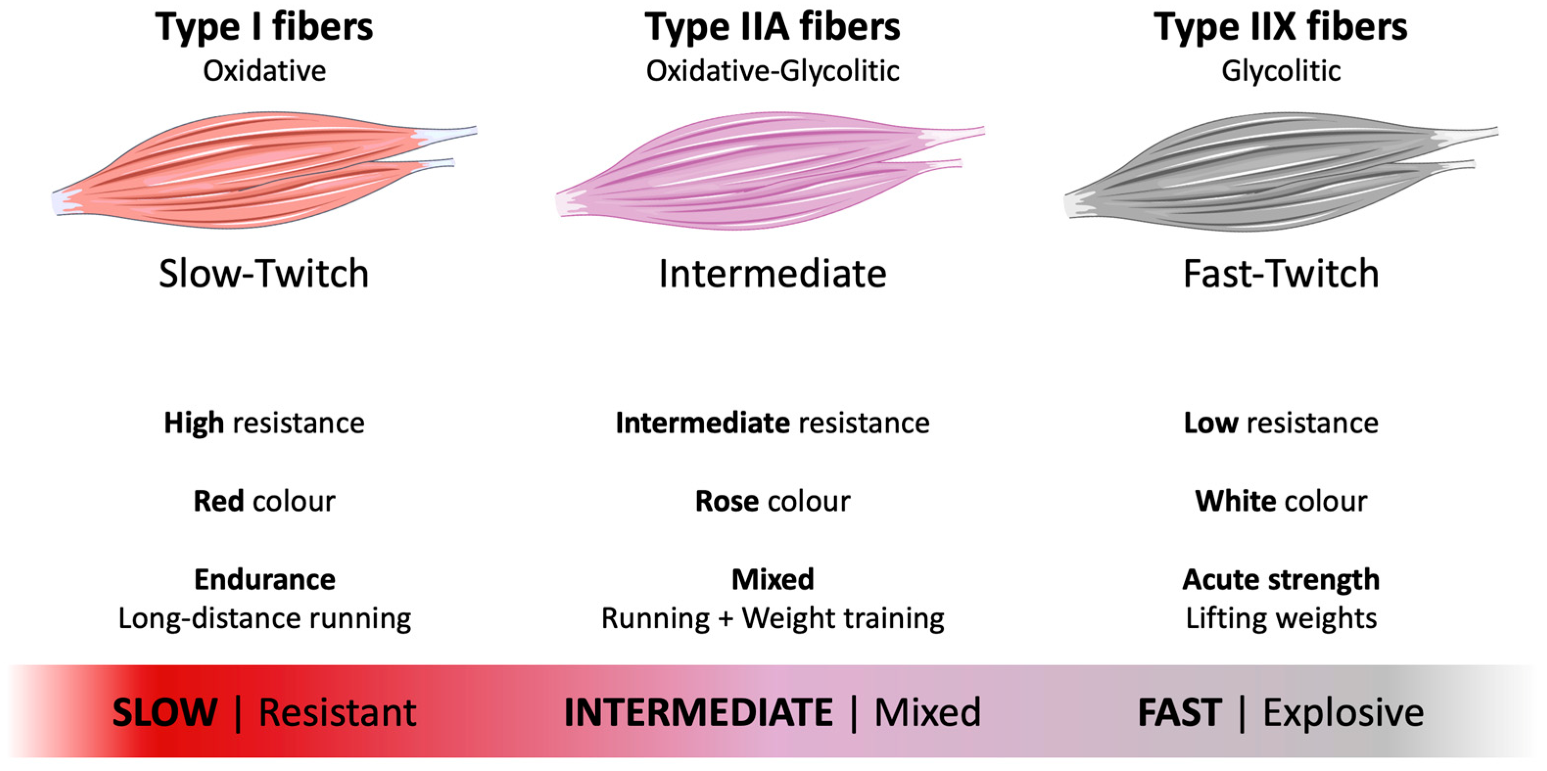
1. Introduction
2. Satellite Cells and Muscle Damage
3. Symptom Duration and Fiber Composition
4. Muscle Fiber Size and Atrophy
5. Physical Activity, Occupation and Loading Patterns
6. Sex and Age Differences
7. Regional Differences
8. Lean Body Mass
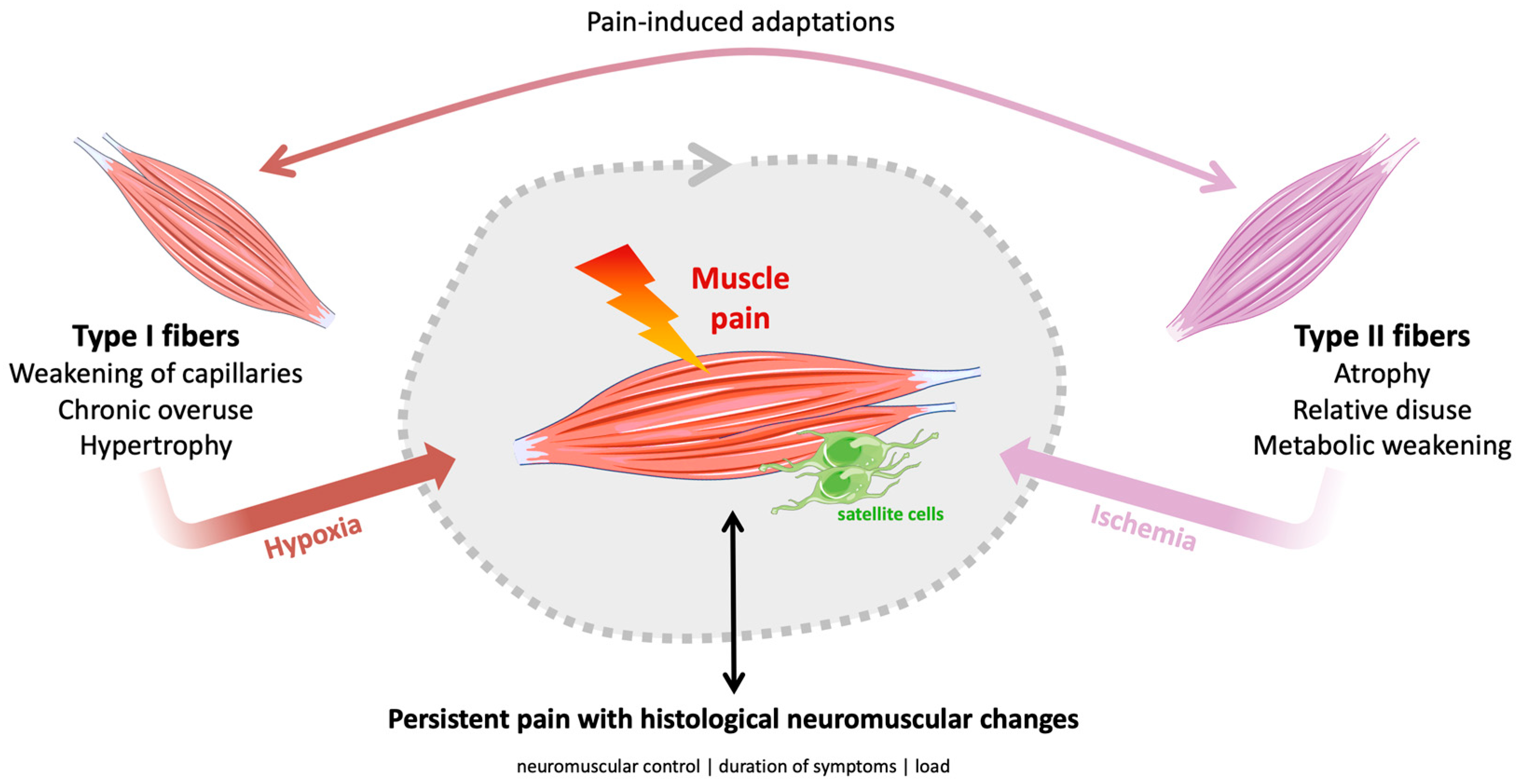
9. Discussion
10. Conclusions
Author Contributions
Funding
Institutional Review Board Statement
Informed Consent Statement
Data Availability Statement
Conflicts of Interest
Abbreviations
| ACTN3 | α-actinine-3 gene |
| ATP | adenosine triphosphate |
| ATPase | adenosine triphosphatase |
| CK | blood creatine kinase |
| CMP | chronic musculoskeletal pain |
| DAMPs | damage-associated molecular patterns |
| ELISA | enzyme-linked immunosorbent assay |
| FM | fibromyalgia |
| MHC | myosin heavy chain |
| MSD | musculoskeletal disorder |
| Pax7 | pair box 7 protein |
| PGC-1α | peroxisome proliferator-activated receptor γ coactivator 1α |
| RCSA | relative cross-sectional area |
References
- Siracusa, J.; Koulmann, N.; Sourdrille, A.; Chapus, C.; Verret, C.; Bourdon, S.; Goriot, M.E.; Banzet, S. Phenotype-Specific Response of Circulating miRNAs Provides New Biomarkers of Slow or Fast Muscle Damage. Front. Physiol. 2018, 9, 684. [Google Scholar] [CrossRef]
- Tellis, C.M.; Rosen, C.; Thekdi, A.; Sciote, J.J. Anatomy and fiber type composition of human interarytenoid muscle. Ann. Otol. Rhinol. Laryngol. 2004, 113, 97–107. [Google Scholar] [CrossRef]
- Burke, R.E.; Levine, D.N.; Zajac, F.E., 3rd. Mammalian motor units: Physiological-histochemical correlation in three types in cat gastrocnemius. Science 1971, 174, 709–712. [Google Scholar] [CrossRef]
- Brooke, M.H.; Kaiser, K.K. Muscle fiber types: How many and what kind? Arch. Neurol. 1970, 23, 369–379. [Google Scholar] [CrossRef]
- Schiaffino, S.; Reggiani, C. Fiber types in mammalian skeletal muscles. Physiol. Rev. 2011, 91, 1447–1531. [Google Scholar] [CrossRef]
- Larsson, L.; Edström, L.; Lindegren, B.; Gorza, L.; Schiaffino, S. MHC composition and enzyme-histochemical and physiological properties of a novel fast-twitch motor unit type. Am. J. Physiol. 1991, 261, C93–C101. [Google Scholar] [CrossRef]
- Barclay, C.J.; Constable, J.K.; Gibbs, C.L. Energetics of fast- and slow-twitch muscles of the mouse. J. Physiol. 1993, 472, 61–80. [Google Scholar] [CrossRef]
- He, Z.-H.; Bottinelli, R.; Pellegrino, M.A.; Ferenczi, M.A.; Reggiani, C. ATP Consumption and Efficiency of Human Single Muscle Fibers with Different Myosin Isoform Composition. Biophys. J. 2000, 79, 945–961. [Google Scholar] [CrossRef]
- Zierath, J.R.; Hawley, J.A. Skeletal muscle fiber type: Influence on contractile and metabolic properties. PLoS Biol. 2004, 2, e348. [Google Scholar] [CrossRef]
- Baker, J.S.; McCormick, M.C.; Robergs, R.A. Interaction among Skeletal Muscle Metabolic Energy Systems during Intense Exercise. J. Nutr. Metab. 2010, 2010, 905612. [Google Scholar] [CrossRef]
- Lievens, E.; Klass, M.; Bex, T.; Derave, W. Muscle fiber typology substantially influences time to recover from high-intensity exercise. J. Appl. Physiol. 2020, 128, 648–659. [Google Scholar] [CrossRef]
- Plotkin, D.L.; Roberts, M.D.; Haun, C.T.; Schoenfeld, B.J. Muscle Fiber Type Transitions with Exercise Training: Shifting Perspectives. Sports 2021, 9, 127. [Google Scholar] [CrossRef]
- Stozer, A.; Vodopivc, P.; Krizancic Bombek, L. Pathophysiology of exercise-induced muscle damage and its structural, functional, metabolic, and clinical consequences. Physiol. Res. 2020, 69, 565–598. [Google Scholar] [CrossRef]
- Proske, U.; Morgan, D.L. Muscle damage from eccentric exercise: Mechanism, mechanical signs, adaptation and clinical applications. J. Physiol. 2001, 537, 333–345. [Google Scholar] [CrossRef]
- Tu, H.; Li, Y.L. Inflammation balance in skeletal muscle damage and repair. Front. Immunol. 2023, 14, 1133355. [Google Scholar] [CrossRef]
- Furrer, R.; Handschin, C. Optimized Engagement of Macrophages and Satellite Cells in the Repair and Regeneration of Exercised Muscle. In Hormones, Metabolism and the Benefits of Exercise; Spiegelman, B., Ed.; Springer: Cham, Switzerland, 2017; pp. 57–66. [Google Scholar]
- Su, Y.; Su, Z. Impact of exercise on immune cell infiltration in muscle tissue: Implications for muscle repair and chronic disease. Clin. Exp. Med. 2025, 25, 306. [Google Scholar] [CrossRef]
- Hody, S.; Croisier, J.L.; Bury, T.; Rogister, B.; Leprince, P. Eccentric Muscle Contractions: Risks and Benefits. Front. Physiol. 2019, 10, 536. [Google Scholar] [CrossRef]
- Mauro, A. Satellite cell of skeletal muscle fibers. J. Biophys. Biochem. Cytol. 1961, 9, 493–495. [Google Scholar] [CrossRef]
- Verdijk, L.B.; Snijders, T.; Drost, M.; Delhaas, T.; Kadi, F.; van Loon, L.J. Satellite cells in human skeletal muscle; from birth to old age. Age 2014, 36, 545–547. [Google Scholar] [CrossRef]
- Almeida, C.F.; Fernandes, S.A.; Ribeiro Junior, A.F.; Keith Okamoto, O.; Vainzof, M. Muscle Satellite Cells: Exploring the Basic Biology to Rule Them. Stem Cells Int. 2016, 2016, 1078686. [Google Scholar] [CrossRef]
- Feige, P.; Brun, C.E.; Ritso, M.; Rudnicki, M.A. Orienting Muscle Stem Cells for Regeneration in Homeostasis, Aging, and Disease. Cell Stem Cell 2018, 23, 653–664. [Google Scholar] [CrossRef]
- Dewi, L.; Lin, Y.C.; Nicholls, A.; Condello, G.; Huang, C.Y.; Kuo, C.H. Pax7+ Satellite Cells in Human Skeletal Muscle After Exercise: A Systematic Review and Meta-analysis. Sports Med. 2023, 53, 457–480. [Google Scholar] [CrossRef]
- Pugh, J.K.; Faulkner, S.H.; Turner, M.C.; Nimmo, M.A. Satellite cell response to concurrent resistance exercise and high-intensity interval training in sedentary, overweight/obese, middle-aged individuals. Eur. J. Appl. Physiol. 2018, 118, 225–238. [Google Scholar] [CrossRef]
- Mackey, A.L.; Andersen, L.L.; Frandsen, U.; Sjøgaard, G. Strength training increases the size of the satellite cell pool in type I and II fibres of chronically painful trapezius muscle in females. J. Physiol. 2011, 589, 5503–5515. [Google Scholar] [CrossRef]
- Treede, R.D.; Rief, W.; Barke, A.; Aziz, Q.; Bennett, M.I.; Benoliel, R.; Cohen, M.; Evers, S.; Finnerup, N.B.; First, M.B.; et al. A classification of chronic pain for ICD-11. Pain 2015, 156, 1003–1007. [Google Scholar] [CrossRef]
- Cieza, A.; Causey, K.; Kamenov, K.; Hanson, S.W.; Chatterji, S.; Vos, T. Global estimates of the need for rehabilitation based on the Global Burden of Disease study 2019: A systematic analysis for the Global Burden of Disease Study 2019. Lancet 2021, 396, 2006–2017, Erratum in Lancet 2021, 397, 198. [Google Scholar] [CrossRef]
- Liu, M.; Rong, J.; An, X.; Li, Y.; Min, Y.; Yuan, G.; Yang, Y.; Li, M. Global, regional, and national burden of musculoskeletal disorders, 1990–2021: An analysis of the global burden of disease study 2021 and forecast to 2035. Front. Public Health 2025, 13, 1562701. [Google Scholar] [CrossRef]
- Prego-Domínguez, J.; Khazaeipour, Z.; Mallah, N.; Takkouche, B. Socioeconomic status and occurrence of chronic pain: A meta-analysis. Rheumatology 2021, 60, 1091–1105. [Google Scholar] [CrossRef]
- James, J.J.; Mellow, M.L.; Bueckers, E.P.; Gutsch, S.B.; Wrucke, D.J.; Pearson, A.G.; Smith, A.E.; Hunter, S.K. Sex differences in human skeletal muscle fiber types and the influence of age, physical activity, and muscle group: A systematic review and meta-analysis. Physiol. Rep. 2025, 13, e70616. [Google Scholar] [CrossRef]
- Eason, J.M.; Schwartz, G.A.; Pavlath, G.K.; English, A.W. Sexually dimorphic expression of myosin heavy chains in the adult mouse masseter. J. Appl. Physiol. 2000, 89, 251–258. [Google Scholar] [CrossRef]
- Larsson, B.; Björk, J.; Elert, J.; Lindman, R.; Gerdle, B. Fibre type proportion and fibre size in trapezius muscle biopsies from cleaners with and without myalgia and its correlation with ragged red fibres, cytochrome-c-oxidase-negative fibres, biomechanical output, perception of fatigue, and surface electromyography during repetitive forward flexions. Eur. J. Appl. Physiol. 2001, 84, 492–502. [Google Scholar] [PubMed]
- Henneman, E.; Olson, C.B. Relations between structure and function in the design of skeletal muscles. J. Neurophysiol. 1965, 28, 581–598. [Google Scholar] [CrossRef]
- Sterling, M.; Jull, G.; Wright, A. The effect of musculoskeletal pain on motor activity and control. J. Pain 2001, 2, 135–145. [Google Scholar] [CrossRef]
- Thorn, S.; Forsman, M.; Zhang, Q.; Taoda, K. Low-threshold motor unit activity during a 1-h static contraction in the trapezius muscle. Int. J. Ind. Ergon. 2002, 30, 225–236. [Google Scholar] [CrossRef]
- Andersen, L.L.; Suetta, C.; Andersen, J.L.; Kjær, M.; Sjøgaard, G. Increased proportion of megafibers in chronically painful muscles. Pain 2008, 139, 588–593. [Google Scholar] [CrossRef]
- Mesquita, R.N.O.; Skarabot, J.; Pearcey, G.E.P. Low-threshold motor units can be a pain during experimental muscle pain. J. Physiol. 2020, 598, 2545–2547. [Google Scholar] [CrossRef]
- Carmona, G.; Guerrero, M.; Cussó, R.; Padullés, J.M.; Moras, G.; Lloret, M.; Bedini, J.L.; Cadefau, J.A. Muscle enzyme and fiber type-specific sarcomere protein increases in serum after inertial concentric-eccentric exercise. Scand. J. Med. Sci. Sports 2015, 25, e547–e557. [Google Scholar] [CrossRef]
- Ureczky, D.; Vácz, G.; Costa, A.; Kopper, B.; Lacza, Z.; Hortobágyi, T.; Tihanyi, J. The effects of short-term exercise training on peak-torque are time- and fiber-type dependent. J. Strength Cond. Res. 2014, 28, 2204–2213. [Google Scholar] [CrossRef] [PubMed]
- Guerrero, M.; Guiu-Comadevall, M.; Cadefau, J.A.; Parra, J.; Balius, R.; Estruch, A.; Rodas, G.; Bedini, J.L.; Cusso, R. Fast and slow myosins as markers of muscle injury. Br. J. Sports Med. 2008, 42, 581–584. [Google Scholar] [CrossRef]
- Bogdanis, G.C.; Tsoukos, A.; Brown, L.E.; Selima, E.; Veligekas, P.; Spengos, K.; Terzis, G. Muscle Fiber and Performance Changes after Fast Eccentric Complex Training. Med. Sci. Sports Exerc. 2018, 50, 729–738. [Google Scholar] [CrossRef]
- Fullen, B.M.; Wittink, H.; De Groef, A.; Hoegh, M.; McVeigh, J.G.; Martin, D.; Smart, K. Musculoskeletal Pain: Current and Future Directions of Physical Therapy Practice. Arch. Rehabil. Res. Clin. Transl. 2023, 5, 100258. [Google Scholar] [CrossRef]
- Behringer, C.; Nagib, N.; Nagib, N. Introduction to multimodal musculoskeletal treatment models: A viewpoint. Discov. Appl. Sci. 2025, 7, 820. [Google Scholar] [CrossRef]
- Dawod, M.S.; Alswerki, M.N.; Alelaumi, A.F.; Alshloul, S.A.; Husami, M.Y.; Sulaiman, S.G.; Al-Azzawi, M.T.; Al-Rawashdah, S.F.; Abualhaj, S. Approach to Adult Patients with Musculoskeletal Complaints and Normal Findings: A Guide for Clinical Practice. Orthop. Res. Rev. 2025, 17, 503–516. [Google Scholar] [CrossRef]
- Mackey, A.L.; Andersen, L.L.; Frandsen, U.; Suetta, C.; Sjøgaard, G. Distribution of myogenic progenitor cells and myonuclei is altered in women with vs. those without chronically painful trapezius muscle. J. Appl. Physiol. 2010, 109, 1920–1929. [Google Scholar] [CrossRef]
- Kadi, F.; Ahlgren, C.; Waling, K.; Sundelin, G.; Thornell, L.E. The effects of different training programs on the trapezius muscle of women with work-related neck and shoulder myalgia. Acta Neuropathol. 2000, 100, 253–258. [Google Scholar] [CrossRef]
- Lindman, R.; Hagberg, M.; Angqvist, K.A.; Söderlund, K.; Hultman, E.; Thornell, L.E. Changes in muscle morphology in chronic trapezius myalgia. Scand J. Work. Environ. Health 1991, 17, 347–355. [Google Scholar] [CrossRef]
- Kadi, F.; Hägg, G.; Håkansson, R.; Holmner, S.; Butler-Browne, G.S.; Thornell, L.E. Structural changes in male trapezius muscle with work-related myalgia. Acta Neuropathol. 1998, 95, 352–360. [Google Scholar] [CrossRef]
- De Meulemeester, K.; Calders, P.; Van Dorpe, J.; De Pauw, R.; Petrovic, M.; Cagnie, B. Morphological Differences in the Upper Trapezius Muscle Between Female Office Workers With and Without Trapezius Myalgia: Facts or Fiction?: A Cross-Sectional Study. Am. J. Phys. Med. Rehabil. 2019, 98, 117–124. [Google Scholar] [CrossRef]
- Weber, B.R.; Uhlig, Y.; Grob, D.; Dvorák, J.; Müntener, M. Duration of pain and muscular adaptations in patients with dysfunction of the cervical spine. J. Orthop. Res. 1993, 11, 805–810. [Google Scholar] [CrossRef] [PubMed]
- Mazis, N.; Papachristou, D.J.; Zouboulis, P.; Tyllianakis, M.; Scopa, C.D.; Megas, P. The effect of different physical activity levels on muscle fiber size and type distribution of lumbar multifidus. A biopsy study on low back pain patient groups and healthy control subjects. Eur. J. Phys. Rehabil. Med. 2009, 45, 459–467. [Google Scholar] [PubMed]
- Purushotham, S.; Hodson, N.; Greig, C.; Gardner, A.; Falla, D. Microscopic changes in the multifidus muscle in people with low back pain associated with lumbar disc herniation. Sci. Rep. 2024, 14, 31927. [Google Scholar] [CrossRef]
- Mannion, A.F.; Weber, B.R.; Dvorak, J.; Grob, D.; Müntener, M. Fibre type characteristics of the lumbar paraspinal muscles in normal healthy subjects and in patients with low back pain. J. Orthop. Res. 1997, 15, 881–887. [Google Scholar] [CrossRef]
- Agten, A.; Stevens, S.; Verbrugghe, J.; Timmermans, A.; Vandenabeele, F. Biopsy samples from the erector spinae of persons with nonspecific chronic low back pain display a decrease in glycolytic muscle fibers. Spine J. 2020, 20, 199–206. [Google Scholar] [CrossRef] [PubMed]
- Mannion, A.F.; Käser, L.; Weber, E.; Rhyner, A.; Dvorak, J.; Müntener, M. Influence of age and duration of symptoms on fibre type distribution and size of the back muscles in chronic low back pain patients. Eur. Spine J. 2000, 9, 273–281. [Google Scholar] [CrossRef]
- Padwal, J.; Berry, D.B.; Hubbard, J.C.; Zlomislic, V.; Allen, R.T.; Garfin, S.R.; Ward, S.R.; Shahidi, B. Regional differences between superficial and deep lumbar multifidus in patients with chronic lumbar spine pathology. BMC Musculoskelet. Disord. 2020, 21, 764. [Google Scholar] [CrossRef]
- Srikuea, R.; Symons, T.B.; Long, D.E.; Lee, J.D.; Shang, Y.; Chomentowski, P.J.; Yu, G.; Crofford, L.J.; Peterson, C.A. Association of fibromyalgia with altered skeletal muscle characteristics which may contribute to postexertional fatigue in postmenopausal women. Arthritis Rheum. 2013, 65, 519–528. [Google Scholar] [CrossRef] [PubMed]
- Sobreira, C.; Marques, W., Jr.; Barreira, A.A. Myalgia as the revealing symptom of multicore disease and fibre type disproportion myopathy. J. Neurol. Neurosurg. Psychiatry 2003, 74, 1317–1319. [Google Scholar] [CrossRef][Green Version]
- Putzu, G.A.; Figarella-Branger, D.; Baeta, A.M.; Lepidi, H.; Pellissier, J.F. Acquired multifocal myofibrillar disruption selective of type II fibres. Neuropathol. Appl. Neurobiol. 1996, 22, 38–43. [Google Scholar] [CrossRef]
- Dickx, N.; Cagnie, B.; Achten, E.; Vandemaele, P.; Parlevliet, T.; Danneels, L. Differentiation between deep and superficial fibers of the lumbar multifidus by magnetic resonance imaging. Eur. Spine J. 2010, 19, 122–128. [Google Scholar] [CrossRef][Green Version]
- Crossman, K.; Mahon, M.; Watson, P.J.; Oldham, J.A.; Cooper, R.G. Chronic low back pain-associated paraspinal muscle dysfunction is not the result of a constitutionally determined “adverse” fiber-type composition. Spine 2004, 29, 628–634. [Google Scholar] [CrossRef]
- Broos, S.; Malisoux, L.; Theisen, D.; Van Thienen, R.; Francaux, M.; Thomis, M.A.; Deldicque, L. The stiffness response of type IIa fibres after eccentric exercise-induced muscle damage is dependent on ACTN3 r577X polymorphism. Eur. J. Sport Sci. 2019, 19, 480–489. [Google Scholar] [CrossRef]
- Vincent, B.; Windelinckx, A.; Nielens, H.; Ramaekers, M.; Van Leemputte, M.; Hespel, P.; Thomis, M.A. Protective role of alpha-actinin-3 in the response to an acute eccentric exercise bout. J. Appl. Physiol. 2010, 109, 564–573. [Google Scholar] [CrossRef]
- Chen, T.C. Effects of a second bout of maximal eccentric exercise on muscle damage and electromyographic activity. Eur. J. Appl. Physiol. 2003, 89, 115–121. [Google Scholar] [CrossRef]
- Mair, J.; Mayr, M.; Müller, E.; Koller, A.; Haid, C.; Artner-Dworzak, E.; Calzolari, C.; Larue, C.; Puschendorf, B. Rapid adaptation to eccentric exercise-induced muscle damage. Int. J. Sports Med. 1995, 16, 352–356. [Google Scholar] [CrossRef]
- Morton, R.W.; Sonne, M.W.; Farias Zuniga, A.; Mohammad, I.Y.Z.; Jones, A.; McGlory, C.; Keir, P.J.; Potvin, J.R.; Phillips, S.M. Muscle fibre activation is unaffected by load and repetition duration when resistance exercise is performed to task failure. J. Physiol. 2019, 597, 4601–4613. [Google Scholar] [CrossRef]
- Zhao, W.P.; Kawaguchi, Y.; Matsui, H.; Kanamori, M.; Kimura, T. Histochemistry and morphology of the multifidus muscle in lumbar disc herniation: Comparative study between diseased and normal sides. Spine 2000, 25, 2191–2199. [Google Scholar] [CrossRef] [PubMed]
- Ge, H.-Y.; Arendt-Nielsen, L.; Farina, D.; Madeleine, P. Gender-specific differences in electromyographic changes and perceived pain induced by experimental muscle pain during sustained contractions of the upper trapezius muscle. Muscle Nerve 2005, 32, 726–733. [Google Scholar] [CrossRef] [PubMed]
- Esbjornsson, M.E.; Dahlstrom, M.S.; Gierup, J.W.; Jansson, E.C. Muscle fiber size in healthy children and adults in relation to sex and fiber types. Muscle Nerve 2021, 63, 586–592. [Google Scholar] [CrossRef]
- Agten, A.; Verbrugghe, J.; Stevens, S.; Eijnde, B.O.; Timmermans, A.; Vandenabeele, F. High Intensity Training Increases Muscle Area Occupied by Type II Muscle Fibers of the Multifidus Muscle in Persons with Non-Specific Chronic Low Back Pain: A Pilot Trial. Appl. Sci. 2021, 11, 3306. [Google Scholar] [CrossRef]
- Verdijk, L.B.; Koopman, R.; Schaart, G.; Meijer, K.; Savelberg, H.H.; van Loon, L.J. Satellite cell content is specifically reduced in type II skeletal muscle fibers in the elderly. Am. J. Physiol. Endocrinol. Metab. 2007, 292, E151–E157. [Google Scholar] [CrossRef]
- Mao, X.; Lv, K.; Qi, W.; Cheng, W.; Li, T.; Sun, Y.; Jin, H.; Pan, H.; Wang, D. Research progress on sarcopenia in the musculoskeletal system. Bone Res. 2025, 13, 78. [Google Scholar] [CrossRef] [PubMed]
- Larsson, L.; Degens, H.; Li, M.; Salviati, L.; Lee, Y.I.; Thompson, W.; Kirkland, J.L.; Sandri, M. Sarcopenia: Aging-Related Loss of Muscle Mass and Function. Physiol. Rev. 2019, 99, 427–511. [Google Scholar] [CrossRef] [PubMed]
- Horlem, T.; Carvalhal, S.R.S.; Bonatto, S.J.R.; Fernandes, L.C. Molecular Framework of the Onset and Progression of Skeletal Muscle Aging. Int. J. Mol. Sci. 2025, 26, 10145. [Google Scholar] [CrossRef]
- Walton, R.G.; Kosmac, K.; Mula, J.; Fry, C.S.; Peck, B.D.; Groshong, J.S.; Finlin, B.S.; Zhu, B.; Kern, P.A.; Peterson, C.A. Human skeletal muscle macrophages increase following cycle training and are associated with adaptations that may facilitate growth. Sci. Rep. 2019, 9, 969. [Google Scholar] [CrossRef]
- Saito, Y.; Chikenji, T.S.; Matsumura, T.; Nakano, M.; Fujimiya, M. Exercise enhances skeletal muscle regeneration by promoting senescence in fibro-adipogenic progenitors. Nat. Commun. 2020, 11, 889. [Google Scholar] [CrossRef]
- Murach, K.A.; Fry, C.S.; Kirby, T.J.; Jackson, J.R.; Lee, J.D.; White, S.H.; Dupont-Versteegden, E.E.; McCarthy, J.J.; Peterson, C.A. Starring or Supporting Role? Satellite Cells and Skeletal Muscle Fiber Size Regulation. Physiology 2018, 33, 26–38. [Google Scholar] [CrossRef]
- Bagley, J.R.; Denes, L.T.; McCarthy, J.J.; Wang, E.T.; Murach, K.A. The myonuclear domain in adult skeletal muscle fibres: Past, present and future. J. Physiol. 2023, 601, 723–741. [Google Scholar] [CrossRef]
- Minerbi, A.; Vulfsons, S. Challenging the Cinderella Hypothesis: A New Model for the Role of the Motor Unit Recruitment Pattern in the Pathogenesis of Myofascial Pain Syndrome in Postural Muscles. Rambam Maimonides Med. J. 2018, 9, e0021-8. [Google Scholar] [CrossRef] [PubMed]
- Steen, J.P.; Jaiswal, K.S.; Kumbhare, D. Myofascial Pain Syndrome: An Update on Clinical Characteristics, Etiopathogenesis, Diagnosis, and Treatment. Muscle Nerve 2025, 71, 889–910. [Google Scholar] [CrossRef]
- Warwick, C.A.; Keyes, A.L.; Woodruff, T.M.; Usachev, Y.M. The complement cascade in the regulation of neuroinflammation, nociceptive sensitization, and pain. J. Biol. Chem. 2021, 297, 101085. [Google Scholar] [CrossRef] [PubMed]
- Barnig, C.; Lutzweiler, G.; Giannini, M.; Lejay, A.; Charles, A.L.; Meyer, A.; Geny, B. Resolution of Inflammation after Skeletal Muscle Ischemia-Reperfusion Injury: A Focus on the Lipid Mediators Lipoxins, Resolvins, Protectins and Maresins. Antioxidants 2022, 11, 1213. [Google Scholar] [CrossRef]
- Ross, J.L.; Queme, L.F.; Shank, A.T.; Hudgins, R.C.; Jankowski, M.P. Sensitization of group III and IV muscle afferents in the mouse after ischemia and reperfusion injury. J. Pain 2014, 15, 1257–1270. [Google Scholar] [CrossRef] [PubMed]
- Gordon, T. Neuromuscular Activity Determines, at Least in Part, the Motoneuron, Nerve and Muscle Properties Under Normal Conditions and After Nerve Injury. Int. J. Mol. Sci. 2025, 26, 6891. [Google Scholar] [CrossRef] [PubMed]
- Murray, G.M.; Sessle, B.J. Pain-sensorimotor interactions: New perspectives and a new model. Neurobiol. Pain 2024, 15, 100150. [Google Scholar] [CrossRef]
- Mastaglia, F.L. The relationship between muscle pain and fatigue. Neuromuscul. Disord. 2012, 22, S178–S180. [Google Scholar] [CrossRef]
- Nardone, A.; Schieppati, M. Shift of activity from slow to fast muscle during voluntary lengthening contractions of the triceps surae muscles in humans. J. Physiol. 1988, 395, 363–381. [Google Scholar] [CrossRef]
- Bai, Z.; Zhang, D.; Liang, D.; Chen, X.; Shi, X.; Chen, S. Effect of Eccentric Training with Different Durations, Intensities, and Contraction Velocities on Upper Limb Muscle Strength: A Meta-Analysis. Life 2025, 15, 456. [Google Scholar] [CrossRef]
- Mannion, A.F.; Taimela, S.; Muntener, M.; Dvorak, J. Active therapy for chronic low back pain part 1. Effects on back muscle activation, fatigability, and strength. Spine 2001, 26, 897–908. [Google Scholar] [CrossRef]
- Wyckelsma, V.L.; Venckunas, T.; Houweling, P.J.; Schlittler, M.; Lauschke, V.M.; Tiong, C.F.; Wood, H.D.; Ivarsson, N.; Paulauskas, H.; Eimantas, N.; et al. Loss of α-actinin-3 during human evolution provides superior cold resilience and muscle heat generation. Am. J. Hum. Genet. 2021, 108, 446–457. [Google Scholar] [CrossRef] [PubMed]
- Seto, J.T.; Chan, S.; Turner, N.; MacArthur, D.G.; Raftery, J.M.; Berman, Y.D.; Quinlan, K.G.; Cooney, G.J.; Head, S.; Yang, N.; et al. The effect of alpha-actinin-3 deficiency on muscle aging. Exp. Gerontol. 2011, 46, 292–302. [Google Scholar] [CrossRef]
- Houweling, P.J.; Papadimitriou, I.D.; Seto, J.T.; Perez, L.M.; Coso, J.D.; North, K.N.; Lucia, A.; Eynon, N. Is evolutionary loss our gain? The role of ACTN3 p.Arg577Ter (R577X) genotype in athletic performance, ageing, and disease. Hum. Mutat. 2018, 39, 1774–1787. [Google Scholar] [CrossRef]
- Lee, F.X.; Houweling, P.J.; North, K.N.; Quinlan, K.G. How does α-actinin-3 deficiency alter muscle function? Mechanistic insights into ACTN3, the ‘gene for speed’. Biochim. Biophys. Acta 2016, 1863, 686–693. [Google Scholar] [CrossRef]
- Lexell, J.; Downham, D. What is the effect of ageing on type 2 muscle fibres? J. Neurol. Sci. 1992, 107, 250–251. [Google Scholar] [CrossRef]
- van Wessel, T.; de Haan, A.; van der Laarse, W.J.; Jaspers, R.T. The muscle fiber type-fiber size paradox: Hypertrophy or oxidative metabolism? Eur. J. Appl. Physiol. 2010, 110, 665–694. [Google Scholar] [CrossRef] [PubMed]
- Agten, A.; Stevens, S.; Verbrugghe, J.; Eijnde, B.O.; Timmermans, A.; Vandenabeele, F. The lumbar multifidus is characterised by larger type I muscle fibres compared to the erector spinae. Anat. Cell Biol. 2020, 53, 143–150. [Google Scholar] [CrossRef] [PubMed]
- Breen, E.; Tang, K.; Olfert, M.; Knapp, A.; Wagner, P. Skeletal muscle capillarity during hypoxia: VEGF and its activation. High Alt. Med. Biol. 2008, 9, 158–166. [Google Scholar] [CrossRef] [PubMed]
- Purushotham, S.; Stephenson, R.S.; Sanderson, A.; Abichandani, D.; Greig, C.; Gardner, A.; Falla, D. Microscopic changes in the spinal extensor musculature in people with chronic spinal pain: A systematic review. Spine J. 2022, 22, 1205–1221. [Google Scholar] [CrossRef]
- Hodges, P.W.; Tucker, K. Moving differently in pain: A new theory to explain the adaptation to pain. Pain 2011, 152, S90–S98. [Google Scholar] [CrossRef]
- Zhang, S.; Wang, Y.; Li, T.; Ma, J.; He, R.; Han, X.; Wu, W.; Wang, C. Relation Between Abnormal Spontaneous Brain Activity and Altered Neuromuscular Activation of Lumbar Paraspinal Muscles in Chronic Low Back Pain. Arch. Phys. Med. Rehabil. 2024, 105, 2107–2117. [Google Scholar] [CrossRef]
- Hides, J.A.; Richardson, C.A.; Jull, G.A. Multifidus muscle recovery is not automatic after resolution of acute, first-episode low back pain. Spine 1996, 21, 2763–2769. [Google Scholar] [CrossRef]
- Newcomer, K.L.; Jacobson, T.D.; Gabriel, D.A.; Larson, D.R.; Brey, R.H.; An, K.N. Muscle activation patterns in subjects with and without low back pain. Arch. Phys. Med. Rehabil. 2002, 83, 816–821. [Google Scholar] [CrossRef]
- Zhu, X.Z.; Parnianpour, M.; Nordin, M.; Kahanovitz, N. Histochemistry and morphology of erector spinae muscle in lumbar disc herniation. Spine 1989, 14, 391–397. [Google Scholar] [CrossRef]
- Rui, Y.; Bai, J.; Perrimon, N. Sarcomere formation occurs by the assembly of multiple latent protein complexes. PLoS Genet. 2010, 6, e1001208. [Google Scholar] [CrossRef]
- Maroni, C.R.; Friedman, M.A.; Zhang, Y.; McClure, M.J.; Fulle, S.; Farber, C.R.; Donahue, H.J. Genetic variability affects the response of skeletal muscle to disuse. J. Musculoskelet. Neuronal Interact. 2021, 21, 387–396. [Google Scholar]
- Leońska-Duniec, A. Genetic Susceptibility to Sport-Related Muscle Injuries: Insights from the Literature and Novel Gene Candidates. Int. J. Mol. Sci. 2025, 26, 11175. [Google Scholar] [CrossRef] [PubMed]
- Marques, I.S.; Tavares, V.; Vieira Neto, B.; Lopes, L.R.; Goes, R.A.; Guimarães, J.A.M.; Perini, J.A.; Medeiros, R. Genetic Variations in Susceptibility to Traumatic Muscle Injuries and Muscle Pain among Brazilian High-Performance Athletes. Int. J. Mol. Sci. 2024, 25, 3300. [Google Scholar] [CrossRef] [PubMed]
- Ruggiero, L.; Manganelli, F.; Santoro, L. Muscle pain syndromes and fibromyalgia: The role of muscle biopsy. Curr. Opin. Support. Palliat. Care 2018, 12, 382–387. [Google Scholar] [CrossRef]
- Carneiro, B.D.; Torres, S.; Costa-Pereira, J.T.; Pozza, D.H.; Tavares, I. Descending Pain Modulation in Fibromyalgia: A Short Review of Mechanisms and Biomarkers. Diagnostics 2025, 15, 2702. [Google Scholar] [CrossRef] [PubMed]
- Chakraborty, S.; Ben-David, R.; Shemer, S. Combating muscle atrophy: Emerging therapeutic targets that are fiber-type-specific. FEBS J. 2025, 292, 6481–6496. [Google Scholar] [CrossRef]
- Booth, F.W.; Roberts, C.K.; Laye, M.J. Lack of exercise is a major cause of chronic diseases. Compr. Physiol. 2012, 2, 1143–1211. [Google Scholar] [CrossRef]
- Hall, E.C.R.; Semenova, E.A.; Bondareva, E.A.; Borisov, O.V.; Andryushchenko, O.N.; Andryushchenko, L.B.; Zmijewski, P.; Generozov, E.V.; Ahmetov, I.I. Association of muscle fiber composition with health and exercise-related traits in athletes and untrained subjects. Biol. Sport 2021, 38, 659–666. [Google Scholar] [CrossRef] [PubMed]
- Carroll, K.M.; Bazyler, C.D.; Bernards, J.R.; Taber, C.B.; Stuart, C.A.; DeWeese, B.H.; Sato, K.; Stone, M.H. Skeletal Muscle Fiber Adaptations Following Resistance Training Using Repetition Maximums or Relative Intensity. Sports 2019, 7, 169. [Google Scholar] [CrossRef]
- Chen, J.; Han, B.; Wu, C. On the superiority of a combination of aerobic and resistance exercise for fibromyalgia syndrome: A network meta-analysis. Front. Psychol. 2022, 13, 949256. [Google Scholar] [CrossRef] [PubMed]
- Smith, J.A.B.; Murach, K.A.; Dyar, K.A.; Zierath, J.R. Exercise metabolism and adaptation in skeletal muscle. Nat. Rev. Mol. Cell Biol. 2023, 24, 607–632. [Google Scholar] [CrossRef]
- Rodriguez-Dominguez, A.J.; Rebollo-Salas, M.; Chillon-Martinez, R.; Rosales-Tristancho, A.; Villa-Del-Pino, I.; Jimenez-Rejano, J.J. The most effective therapeutic exercises for pain intensity in women with fibromyalgia: A systematic review and network meta-analysis. Braz. J. Phys. Ther. 2025, 29, 101226. [Google Scholar] [CrossRef]
- Liu, C.J.; Latham, N.K. Progressive resistance strength training for improving physical function in older adults. Cochrane Database Syst. Rev. 2009, 2009, CD002759. [Google Scholar] [CrossRef] [PubMed]
- Weakley, J.; Schoenfeld, B.J.; Ljungberg, J.; Halson, S.L.; Phillips, S.M. Physiological Responses and Adaptations to Lower Load Resistance Training: Implications for Health and Performance. Sports Med.-Open 2023, 9, 28. [Google Scholar] [CrossRef]
- Moro, T.; Brightwell, C.R.; Volpi, E.; Rasmussen, B.B.; Fry, C.S. Resistance exercise training promotes fiber type-specific myonuclear adaptations in older adults. J. Appl. Physiol. 2020, 128, 795–804. [Google Scholar] [CrossRef]
- Molmen, K.S.; Almquist, N.W.; Skattebo, O. Effects of Exercise Training on Mitochondrial and Capillary Growth in Human Skeletal Muscle: A Systematic Review and Meta-Regression. Sports Med. 2025, 55, 115–144. [Google Scholar] [CrossRef]
- Ding, Y.; Wan, Q.; Kim, J.C.; Liu, W.; Ji, W. The role of exercise induced capillarization adaptations in skeletal muscle aging: A systematic review. Front. Physiol. 2025, 16, 1681184. [Google Scholar] [CrossRef]
- Baroncini, A.; Maffulli, N.; Manocchio, N.; Bossa, M.; Foti, C.; Schäfer, L.; Klimuch, A.; Migliorini, F. Active and passive physical therapy in patients with chronic low-back pain: A level I Bayesian network meta-analysis. J. Orthop. Traumatol. 2025, 26, 66. [Google Scholar] [CrossRef]
- De la Corte-Rodriguez, H.; Roman-Belmonte, J.M.; Resino-Luis, C.; Madrid-Gonzalez, J.; Rodriguez-Merchan, E.C. The Role of Physical Exercise in Chronic Musculoskeletal Pain: Best Medicine-A Narrative Review. Healthcare 2024, 12, 242. [Google Scholar] [CrossRef]
- Choi, H.K.; Gwon, H.J.; Kim, S.R.; Park, C.S.; Cho, B.J. Effects of active rehabilitation therapy on muscular back strength and subjective pain degree in chronic lower back pain patients. J. Phys. Ther. Sci. 2016, 28, 2700–2702. [Google Scholar] [CrossRef]
- Kerbaul, F.; Brousse, M.; Collart, F.; Pellissier, J.F.; Planche, D.; Fernandez, C.; Gouin, F.; Guidon, C. Combination of histopathological and electromyographic patterns can help to evaluate functional outcome of critical ill patients with neuromuscular weakness syndromes. Crit. Care 2004, 8, R358–R366. [Google Scholar] [CrossRef] [PubMed]
- Imrani, L.; Boudaoud, S.; Lahaye, C.; Moreau, C.; Ghezal, M.; Ben Manaa, S.; Doulazmi, M.; Laforet, J.; Marin, F.; Kinugawa, K. High-density Surface Electromyography as Biomarker of Muscle Aging. J. Gerontol. A Biol. Sci. Med. Sci. 2023, 78, 25–33. [Google Scholar] [CrossRef] [PubMed]
- Shandiz, E.; Gaskell, A.; Ingram, M.; Trajano, G.S.; Fernandes, G.L.; Henkin, J.S.; McCombe, P.A.; Henderson, R.D. Applying high-density surface EMG to the study of neuromuscular disorders: A systematic review. Clin. Neurophysiol. 2025, 179, 2110983. [Google Scholar] [CrossRef] [PubMed]
- Chen, Z.J.; Huang, X.L.; Xia, N.; Gu, M.H.; Xu, J.; Lu, M.; Chen, H.; Xiong, C.H.; Chen, Y. Next-Generation Neurotechnologies Inspired by Motor Primitive Model for Restoring Human Natural Movement. Research 2025, 8, 0942. [Google Scholar] [CrossRef] [PubMed]



| Reference/Type | Population | Type of Pain | Evaluation Method | Results |
|---|---|---|---|---|
| [25] Randomized controlled trial | 42 women with trapezius myalgia: monotonous and repetitive work tasks | Chronic trapezius myalgia | Immunohistochemistry | Type I fibers: increase of 65% in satellite cells; Type II fibers: increase of 164% in satellite cells |
| [45] Experimental, cross-sectional | 62 female office workers: 42 with myalgia in trapezius muscle, with monotonous and repetitive work tasks | Chronic trapezius myalgia | Fluorescent laminin staining | Type I fibers contained more satellite cells than Type II fibers; Type II fibers, less frequently recruited during sustained low-intensity activity, had fewer satellite cells and showed smaller fiber areas indicative of atrophy |
| [46] Randomized controlled trial | 21 women with work-related neck and shoulder myalgia divided into three 10-week training programs | Chronic Trapezius myalgia | Enzyme-immunohistochemical analysis | Strength training increased Type IIA area (+49%) and proportion Coordination decreased Type I area; there was also an increased capillarization in Type I and IIA |
| [47] Case–control | 10 female patients with chronic trapezius myalgia due static and repetitive work tasks and 5 healthy females | Chronic Trapezius myalgia | Enzyme histochemical, immunohistochemical and biochemical analyses | Patients had significantly larger Type I fibers and a lower capillary-to-fiber area ratio for Type I and Type IIA fibers |
| [36] Case–control study | 42 female workers with trapezius myalgia and 20 female matched workers without | Trapezius myalgia | Histochemical | Significantly higher proportion of Type I megafibers in females with trapezius myalgia and poor capillarization |
| [32] Cross-sectional case–control study | 25 female cleaners with trapezius myalgia and 25 without; 21 healthy female teachers without repetitive or static muscle work | Trapezius myalgia | Electromyogram and Immunohistochemistry | Smaller cross-sectional area of Type II fibers in cleaners compared to teachers; no difference in Type I fibers |
| [48] Case–control study | 10 male forest machine operators with myalgia, 9 without myalgia, and 6 healthy controls | Trapezius myalgia | Histochemistry and immunohistochemistry | Men with myalgia showed significant Type IIA fiber hypertrophy with a proportionally matched increase in capillarization showing an active injury-regeneration cycle |
| [49] Cross-sectional study | 17 female office workers with myalgia and 15 healthy controls | Trapezius myalgia | Major histocompatibility complex immunohistochemistry | Higher proportions of Type IIA and IIA/IIX fibers, and lower proportions of Type I fibers in the myalgia group No differences in fiber size between groups, but Type I fibers were significantly larger than Type II fibers in both groups |
| [50] Observational study | 24 patients undergoing surgery for cervical dysfunction with severe neck pain | Chronic neck pain | Histochemical methods | Symptom duration < 16 months showed higher proportion of transitional Type IIC fibers (active transformation) Symptom duration > 20 months lacked Type IIC fibers |
| [51] Cross-sectional case–control study | 64 patients with low back pain, 17 healthy control individuals | Low back pain | Histochemical | Low back pain showed a higher Type II fiber proportion The physical activity level did not influence fiber type and diameter |
| [52] Observational study | 30 healthy adults (11 men) undergoing microdiscectomy for lumbar disc herniation | Low back pain | Immunohistochemistry and epifluorescence | Affected multifidus contained a higher proportion of pathological fibers Higher pain intensity and disability linked to smaller Type I fibers |
| [53] Cross-sectional controlled study | 21 low back pain patients and 21 healthy controls | Low back pain | Histochemical | Patients had more Type IIB than Type I fibers; early-stage (<1 year) pain was linked to higher Type IIC proportions, while long-term (>3 years) pain showed marked reduction |
| [54] Cross sectional study | 20 chronic low back pain and 18 healthy controls | Low back pain | Immunofluorescence | Pain is associated with fewer Type IIB fibers and a shift toward a more aerobic profile, with larger Type I and smaller Type IIB cross-sectional areas |
| [55] Experimental study | 59 patients (30 women) with chronic low back pain | Chronic low back pain | Histochemical and magnetic resonance | Type I: Longer symptoms, lower proportion; higher in pathological biopsies; Female > Male Type IIA: Unaffected by symptom duration; diameter strongly tied to pain. Type IIX: Longer symptoms, higher proportion + stronger glycolytic profile; diameter correlates with pain; Male > Female Type IIC: Very low in chronic cases, Male > Female |
| [56] Observational study | 16 patients undergoing lumbar spinal surgery | Chronic low back pain | Histochemical and MRI | No significant differences in fiber type distribution, size, fat infiltration, or muscle degeneration between the superficial and deep multifidus Chronic degeneration affects the multifidus globally |
| [57] Experimental Study | 37 healthy postmenopausal women with (n = 14) and without (n = 23) Fibromyalgia (FM) | FM with a fatiguing exercise | Histochemistry, immunohistochemistry and electron microscopic | FM patients show similar Type I/II proportions, but greater fiber-size variability, more small fibers, and lower capillary density |
| [58] Case series | 24-year-old man, 14-year-old male, and 47-year-old women | Myalgia | Histochemistry and immunohistochemistry | Type I fibers predominance with a selective uniform atrophy or a predominant multiple area of focal myofibrillar degeneration |
| [59] Case series | 10-year-old girl, 22-year-old man, and 55-year-old man | Myalgia and cramps | Electromyogram, histochemistry, immunohistochemistry and electron microscopic | Acquired multifocal myofibrillar disorganization selectively affects Type II muscle fibers |
| Reference/Type | Population | Type Muscle Injury | Evaluation Method | Results |
|---|---|---|---|---|
| [60] Cross-sectional experimental study | 15 young males | Experimentally induced low back muscle pain with hypertonic saline solution | Magnetic resonance | Deep multifidus fibers are predominantly slow twitch compared to the superficial layer; no differential recruitment has been found following trunk extension with and without pain induction |
| [61] Investigative case–control study | 35 males with chronic low back pain and 32 control | Excessive paraspinal muscle fatigue by exercise | Electromyography, histomorphometry and immunohistochemistry | Paraspinal muscle dysfunction did not stem from a constitutionally predetermined adverse fiber type profile |
| [39] Experimental research article | Nine healthy sedentary men not involved in strenuous eccentric exercise in past 6 months | Eccentric exercise induced muscle damage | Immunohistochemistry | Damage to Type II preceded damage to Type I fibers |
| [62] Experimental study | Eight non-athletic young men | Intensive eccentric knee flexion exercise | Muscle biopsies with suction, blood, and pain scores | Type IIA fibers exhibit an 8% drop in peak force 5 h post-eccentric exercise, with no changes in Type I fibers; their stiffness response is influenced by the ACTN3 R577X polymorphism |
| [63] Experimental study | 20 healthy young men | Eccentric exercise | Blood creatine kinase | The absence of α-actinin-3 is associated with greater muscle damage in Type II fibers |
| [64] Case–control study | 26 healthy male college students divide in two groups | Maximal eccentric exercise | Electromyographic activity | Reduced activation of fast-twitch motor units during the second eccentric bout |
| [65] Experimental study | Two groups of 11 healthy male volunteers | Eccentric exercise-induced muscle damage | CK, slow-twitch skeletal myosin heavy chains and cardiac troponin I | Clear damage was seen in Type I fibers, yet CK levels imply that Type II fibers were also involved, likely to a comparable extent |
| [38] Experimental study | 10 healthy men, no myotendinous injuries and no training program | Exercise induced muscle damage | Blood sampling-ELISA | Concentric-eccentric inertial exercise: sarcomere disruption in Type II fibers, while Type I fibers remain unaffected |
| [66] Experimental study | 10 recreationally trained young men | Resistance exercise | Biopsies (glycogen depletion in Type I and II fibers), EMG, and anabolic signaling markers | Type I and II fibers were similarly activated regardless of load or repetition duration when exercise was performed to task failure, indicating that full motor unit recruitment occurs independent of load magnitude |
| Aspect | Type I (Slow Twitch) Fibers | Type II (Fast Twitch) Fibers (IIA/IIX) | Type IIC (Transitional) Fibers |
|---|---|---|---|
| Satellite cell response to strength training | ↑ satellite cells | ↑↑ satellite cells Stronger regenerative response | Can be influenced |
| Damage patterns | Common in repetitive work, hypertrophied megafibers with poor capillarization | Often damaged first in eccentric/concentric-eccentric exercise, sarcomere disruption and reduced force | May increase transiently |
| Symptoms (low back pain) | Lower proportion with longer symptoms; presence of Type I megafibers linked to chronicity | Longer symptoms linked to ↑ Type IIX and glycolytic profile; later shift sometimes toward more aerobic profile; contradictory findings across studies | Rare in chronic pain; higher proportions seen in early (<1 year) symptoms suggesting early fiber transformation |
| Fiber size and atrophy | Chronic pain associated with smaller Type I fibers | Atrophy often more pronounced; reduced strength potential; many studies report smaller Type II fibers | Not typically described as selectively atrophied |
| Physical activity influence | No clear differences across activity levels | No clear differences across activity levels | Not found |
| Sex differences | Women tend to have more Type I fibers; more megafibers in some pain conditions. | Men tend to have more Type IIX fibers and larger fiber diameter | No clear evidence, probably fewer in women |
| Age tendencies | Tendency toward higher proportion with aging | Aging tends to reduce fiber size and proportion, especially Type IIX | Not found |
Disclaimer/Publisher’s Note: The statements, opinions and data contained in all publications are solely those of the individual author(s) and contributor(s) and not of MDPI and/or the editor(s). MDPI and/or the editor(s) disclaim responsibility for any injury to people or property resulting from any ideas, methods, instructions or products referred to in the content. |
© 2026 by the authors. Licensee MDPI, Basel, Switzerland. This article is an open access article distributed under the terms and conditions of the Creative Commons Attribution (CC BY) license.
Share and Cite
Cardia, M.L.; Carneiro, B.D.; Tavares, I.; Pozza, D.H. Skeletal Fiber Type in Muscle Pain and Dysfunction. Biomedicines 2026, 14, 794. https://doi.org/10.3390/biomedicines14040794
Cardia ML, Carneiro BD, Tavares I, Pozza DH. Skeletal Fiber Type in Muscle Pain and Dysfunction. Biomedicines. 2026; 14(4):794. https://doi.org/10.3390/biomedicines14040794
Chicago/Turabian StyleCardia, Maria Lopes, Bruno Daniel Carneiro, Isaura Tavares, and Daniel Humberto Pozza. 2026. "Skeletal Fiber Type in Muscle Pain and Dysfunction" Biomedicines 14, no. 4: 794. https://doi.org/10.3390/biomedicines14040794
APA StyleCardia, M. L., Carneiro, B. D., Tavares, I., & Pozza, D. H. (2026). Skeletal Fiber Type in Muscle Pain and Dysfunction. Biomedicines, 14(4), 794. https://doi.org/10.3390/biomedicines14040794