Pachychoroid-Related Pigment Epithelial Detachment Treated with Photodynamic Therapy




Abstract
1. Introduction
2. Material and Methods
Statistical Analysis
3. Results
- BCVA and age: rho = 0.37, p = 0.0196
- BCVA and disease duration: rho = 0.49, p = 0.0017
- BCVA and PED height: rho = 0.22, p = 0.1753
- BCVA and CST: rho = −0.12, p = 0.4802
- BCVA and MST: rho = −0.35, p = 0.0314
- BCVA and MV: rho = −0.35, p = 0.0314
- BCVA and choroidal thickness: rho = 0.10, p = 0.5439
- BCVA and PDT spot diameter: rho = 0.54, p = 0.0003
4. Discussion
Limitations of the Study
5. Conclusions
Supplementary Materials
Author Contributions
Funding
Institutional Review Board Statement
Informed Consent Statement
Data Availability Statement
Conflicts of Interest
Abbreviations
| BCVA | Best-Corrected Visual Acuity |
| C | Central |
| CFP | Color Fundus Photography |
| CSC | Central Serous Chorioretinopathy |
| CST | Central Subfoveal Retinal Thickness |
| FA | Fluorescein Angiography |
| FAF | Fundus Autofluorescence |
| GEE | General Estimating Equations |
| hd-PDT | Half-Dose Photodynamic Therapy |
| ICGA | Indocyanine Green Angiography |
| logMAR | Logarithm of the Minimum Angle of Resolution |
| M | Mean |
| Me | Median |
| MNV | Macular Neovascularization |
| MST | Mean Subfoveal Retinal Thickness |
| MV | Macular Volume |
| n | Number |
| O | Overall |
| OCTA | Optical Coherence Tomography Angiography |
| P | Paramacular |
| PDT | Photodynamic Therapy |
| PED | Pigment Epithelial Detachment |
| PPE | Pachychoroid Pigment Epitheliopathy |
| PPS | Peripapillary Pachychoroid Syndrome |
| Q | Quartiles |
| RPE | Retinal Pigment Epithelium |
| SD | Standard Deviation |
| SD-OCT | Spectral-Domain Optical Coherence Tomography |
| SFCT | Subfoveal Choroidal Thickness |
| SRF | Subretinal Fluid |
References
- Warrow, D.J.; Hoang, Q.V.; Freund, K.B. Pachychoroid pigment epitheliopathy. Retina 2013, 33, 1659–1672. [Google Scholar] [CrossRef] [Scilit] [PubMed]
- Akkaya, S. Spectrum of pachychoroid diseases. Int. Ophthalmol. 2018, 38, 2239–2246. [Google Scholar] [CrossRef] [Scilit] [PubMed]
- Siedlecki, J.; Schworm, B.; Priglinger, S.G. The Pachychoroid Disease Spectrum-and the Need for a Uniform Classification System. Ophthalmol. Retin. 2019, 3, 1013–1015. [Google Scholar] [CrossRef] [Scilit] [PubMed]
- Cheung, C.M.G.; Dansingani, K.K.; Koizumi, H.; Lai, T.Y.; Sivaprasad, S.; Boon, C.J.; Van Dijk, E.H.; Chhablani, J.; Lee, W.K.; Freund, K.B. Pachychoroid disease: An updated review. Eye 2024, 38, 819–834. [Google Scholar] [CrossRef] [Scilit]
- Mazzeo, T.J.M.M.; Leber, H.M.; da Silva, A.G.; Freire, R.C.M.; Barbosa, G.C.S.; Criado, G.G.; Jacob, G.A.V.; Machado, C.G.; Gomes, A.M.V. Pachychoroid disease spectrum: Review article. Graefe’s Arch. Clin. Exp. Ophthalmol. 2021, 260, 723–735. [Google Scholar] [CrossRef] [Scilit] [PubMed]
- Gupta, B.; Mohamed, M.D. Photodynamic therapy for variant central serous chorioretinopathy: Efficacy and side effects. Ophthalmologica 2011, 225, 207–210. [Google Scholar] [CrossRef] [Scilit]
- Feenstra, H.M.A.; van Dijk, E.H.C.; Cheung, C.M.G.; Ohno-Matsui, K.; Lai, T.Y.Y.; Koizumi, H.; Larsen, M.; Querques, G.; Downes, S.M.; Yzer, S.; et al. Central serous chorioretinopathy: An evidence-based treatment guideline. Prog. Retin. Eye Res. 2024, 101, 101236. [Google Scholar] [CrossRef] [Scilit]
- Kim, Y.J.; Sivaprasad, S.; Aslam, T.; Jaki Mekjavić, P.; Balčiūnienė, V.J.; Visser, L.; Joussen, A.M.; Yoon, Y.H.; Lai, T.Y.Y.; Okada, A.A. Treatment of central serous chorioretinopathy: New options for an old disease. Eye 2025, 39, 2375–2388. [Google Scholar] [CrossRef] [Scilit]
- Radke, N.V.; van Dijk, E.H.C.; Spaide, R.F.; Holz, F.G.; Koizumi, H.; Freund, K.B.; Subhi, Y.; Lange, C.; Singh, S.R.; Chen, H.; et al. International consensuses and guidelines on central serous chorioretinopathy (CSC) by the Asia Pacific Vitreo-retina Society (APVRS), the Academy of Asia-Pacific Professors of Ophthalmology (AAPPO) and the Academia Retina Internationalis (ARI). Asia Pac. J. Ophthalmol. 2025, 14, 100252. [Google Scholar] [CrossRef] [Scilit]
- Arif, F.; Pryds, A.; Larsen, M. Isolated pigment epithelium detachment: Relation to central serous chorioretinopathy and effect of photodynamic therapy. Acta Ophthalmol. 2018, 96, 821–827. [Google Scholar] [CrossRef] [Scilit]
- Goto, S.; Gomi, F.; Ueno, C.; Nishida, K. Reduced-fluence photodynamic therapy for subfoveal serous pigment epithelial detachment with choroidal vascular hyperpermeability. Am. J. Ophthalmol. 2012, 154, 865–871.e1. [Google Scholar] [CrossRef] [Scilit]
- Hwang, S.; Kang, S.W.; Kim, S.J.; Jang, J.W.; Kim, K.T. Photodynamic therapy for symptomatic subfoveal retinal pigment epithelial detachment in central serous chorioretinopathy: Outcomes and prognostic factors. Retina 2019, 39, 1117–1124. [Google Scholar] [CrossRef] [Scilit] [PubMed]
- Kishimoto-Kishi, M.; Miki, A.; Oka, T.; Ikeuchi, E.; Masuda, R.; Yamada, H.; Imai, H.; Nakamura, M. Half-time photodynamic therapy for serous pigment epithelium detachment: Anatomical and functional outcomes. Photodiagnosis Photodyn. Ther. 2025, 56, 105269. [Google Scholar] [CrossRef] [Scilit] [PubMed]
- Tang, J.; Han, X.; Tang, R.; Li, M.; Wang, Z.; Zhao, M.; Qu, J. Case series: Pachychoroid pigment epitheliopathy transformed to polypoidal choroidal vasculopathy after long-term follow-up. BMC Ophthalmol. 2022, 22, 272. [Google Scholar] [CrossRef] [Scilit] [PubMed]
- Iacono, P.; Da Pozzo, S.; Varano, M.; Parravano, M. Photodynamic Therapy with Verteporfin for Chronic Central Serous Chorioretinopathy: A Review of Data and Efficacy. Pharmaceuticals 2020, 13, 349. [Google Scholar] [CrossRef] [Scilit]
- Kojima, H.; Shiragami, C.; Yamashita, A.; Miyoshi, Y.; Osaka, R.; Ono, A.; Suzuma, K. Increased macular atrophy area with photodynamic therapy over intravitreal aflibercept at 2-year follow-up of pachychoroid neovasculopathy. Jpn. J. Ophthalmol. 2025, 69, 529–535. [Google Scholar] [CrossRef] [Scilit]
- Hsu, S.T.; Chen, L.J.; Chan, W.C.; Lai, Y.J.; Chiu, F.Y.; Chiu, N.; Tsai, H.L. Long-Term retrospective analysis of retinal pigment epithelium atrophy and secondary hyperplasia following verteporfin photodynamic therapy. Photodiagnosis Photodyn. Ther. 2025, 53, 104576. [Google Scholar] [CrossRef] [Scilit]
- Pérez-García, P.; Oribio-Quinto, C.; Gómez-Calleja, V.; Moreno-Morillo, F.J.; Burgos-Blasco, B.; Fernández-Vigo, J.I. Fuji sign: Prevalence and predictive power to photodynamic therapy in chronic central serous chorioretinopathy. Photodiagnosis Photodyn. Ther. 2023, 42, 103316. [Google Scholar] [CrossRef] [Scilit]
- Khandhadia, S.; Thulasidharan, S.; Hoang, N.T.V.; Ibrahim, S.A.; Ouyang, Y.; Lotery, A. Real world outcomes of photodynamic therapy for chronic central serous chorioretinopathy. Eye 2023, 37, 2548–2553. [Google Scholar] [CrossRef] [Scilit]
- Nakamura, K.; Takeuchi, J.; Kataoka, K.; Ota, H.; Asai, K.; Nakano, Y.; Horiguchi, E.; Taki, Y.; Ito, Y.; Terasaki, H.; et al. Effects of half-dose photodynamic therapy on chronic central serous chorioretinopathy with or without macular neovascularization assessed using optical coherence tomography angiography. Retina 2022, 42, 2346–2353. [Google Scholar] [CrossRef] [Scilit]
- Iovino, C.; Peiretti, E.; Tatti, F.; Querques, G.; Borrelli, E.; Sacconi, R.; Chhablani, J.; Agrawal, H.; Boon, C.J.F.; van Dijk, E.H.C.; et al. Photodynamic therapy as a treatment option for peripapillary pachychoroid syndrome: A pilot study. Eye 2022, 36, 716–723. [Google Scholar] [CrossRef] [Scilit] [PubMed]
- Daruich, A.; Matet, A.; Marchionno, L.; De Azevedo, J.D.; Ambresin, A.; Mantel, I.; Behar-Cohen, F. Acute central serous chorioretinopathy: Factors influencing episode duration. Retina 2017, 37, 1905–1915. [Google Scholar] [CrossRef] [Scilit] [PubMed]
- Zhou, X.; Komuku, Y.; Araki, T.; Hozumi, K.; Terasaki, H.; Miki, A.; Kuwayama, S.; Niki, M.; Matsubara, H.; Kinoshita, T.; et al. A multicentre study of the risk factors associated with recurrence of central serous chorioretinopathy. Acta Ophthalmol. 2022, 100, e1729–e1736. [Google Scholar] [CrossRef] [Scilit] [PubMed]
- Kiraly, P.; Smrekar, J.; Jaki Mekjavić, P. Biomarkers predicting central serous chorioretinopathy episode persistence. Eur. J. Ophthalmol. 2022, 33, 1116–1124. [Google Scholar] [CrossRef] [Scilit]
- Venkatesh, R.; Prabhu, V.; Hande, P.; Tendulkar, K.; Vidyasagar, S.; Kathare, R.; Reddy, N.G.; Yadav, N.K.; Sirsikar, A.; Biradar, P.; et al. Controversies and conundrums in pachychoroid spectrum disorders: A structured diagnostic approach. Surv. Ophthalmol. 2026, 71, 25–34. [Google Scholar] [CrossRef] [Scilit]
- Funatsu, R.; Sonoda, S.; Terasaki, H.; Kakiuchi, N.; Shiihara, H.; Hirokawa, M.; Tanabe, Y.; Ishida, M.; Sakamoto, T. An analysis of vortex veins using a 3-dimensional eye model based upon ultra-wide field images. Investig. Ophthalmol. Vis. Sci. 2020, 61, 22333. [Google Scholar]
- Bacci, T.; Oh, D.J.; Singer, M.; Sadda, S.; Freund, K.B. Ultra-widefield indocyanine green angiography reveals patterns of choroidal venous insufficiency influencing pachychoroid disease. Investig. Ophthalmol. Vis. Sci. 2022, 63, 17. [Google Scholar] [CrossRef] [Scilit]
- Castro-Navarro, V.; Behar-Cohen, F.; Chang, W.; Joussen, A.M.; Lai, T.Y.Y.; Navarro, R.; Pearce, I.; Yanagi, Y.; Okada, A.A. Pachychoroid: Current concepts on clinical features and pathogenesis. Graefe’s Arch. Clin. Exp. Ophthalmol. 2021, 259, 1385–1400. [Google Scholar] [CrossRef] [Scilit]
- Kiciński, K.; Gawęcki, M. Choroidal and Retinal Thicknesses in Healthy Eyes Measured with Ultra-Wide-Field Optical Coherence Tomography. Diagnostics 2024, 14, 1114. [Google Scholar] [CrossRef] [Scilit]
- Xie, J.; Ye, L.; Chen, Q.; Shi, Y.; Hu, G.; Yin, Y.; Zou, H.; Zhu, J.; Fan, Y.; He, J.; et al. Choroidal Thickness and Its Association with Age, Axial Length, and Refractive Error in Chinese Adults. Investig. Ophthalmol. Vis. Sci. 2022, 63, 34. [Google Scholar] [CrossRef] [Scilit]
- Haimovici, R.; Koh, S.; Gagnon, D.R.; Lehrfeld, T.; Wellik, S. Central Serous Chorioretinopathy Case-Control Study Group. Risk factors for central serous chorioretinopathy: A case-control study. Ophthalmology 2004, 111, 244–249. [Google Scholar] [CrossRef] [Scilit]
- Spaide, R.F.; Campeas, L.; Haas, A.; Yannuzzi, L.A.; Fisher, Y.L.; Guyer, D.R.; Slakter, J.S.; Sorenson, J.A.; Orlock, D.A. Central serous chorioretinopathy in younger and older adults. Ophthalmology 1996, 103, 2070–2079. [Google Scholar] [CrossRef] [Scilit]
- Tittl, M.K.; Spaide, R.F.; Wong, D.; Pilotto, E.; Yannuzzi, L.A.; Fisher, Y.L.; Freund, B.; Guyer, D.R.; Slakter, J.S.; Sorenson, J.A. Systemic findings associated with central serous chorioretinopathy. Am. J. Ophthalmol. 1999, 128, 63–68. [Google Scholar] [CrossRef] [Scilit]
- Yagi, M.; Miyake, M.; Mori, Y.; Hosoda, Y.; Takahashi, A.; Muraoka, Y.; Ueda-Arakawa, N.; Miyata, M.; Yamashiro, K.; Tamura, H.; et al. Natural Course of Pachychoroid Pigment Epitheliopathy. Ophthalmol. Sci. 2022, 2, 100201. [Google Scholar] [CrossRef] [Scilit]
- Karacorlu, M.; Ersoz, M.G.; Arf, S.; Hocaoglu, M.; Sayman Muslubas, I. Long-term follow-up of pachychoroid pigment epitheliopathy and lesion characteristics. Graefe’s Arch. Clin. Exp. Ophthalmol. 2018, 256, 2319–2326. [Google Scholar] [CrossRef] [Scilit]
- Fong, A.H.C.; Zhao, J.; Wong, W.M.; Fenner, B.; Sim, S.; Tan, A.C.S.; Su, X.Y.; Teo, K.Y.C.; Cheung, C.M.G. Incidence and risk factors for progression of pachychoroid disease spectrum. Retina 2025, 45, 426–434. [Google Scholar] [CrossRef] [Scilit]



| Parameter | n (%) |
|---|---|
| No. of participants | 27 (n/a) |
| No. of eyes | 34 (n/a) |
| Gender | |
| 4 (14.81) |
| 23 (85.19) |
| Localization of PED | |
| 22 (64.71) |
| 12 (35.29) |
| Side | |
| 14 (41.18) |
| 20 (58.82) |
| M (SD), Me (Q1–Q3) | |
| Age [years] | 45.57 (6.76), 44 (42–50) |
| Disease duration [months] | 34.13 (32.28), 18 (12–48) |
| BCVA [logMAR] | 0.22 (0.24), 0.15 (0.10–0.30) |
| Focal diameter of PDT spot [µm] | 3685 (1131), 4000 (2500–4500) |
| CST [µm] | 341.44 (137.35), 302 (272–359) |
| MST [µm] | 322.79 (34.24), 318 (304–336) |
| MV [mm3] | 9.13 (0.97), 8.98 (8.59–9.49) |
| SFCT [µm] | 595.41 (73.83), 596 (546–650) |
| PED height [µm] | 198.94 (125.55), 184 (157–184) |
| Central | Paramacular | p Value | |
|---|---|---|---|
| n (%) | |||
| No. of eyes | 22 (64.71) | 12 (35.29) | n/a |
| Gender | |||
| 3 (13.64) | 1 (8.33) | >0.9999 |
| 19 (86.36) | 11 (91.67) | |
| M (SD), Me (Q1–Q3) | |||
| Age [years] | 45.81 (6.56), 44 (42–48) | 48.86 (7.36), 50 (44–52) | 0.9037 |
| Disease duration [months] | 40.95 (37.00), 36 (12–48) | 20.50 (12.52), 17 (12–24) | 0.0135 |
| BCVA [logMAR] | 0.26 (0.26), 0.15 (0.10–0.30) | 0.15 (0.17), 0.13 (0.00–0.25) | 0.0483 |
| PDT spot size [µm] | 3513 (1067), 3750 (2500–4500) | 4000 (1225), 4500 (3250–4500) | 0.0820 |
| CST [µm] | 372.82 (158.48), 330 (287–379) | 283.92 (55.42), 276 (264–311) | 0.0007 |
| MST [µm] | 329.64 (39.43), 323 (307–338) | 310.25 (16.86), 306 (300–321) | 0.0105 |
| MV [mm3] | 9.32 (1.12), 9.11 (8.68–9.55) | 8.77 (0.48), 8.65 (8.50–9.06) | 0.0153 |
| SFCT [µm] | 601.54 (84.00), 601 (517–658) | 584.17 (51.66), 582 (559–616) | 0.3891 |
| PED height [µm] | 235.36 (131.38), 202 (148–310) | 132.17 (82.58), 97 (76–199) | 0.0005 |
| Affected Eye | Fellow Eye | p Value | |
| M (SD), Me (Q1–Q3) | |||
| BCVA [logMAR] | 0.20 (0.21), 0.15 (0.10–0.20) | 0.31 (0.52), 0.10 (0.00–0.40) | 0.5228 |
| CST [µm] | 368.10 (170.00), 311 (279–402) | 309.37 (94.90), 287 (263–300) | 0.0412 |
| MST [µm] | 332.74 (40.47), 323 (312–338) | 335.79 (65.85), 311 (307–328) | 0.2064 |
| MV [mm3] | 9.40 (1.15), 9.12 (8.81–9.56) | 9.49 (1.86), 8.79 (8.68–9.26) | 0.2101 |
| SFCT [µm] | 595.42 (80.34), 583 (559–650) | 509.95 (54.85), 495 (469–531) | 0.0002 |
| Study Group and Phase | Statistical Parameter | p Value | ||||
|---|---|---|---|---|---|---|
| M | SD | Me | Q1–Q3 | Repeated | Between Group | |
| Baseline O | 0.22 | 0.24 | 0.15 | 0.10–0.30 | ||
| 1 m O | 0.13 | 0.18 | 0.10 | 0.00–0.15 | ||
| 6 m O | 0.10 | 0.16 | 0.05 | 0.00–0.10 | <0.0001 | |
| Baseline C | 0.27 | 0.27 | 0.15 | 0.10–0.35 | ||
| 1 m C | 0.16 | 0.21 | 0.10 | 0.05–0.15 | ||
| 6 m C | 0.11 | 0.19 | 0.05 | 0.00–0.13 | <0.0001 | |
| Baseline P | 0.15 | 0.17 | 0.13 | 0.00–0.25 | ||
| 1 m P | 0.09 | 0.13 | 0.05 | 0.00–0.10 | ||
| 6 m P | 0.07 | 0.11 | 0.03 | 0.00–0.08 | 0.0206 | 0.2097 |
| Study Group and Phase | Statistical Parameter | p Value | ||||
|---|---|---|---|---|---|---|
| M | SD | Me | Q1–Q3 | Repeated | Between Group | |
| Baseline O | 341.44 | 137.35 | 302 | 272–359 | ||
| 1 m O | 278.21 | 92.39 | 264 | 237–288 | ||
| 6 m O | 268.69 | 46.47 | 264 | 242–287 | 0.0001 | |
| Baseline C | 372.82 | 158.48 | 330 | 287–379 | ||
| 1 m C | 287.68 | 112.83 | 258 | 236–291 | ||
| 6 m C | 266.80 | 48.41 | 259 | 235–291 | <0.0001 | |
| Baseline P | 283.92 | 55.42 | 276 | 264–31 | ||
| 1 m P | 260.83 | 28.14 | 271 | 246–278 | ||
| 6 m P | 271.83 | 44.97 | 269 | 256–279 | 0.6397 | 0.0116 |
| Study Group and Phase | Statistical Parameter | p Value | |||||
|---|---|---|---|---|---|---|---|
| M | SD | Me | Q1–Q3 | Repeated | Between-Group | ||
| BCVA [logMAR] | Baseline N | 0.11 | 0.06 | 0.10 | 0.10–0.15 | ||
| 1 m N | 0.06 | 0.05 | 0.05 | 0.00–0.10 | |||
| 6 m N | 0.03 | 0.05 | 0.025 | 0.00–0.05 | <0.0001 | ||
| Baseline P | 0.27 | 0.26 | 0.20 | 0.10–0.40 | |||
| 1 m P | 0.17 | 0.20 | 0.10 | 0.02–0.25 | |||
| 6 m P | 0.13 | 0.18 | 0.05 | 0.00–0.18 | <0.0001 | 0.0124 | |
| CST [µm] | Baseline N | 324.60 | 58.08 | 311 | 279–359 | ||
| 1 m N | 276.30 | 22.29 | 278 | 259–291 | |||
| 6 m N | 282.10 | 26.02 | 282 | 263–293 | <0.0001 | ||
| Baseline P | 348.46 | 157.98 | 302 | 271–360 | |||
| 1 m P | 279.00 | 108.55 | 257 | 223–286 | |||
| 6 m P | 300.46 | 135.84 | 260 | 228–287 | 0.0002 | 0.5960 | |
| MST [µm] | Baseline N | 326.00 | 18.32 | 329 | 312–336 | ||
| 1 m N | 318.30 | 11.99 | 321 | 311–329 | |||
| 6 m N | 318.70 | 11.72 | 321 | 309–328 | 0.0001 | ||
| Baseline P | 321.46 | 38.79 | 314 | 303–331 | |||
| 1 m P | 302.83 | 20.47 | 303 | 290–313 | |||
| 6 m P | 304.00 | 27.16 | 304 | 286–311 | <0.0001 | 0.0514 | |
| MV [mm3] | Baseline N | 9.21 | 0.52 | 9.32 | 8.82–9.49 | ||
| 1 m N | 9.00 | 0.34 | 9.08 | 8.80–9.30 | |||
| 6 m N | 9.00 | 0.34 | 9.08 | 8.74–9.28 | <0.0001 | ||
| Baseline P | 9.09 | 1.10 | 8.88 | 8.56–9.36 | |||
| 1 m P | 8.56 | 0.58 | 8.57 | 8.22–8.85 | |||
| 6 m P | 8.60 | 0.77 | 8.59 | 8.10–8.78 | <0.0001 | 0.0567 | |
| SFCT [µm] | Baseline N | 610.70 | 78.63 | 606 | 583–657 | ||
| 1 m N | 634.60 | 68.28 | 645 | 638–664 | |||
| 6 m N | 634.90 | 56.98 | 624 | 605–681 | 0.4493 | ||
| Baseline P | 589.04 | 70.80 | 574 | 542–626 | |||
| 1 m P | 593.50 | 70.03 | 578 | 545–642 | |||
| 6 m P | 573.79 | 63.63 | 575 | 515–620 | 0.1131 | 0.0055 | |
| PED height [µm] | Baseline N | 182.10 | 95.78 | 175 | 103–221 | ||
| 1 m N | 5.50 | 16.93 | 0 | 0–0 | |||
| 6 m N | 18.60 | 40.89 | 0 | 0–0 | <0.0001 | ||
| Baseline P | 205.96 | 135.12 | 184 | 88–279 | |||
| 1 m P | 73.75 | 147.05 | 0 | 0–63 | |||
| 6 m P | 69.54 | 144.73 | 0 | 0–77 | <0.0001 | 0.1150 | |
| Resorbed | Unresorbed | p Value | |
|---|---|---|---|
| n (%) | |||
| No. of eyes | 25 (73.52) | 9 (26.47) | n/a |
| Gender | |||
| 3 (16.67) | 1 (11.11) | >0.9999 |
| 15 (83.33) | 8 (88.89) | |
| M (SD), Me (Q1–Q3) | |||
| Age [y] | 45.20 (6.35), 44 (41–48) | 47.00 (7.31), 47 (41–50) | 0.4438 |
| Disease duration [m] | 30.09 (32.13), 18 (12–36) | 36.56 (34.44), 18 (15–48) | 0.3404 |
| BCVA [logMAR] | 0.15 (0.12), 0.10 (0.10–0.20) | 0.41 (0.36), 0.20 (0.15–0.60) | 0.0044 |
| Focal diameter [µm] | 3484 (1181), 3500 (2500–4500) | 4244 (783), 4500 (4000–4500) | 0.0117 |
| CST [µm] | 313.20 (103.00), 287 (270–326) | 419.89 (191.25), 350 (330–402) | 0.0001 |
| MST [µm] | 318.84 (20.59), 317 (304–333) | 333.78 (58.14), 319 (302–339) | 0.6642 |
| MV [mm3] | 9.01 (0.58), 8.95 (8.61–9.42) | 9.45 (1.64), 9.09 (8.54–9.59) | 0.6443 |
| SFCT [µm] | 600.52 (76.45), 604 (560–650) | 581.22 (68.12), 568 (517–627) | 0.4275 |
| PED height [µm] | 178.16 (124.45), 159 (87–215) | 256.67 (116.03), 238 (163–314) | 0.0053 |
| Study (Year) | Material | PDT Protocol | PED Resolution (%) and Other Findings | Visual Acuity Outcomes |
|---|---|---|---|---|
| Gupta & Mohamed (2011) [6] | 3 eyes of 3 patients | Standard PDT (full dose) | 100% complete; RPE mottling in all cases post PDT | BCVA improved (2 cases) or stable (1 case) |
| Goto et al. (2012) [11] | 15 eyes of 15 patients | Reduced-fluence PDT | 93% (14 eyes) complete at 1–3 months; 100% overall improvement (reduction in PED height in 1 eye); significant reduction in subfoveal choroidal thickness; no complications | Significant improvement of mean BCVA from 0.08 to −0.01 logMAR at 3 months |
| Arif et al. (2018) [10] | 9 eyes of 9 patients | Half-dose (1 eye)/standard dose (8 eyes) PDT | 78% (7 out of 9 complete after 1 session); 1 eye complete after 3 sessions, 1 eye persisted despite treatment, no complications | Mean BCVA improved significantly from 0.8 to 1.0 Snellen |
| Hwang et al. (2019) [12] | 35 eyes of 28 patients | Reduced-fluence PDT | 80% complete resolution at 1 month sustained at the end of follow-up (mean 10.4 months) | Significant BCVA improvement from 0.15 to 0.09 logMAR at 3 months |
| Kishimoto-Kishi et al. (2025) [13] | 17 eyes of 17 patients | Half-time PDT | 64.7% (11 eyes) complete, 11.7% partial (2 eyes), 23.5% persisted unchanged (4 eyes) at 6 months post PDT; no complications | Significant mean BCVA improvement at 6 months from 0.05 to −0.02 logMAR, especially in foveal PED |
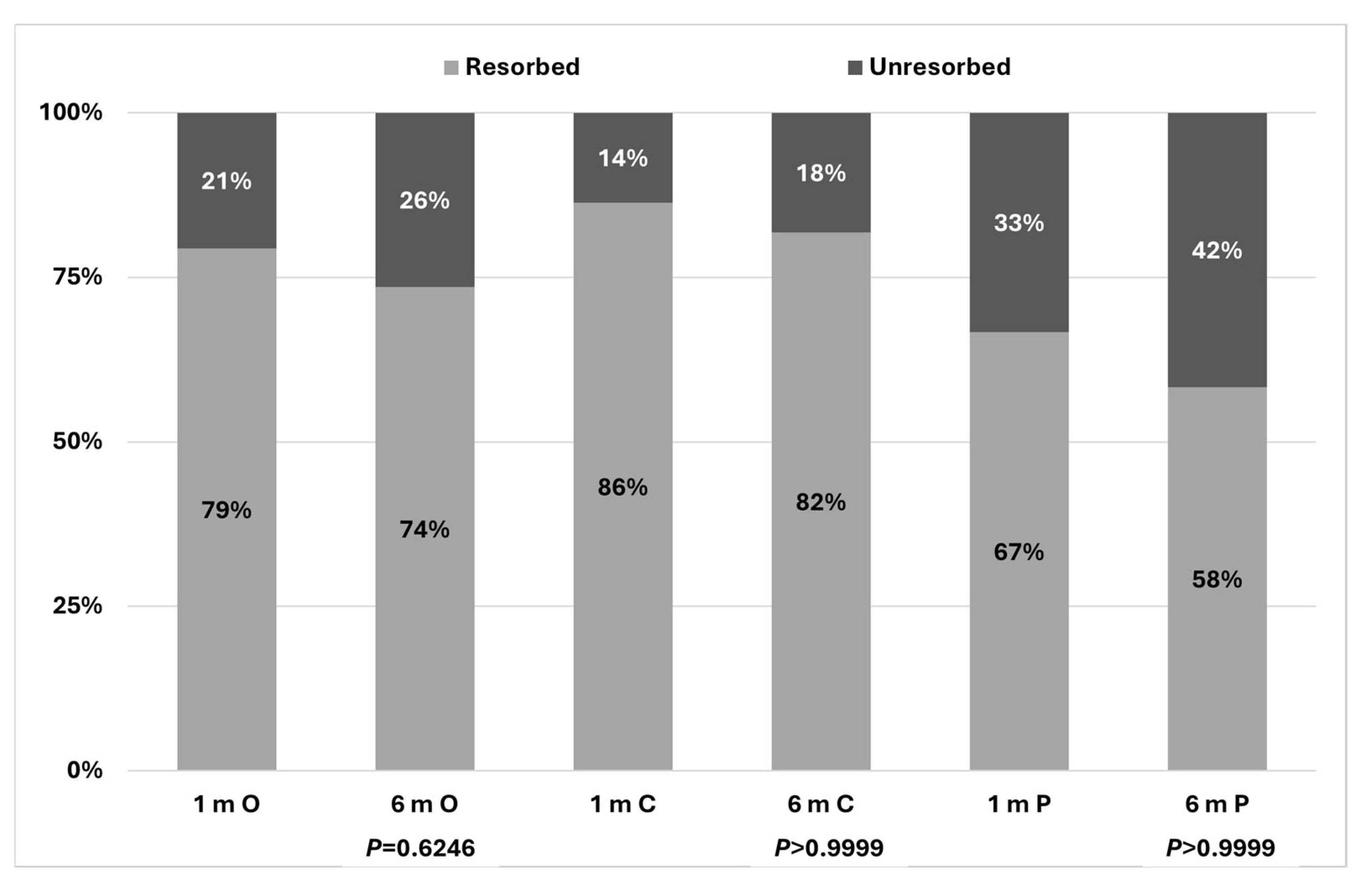
| Gawęcki et al. (2026) (present study) | 34 eyes of 27 patients; 22 eyes central, 12 eyes paramacular PED | Half-dose PDT | Complete resolution in 79.4% at 1 month and 73.5% at 6 months | BCVA improvement significant from 0.22 to 0.1 logMAR at 6 months |
Disclaimer/Publisher’s Note: The statements, opinions and data contained in all publications are solely those of the individual author(s) and contributor(s) and not of MDPI and/or the editor(s). MDPI and/or the editor(s) disclaim responsibility for any injury to people or property resulting from any ideas, methods, instructions or products referred to in the content. |
© 2026 by the authors. Licensee MDPI, Basel, Switzerland. This article is an open access article distributed under the terms and conditions of the Creative Commons Attribution (CC BY) license.
Share and Cite
Gawęcki, M.; Mach, K.; Kiciński, K.; Grzybowski, A. Pachychoroid-Related Pigment Epithelial Detachment Treated with Photodynamic Therapy. Biomedicines 2026, 14, 620. https://doi.org/10.3390/biomedicines14030620
Gawęcki M, Mach K, Kiciński K, Grzybowski A. Pachychoroid-Related Pigment Epithelial Detachment Treated with Photodynamic Therapy. Biomedicines. 2026; 14(3):620. https://doi.org/10.3390/biomedicines14030620
Chicago/Turabian StyleGawęcki, Maciej, Karolina Mach, Krzysztof Kiciński, and Andrzej Grzybowski. 2026. "Pachychoroid-Related Pigment Epithelial Detachment Treated with Photodynamic Therapy" Biomedicines 14, no. 3: 620. https://doi.org/10.3390/biomedicines14030620
APA StyleGawęcki, M., Mach, K., Kiciński, K., & Grzybowski, A. (2026). Pachychoroid-Related Pigment Epithelial Detachment Treated with Photodynamic Therapy. Biomedicines, 14(3), 620. https://doi.org/10.3390/biomedicines14030620