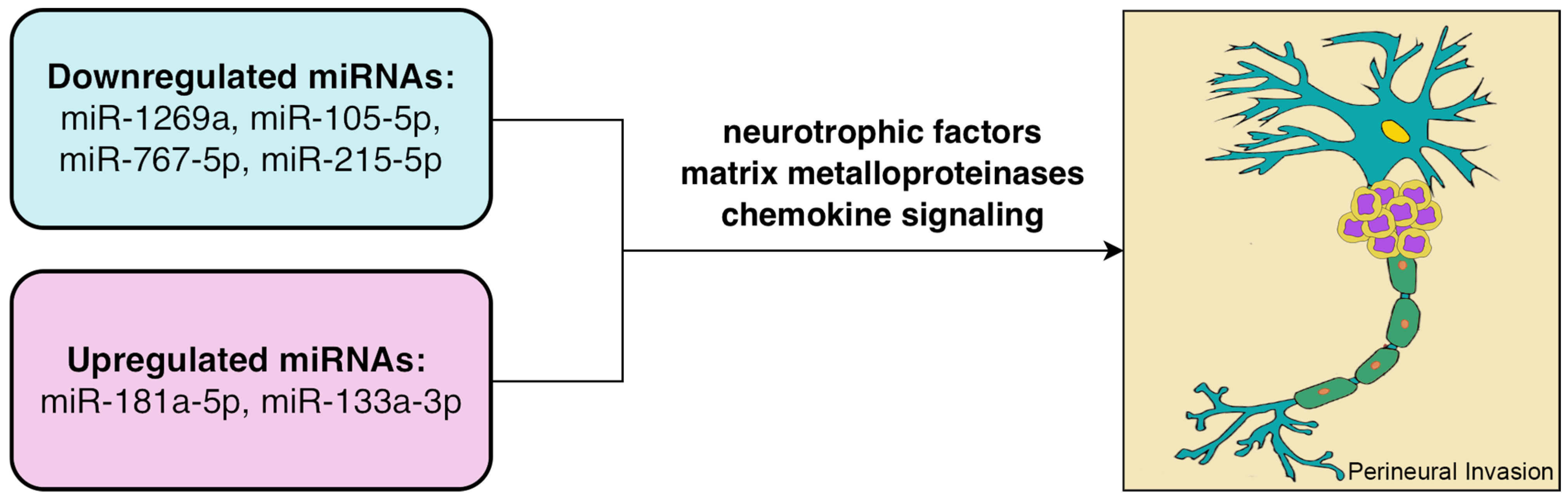
Perineural Invasion in Early-Stage Cervical Cancer: Marker of Aggressive Pathology and Increased Recurrence Risk


Abstract
1. Introduction
2. Materials and Methods
2.1. Study Design and Patient Cohorts
2.2. PNI Assessment
2.3. Molecular Analysis
2.4. Statistical Analysis
3. Results
3.1. Clinical and Pathological Characteristics
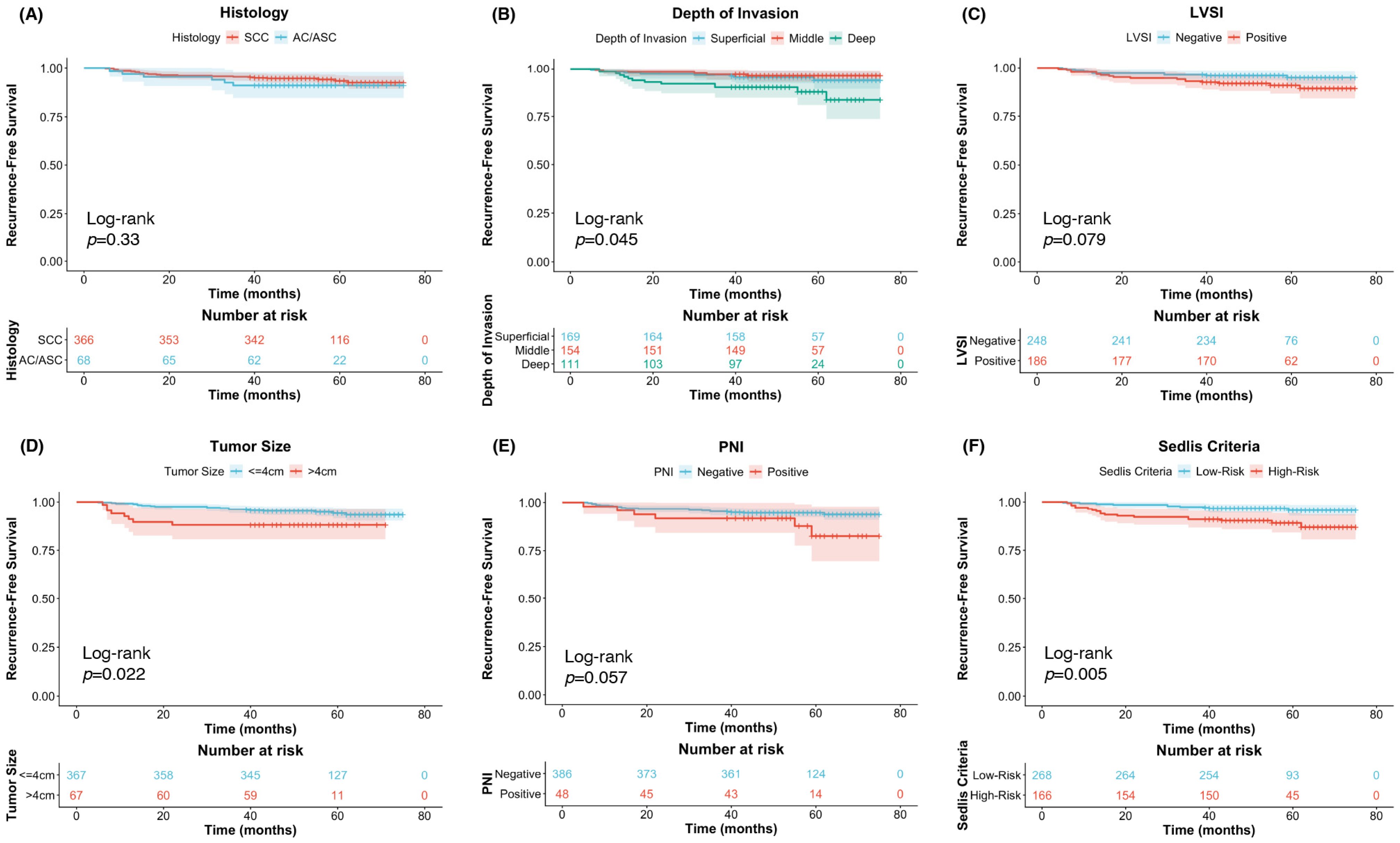
3.2. Prognostic Factors for Survival
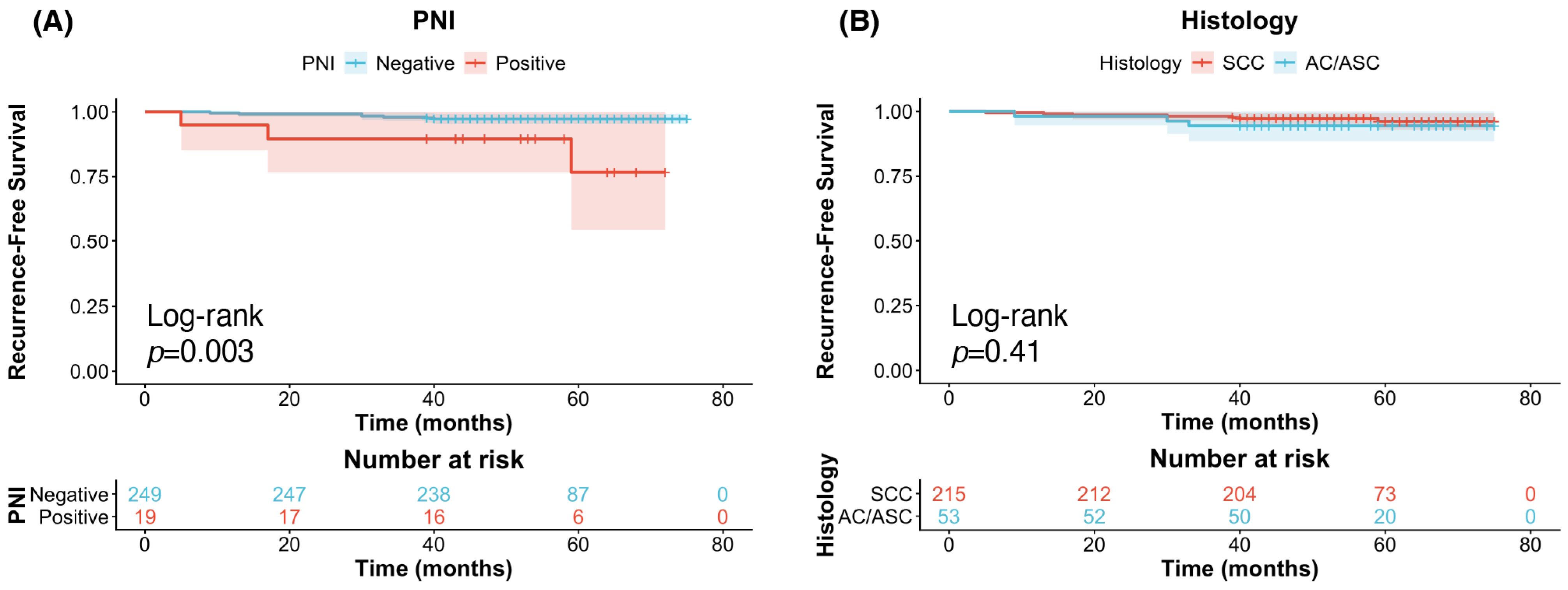
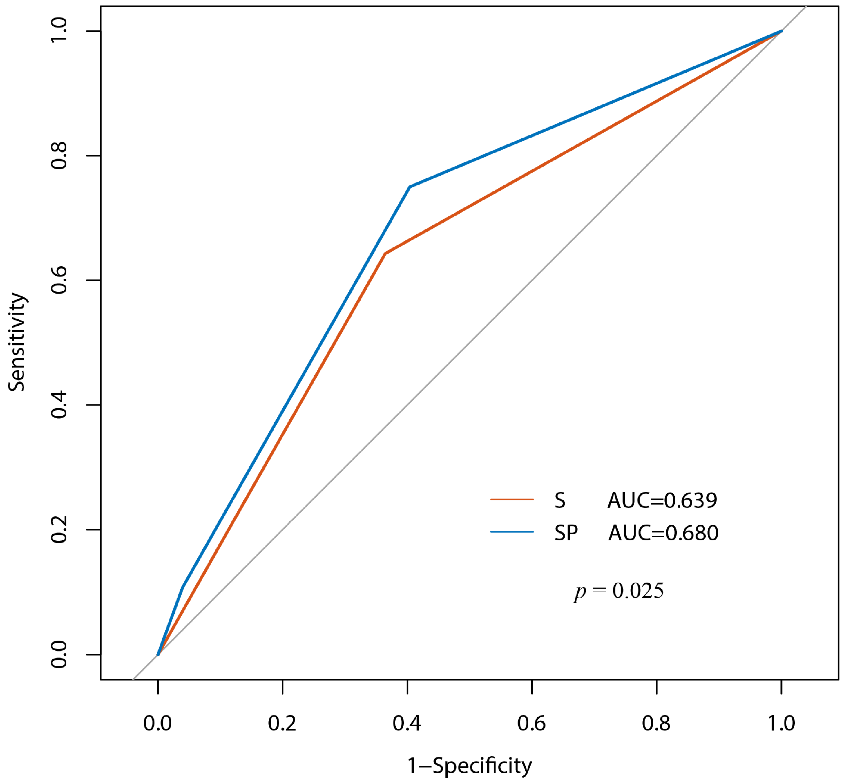
3.3. Prognostic Value of PNI in Patients Not Meeting the Sedlis Criteria
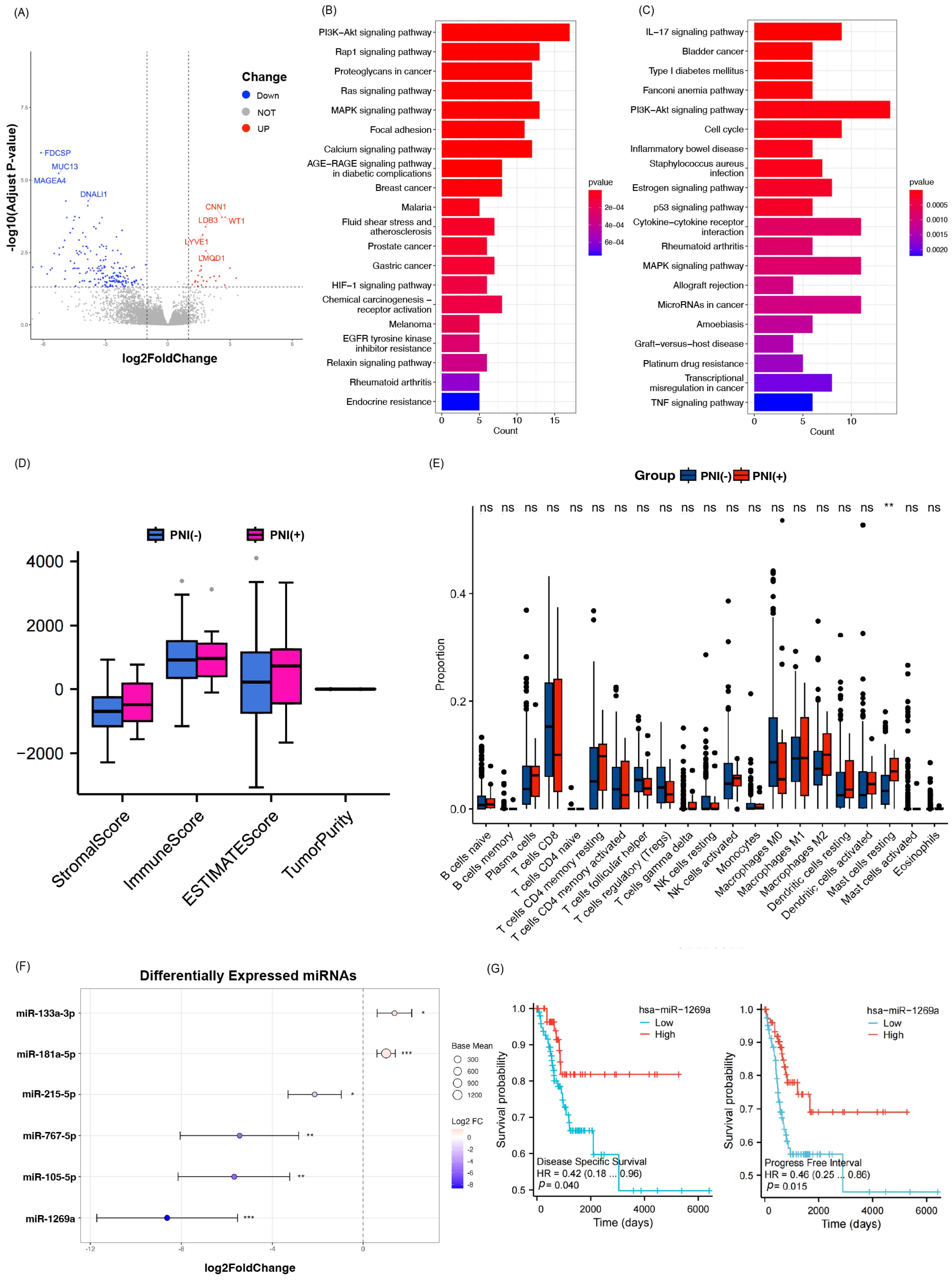
3.4. Immune Microenvironment and Gene Expression
3.5. PNI-Associated miRNAs
3.6. Prognostic Role of hsa-miR-1269a
4. Discussion
5. Conclusions
Author Contributions
Funding
Institutional Review Board Statement
Informed Consent Statement
Data Availability Statement
Conflicts of Interest
Abbreviations
| PNI | Perineural Invasion |
| LVSI | Lymphovascular Invasion |
| RFS | Recurrence-Free Survival |
| OS | Overall Survival |
| CESC | Cervical Squamous Cell Carcinoma |
| FIGO | The International Federation of Gynecology and Obstetrics |
| LNM | Lymph Node Metastasis |
| OR | Odds Ratio |
| CI | Confidence Interval |
| DEGs | Differentially Expressed Genes |
References
- Siegel, R.L.; Miller, K.D.; Wagle, N.S.; Jemal, A. Cancer statistics, 2023. CA Cancer J. Clin. 2023, 73, 17–48. [Google Scholar] [CrossRef] [Scilit]
- Sedlis, A.; Bundy, B.N.; Rotman, M.Z.; Lentz, S.S.; Muderspach, L.I.; Zaino, R.J. A randomized trial of pelvic radiation therapy versus no further therapy in selected patients with stage IB carcinoma of the cervix after radical hysterectomy and pelvic lymphadenectomy: A Gynecologic Oncology Group Study. Gynecol. Oncol. 1999, 73, 177–183. [Google Scholar] [CrossRef] [Scilit]
- Liebig, C.; Ayala, G.; Wilks, J.A.; Berger, D.H.; Albo, D. Perineural invasion in cancer: A review of the literature. Cancer 2009, 115, 3379–3391. [Google Scholar] [CrossRef] [Scilit]
- Takahashi, H.; Ohigashi, H.; Ishikawa, O.; Gotoh, K.; Yamada, T.; Nagata, S.; Tomita, Y.; Eguchi, H.; Doki, Y.; Yano, M. Perineural invasion and lymph node involvement as indicators of surgical outcome and pattern of recurrence in the setting of preoperative gemcitabine-based chemoradiation therapy for resectable pancreatic cancer. Ann. Surg. 2012, 255, 95–102. [Google Scholar] [CrossRef] [Scilit]
- Ozcan, F. Correlation of perineural invasion on radical prostatectomy specimens with other pathologic prognostic factors and PSA failure. Eur. Urol. 2001, 40, 308–312. [Google Scholar] [CrossRef] [Scilit]
- Shen, F.Z.; Zhang, B.Y.; Feng, Y.J.; Jia, Z.X.; An, B.; Liu, C.C.; Deng, X.Y.; Kulkarni, A.D.; Lu, Y. Current research in perineural invasion of cholangiocarcinoma. J. Exp. Clin. Cancer Res. 2010, 29, 24. [Google Scholar] [CrossRef] [Scilit]
- Shimada, K.; Nara, S.; Esaki, M.; Sakamoto, Y.; Kosuge, T.; Hiraoka, N. Intrapancreatic nerve invasion as a predictor for recurrence after pancreaticoduodenectomy in patients with invasive ductal carcinoma of the pancreas. Pancreas 2011, 40, 464–468. [Google Scholar] [CrossRef] [Scilit]
- Zhang, F.; Chen, H.; Luo, D.; Xiong, Z.; Li, X.; Yin, S.; Jin, L.; Chen, S.; Peng, J.; Lian, L. Lymphovascular or perineural invasion is associated with lymph node metastasis and survival outcomes in patients with gastric cancer. Cancer Med. 2023, 12, 9401–9408. [Google Scholar] [CrossRef] [Scilit]
- Cai, G.; Zhang, S.; Gao, S.; Deng, T.; Huang, H.; Feng, Y.; Wan, T. What is the impact of perineural invasion on the prognosis of cervical cancer: A systematic review and meta-analysis. BMC Cancer 2025, 25, 491. [Google Scholar] [CrossRef] [Scilit]
- Memarzadeh, S.; Natarajan, S.; Dandade, D.P.; Ostrzega, N.; Saber, P.A.; Busuttil, A.; Lentz, S.E.; Berek, J.S. Lymphovascular and perineural invasion in the parametria: A prognostic factor for early-stage cervical cancer. Obstet. Gynecol. 2003, 102, 612–619. [Google Scholar] [CrossRef] [Scilit]
- Horn, L.C.; Meinel, A.; Fischer, U.; Bilek, K.; Hentschel, B. Perineural invasion in carcinoma of the cervix uteri--prognostic impact. J. Cancer Res. Clin. Oncol. 2010, 136, 1557–1562. [Google Scholar] [CrossRef] [Scilit]
- Long, Y.; Yao, D.S.; Wei, Y.S.; Wu, G.T. Effects of Nerve Growth Factor Expression on Perineural Invasion and Worse Prognosis in Early-Stage Cervical Cancer. Chin. Med. J. 2018, 131, 2360–2363. [Google Scholar] [CrossRef] [Scilit]
- Zhang, G.; Yang, Y.; Zhu, Y.; Cui, L.; Jia, S.; Shi, Y.; Song, S.; Xu, S. Evidence of perineural invasion on early-stage cervical cancer and prognostic significance. Zhonghua Fu Chan Ke Za Zhi 2015, 50, 673–678. [Google Scholar]
- Wan, T.; Tu, H.; Liu, L.; Huang, H.; Feng, Y.; Liu, J. Perineural Invasion Should Be Regarded as an Intermediate-Risk Factor for Recurrence in Surgically Treated Cervical Cancer: A Propensity Score Matching Study. Dis. Markers 2021, 2021, 1375123. [Google Scholar] [CrossRef] [Scilit]
- Fernandez-Nogueira, P.; Linzoain-Agos, P.; Cueto-Remacha, M.; De la Guia-Lopez, I.; Recalde-Percaz, L.; Parcerisas, A.; Gascon, P.; Carbo, N.; Gutierrez-Uzquiza, A.; Fuster, G.; et al. Role of semaphorins, neuropilins and plexins in cancer progression. Cancer Lett. 2024, 606, 217308. [Google Scholar] [CrossRef] [Scilit]
- Silverman, D.A.; Martinez, V.K.; Dougherty, P.M.; Myers, J.N.; Calin, G.A.; Amit, M. Cancer-Associated Neurogenesis and Nerve-Cancer Cross-talk. Cancer Res. 2021, 81, 1431–1440. [Google Scholar] [CrossRef] [Scilit]
- Zhu, Z.; Zhang, X.; Guo, H.; Fu, L.; Pan, G.; Sun, Y. CXCL13-CXCR5 axis promotes the growth and invasion of colon cancer cells via PI3K/AKT pathway. Mol. Cell. Biochem. 2015, 400, 287–295. [Google Scholar] [CrossRef] [Scilit]
- Xia, Y.; Jiang, T.; Li, Y.; Gu, C.; Lv, J.; Lu, C.; Xu, P.; Fang, L.; Chen, Z.; Liu, H.; et al. circVAPA-rich small extracellular vesicles derived from gastric cancer promote neural invasion by inhibiting SLIT2 expression in neuronal cells. Cancer Lett. 2024, 592, 216926. [Google Scholar] [CrossRef] [Scilit]
- Baraldi, J.H.; Martyn, G.V.; Shurin, G.V.; Shurin, M.R. Tumor Innervation: History, Methodologies, and Significance. Cancers 2022, 14, 1979. [Google Scholar] [CrossRef] [Scilit]
- Zhang, M.; Xin, L.; Cheng, B.; Yan, B.; Zhen, J.; Yang, C.; Ma, L.; Hou, Q. Clinical Value Analysis of Serum TK1, SCC-Ag, and MUC-1 in the Diagnosis and Prognosis Evaluation of Cervical Cancer. Altern. Ther. Health Med. 2023, 29, 788–792. [Google Scholar]
- Ratan, C.; Cicily, K.D.D.; Nair, B.; Nath, L.R. MUC Glycoproteins: Potential Biomarkers and Molecular Targets for Cancer Therapy. Curr. Cancer Drug Targets 2021, 21, 132–152. [Google Scholar] [CrossRef] [Scilit]
- Mancusi de Carvalho, J.P.; Salim, R.C.; Carvalho, F.M.; Nogueira Dias Genta, M.L.; Baracat, E.C.; Carvalho, J.P. L1 cell adhesion molecule (L1CAM) in stage IB cervical cancer: Distinct expression in squamous cell carcinomas and adenocarcinomas. J. Clin. Pathol. 2020, 73, 748–753. [Google Scholar] [CrossRef] [Scilit]
- Benbrook, D.M.; Deng, W.; Gold, M.A.; Rai, R.; Conrad, R.; van der Wel, H.; Husain, S.; Moore, K.; Spirtos, N.; Jackson, A.L.; et al. Association of Sialyl Tn antigen with cervical cancer lymph node status: An NRG oncology/GOG study. Gynecol. Oncol. 2023, 171, 67–75. [Google Scholar] [CrossRef] [Scilit]
- Gao, X.; Wang, Q.; Huang, T.; Xu, C.; Yang, X.; Zhang, L.; Wang, J.; Yang, L.; Zheng, X.; Fan, Q.; et al. Cervical cancer-produced neuromedin-B reprograms Schwann cells to initiate perineural invasion. Cell Death Dis. 2024, 15, 636. [Google Scholar] [CrossRef] [Scilit]
- Chen, G.; Zheng, Z.; Sun, H.; You, J.; Chu, J.; Gao, J.; Qiu, L.; Liu, X. Dedifferentiated Schwann cells promote perineural invasion mediated by the PACAP paracrine signalling in cervical cancer. J. Cell Mol. Med. 2023, 27, 3692–3705. [Google Scholar] [CrossRef] [Scilit]
- Wan, T.; Deng, T.; Peng, X.; Cai, G.; Huang, H.; Ling, Y.; Gao, S.; Li, H.; Yu, D.; Li, H.; et al. Comprehensive Multi-Omic Characterization of Perineural Invasion in Cervical Cancer Reveals Diagnostic Markers, Molecular Drivers, and Therapeutic Strategies. Cancer Res. 2025. [Google Scholar] [CrossRef] [Scilit]
- Liu, J.; Zhang, Y.; Zhao, J.; Yang, Z.; Li, D.; Katirai, F.; Huang, B. Mast cell: Insight into remodeling a tumor microenvironment. Cancer Metastasis Rev. 2011, 30, 177–184. [Google Scholar] [CrossRef] [Scilit]
- Forsythe, P. Mast Cells in Neuroimmune Interactions. Trends Neurosci. 2019, 42, 43–55. [Google Scholar] [CrossRef] [Scilit]
- Keser, S.H.; Kandemir, N.O.; Ece, D.; Gecmen, G.G.; Gul, A.E.; Barisik, N.O.; Sensu, S.; Buyukuysal, C.; Barut, F. Relationship of mast cell density with lymphangiogenesis and prognostic parameters in breast carcinoma. Kaohsiung J. Med. Sci. 2017, 33, 171–180. [Google Scholar] [CrossRef] [Scilit]
- He, S.; Chen, C.H.; Chernichenko, N.; He, S.; Bakst, R.L.; Barajas, F.; Deborde, S.; Allen, P.J.; Vakiani, E.; Yu, Z.; et al. GFRalpha1 released by nerves enhances cancer cell perineural invasion through GDNF-RET signaling. Proc. Natl. Acad. Sci. USA 2014, 111, E2008–E2017. [Google Scholar] [CrossRef] [Scilit]
- He, S.; He, S.; Chen, C.H.; Deborde, S.; Bakst, R.L.; Chernichenko, N.; McNamara, W.F.; Lee, S.Y.; Barajas, F.; Yu, Z.; et al. The chemokine (CCL2-CCR2) signaling axis mediates perineural invasion. Mol. Cancer Res. 2015, 13, 380–390. [Google Scholar] [CrossRef] [Scilit]
- Chen, X.; An, Y.; Zhang, Y.; Xu, D.; Chen, T.; Yang, Y.; Chen, W.; Wu, D.; Zhang, X. CCL26 is upregulated by nab-paclitaxel in pancreatic cancer-associated fibroblasts and promotes PDAC invasiveness through activation of the PI3K/AKT/mTOR pathway. Acta Biochim. Biophys. Sin. 2021, 53, 612–619. [Google Scholar] [CrossRef] [Scilit]
- Wang, J.; Li, Q.; Liang, F.; Du, X.; Song, P.; Wu, T.; Chen, R.; Lin, X.; Liu, Q.; Hu, H.; et al. Dickkopf-1 drives perineural invasion via PI3K-AKT signaling pathway in head and neck squamous cancer. MedComm 2024, 5, e518. [Google Scholar] [CrossRef] [Scilit]
- Kim, J.H.; Lee, K.W.; Ahn, D.G.; Oh, K.Y.; Yoon, H.J. Clinical significance of L1CAM expression and its biological role in the progression of oral squamous cell carcinoma. Oncol. Rep. 2023, 49, 67. [Google Scholar] [CrossRef] [Scilit]
- Hansen, F.J.; David, P.; Akram, M.; Knoedler, S.; Mittelstadt, A.; Merkel, S.; Podolska, M.J.; Swierzy, I.; Rossdeutsch, L.; Klosch, B.; et al. Circulating Monocytes Serve as Novel Prognostic Biomarker in Pancreatic Ductal Adenocarcinoma Patients. Cancers 2023, 15, 363. [Google Scholar] [CrossRef] [Scilit]
- Bostanghadiri, N.; Razavi, S.; Shariati, A.; Talebi, M.; Mirkalantari, S.; Emami Razavi, A.; Darban-Sarokhalil, D. Exploring the interplay between Fusobacterium nucleatum with the expression of microRNA, and inflammatory mediators in colorectal cancer. Front. Microbiol. 2023, 14, 1302719. [Google Scholar] [CrossRef] [Scilit]
- Zhou, Y.; Qin, Y.; Ma, J.; Li, Z.; Heng, W.; Zhang, L.; Liu, H.; Li, R.; Zhang, M.; Peng, Q.; et al. Heat-killed Prevotella intermedia promotes the progression of oral squamous cell carcinoma by inhibiting the expression of tumor suppressors and affecting the tumor microenvironment. Exp. Hematol. Oncol. 2024, 13, 33. [Google Scholar] [CrossRef] [Scilit]
- Kori, M.; Yalcin Arga, K. Potential biomarkers and therapeutic targets in cervical cancer: Insights from the meta-analysis of transcriptomics data within network biomedicine perspective. PLoS ONE 2018, 13, e0200717. [Google Scholar] [CrossRef] [Scilit]
- Xie, Z.; Zhong, C.; Duan, S. miR-1269a and miR-1269b: Emerging Carcinogenic Genes of the miR-1269 Family. Front. Cell Dev. Biol. 2022, 10, 809132. [Google Scholar] [CrossRef] [Scilit]
- Xu, H.; Zhu, J.; Hu, C.; Song, H.; Li, Y. Inhibition of microRNA-181a may suppress proliferation and invasion and promote apoptosis of cervical cancer cells through the PTEN/Akt/FOXO1 pathway. J. Physiol. Biochem. 2016, 72, 721–732. [Google Scholar] [CrossRef] [Scilit]
- Liu, W.L.; Wang, H.X.; Shi, C.X.; Shi, F.Y.; Zhao, L.Y.; Zhao, W.; Wang, G.H. MicroRNA-1269 promotes cell proliferation via the AKT signaling pathway by targeting RASSF9 in human gastric cancer. Cancer Cell. Int. 2019, 19, 308. [Google Scholar] [CrossRef] [Scilit]
- Bu, P.; Wang, L.; Chen, K.Y.; Rakhilin, N.; Sun, J.; Closa, A.; Tung, K.L.; King, S.; Kristine Varanko, A.; Xu, Y.; et al. miR-1269 promotes metastasis and forms a positive feedback loop with TGF-beta. Nat. Commun. 2015, 6, 6879. [Google Scholar] [CrossRef] [Scilit]
- Yin, Y.; Du, L.; Li, X.; Zhang, X.; Gao, Y. miR-133a-3p suppresses cell proliferation, migration, and invasion and promotes apoptosis in esophageal squamous cell carcinoma. J. Cell. Physiol. 2019, 234, 12757–12770. [Google Scholar] [CrossRef] [Scilit]
- Tang, Y.; Pan, J.; Huang, S.; Peng, X.; Zou, X.; Luo, Y.; Ren, D.; Zhang, X.; Li, R.; He, P.; et al. Downregulation of miR-133a-3p promotes prostate cancer bone metastasis via activating PI3K/AKT signaling. J. Exp. Clin. Cancer Res. 2018, 37, 160. [Google Scholar] [CrossRef] [Scilit]
- Zhang, M.; Xian, H.C.; Dai, L.; Tang, Y.L.; Liang, X.H. MicroRNAs: Emerging driver of cancer perineural invasion. Cell. Biosci. 2021, 11, 117. [Google Scholar] [CrossRef] [Scilit]



| Characteristics | PNI | p-Value | ||
|---|---|---|---|---|
| Negative (n = 441) | Positive (n = 58) | |||
| Age, years | <60 | 400 (90.7) | 52 (89.7) | 0.797 |
| ≥60 | 41 (9.3) | 6 (10.3) | ||
| Conization | no | 402 (91.2) | 55 (94.8) | 0.455 |
| yes | 39 (8.8) | 3 (5.2) | ||
| FIGO stage | IB | 369 (83.7) | 49 (84.5) | 0.875 |
| IIA | 72 (16.3) | 9 (15.5) | ||
| Tumor size, cm | ≤2 | 160 (36.3) | 10 (17.2) | 0.002 |
| 2~4 | 206 (46.7) | 29 (50.0) | ||
| >4 | 75 (17.0) | 19 (32.8) | ||
| Histology | SCC | 373 (84.6) | 47 (81.0) | 0.487 |
| AC/ASC | 68 (15.4) | 11 (19.0) | ||
| Grade | G1 and G2 | 426 (96.6) | 54 (93.1) | 0.260 |
| G3 | 15 (3.4) | 4 (6.9) | ||
| Vaginal invasion | negative | 350 (79.4) | 38 (65.5) | 0.017 |
| positive | 91 (20.6) | 20 (34.5) | ||
| Depth of invasion | superficial 1/3 | 171 (38.8) | 9 (15.5) | <0.001 |
| middle 1/3 | 154 (34.9) | 17 (29.3) | ||
| deep 1/3 | 116 (26.3) | 32 (55.2) | ||
| LVSI | negative | 244 (55.3) | 24 (41.4) | 0.045 |
| positive | 197 (44.7) | 34 (58.6) | ||
| LNM | negative | 386 (87.5) | 48 (82.8) | 0.310 |
| positive | 55 (12.5) | 10 (17.2) | ||
| 5-year RFS | alive | 405 (91.8) | 50 (86.2) | 0.155 |
| dead | 36 (8.2) | 8 (13.8) | ||
| 5-year OS | alive | 426 (96.6) | 54 (93.1) | 0.260 |
| dead | 15 (3.4) | 4 (6.9) | ||
| Follow-up time, months | 54.29 (46.00–63.00) | 52.62 (44.75–61.25) | ||
| Characteristics | 5-Year RFS | p-Value | 5-Year OS | p-Value | |
|---|---|---|---|---|---|
| Univariate Analysis OR (95% CI) | Univariate Analysis OR (95% CI) | ||||
| Age, years | <60 | 1 | 0.124 | 1 | 0.534 |
| ≥60 | 0.207 (0.028–1.537) | 0.524 (0.068–4.017) | |||
| Conization | no | 1 | 0.866 | 1 | 0.736 |
| yes | 1.097 (0.373–3.232) | 1.294 (0.289–5.803) | |||
| FIGO stage | IB | 1 | 0.103 | 1 | 0.232 |
| IIA | 1.833 (0.885–3.798) | 1.898 (0.664–5.426) | |||
| Tumor size, cm | ≤2 | 1 | 0.018 | 1 | 0.003 |
| 2~4 | 1.664 (0.738–3.751) | 0.219 | 5.956 (0.738–48.075) | 0.094 | |
| >4 | 3.397 (1.424–8.101) | 0.006 | 20.119 (2.533–159.793) | 0.005 | |
| Histology | SCC | 1 | 0.193 | 1 | 0.210 |
| AC/ASC | 1.645 (0.777–3.484) | 1.959 (0.685–5.604) | |||
| Grade | G1 and G2 | 1 | 0.582 | 1 | 0.737 |
| G3 | 0.565 (0.074–4.333) | 1.426 (0.180–11.278) | |||
| Vaginal invasion | negative | 1 | 0.225 | 1 | 0.126 |
| positive | 1.5228 (0.770–3.031) | 2.109 (0.810–5.492) | |||
| Depth of invasion | superficial 1/3 | 1 | 0.010 | 1 | 0.205 |
| middle 1/3 | 1.283 (0.539–3.052) | 0.573 | 1.600 (0.444–5.771) | 0.473 | |
| deep 1/3 | 1.968 (1.358–6.489) | 0.006 | 2.849 (0.859–9.446) | 0.087 | |
| LVSI | negative | 1 | 0.001 | 1 | 0.021 |
| positive | 3.040 (1.550–5.963) | 3.394 (1.203–9.571) | |||
| PNI | negative | 1 | 0.160 | 1 | 0.201 |
| positive | 1.800 (0.792–4.089) | 2.104 (0.674–6.569) | |||
| LNM | negative | 1 | <0.001 | 1 | 0.004 |
| positive | 4.735 (2.394–9.365) | 4.244 (1.606–11.215) | |||
| Sedlis criteria | low-risk | 1 | <0.001 | 1 | 0.003 |
| high-risk | 3.711 (1.890–7.284) | 5.528 (1.808–16.908) |
| Characteristics | 5-Year RFS | p-Value | 5-Year OS | p-Value | |
|---|---|---|---|---|---|
| Multivariate Analysis OR (95% CI) | Multivariate Analysis OR (95% CI) | ||||
| Tumor size, cm | ≤2 | Reference | 0.567 | Reference | 0.021 |
| 2~4 | 1.290 (0.544,3.060) | 0.564 | 5.123 (0.611,42.931) | 0.132 | |
| >4 | 1.714 (0.631,4.652) | 0.290 | 14.812 (1.698,129.218) | 0.015 | |
| Depth of invasion | superficial 1/3 | Reference | 0.639 | Reference | 0.865 |
| middle 1/3 | 0.924 (0.369,2.312) | 0.866 | 0.932 (0.244,3.554) | 0.918 | |
| deep 1/3 | 1.350 (0.540,3.378) | 0.521 | 0.713 (0.181,2.812) | 0.628 | |
| LVSI | negative | Reference | Reference | ||
| positive | 2.204 (1.071,4.534) | 0.032 | 2.383 (0.792,7.175) | 0.122 | |
| PNI | negative | Reference | Reference | ||
| positive | 1.261 (0.519,3.063) | 0.608 | 1.366 (0.407,4.587) | 0.613 | |
| LNM | negative | Reference | Reference | ||
| positive | 3.176 (1.505,6.700) | 0.002 | 2.391 (0.797,7.175) | 0.120 | |
Disclaimer/Publisher’s Note: The statements, opinions and data contained in all publications are solely those of the individual author(s) and contributor(s) and not of MDPI and/or the editor(s). MDPI and/or the editor(s) disclaim responsibility for any injury to people or property resulting from any ideas, methods, instructions or products referred to in the content. |
© 2026 by the authors. Licensee MDPI, Basel, Switzerland. This article is an open access article distributed under the terms and conditions of the Creative Commons Attribution (CC BY) license.
Share and Cite
Tan, L.; Li, H.; Liu, T.; Mao, W.; Song, Y.; Zhao, D. Perineural Invasion in Early-Stage Cervical Cancer: Marker of Aggressive Pathology and Increased Recurrence Risk. Biomedicines 2026, 14, 591. https://doi.org/10.3390/biomedicines14030591
Tan L, Li H, Liu T, Mao W, Song Y, Zhao D. Perineural Invasion in Early-Stage Cervical Cancer: Marker of Aggressive Pathology and Increased Recurrence Risk. Biomedicines. 2026; 14(3):591. https://doi.org/10.3390/biomedicines14030591
Chicago/Turabian StyleTan, Lihua, Hongyao Li, Tianyi Liu, Wei Mao, Yan Song, and Dan Zhao. 2026. "Perineural Invasion in Early-Stage Cervical Cancer: Marker of Aggressive Pathology and Increased Recurrence Risk" Biomedicines 14, no. 3: 591. https://doi.org/10.3390/biomedicines14030591
APA StyleTan, L., Li, H., Liu, T., Mao, W., Song, Y., & Zhao, D. (2026). Perineural Invasion in Early-Stage Cervical Cancer: Marker of Aggressive Pathology and Increased Recurrence Risk. Biomedicines, 14(3), 591. https://doi.org/10.3390/biomedicines14030591