Bisphenol F and Steatotic Liver Disease: Resolving the PXR Paradox Through Stress Pathway Mechanisms
 ,
,
Abstract
1. Introduction
2. Steatotic Liver Disease and Metabolic Dysfunction-Associated Steatotic Liver Disease
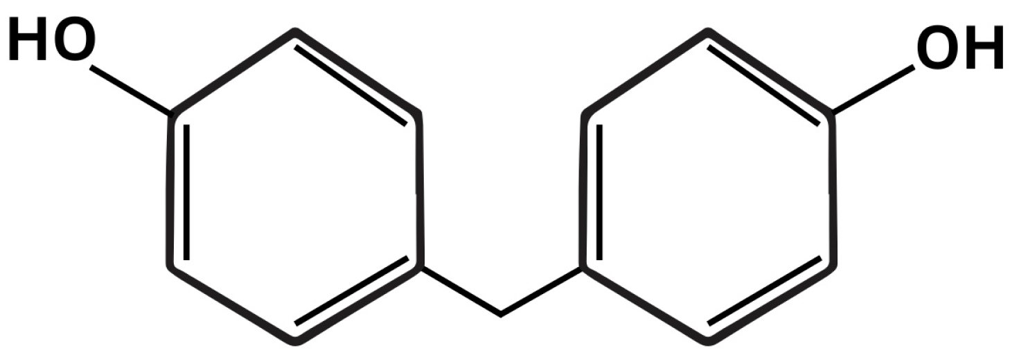
3. Bisphenol F
4. Mechanisms Involved in Bisphenol F-Induced SLD
4.1. The PXR-Dependent Axis: Conceptual Framework and Limited BPF Evidence
4.1.1. PXR as a Master Metabolic Regulator—General Framework
4.1.2. BPF Interaction with PXR: Weak Direct Agonism
4.2. PXR-Independent Mechanisms: Integrated Mechanistic Framework
4.2.1. Oxidative Stress and ER Dysfunction: Primary Initiating Events
4.2.2. Mitochondrial Dysfunction and Energy Imbalance: A Central Hub
4.2.3. Inflammation: Convergence of Innate Immune Activation and Hepatocyte Injury
4.2.4. Post-Translational Modifications: Lipogenic Reprogramming Independent of PXR Activation
4.2.5. Epigenetic Modifications: Heritable Reprogramming and Metabolic Memory
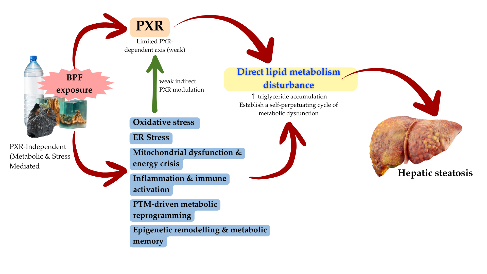
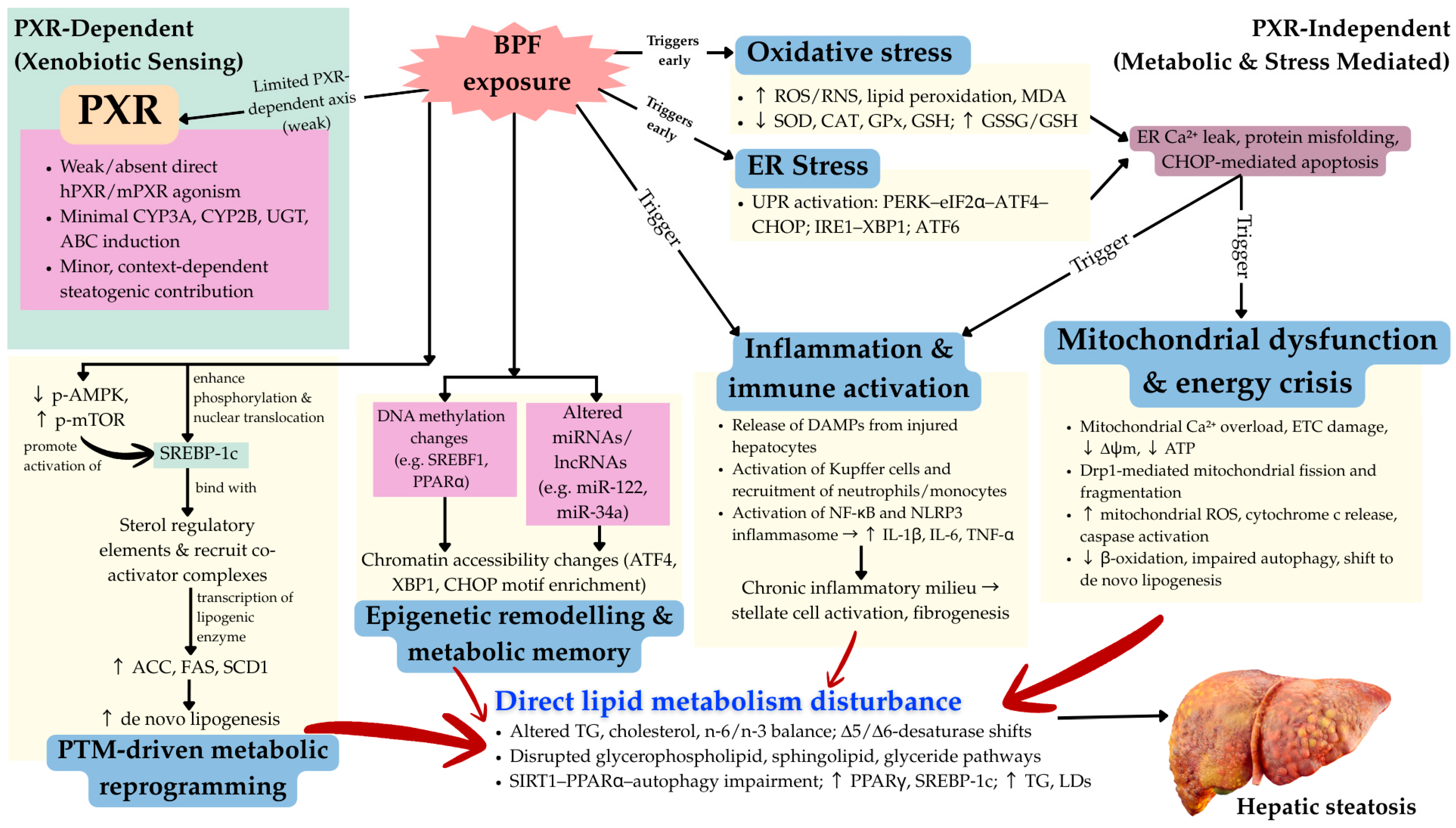
4.2.6. Integration: Multi-Hit Mechanistic Model
5. BPF Induced Disruption of Lipid Metabolism
6. Discussion
6.1. The PXR Paradox and Its Implications
6.2. Evidence Hierarchy: Well-Supported Versus Speculative Mechanisms

| Mechanisms | PXR Dependence | Evidence Strength | In Vitro OR In Vivo Studies | Major Limitations | Functional Validation | References |
|---|---|---|---|---|---|---|
| Oxidative stress and ROS elevation | None (direct) | Strong | Both robust | Non-physiological BPF concentrations (in vitro); limited time-course studies; unclear dose–response relationship for environmentally relevant exposures | Antioxidant enzyme activity measured; GSH/GSSG ratios quantified; MDA/lipid peroxidation confirmed | Zhang et al. [6], Meng et al. [12], Aykut et al. [43], Linillos-Pradillo et al. [44], Lința et al. [63], Sangwan et al. [95], Charles & Prince [96] |
| ER stress and UPR activation | None (direct) | Strong | Both; strong multi-omics | Chromatin accessibility data largely correlative; limited mechanistic link between UPR branch activation and specific outcomes; no UPR component knockdown studies | Transcriptomic evidence for ATF6, PERK, IRE1 branch activation; multi-omics enrichment for ER-related transcription factors | Fan et al. [8], Luo et al. [60], Chipurupalli et al. [97] |
| Drp1-mediated mitochondrial fission | None (direct) | Strong | In vivo primarily | Limited to one key study; Drp1 inhibition only tested in one model; sex differences not examined; age-dependence unclear | Drp1 inhibition restores mitochondrial integrity and reduces lipid deposition | Zhang et al. [6] |
| NLRP3/NF-κB inflammatory activation | None (direct) | Strong | Both; consistent across models | Mostly descriptive (cytokine measurement); limited mechanistic dissection of NLRP3 vs. NF-κB contributions; inflammasome component knockdown not tested; microbiota role speculative | Cytokine elevation replicated; NLRP3 activation | Zhang et al. [6], Linillos-Pradillo et al. [44], Taru et al. [98], Jee et al. [73], Peinado et al. [76], Marques et al. [77] |
| SREBP-1c PTM and lipogenic reprogramming | None (direct) | Moderate | Both (limited In vivo) | Only one comprehensive PTM study (Xue et al.); limited mechanistic detail on which PTMs activate SREBP-1c; no SREBP-1c knockout rescue; unclear whether PTMs are primary drivers or consequences | SREBP-1c nuclear translocation and lipogenic enzyme upregulation demonstrated | Cao et al. [99] |
| AMPK–mTOR dysregulation | None (direct) | Moderate | Both (limited in vivo) | Phosphorylation status measured but causality not tested (no pAMPK/mTOR manipulation); relationship to energy deficit unclear; tissue-specific effects not examined | pAMPK decrease and p-mTOR increase replicated; but no pharmacological/genetic intervention | Xue et al. [45], Yang et al. [83] |
| Autophagy disruption and SIRT1–PPARα impairment | None (direct) | Moderate | Both (limited in vivo) | Single study; no ATG gene knockout/knockdown; SIRT1/PPARα inhibition not tested; unclear whether autophagy impairment is primary or secondary to mitochondrial dysfunction | Autophagy marker changes (LC3, p62, ATG genes) documented; lipid droplet accumulation confirmed | Wang et al. [54] |
| Epigenetic remodelling (DNA methylation, chromatin accessibility, miR dysregulation) | None (direct) | Moderate–weak | Both; limited in vivo | Correlative multi-omics data only; no CRISPR-based epigenetic editing to test causality; no pharmacologic epigenetic modifier studies; transgenerational effects entirely speculative; unclear whether epigenetic changes are drivers or consequences of stress | Chromatin accessibility and transcriptomic changes documented; no functional validation of epigenetic causality | Fan et al. [8], Meng et al. [12], Ramirez et al. [87], Merrel et al. [100] |
| Indirect PXR modulation via oxidative/metabolic stress | Indirect (non-ligand-dependent) | Weak/speculative | Primarily in vivo | No direct evidence that PXR knockout/antagonism attenuates BPF-induced SLD; In vitro PXR activation assays consistently negative; mechanistic link between oxidative stress and PXR modulation not demonstrated; species-specific differences unexplained; no humanized PXR model tested | CYP3A and PPARγ induction observed in vivo, but causality to PXR unproven; could reflect CAR or PPAR-driven responses | Sun et al. [33], De Battistis et al. [52], Xue et al. [45], Niu et al. [51] |
| Direct PXR ligand-dependent activation | Direct (ligand- dependent) | Not supported | In vitro only (negative findings) | Multiple independent In vitro studies show NO hPXR or mPXR activation by BPF; structural differences (loss of methyl groups) explain reduced affinity; no in vivo validation of ligand-dependent mechanism; contradicts weak PXR binding and agonism data | Transient transfection reporter assays negative for BPF; structural model predicts poor binding | Sui Et Al. [40], Grimaldi Et Al. [41] |
6.3. Why PXR-Independent Pathways Dominate
6.4. Critical Knowledge Gaps and Research Priorities
6.5. Regulatory and Public Health Implications
6.6. Therapeutic Implications
| Bisphenols | PXR Activation (In Vitro/In Vivo) | Structural Determinants | Key Findings | Potential Indirect Mechanisms | Authors |
|---|---|---|---|---|---|
| Bisphenol A (BPA) | Strong activator of hPXR; No effect on mPXR | Two methyl groups on bridge; para-hydroxy phenyl rings | Induces CYP3A4, CD36, PPARγ; triggers steatosis and hyperlipidemia | Direct ligand binding; Energetically stable direct ligand binding (LBD) fit | Sui Et Al. [40,105] |
| Bisphenol F (BPF) | Weak or no activation of hPXR/mPXR | Missing methyl groups; reduced hydrophobic complementarity | Mimics downstream PXR responses (CYP, lipogenic gene upregulation); causes SLD phenotypes | Indirect activation via oxidative stress, FXR–PXR crosstalk and CAR pathways | Ji et al. [24]; Han et al. [106]; Grimaldi Et Al. [55] |
| Bisphenol S (BPS) | No activation of hPXR or mPXR | Sulfone linker (SO2) disrupts hydrogen bond with Ser247 | Weaker lipid-accumulating effect in rodent models | Alters AhR/ER signalling; no PXR dependence | Fang et al. [107]; Han et al. [106] |
7. Conclusions
Author Contributions
Funding
Institutional Review Board Statement
Informed Consent Statement
Data Availability Statement
Acknowledgments
Conflicts of Interest
Abbreviations
| BPF | Bisphenol F |
| PXR | Pregnane-X Receptor |
| FXR | Farnesoid X Receptor |
| LXR | Liver X Receptor |
| CAR | Constitutive Androstane Receptor |
| CYP3A4 | cytochrome P450 3A4 |
| ↑ | Increase |
| ↓ | Decrease |
| PTMs | Post-Translational Modifications |
| MDA | Malondialdehyde |
| SOD | Superoxide dismutase |
| CAT | Catalase |
| GSH | Glutathione |
| GST | Glutathione S transferase |
| GPx | Glutathione peroxidase |
| GRx | Glutathione reductase |
| µM | Micro molar |
| h | Hour |
| GSSG | Glutathione Disulfide |
| LPO | Lipid peroxidation |
| NLRP3 | NLR family pyrin domain containing 3 |
| PyCARD | PYRIN domain and a caspase-recruitment domain |
| CASP1 | gene that codes for Caspase-1 |
| IL-1β | Interleukin 1 beta |
| IL-18 | Interleukin-18 |
| IFN-γ | Interferon gamma |
| TNF-α | Tumor necrosis factor alpha |
| Drp1 | Dynamin-Related Protein 1 |
| Acox1 | Acyl-CoA oxidase 1 |
| PMP70 | Peroxisomal membrane protein 70 |
| Pex5 | Peroxisomal Biogenesis Factor 5 |
| Pex19 | Peroxisomal Biogenesis Factor 19 |
| PPARα | Peroxisome proliferator-activated receptor alpha |
| CPT1α | Carnitine Palmitoyltransferase 1 alpha |
| ACSL1 | Acyl-CoA Synthetase Long Chain Family Member 1 |
| CoxIV | Cytochrome c oxidase subunit IV |
| SHP | Small Heterodimer Partner |
| FA | Fatty acid |
| TG | Triglyceride |
| ACC | Acetyl-CoA Carboxylase |
| FAS | Fatty Acid Synthase |
| SREBP-1c | Sterol Regulatory Element-Binding Protein-1c |
| CEBPA | CCAAT-Enhancer-Binding Protein Alpha |
| PPARG | Peroxisome Proliferator-Activated Receptor Gamma |
| SCD1 | Stearoyl-CoA Desaturase 1 |
| AGPAT1 | 1-acylglycerol-3-phosphate O-acyltransferase 1 |
| DLK1 | Delta-like 1 homolog |
| AMPK | AMP-activated protein kinase |
| p-mTOR | phosphorylated mTOR |
References
- Gan, C.; Yuan, Y.; Shen, H.; Gao, J.; Kong, X.; Che, Z.; Guo, Y.; Wang, H.; Dong, E.; Xiao, J.; et al. Liver diseases: Epidemiology, causes, trends and predictions. Signal Transduct. Target. Ther. 2025, 10, 33. [Google Scholar] [CrossRef] [PubMed]
- Kan, C.; Zhang, K.; Wang, Y.; Zhang, X.; Liu, C.; Ma, Y.; Hou, N.; Huang, N.; Han, F.; Sun, X. Global burden and future trends of metabolic dysfunction-associated Steatotic liver disease: 1990–2021 to 2045. Ann. Hepatol. 2025, 30, 101898. [Google Scholar] [CrossRef] [PubMed]
- Li, L.; Lin, W.; Wang, Z.; Huang, R.; Xia, H.; Li, Z.; Deng, J.; Ye, T.; Huang, Y.; Yang, Y. Hormone Regulation in Testicular Development and Function. Int. J. Mol. Sci. 2024, 25, 5805. [Google Scholar] [CrossRef] [PubMed]
- Wang, J.; Yu, P.; Xie, X.; Wu, L.; Zhou, M.; Huan, F.; Jiang, L.; Gao, R. Bisphenol F induces nonalcoholic fatty liver disease-like changes: Involvement of lysosome disorder in lipid droplet deposition. Environ. Pollut. 2021, 271, 116304. [Google Scholar] [CrossRef]
- Rajak, S.; Raza, S.; Tewari, A.; Sinha, R.A. Environmental toxicants and NAFLD: A neglected yet significant relationship. Dig. Dis. Sci. 2022, 67, 3497–3507. [Google Scholar] [CrossRef]
- Zhang, L.; Xie, X.; Tao, J.; Wang, S.; Hu, M.; Wang, X.; Yu, Z.; Xu, L.; Lin, Y.; Wu, W.; et al. Mystery of bisphenol F-induced nonalcoholic fatty liver disease-like changes: Roles of Drp1-mediated abnormal mitochondrial fission in lipid droplet deposition. Sci. Total Environ. 2023, 904, 166831. [Google Scholar] [CrossRef]
- Li, C.-L.; Yao, Z.-Y.; Zhang, Y.-F.; Cui, X.-T.; Sun, A.; Cao, J.-Y.; Wang, Z.-S. Bisphenols exposure and non-alcoholic fatty liver disease: From environmental trigger to molecular pathogenesis. Front. Endocrinol. 2025, 16, 1606654. [Google Scholar] [CrossRef]
- Fan, Y.; Li, S.; Yang, X.; Bai, S.; Tang, M.; Zhang, X.; Lu, C.; Ji, C.; Du, G.; Qin, Y. Multi-omics approach characterizes the role of Bisphenol F in disrupting hepatic lipid metabolism. Environ. Int. 2024, 187, 108690. [Google Scholar] [CrossRef]
- Xu, Y.; An, Z.; Wang, S.; Ni, Y.; Zhou, M.; Feng, Q.; Gou, X.; Xu, M.; Qi, Y. Dual role of pregnane X receptor in nonalcoholic fatty liver disease. Curr. Mol. Pharmacol. 2024, 17, E18761429259143. [Google Scholar] [CrossRef]
- Fritsche, K.; Ziková-Kloas, A.; Marx-Stoelting, P.; Braeuning, A. Metabolism-disrupting chemicals affecting the liver: Screening, testing, and molecular pathway identification. Int. J. Mol. Sci. 2023, 24, 2686. [Google Scholar] [CrossRef]
- Ikeda, K.; Horie, K.; Inoue, S.; Blumberg, B. Physiological Functions and Pathological Roles of PXR. J. Endocr. Soc. 2025, 9, bvaf119. [Google Scholar] [CrossRef] [PubMed]
- Meng, Z.; Tian, S.; Yan, J.; Jia, M.; Yan, S.; Li, R.; Zhang, R.; Zhu, W.; Zhou, Z. Effects of perinatal exposure to BPA, BPF and BPAF on liver function in male mouse offspring involving in oxidative damage and metabolic disorder. Environ. Pollut. 2019, 247, 935–943. [Google Scholar] [CrossRef] [PubMed]
- Israelsen, M.; Francque, S.; Tsochatzis, E.A.; Krag, A. Steatotic liver disease. Lancet 2024, 404, 1761–1778. [Google Scholar] [CrossRef]
- Shiha, G.; Alswat, K.; Al Khatry, M.; Sharara, A.I.; Örmeci, N.; Waked, I.; Benazzouz, M.; Al-Ali, F.; Hamed, A.E.; Hamoudi, W.; et al. Nomenclature and definition of metabolic-associated fatty liver disease: A consensus from the Middle East and north Africa. Lancet Gastroenterol. Hepatol. 2021, 6, 57–64. [Google Scholar] [CrossRef] [PubMed]
- Chan, W.-K.; Chuah, K.-H.; Rajaram, R.B.; Lim, L.-L.; Ratnasingam, J.; Vethakkan, S.R. Metabolic dysfunction-associated steatotic liver disease (MASLD): A state-of-the-art review. J. Obes. Metab. Syndr. 2023, 32, 197. [Google Scholar] [CrossRef]
- Marchesini, G.; Vettor, R.; Pinzani, M. MASLD emerging from the fog of fatty liver. J. Hepatol. 2024, 80, 178–180. [Google Scholar] [CrossRef]
- Mostafavi Abdolmaleky, H.; Zhou, J.-R. Gut microbiota dysbiosis, oxidative stress, inflammation, and epigenetic alterations in metabolic diseases. Antioxidants 2024, 13, 985. [Google Scholar] [CrossRef]
- Melaram, R. Environmental risk factors implicated in liver disease: A mini-review. Front. Public Health 2021, 9, 683719. [Google Scholar] [CrossRef]
- Mosca, A.; Manco, M.; Braghini, M.R.; Cianfarani, S.; Maggiore, G.; Alisi, A.; Vania, A. Environment, endocrine disruptors, and fatty liver disease associated with metabolic dysfunction (MASLD). Metabolites 2024, 14, 71. [Google Scholar] [CrossRef]
- Beier, J.I.; Luo, J.; Vanderpuye, C.-M.; Brizendine, P.; Muddasani, P.; Bolatimi, O.; Heinig, S.A.; Ekuban, F.A.; Siddiqui, H.; Ekuban, A. Environmental Pollutants, Occupational Exposures, and Liver Disease. Semin. Liver Dis. 2025, 45, 148–166. [Google Scholar] [CrossRef]
- Fouyet, S.; Olivier, E.; Leproux, P.; Dutot, M.; Rat, P. Bisphenol A, bisphenol F, and bisphenol S: The bad and the ugly. Where is the good? Life 2021, 11, 314. [Google Scholar] [CrossRef] [PubMed]
- Shamhari, A.A.; Abd Hamid, Z.; Budin, S.B.; Shamsudin, N.J.; Taib, I.S. Bisphenol a and its analogues deteriorate the hormones physiological function of the male reproductive system: A mini-review. Biomedicines 2021, 9, 1744. [Google Scholar] [CrossRef] [PubMed]
- Jefferi, N.E.S.; Shamhari, A.A.; Abd Hamid, Z.; Budin, S.B.; Taib, I.S. Interlinkage between inflammation, oxidative stress, and endoplasmic reticulum stress in bisphenols-induced testicular steroidogenesis disturbance: A mini review. Int. J. Reprod. Biomed. 2025, 23, 17. [Google Scholar] [CrossRef] [PubMed]
- Ji, Z.; Liu, J.; Sakkiah, S.; Guo, W.; Hong, H. BPA replacement compounds: Current status and perspectives. ACS Sustain. Chem. Eng. 2021, 9, 2433–2446. [Google Scholar] [CrossRef]
- Bousoumah, R.; Leso, V.; Iavicoli, I.; Huuskonen, P.; Viegas, S.; Porras, S.P.; Santonen, T.; Frery, N.; Robert, A.; Ndaw, S. Biomonitoring of occupational exposure to bisphenol A, bisphenol S and bisphenol F: A systematic review. Sci. Total Environ. 2021, 783, 146905. [Google Scholar] [CrossRef]
- Ni, L.; Zhong, J.; Chi, H.; Lin, N.; Liu, Z. Recent advances in sources, migration, public health, and surveillance of bisphenol A and its structural analogs in canned foods. Foods 2023, 12, 1989. [Google Scholar] [CrossRef]
- Estévez-Danta, A.; Montes, R.; Prieto, A.; Santos, M.M.; Orive, G.; Lertxundi, U.; Quintana, J.B.; Rodil, R. Wastewater-based epidemiology methodology to investigate human exposure to bisphenol A, bisphenol F and bisphenol S. Water Res. 2024, 261, 122016. [Google Scholar] [CrossRef]
- Schiano, M.E.; Sodano, F.; Magli, E.; Corvino, A.; Fiorino, F.; Rimoli, M.G.; Seccia, S.; Albrizio, S. Quantitative determination of BPA, BPB, BPF and BPS levels in canned legumes from Italian market. Food Chem. 2023, 416, 135642. [Google Scholar] [CrossRef]
- Long, F.; Ren, Y.; Bi, F.; Wu, Z.; Zhang, H.; Li, J.; Gao, R.; Liu, Z.; Li, H. Contamination Characterization, Toxicological Properties, and Health Risk Assessment of Bisphenols in Multiple Media: Current Research Status and Future Perspectives. Toxics 2025, 13, 109. [Google Scholar] [CrossRef]
- Buckley, J.P.; Kuiper, J.R.; Bennett, D.H.; Barrett, E.S.; Bastain, T.; Breton, C.V.; Chinthakindi, S.; Dunlop, A.L.; Farzan, S.F.; Herbstman, J.B.; et al. Exposure to contemporary and emerging chemicals in commerce among pregnant women in the United States: The Environmental Influences on Child Health Outcome (ECHO) Program. Environ. Sci. Technol. 2022, 56, 6560–6573. [Google Scholar] [CrossRef]
- Guo, T.L.; Eldefrawy, F.; Guo, K.M. Liver Toxicity Induced by Exposure to Bisphenol Analogs at Environmentally Relevant Levels: Insights from a Literature Review on Multiple Species. Livers 2025, 5, 24. [Google Scholar] [CrossRef]
- Lee, S.; An, K.S.; Kim, H.J.; Noh, H.J.; Lee, J.; Lee, J.; Song, K.S.; Chae, C.; Ryu, H.Y. Pharmacokinetics and toxicity evaluation following oral exposure to bisphenol F. Arch. Toxicol. 2022, 96, 1711–1728. [Google Scholar] [CrossRef] [PubMed]
- Sun, F.; Huang, Y.; Chen, H.; Huang, J.; Zhang, L.; Wei, S.; Liu, F.; Chen, D.; Huang, W. BPA and its alternatives BPF and BPAF exaggerate hepatic lipid metabolism disorders in male mice fed a high fat diet. Sci. Total Environ. 2023, 867, 161521. [Google Scholar] [CrossRef] [PubMed]
- Moon, H.J.; Shin, H.S.; Lee, S.H.; Hong, E.-J.; Ahn, C.; Yoo, Y.-M.; Jeung, E.-B.; Lee, G.-S.; An, B.-S.; Jung, E.-M.; et al. Effects of prenatal bisphenol S and bisphenol F exposure on behavior of offspring mice. Anim. Cells Syst. 2023, 27, 260–271. [Google Scholar] [CrossRef] [PubMed]
- EPA. Bisphenol A Alternatives in Thermal Paper, Final Report; United States Environmental Protection Agency: Washington, DC, USA, 2016.
- Qiu, W.; Liu, S.; Chen, H.; Luo, S.; Xiong, Y.; Wang, X.; Xu, B.; Zheng, C.; Wang, K.-J. The comparative toxicities of BPA, BPB, BPS, BPF, and BPAF on the reproductive neuroendocrine system of zebrafish embryos and its mechanisms. J. Hazard. Mater. 2021, 406, 124303. [Google Scholar] [CrossRef]
- Conroy-Ben, O.; Garcia, I.; Teske, S.S. In silico binding of 4, 4′-bisphenols predicts in vitro estrogenic and antiandrogenic activity. Environ. Toxicol. 2018, 33, 569–578. [Google Scholar] [CrossRef]
- Park, C.; Song, H.; Choi, J.; Sim, S.; Kojima, H.; Park, J.; Iida, M.; Lee, Y. The mixture effects of bisphenol derivatives on estrogen receptor and androgen receptor. Environ. Pollut. 2020, 260, 114036. [Google Scholar] [CrossRef]
- Ullah, A.; Pirzada, M.; Afsar, T.; Razak, S.; Almajwal, A.; Jahan, S. Effect of bisphenol F, an analog of bisphenol A, on the reproductive functions of male rats. Environ. Health Prev. Med. 2019, 24, 41. [Google Scholar] [CrossRef]
- Sui, Y.; Ai, N.; Park, S.-H.; Rios-Pilier, J.; Perkins, J.T.; Welsh, W.J.; Zhou, C. Bisphenol A and its analogues activate human pregnane X receptor. Environ. Health Perspect. 2012, 120, 399–405. [Google Scholar] [CrossRef]
- Grimaldi, M.; Boulahtouf, A.; Toporova, L.; Balaguer, P. Functional profiling of bisphenols for nuclear receptors. Toxicology 2019, 420, 39–45. [Google Scholar] [CrossRef]
- Lv, Y.; Luo, Y.Y.; Ren, H.W.; Li, C.J.; Xiang, Z.X.; Luan, Z.L. The role of pregnane X receptor (PXR) in substance metabolism. Front. Endocrinol. 2022, 13, 959902. [Google Scholar] [CrossRef] [PubMed]
- Aykut, H.; Kaptaner, B. In vitro effects of bisphenol F on antioxidant system indicators in the isolated hepatocytes of rainbow trout (Oncorhyncus mykiss). Mol. Biol. Rep. 2021, 48, 2591–2599. [Google Scholar] [CrossRef] [PubMed]
- Linillos-Pradillo, B.; Rancan, L.; Murias, J.G.; Schlumpf, M.; Lichtensteiger, W.; Tresguerres, J.; Vara, E.; Paredes, S.D. Oxidative stress increases in liver of lactating rats after BPF-low-dose exposure: Perinatal effects in the offspring. Sci. Rep. 2023, 13, 11229. [Google Scholar] [CrossRef]
- Xue, J.; Zhang, L.; Tao, J.; Xie, X.; Wang, X.; Wu, L.; Du, S.; Tan, N.; Jin, Y.; Ju, J.; et al. A novel bellidifolin intervention mitigates nonalcoholic fatty liver disease-like changes induced by bisphenol F. J. Biomed. Res. 2024, 38, 451. [Google Scholar] [CrossRef] [PubMed]
- Gee, R.R.F.; Huber, A.D.; Chen, T. Regulation of PXR in drug metabolism: Chemical and structural perspectives. Expert Opin. Drug Metab. Toxicol. 2024, 20, 9–23. [Google Scholar] [CrossRef]
- Bi, Y.; Liu, S.; Wang, L.; Peng, D.; Chen, W.; Zhang, Y.; Wang, Y. Mechanisms and Therapeutic Advances of PXR in Metabolic Diseases and Cancer. Int. J. Mol. Sci. 2025, 26, 8029. [Google Scholar] [CrossRef]
- Sayaf, K.; Zanotto, I.; Russo, F.P.; Gabbia, D.; De Martin, S. The nuclear receptor PXR in chronic liver disease. Cells 2021, 11, 61. [Google Scholar] [CrossRef]
- Pelletier, G.; Wang, G.S.; Wawrzynczak, A.; Rigden, M.; Aranda-Rodriguez, R.; Caldwell, D. Direct Comparison of the Impacts of Bisphenol A, Bisphenol F, and Bisphenol S in a Male Rat 28-Day Oral Exposure Study. Int. J. Toxicol. 2025, 10915818251378990. [Google Scholar] [CrossRef]
- Rosenmai, A.K.; Dybdahl, M.; Pedersen, M.; Alice van Vugt-Lussenburg, B.M.; Wedebye, E.B.; Taxvig, C.; Vinggaard, A.M. Are structural analogues to bisphenol a safe alternatives? Toxicol. Sci. 2014, 139, 35–47. [Google Scholar] [CrossRef]
- Niu, X.; Wu, T.; Li, G.; Gu, X.; Tian, Y.; Cui, H. Insights into the critical role of the PXR in preventing carcinogenesis and chemotherapeutic drug resistance. Int. J. Biol. Sci. 2022, 18, 742. [Google Scholar] [CrossRef]
- De Battistis, F.; Djordjevic, A.B.; Saso, L.; Mantovani, A. Constitutive androstane receptor, liver pathophysiology and chemical contaminants: Current evidence and perspectives. Front. Endocrinol. 2025, 16, 1472563. [Google Scholar] [CrossRef]
- Chai, S.C.; Chen, T. Pregnane X Receptor: Understanding Its Function and Activity at the Molecular Level. In Nuclear Receptors: The Art and Science of Modulator Design and Discovery; Springer: Cham, Switzerland, 2021; pp. 179–198. [Google Scholar]
- Wang, L.; Li, X.; Tao, J.; Li, F.; Wang, W.; Ji, G.; Wang, J.; Yan, W.; Gao, R. Bisphenol F disrupts lipophagy and lysosomal acidification via ATGL-SIRT1-PPARα signaling in NAFLD-like hepatic changes. Ecotoxicol. Environ. Saf. 2025, 303, 119000. [Google Scholar] [CrossRef] [PubMed]
- Wang, H.; Qi, S.; Mu, X.; Yuan, L.; Li, Y.; Qiu, J. Bisphenol F induces liver-gut alteration in zebrafish. Sci. Total Environ. 2022, 851, 157974. [Google Scholar] [CrossRef] [PubMed]
- Rodríguez-Sanabria, J.S.; Escutia-Gutiérrez, R.; Rosas-Campos, R.; Armendáriz-Borunda, J.S.; Sandoval-Rodríguez, A. An update in epigenetics in metabolic-associated fatty liver disease. Front. Med. 2022, 8, 770504. [Google Scholar] [CrossRef] [PubMed]
- D’Souza, L.C.; Paithankar, J.G.; Stopper, H.; Pandey, A.; Sharma, A.; Signaling, R. Environmental chemical-induced reactive oxygen species generation and immunotoxicity: A comprehensive review. Antioxid. Redox Signal. 2024, 40, 691–714. [Google Scholar] [CrossRef]
- Allameh, A.; Niayesh-Mehr, R.; Aliarab, A.; Sebastiani, G.; Pantopoulos, K. Oxidative stress in liver pathophysiology and disease. Antioxidants 2023, 12, 1653. [Google Scholar] [CrossRef]
- Zhou, Z.-S.; Li, M.-X.; Liu, J.; Jiao, H.; Xia, J.-M.; Shi, X.-J.; Zhao, H.; Chu, L.; Liu, J.; Qi, W.; et al. Competitive oxidation and ubiquitylation on the evolutionarily conserved cysteine confer tissue-specific stabilization of Insig-2. Nat. Commun. 2020, 11, 379. [Google Scholar] [CrossRef]
- Luo, Y.; Jiao, Q.; Chen, Y. Targeting endoplasmic reticulum stress—The responder to lipotoxicity and modulator of non-alcoholic fatty liver diseases. Expert Opin. Ther. Targets 2022, 26, 1073–1085. [Google Scholar] [CrossRef]
- Godoy, J.A.; Rios, J.A.; Picón-Pagès, P.; Herrera-Fernández, V.; Swaby, B.; Crepin, G.; Vicente, R.; Fernández-Fernández, J.M.; Muñoz, F.J. Mitostasis, calcium and free radicals in health, aging and neurodegeneration. Biomolecules 2021, 11, 1012. [Google Scholar] [CrossRef]
- LeFort, K.R.; Rungratanawanich, W.; Song, B.-J. Contributing roles of mitochondrial dysfunction and hepatocyte apoptosis in liver diseases through oxidative stress, post-translational modifications, inflammation, and intestinal barrier dysfunction. Cell. Mol. Life Sci. 2024, 81, 34. [Google Scholar] [CrossRef]
- Lința, A.V.; Lolescu, B.M.; Ilie, C.A.; Vlad, M.; Blidișel, A.; Sturza, A.; Borza, C.; Muntean, D.M.; Crețu, O.M. Liver and Pancreatic Toxicity of Endocrine-Disruptive Chemicals: Focus on Mitochondrial Dysfunction and Oxidative Stress. Int. J. Mol. Sci. 2024, 25, 7420. [Google Scholar] [CrossRef] [PubMed]
- Arumugam, M.K.; Gopal, T.; Kalari Kandy, R.R.; Boopathy, L.K.; Perumal, S.K.; Ganesan, M.; Rasineni, K.; Donohue, T.M., Jr.; Osna, N.A.; Kharbanda, K. Mitochondrial dysfunction-associated mechanisms in the development of chronic liver diseases. Biology 2023, 12, 1311. [Google Scholar] [CrossRef] [PubMed]
- Kumar, S.; Kini, R.D.; Kumar, N.A.; Gokul, M.; Pai, V.M.; Nayak, S. Potential role of vitamin C as an antioxidant on bisphenol (BPA) Induced oxidative stress in wistar rats. J. Pharm. Technol. 2023, 16, 2142–2146. [Google Scholar] [CrossRef]
- Napolitano, G.; Fasciolo, G.; Venditti, P. Mitochondrial management of reactive oxygen species. Antioxidants 2021, 10, 1824. [Google Scholar] [CrossRef]
- Lu, Q.; Tian, X.; Wu, H.; Huang, J.; Li, M.; Mei, Z.; Zhou, L.; Xie, H.; Zheng, S. Metabolic changes of hepatocytes in NAFLD. Front. Physiol. 2021, 12, 710420. [Google Scholar] [CrossRef]
- Panov, A.V.; Mayorov, V.I.; Dikalov, S.I. Role of fatty acids β-oxidation in the metabolic interactions between organs. Int. J. Mol. Sci. 2024, 25, 12740. [Google Scholar] [CrossRef]
- Ferré, P.; Phan, F.; Foufelle, F. SREBP-1c and lipogenesis in the liver: An update. Biochem. J. 2021, 478, 3723–3739. [Google Scholar] [CrossRef]
- Zhang, S.; Peng, X.; Yang, S.; Li, X.; Huang, M.; Wei, S.; Liu, J.; He, G.; Zheng, H.; Yang, L.; et al. The regulation, function, and role of lipophagy, a form of selective autophagy, in metabolic disorders. Cell Death Dis. 2022, 13, 132. [Google Scholar] [CrossRef]
- Li, F.; Guan, Z.; Gao, Y.; Bai, Y.; Zhan, X.; Ji, X.; Xu, J.; Zhou, H.; Rao, Z. ER stress promotes mitochondrial calcium overload and activates the ROS/NLRP3 axis to mediate fatty liver ischemic injury. Hepatol. Commun. 2024, 8, e0399. [Google Scholar] [CrossRef]
- Zhou, Y.; Long, D.; Zhao, Y.; Li, S.; Liang, Y.; Wan, L.; Zhang, J.; Xue, F.; Feng, L. Oxidative stress-mediated mitochondrial fission promotes hepatic stellate cell activation via stimulating oxidative phosphorylation. Cell Death Dis. 2022, 13, 689. [Google Scholar] [CrossRef]
- Jee, Y.-M.; Lee, J.-Y.; Ryu, T. Chronic Inflammation and Immune Dysregulation in Metabolic-Dysfunction-Associated Steatotic Liver Disease Progression: From Steatosis to Hepatocellular Carcinoma. Biomedicines 2025, 13, 1260. [Google Scholar] [CrossRef] [PubMed]
- Ma, D.W.; Ha, J.; Yoon, K.S.; Kang, I.; Choi, T.G.; Kim, S.S. Innate immune system in the pathogenesis of non-alcoholic fatty liver disease. Nutrients 2023, 15, 2068. [Google Scholar] [CrossRef] [PubMed]
- Wang, Y.; Liu, Y.; Chen, D.; Liu, L.; Sun, L.; Zhang, L. Therapeutic targeting of myeloid cells in liver fibrosis: Mechanisms and clinical prospects. Anim. Model. Exp. Med. 2025, 8, 1215–1228. [Google Scholar] [CrossRef] [PubMed]
- Peinado, F.M.; Iribarne-Durán, L.M.; Artacho-Cordón, F. Human exposure to bisphenols, parabens, and benzophenones, and its relationship with the inflammatory response: A systematic review. Int. J. Mol. Sci. 2023, 24, 7325. [Google Scholar] [CrossRef]
- Marques, P.; Francisco, V.; Martínez-Arenas, L.; Carvalho-Gomes, Â.; Domingo, E.; Piqueras, L.; Berenguer, M.; Sanz, M.-J. Overview of cellular and soluble mediators in systemic inflammation associated with non-alcoholic fatty liver disease. Int. J. Mol. Sci. 2023, 24, 2313. [Google Scholar] [CrossRef]
- Pang, Y.; Wu, D.; Ma, Y.; Cao, Y.; Liu, Q.; Tang, M.; Pu, Y.; Zhang, T. Reactive oxygen species trigger NF-κB-mediated NLRP3 inflammasome activation involvement in low-dose CdTe QDs exposure-induced hepatotoxicity. Redox Biol. 2021, 47, 102157. [Google Scholar] [CrossRef]
- Li, Y.; Li, L.; Zhang, Y.; Lu, J.; Tang, X.; Bi, C.; Qu, Y.; Chai, J. Clinical and pathological characteristics of metabolic dysfunction-associated steatotic liver disease and the key role of epigenetic regulation: Implications for molecular mechanism and treatment. Ther. Adv. Endocrinol. Metab. 2025, 16, 20420188251321602. [Google Scholar] [CrossRef]
- Ramazi, S.; Zahiri, J. Post-translational modifications in proteins: Resources, tools and prediction methods. Database 2021, 2021, baab012. [Google Scholar] [CrossRef]
- Bou Matar, D.; Zhra, M.; Nassar, W.K.; Altemyatt, H.; Naureen, A.; Abotouk, N.; Elahi, M.A.; Aljada, A. Adipose tissue dysfunction disrupts metabolic homeostasis: Mechanisms linking fat dysregulation to disease. Front. Endocrinol. 2025, 16, 1592683. [Google Scholar] [CrossRef]
- Jefferi, N.E.S.; Shamhari, A.A.; Hamid, Z.A.; Budin, S.B.; Zulkifly, A.M.Z.; Roslan, F.N.; Taib, I.S. Knowledge gap in understanding the steroidogenic acute regulatory protein regulation in steroidogenesis following exposure to bisphenol A and its analogues. Biomedicines 2022, 10, 1281. [Google Scholar] [CrossRef]
- Yang, Y.; Wu, J.; Zhou, W.; Ji, G.; Dang, Y. Protein post translational modifications in metabolic diseases: Basic concepts and targeted therapies. MedComm 2024, 5, e752. [Google Scholar] [CrossRef] [PubMed]
- Hlady, R.A.; Robertson, K.D. Epigenetic memory of environmental exposures as a mediator of liver disease. Hepatology 2024, 80, 451–464. [Google Scholar] [CrossRef] [PubMed]
- Abdel Halim, A.S.; Rudayni, H.A.; Chaudhary, A.A.; Ali, M.A. MicroRNAs: Small molecules with big impacts in liver injury. J. Cell. Physiol. 2023, 238, 32–69. [Google Scholar] [CrossRef] [PubMed]
- Cione, E.; Abrego Guandique, D.M.; Caroleo, M.C.; Luciani, F.; Colosimo, M.; Cannataro, R. Liver damage and microRNAs: An update. Curr. Issues Mol. Biol. 2022, 45, 78–91. [Google Scholar] [CrossRef]
- Wang, J.J.; Chen, X.Y.; Zhang, Y.R.; Shen, Y.; Zhu, M.L.; Zhang, J.; Zhang, J.J. Role of genetic variants and DNA methylation of lipid metabolism-related genes in metabolic dysfunction-associated steatotic liver disease. Front. Physiol. 2025, 16, 1562848. [Google Scholar] [CrossRef]
- Ramírez, V.; Gálvez-Ontiveros, Y.; Porras-Quesada, P.; Martinez-Gonzalez, L.J.; Rivas, A.; Alvarez-Cubero, M.J. Metabolic pathways, alterations in miRNAs expression and effects of genetic polymorphisms of bisphenol a analogues: A systematic review. Environ. Res. 2021, 197, 111062. [Google Scholar] [CrossRef]
- Tauil, R.B.; Golono, P.T.; de Lima, E.P.; de Alvares Goulart, R.; Guiguer, E.L.; Bechara, M.D.; Nicolau, C.C.; Yanaguizawa Junior, J.L.; Fiorini, A.M.; Méndez-Sánchez, N.; et al. Metabolic-Associated fatty liver disease: The influence of oxidative stress, inflammation, mitochondrial dysfunctions, and the role of polyphenols. Pharmaceuticals 2024, 17, 1354. [Google Scholar] [CrossRef]
- Di Ciaula, A.; Passarella, S.; Shanmugam, H.; Noviello, M.; Bonfrate, L.; Wang, D.Q.H.; Portincasa, P. Nonalcoholic fatty liver disease (NAFLD). Mitochondria as players and targets of therapies? Int. J. Mol. Sci. 2021, 22, 5375. [Google Scholar] [CrossRef]
- Geng, Y.; Faber, K.N.; de Meijer, V.E.; Blokzijl, H.; Moshage, H. How does hepatic lipid accumulation lead to lipotoxicity in non-alcoholic fatty liver disease? Hepatol. Int. 2021, 15, 21–35. [Google Scholar] [CrossRef]
- Garbuzenko, D.V. Pathophysiological mechanisms of hepatic stellate cells activation in liver fibrosis. World J. Clin. Cases 2022, 10, 3662. [Google Scholar] [CrossRef]
- Wang, X.; Wu, L.; Tao, J.; Ye, H.; Wang, J.; Gao, R.; Liu, W. A lipidomic approach to bisphenol F-induced non-alcoholic fatty liver disease-like changes: Altered lipid components in a murine model. Environ. Sci. Pollut. Res. 2023, 30, 112644–112659. [Google Scholar] [CrossRef] [PubMed]
- Rigalli, J.P.; Theile, D.; Nilles, J.; Weiss, J. Regulation of pxr function by coactivator and corepressor proteins: Ligand binding is just the beginning. Cells 2021, 10, 3137. [Google Scholar] [CrossRef] [PubMed]
- Sangwan, S.; Bhattacharyya, R.; Banerjee, D. Plastic compounds and liver diseases: Whether bisphenol A is the only culprit. Liver Int. 2024, 44, 1093–1105. [Google Scholar] [CrossRef] [PubMed]
- Charles, D.A.; Prince, S.E. Deciphering the molecular mechanism of NLRP3 in BPA-mediated toxicity: Implications for targeted therapies. Heliyon 2024, 10, e28917. [Google Scholar] [CrossRef]
- Chipurupalli, S.; Samavedam, U.; Robinson, N. Crosstalk between ER stress, autophagy and inflammation. Front. Med. 2021, 8, 758311. [Google Scholar] [CrossRef]
- Taru, V.; Szabo, G.; Mehal, W.; Reiberger, T. Inflammasomes in chronic liver disease: Hepatic injury, fibrosis progression and systemic inflammation. J. Hepatol. 2024, 81, 895–910. [Google Scholar] [CrossRef]
- Cao, S.; Liu, M.; Sehrawat, T.S.; Shah, V.H. Regulation and functional roles of chemokines in liver diseases. Nat. Rev. Gastroenterol. Hepatol. 2021, 18, 630–647. [Google Scholar] [CrossRef]
- Merrell, A.J.; Peng, T.; Li, J.; Sun, K.; Li, B.; Katsuda, T.; Grompe, M.; Tan, K.; Stanger, B.Z. Dynamic transcriptional and epigenetic changes drive cellular plasticity in the liver. Hepatology 2021, 74, 444–457. [Google Scholar] [CrossRef]
- Xie, Y.; Xu, M.; Deng, M.; Li, Z.; Wang, P.; Ren, S.; Guo, Y.; Ma, X.; Fan, J.; Billiar, T.R.; et al. Activation of pregnane X receptor sensitizes mice to hemorrhagic shock–induced liver injury. Hepatology 2019, 70, 995–1010. [Google Scholar] [CrossRef]
- Singh, A.; Faccenda, D.; Campanella, M. Pharmacological advances in mitochondrial therapy. EBioMedicine 2021, 65. [Google Scholar] [CrossRef]
- Ćurčić, I.B.; Kizivat, T.; Petrović, A.; Smolić, R.; Tabll, A.; Wu, G.Y.; Smolić, M. Therapeutic perspectives of IL1 family members in liver diseases: An update. J. Clin. Transl. Hepatol. 2022, 10, 1186. [Google Scholar] [CrossRef]
- Sorrenti, V.; Benedetti, F.; Buriani, A.; Fortinguerra, S.; Caudullo, G.; Davinelli, S.; Zella, D.; Scapagnini, G. Immunomodulatory and antiaging mechanisms of resveratrol, rapamycin, and metformin: Focus on mTOR and AMPK signaling networks. Pharmaceuticals 2022, 15, 912. [Google Scholar] [CrossRef]
- Sui, Y.; Meng, Z.; Chen, J.; Liu, J.; Hernandez, R.; Gonzales, M.B.; Gwag, T.; Morris, A.J.; Zhou, C. Effects of dicyclohexyl phthalate exposure on PXR activation and lipid homeostasis in mice. Environ. Health Perspect. 2021, 129, 127001. [Google Scholar] [CrossRef]
- Han, Y.; Liu, Y.; Wang, M.; Xue, Y. Effects of BPZ, BPC, BPF, and BPS exposure on adult zebrafish (Danio rerio): Accumulation, oxidative stress, and gene expression. Int. J. Environ. Res. Public Health 2022, 19, 15784. [Google Scholar] [CrossRef]
- Fang, Z.; Gao, Y.; Wu, X.; Xu, X.; Sarmah, A.K.; Bolan, N.; Gao, B.; Shaheen, S.M.; Rinklebe, J.; Ok, Y.S.; et al. A critical review on remediation of bisphenol S (BPS) contaminated water: Efficacy and mechanisms. Crit. Rev. Environ. Sci. Technol. 2020, 50, 476–522. [Google Scholar] [CrossRef]


| Concentration of BPF | Duration of Exposure | Animal/Cells | Key Findings | Proposed Key Mechanisms | Authors |
|---|---|---|---|---|---|
| 0, 15.63, 31.25, 62.50, 125, 250, and 500 µM | 24 h | Hepatocytes isolated from the liver of rainbow trout (Oncorhynchus mykiss) (ex vivo) | ↑ MDA: 15.63–250 µM ↑ SOD: 15.63–500 µM ↓ CAT: 15.63–500 µM ↑ GSH: 15.63; ↓ 500 µM GST: no changes ↑ GPx: 31.25–500 µM | Oxidative stress induction due to excessive ROS and LPO production; imbalance in antioxidant defences (SOD, CAT, GPx) leads to hepatocellular oxidative injury and lipid peroxidation. | Aykut & Kaptanur [43] |
| BPF-low-dose (LBPF) 0.0365 mg/kg b.w./day, and BPF-high-dose (HBPF) 3.65 mg/kg b.w./day | Diet (Total exposure days: 53 days) Premating— 14 days Mating— 10 days Pregnant— 23 days PND—6 days | Thirty-six female (eight weeks of age) and eighteen male (ten weeks of age) Long Evans rats (offspring) Perinatal Exposure | LBPF ↓: CAT, SOD, GPx, GRx, GST, GSH; ↑ GSSG, GSSG/GSH ratio, MDA, LPO ↑ Activation of NLRP3 inflammasome components (NLRP3, PyCARD, CASP1), ↑ pro-inflammatory cytokines (IL-1β, IL-18, IFN-γ, and TNF-α) | NLRP3 inflammasome activation driven by oxidative stress and ER stress; excessive ROS and lipid peroxidation trigger pro-inflammatory cytokine production (IL-1β, TNF-α), promoting steatohepatitis. | Linillos-Pradillo et al. [44] |
| In vitro: 10 µM In vivo: 0.05, 0.2 and 0.5 mg/kg | In vitro: 24 h In vivo: 30 consecutive days via gavage. | In vitro: human hepatocytes (L02) In vivo: Male C57BL/6J mice, aged 5–6 weeks (n = 6)(young adult) | Both L02 and mouse liver— ↑ MDA ↑ Drp1 ↓ mitochondrial ATP production capacity Lipid gene metabolism (↓ Acox1, PMP70, Pex5, Pex19) Mitochondrial-related lipid metabolism (↓ PPARα, CPT1α, ACSL1, Sod2, ↑ CoxIV) Promote aberrant mitochondrial fission and fusion Mouse liver: Accumulation of lipid droplets in liver | Mitochondrial-peroxisomal dysfunction via PPARα/CPT1α downregulation, resulting in disrupted β-oxidation, ATP depletion, and aberrant fission–fusion cycles (↑ Drp1) leading to lipid accumulation. | Zhang et al. [6] |
| 100 ng/g bw/day | 7th day of pregnancy to the 21st day after delivery. | Primigravida pregnant ICR mice (n = 8) (male mouse offspring) | ↓ GSH, CAT ↓ mRNA gene expression (Sod1, Sod2, Cat) Weak activator of the Fxr-Shp pathway | Antioxidative enzyme suppression and impaired FXR-SHP signalling, diminishing bile acid regulation and mitochondrial defence against oxidative insults, promoting steatogenesis. | Meng et al. [12] |
| In vivo: 50 mg/kg/day In vitro: 10 µM | 30 days via subcutaneous injection | In vivo: Male C57BL6 mice (8 weeks old) In vitro: HepG2 cells | ↑ TG ↑ Fatty acids (9,12-Octadecadienoic acid, arachidonic acid, Dodecanoic acid, Eicosapentaenoic acid, Heptadecanoic acid, Palmitelaidic acid, Palmitic acid, α-Linolenic acid | Lipogenesis activation involving SREBP-1c/PPARγ upregulation and fatty acid elongation; disruption of lipid homeostasis, promoting triglyceride accumulation and hepatic steatosis. | Wang et al. [4] |
| 0.05, 5 mg/kg bw | 0.05 mg/kg bw BPF for 8 weeks 5 mg/kg bw BPF for 1st week; 0.05 mg/kg bw BPF for 7 weeks under HFD | HFD mice | ↑ SOD, ↓ MDA Lipid metabolism disturbance: ↓ TGs, Cholesterol Changes in fatty acid metabolism: ↑ N-6 FAs, ↓ N-3 FAs | Disruption of fatty acid metabolism and desaturation ratio (Δ5/Δ6-desaturases) changes; demonstrates modulation of lipid remodelling and oxidative enzyme activity under combined metabolic stress. | Sun et al. [33] |
| 0.5, 5, 50 µg/L | embryonic stage for 180 days | Wild-type (AB) zebrafish | 5, 50 µg/L hepatic fibrosis and steatosis BPF shift microbiome composition in intestinal | Gut–liver axis modulation—altered bile acid metabolism and microbiota composition promoting endotoxin-driven hepatic inflammation and fibrosis. | Wang et al. [55] |
| 0.00, 0.04, 0.40, and 4.00 mg/kg | 30 consecutive days | C57BL/6J mice (7 weeks old) NAFLD in volunteers and patients with NAFLD | BPF ↑ lipid droplet and deposition in mouse liver (histology) ↑ TG: 0.04, 0.40, and 4.00 mg/kg ↓ glycerides: 4.00 mg/kg ↓ Sterol esters: 0.04, 4.00 mg/kg ↓ Phospholipids: 4.00 mg/kg ↓ Sphingolipids: 0.04, 0.40, and 4.00 mg/kg ↓ Fatty acyl: 4.00 mg/kg ↑ [BPF] in moderate to severe fatty liver ↑ TG | Lipidomic reprogramming—disturbance in glycerophospholipid and sphingolipid metabolism, impairing membrane integrity and signalling pathways that regulate hepatic lipogenesis. | Wang et al. [54] |
| In vitro: 10 μmol/L In vivo: 200 μg/kg body weight | In vitro: 24 h In vivo: 30 consecutive days via gavage | In vitro: HepG2 cells In vivo: C57BL/6J mice (male, 5 to 6 weeks old) | In vitro: lipid droplet accumulation increased and enlarged lipid droplets in HepG2 cells ↑ TGs, ↓ Cholesterol mRNA and protein levels-upregulated in ACC, FAS, SREBP-1c, CEBPA, PPARγ, SCD1, and AGPAT1 (lipogenesis), down-regulated DLK1 (anti-lipogenesis) ↓ phosphorylation of AMPK, ↑ p-mTOR In vivo: mRNA and protein levels-upregulated in ACC, FAS, SREBP-1c, C/EBPα, PPARγ, and SCD1 (lipogenesis) ↓ phosphorylation of AMPK, ↑ p-mTOR lipid droplet accumulation ↑ TGs, ↓ Cholesterol | Pro-lipogenic signalling via AMPK/mTOR suppression; enhanced SREBP-1c and FAS transcription promoting lipid accumulation; disrupted DLK1-mediated inhibitory feedback in lipogenesis. | Xue et al. [45] |
| In vitro: 100 μM In vivo: low dose (100 μg/kg bw/day) high dose (1000 μg/kg bw/day) | In vitro: 24 h In vivo: Three months | In vitro: AML12 cell line In vivo: Eight-week-old C57BL6 male mice | In vitro: mRNA expression levels of markers of lipid metabolism (upregulation Acot13, Cpt1a, Acox1, Acox2, Atf3) and ER stress (upregulation Atf6, Ern1, Eif2a and downregulation of Ddit3, Atf4, Cebpb). In vivo: Metabolomics findings (Reprogram the chromatin accessibility and enhancer landscape in the liver) | Epigenetic remodelling of lipid metabolism genes through altered histone acetylation and enhancer accessibility; persistent transcriptional reprogramming linked to hepatic lipid imbalance via ER stress | Fan et al. [8] |
| In vitro: 10 μM In vivo: 200 μg/kg/day | In vitro: 24 h In vivo: oral gavage daily for a consecutive 30-day period | In vitro: HepG2 In vivo: Male C57BL/6 mice (5–6 weeks old) | In vitro: Lipid droplet accumulates in hepG2 cells ↑ TG content mRNA levels of autophagy-related genes, SIRT1 and PPARα in HepG2 cells (↑ ATG5, ATG7, ATG14, Beclin-1, LC3, p62, Beclin-1; ↓ SIRT1, LAMP1, TFEB, PPARα, ATGL) In vivo: Lipid droplet accumulates in hepG2 cells ↑ TG content mRNA levels of autophagy-related genes (↓ SIRT1, LAMP1, PPARα, ATGL; ↑ p62, Beclin-1, ATG5, LC3) | Autophagy dysregulation—impaired SIRT1–PPARα–autophagy pathway reduces lipid degradation capacity; increased LC3/p62 accumulation signals defective autophagic clearance driving steatosis. | Wang et al. [87] |
Disclaimer/Publisher’s Note: The statements, opinions and data contained in all publications are solely those of the individual author(s) and contributor(s) and not of MDPI and/or the editor(s). MDPI and/or the editor(s) disclaim responsibility for any injury to people or property resulting from any ideas, methods, instructions or products referred to in the content. |
© 2025 by the authors. Licensee MDPI, Basel, Switzerland. This article is an open access article distributed under the terms and conditions of the Creative Commons Attribution (CC BY) license.
Share and Cite
AbdalHussin, E.A.; Abd Hamid, Z.; Md Idris, M.H.; Omar, M.H.; Taib, I.S. Bisphenol F and Steatotic Liver Disease: Resolving the PXR Paradox Through Stress Pathway Mechanisms. Biomedicines 2026, 14, 30. https://doi.org/10.3390/biomedicines14010030
AbdalHussin EA, Abd Hamid Z, Md Idris MH, Omar MH, Taib IS. Bisphenol F and Steatotic Liver Disease: Resolving the PXR Paradox Through Stress Pathway Mechanisms. Biomedicines. 2026; 14(1):30. https://doi.org/10.3390/biomedicines14010030
Chicago/Turabian StyleAbdalHussin, Enwar Abdalkarim, Zariyantey Abd Hamid, Muhd Hanis Md Idris, Maizatul Hasyima Omar, and Izatus Shima Taib. 2026. "Bisphenol F and Steatotic Liver Disease: Resolving the PXR Paradox Through Stress Pathway Mechanisms" Biomedicines 14, no. 1: 30. https://doi.org/10.3390/biomedicines14010030
APA StyleAbdalHussin, E. A., Abd Hamid, Z., Md Idris, M. H., Omar, M. H., & Taib, I. S. (2026). Bisphenol F and Steatotic Liver Disease: Resolving the PXR Paradox Through Stress Pathway Mechanisms. Biomedicines, 14(1), 30. https://doi.org/10.3390/biomedicines14010030