Article

Characterization of the Anticholinesterase and Antioxidant Properties of Phytochemicals from Moringa oleifera as a Potential Treatment for Alzheimer’s Disease

1
Department of Pharmacology, College of Pharmacy, Najran University, P.O. Box 1988, Najran 66462, Saudi Arabia
2
Clinical Toxicology Research Group, School of Medicine, Royal Derby Hospital Centre, University of Nottingham, Derby DE22 3DT, UK
3
Children’s Brain Tumour Research Centre, School of Medicine, Biodiscovery Institute, University of Nottingham, Nottingham NG7 2RD, UK
4
Centre for Analytical Bioscience, Advanced Materials and Healthcare Technologies Division, School of Pharmacy, University of Nottingham, Nottingham NG7 2RD, UK
5
College of Pharmacy, Kyungpook National University, Daegu 41566, Republic of Korea
*
Author to whom correspondence should be addressed.
Biomedicines 2025, 13(9), 2148; https://doi.org/10.3390/biomedicines13092148
Submission received: 27 July 2025 / Revised: 21 August 2025 / Accepted: 1 September 2025 / Published: 3 September 2025
(This article belongs to the Section Drug Discovery, Development and Delivery)

Abstract

Background/Objectives: Alzheimer’s disease (AD) is the most prevalent form of dementia and is characterized by a decline in cognition that may be due, in part, to deficient cholinergic signalling. Cholinesterase inhibitors (ChEIs) are the first-line pharmacotherapies for treating the diminished cholinergic function in AD patients. Plant phytochemicals may provide useful ChEIs and mitigate other elements of AD pathology, including oxidative stress. Methods: Herein, the phytochemicals present in Moringa oleifera aqueous and methanolic extracts were identified by LC-MS/MS and the potential of several phytochemicals (4-O-caffeoylquinic acid (4-CQA), quercetin 3-β-D-glucoside (Q3-β-D), chlorogenic acid (CGA), and rutin) to act as ChEIs and antioxidants was assessed. Results: The phytochemicals inhibited human acetylcholinesterase (AChE) in the following order of potency: 4-CQA > Q3-β-D > CGA > rutin; for AChE from Electrophorus electricus, the order of potency was Q3-β-D > 4-CQA > CGA > rutin. For human butyrylcholinesterase (hBuChE), the order of potency was rutin > 4-CQA > Q3-β-D > CGA and for equine serum BuChE, it was 4-CQA > Q3-β-D > rutin > CGA. Molecular docking validated the binding of the phytochemicals to cholinesterases, with binding affinities comparable to or higher than those of ChEI drugs. All the phytochemicals displayed potent radical-scavenging and antioxidant activities across six assays. 4-CQA was the most effective antioxidant in three of the assays. Conclusions: M. oleifera contains phytochemicals with weak ChEI activity and potent antioxidant capacity, with potential use as nutraceuticals to treat the cholinergic signalling deficit and oxidative stress that typifies AD pathology.

1. Introduction

Dementia is a leading cause of morbidity and mortality among the elderly. It poses a substantive global health challenge, with the number of people living with dementia estimated to rise to 78 million by 2030 [1]. Alzheimer’s disease (AD) is the most common form of dementia, accounting for 60‒70% of cases [2]. AD is an irreversible neurodegenerative disease with symptoms that include depression, cognitive impairment, confusion, and memory loss. These symptoms typically arise after the age of 65 and become progressively worse, resulting in premature death [3,4,5]. AD has an idiopathic aetiology, although several risk factors in addition to ageing can confer an increased relative risk of disease development. These include genetic predisposition, including mutations in the apolipoprotein E (APOE) gene [6,7], and exposure to environmental toxins such as pesticides [8].
At autopsy, patients with AD typically display atrophy in the entorhinal cortex and hippocampus, consistent with the reduced functionality of these brain regions and the associated symptomology (memory impairment) displayed by patients [9]. Based on post-mortem analyses, the key histopathological features of AD include the deposition and accumulation of extracellular amyloid-beta (Aβ) and intracellular (hyperphosphorylated) tau proteins, leading to intracellular neurofibrillary tangles (NFTs). These may show neurotoxic prion-like spreading within brain tissues [9,10,11]. AD, like other neurodegenerative diseases, is also pathologically influenced by neuroinflammation and the production of excessive reactive oxygen species (ROS) and the associated redox stress [12,13,14]. Cellular oxidative stress arises when reactive free radicals overwhelm the cell’s ability to dissipate them using endogenous enzymatic and non-enzymatic antioxidants [12,13,14].
Current AD treatments are limited to managing symptoms and slowing disease progression. The approved pharmacotherapies for AD are cholinesterase inhibitors (ChEIs) (donepezil, galantamine, and rivastigmine) and memantine, an N-methyl-D-aspartate (NMDA) receptor antagonist. The rationale for the use of ChEIs is to restore the acetylcholine signalling deficit (by sustaining acetylcholine levels) in AD patients and thereby increase cholinergic neurotransmission. Memantine attenuates the pathological NMDA receptor excitotoxicity in AD caused by elevated levels of glutamate [15,16]. Although other therapies have been developed, including biological therapies such as anti-amyloid monoclonal antibodies [15,16], pharmacotherapy remains the mainstay, cost-effective treatment. However, neither ChEIs nor memantine are efficacious in all patients and they can elicit undesired side effects [15]. Therefore, it is imperative to develop new ChEIs and compounds that are able to counter other elements of AD pathophysiology, including cellular redox stress.
Plants and traditional remedies and medicines are still widely used, particularly in some low- to middle-income countries [17]. Moringa oleifera, from the Moringaceae family, is known as the ‘miracle tree’, and is widely distributed in Asia and Africa and exploited for its plant extracts and phytochemicals with broad pharmacological properties and ethnomedicinal health benefits [18,19,20]. Previously, we have shown that plant extracts from M. oleifera leaves display concentration-dependent antioxidant and potent ChEI activities in vitro [21], consistent with the neuroprotective and cognitive-enhancing abilities reported in vivo [22,23,24,25]. Collectively, these studies and others suggest that M. oleifera could counter the cholinergic deficit associated with AD and other neurodegenerative diseases [26] due to its high levels of bioactive compounds [17,26].
In this study, to characterize the potentially bioactive constituents of M. oleifera, the phytochemicals in aqueous and ethanolic extracts of M. oleifera were identified. Subsequently, the in vitro ChEI activity, binding affinity to cholinesterases (using molecular modelling), and antioxidant capabilities of several phytochemicals were characterized.

2. Materials and Methods

2.1. Reagents and Chemicals

Unless specified otherwise, all chemicals were purchased from Sigma (Poole, UK), including the phytochemicals, 4-O-caffeoylquinic acid (product 65969), quercetin 3-β-D-glucoside (product 17793), chlorogenic acid (product PHR2202), and rutin (product R5143).

2.2. Liquid Chromatography–Mass Spectrometry Analysis

Ethanolic and aqueous extracts of M. oleifera were produced according to the methods of a previous publication [27]. The extracts were diluted in methanol at concentrations of 10–300 mg/mL before being separated using a Dionex UltiMate 3000 high-performance liquid chromatography (HPLC) system coupled to a Q-Exactive Plus hybrid quadrupole-orbitrap mass spectrometer with a heated electrospray ionization (HESI) source (Thermo Fisher Scientific, Hemel Hempstead, UK). Liquid chromatography–mass spectrometry (LC-MS) was performed as described in a previous publication [27].

2.3. Assessment of Cholinesterase Inhibitory Activity

The ability of agents to inhibit the activity of human acetylcholinesterase (hAChE) (C1682, Sigma, Irvine, UK), human butyrylcholinesterase (hBuChE) (B4186, Sigma, Irvine, UK), acetylcholinesterase from Electrophorus electricus (electric eel) (eeAChE) (C3389, Sigma, Irvine, UK), and butyrylcholinesterase from horse (equine) serum (esBuChE) (C7512, Sigma, Irvine, UK) were measured using a modified version of Ellman’s method [28] that was adapted to be read with a 96-well microplate reader (VersaMax Molecular Devices, San Jose, CA, USA) [29]. A 5 min kinetic assay was performed with each phytochemical using 3 µL of either hAChE or eeAChE for a final enzyme concentration of 0.44 U/mL in a total volume of 200 µL (diluted with 0.1 M phosphate-buffered saline (PBS)). For hBuChE and esBuChE, a final concentration of 0.5 U/mL was used in each assay due to the slower kinetic response of BuChE during the first five minutes of the reaction compared to AChE. The assays were conducted in a final volume of 200 µL, which consisted of 150 µL of 0.01 M 5,5-dithio-bis(2-nitrobenzoic) acid (DTNB), 46 µL of 0.1 M PBS or inhibitor, and 4 µL of substrate (either acetylthiocholine iodide (ATCI) for AChE or butyrylthiocholine iodide (BTCI) for BuChE). Following incubation with phytochemicals or positive control ChEIs, the resulting thiocholine reacted with 5,5-dithiobis-(2-nitrobenzoate) ions to form yellow 5-thio-2-nitrobenzoate (TNB2) anions. This product was quantified using visible spectroscopy to assess ChE inhibition, with absorbance readings measured at 412 nm. The phytochemicals were tested at 75, 100, 500, 750, and 1000 µM concentrations. ChE inhibition was assessed after a pre-incubation of 20 min at room temperature protected from light using aluminium foil. The enzymatic reaction was initiated by adding 4 µL of substrate using a multi-channel automated pipette, before readings were taken every 30 s for 5 min using the kinetic assay of a Multiskan Spectrum (Thermo Electron Corporation, Vantaa, Finland). Nonlinear regression was performed using GraphPad Prism V.10 (San Diego, CA, USA; https://www.graphpad.com/scientific-software/prism/) to calculate the actual or theoretical concentration of inhibitors that inhibited cholinesterase activity by 50% (IC50).

2.4. In Silico Molecular Docking

Molecular modelling investigations were conducted using MOE 2015.10, a software provided by Chemical Computing Group Inc. (Montreal, QC, Canada). In silico molecular docking experiments were conducted to investigate the interactions of human acetylcholinesterase (hAChE) and butyrylcholinesterase (hBuChE) with selected phytochemical ligands. Energy-minimized ligands were superimposed onto the co-crystallized structures of donepezil (E20) and N-(1-(2,3-dihydro-1H-inden-2-yl)piperidin-3-yl)methyl-N-(2-(dimethylamino)ethyl)-2-naphthamide (92H), as represented by the Protein Data Bank (PDB) entries 7E3H and 5NN0, respectively (https://www.rcsb.org/) The co-crystallized ligands were removed prior to docking. Molecular docking was performed using MOE 2015.10, the default Triangle Matcher placement method, and the GBVI/WSA ∆G scoring function to estimate the free energy of ligand binding. Gasteiger–Hückel charges were applied, and water molecules were excluded from the docking simulations. The 3D structures of the ligands were generated and energy-minimized within MOE using its integrated force field. Ligand–enzyme complexes exhibiting the lowest binding energy scores, indicative of the highest binding affinities, were selected for further analysis.

2.5. Antioxidant Assays

2.5.1. 2,2-Diphenyl-1-picrylhydrazyl (DPPH) Free Radical-Scavenging Assay

The 2,2-diphenyl-1-picrylhydrazyl radical (DPPH•)-scavenging assay, which measures the relative ability of an antioxidant to neutralize the DPPH radical, was performed according to the method of a previous publication [30]. The DPPH radical’s unpaired electron absorbs strongly at 517 nm. Phytochemicals (20 µL) at final concentrations of 10, 50, 75, 500, 750, and 1000 µM and 180 µL of 0.1 mM DPPH (prepared in ethanol) were added into a 96-well microtiter plate. The negative control consisted of 20 µL of ultrapure water and the positive controls were the known antioxidants gallic acid and α-tocopherol (vitamin E). The samples were incubated at 37 °C for 40 min protected from the light using aluminium foil, and then the remaining DPPH radicals were quantified by spectrophotometry at 517 nm (Multiskan Spectrum, Thermo Electron Corporation, Vantaa, Finland) as an endpoint measurement. All samples were examined in triplicate. The negative control adsorption values were subtracted from the measured values. The percentage inhibition of DPPH was calculated using the following equation:
DPPH• scavenging (%) = 100 × (A0 − A1)/A0
where (A0) denotes the absorbance of the control reaction and (A1) denotes the absorbance of the reactions with the phytochemicals.

2.5.2. Ferric Reducing Antioxidant Power (FRAP) Assay

The potential of phytochemicals to reduce ferric (Fe3+) ions to ferrous (Fe2+) ions was used as a measure of the relative reducing (antioxidant) capacity using the method of a previous publication [30]. The phytochemicals were tested at final concentrations of 10, 50, 75, 500, 750, and 1000 µM. To 400 µL of PBS, 4 µL of each of the phytochemicals or controls was added and then this was mixed with 250 µL of 1% potassium ferricyanide, and then the sample was incubated at 50 °C for 20 min, before 250 µL of 10% (w/v) trichloroacetic acid was added and the mixture was thoroughly vortexed. The samples were then centrifugated for 10 min at 3000 rpm. A 100 µL volume of the supernatant was removed and transferred into a 96-well microtiter plate. A 100 µL volume of ultrapure water was added along with 20 µL of a freshly prepared 0.1% ferric chloride solution. The level of Perl’s Prussian blue produced in the reaction was read at 700 nm using a Varioskan™ LUX multimode microplate reader (ThermoFisher, Stafford, UK), and the antioxidant ability was compared with that of gallic acid and L-ascorbic acid.

2.5.3. Lipid Peroxidation Inhibition (LPI) Assay

The ability of the phytochemicals to inhibit lipid peroxidation was measured according to the method of ALNasser et al. (2022) [30]. A 100 µL volume of bovine brain extract type I (% mg/mL), Folch fraction I (Sigma, B1502), was mixed with 100 µL of the phytochemicals (final concentrations of 10, 50, 75, 500, 750, and 1000 µM), 40 µL of ultrapure water, and 30 µL of PBS, which were then combined with 100 µL of 5 mM sodium nitroprusside as the pro-oxidant. Following that, the samples were incubated at 37 °C for 2 h and then 500 µL of acetic acid, 300 µL of 8.1% sodium dodecyl sulphate, and 500 µL of 0.8% thiobarbituric acid were added to the mixture. This mixture was subsequently incubated at 85 °C for 45 min to facilitate the formation of a coloured malondialdehyde (MDA) product. After the incubation, the samples were cooled, and then 200 µL from each sample was transferred into a 96-well microtiter plate and the absorbance of the coloured MDA product was measured at 532 nm using a VarioskanTM LUX multimode microplate reader (ThermoFisher, Stafford, UK). Gallic acid and L-ascorbic acid were similarly processed to provide a reference as known antioxidants. The percentage of LPI was determined using the same formula that was used to calculate the DPPH free radical-scavenging activity.

2.5.4. Hydroxyl Radical-Scavenging Assay

The ability of the phytochemicals to scavenge the hydroxyl radical (•OH) produced by a Fenton reaction using an Fe3+–ascorbate–ethylenediaminetetraacetic acid–H2O2 system was performed according to the method of a previous publication [30]. The reaction mixture had a final volume of 200 µL, which consisted of 50 µL of 2-deoxy-2-ribose sugar (12 mM), 20 µL of fresh ferric chloride (FeCl3) (1 mM), 20 µL of ethylenediaminetetraacetic acid (1 mM), 30 µL of PBS, 50 µL of H2O2 (8 mM), 20 µL of L-ascorbic acid (1 mM), and 10 µL of the phytochemical at concentrations of 10, 50, 75, 500, 750, and 1000 µM. After incubating for 45 min at 37 °C, the reaction mixture was supplemented with 40 µL of a mixture of 2-thiobarbituric acid (TBA) (0.5% in 0.025 M sodium hydroxide solution) and 2.8% trichloroacetic acid, and then the mixture was incubated for 15 min at 85 °C to generate a pink chromogen. After cooling, 200 µL of this solution was transferred into a 96-well microtiter plate, and the absorbance was read at 532 nm using a VarioskanTM LUX multimode microplate reader (ThermoFisher, Stafford, UK). Gallic acid and L-ascorbic acid were similarly processed and used as reference antioxidant standards. The percentage of inhibition and free radical-scavenging activity was calculated according to the equation used to calculate the DPPH radical-scavenging activity.

2.5.5. 2,2′-Azino-bis(3-ethylbenzthiazoline-6-sulfonic acid) (ABTS) Radical-Scavenging Assay

An assessment of the relative ability of the phytochemicals to scavenge the ABTS radical cation (ABTS•+) was undertaken using a spectrophotometric assay according to the method of a previous publication [30]. Potassium persulfate (2.45 mM) and ABTS (7 mM) solutions were prepared in ultrapure water and then a working solution was generated by combining 3 mL of each solution and incubating the reaction for 12–16 h at room temperature (25 °C) in the dark. The solution was then diluted by combining 1 mL of the ABTS radical solution with 25 mL of PBS to achieve an absorbance of 0.70 at 750 nm, which was measured using a VarioskanTM LUX multimode microplate reader (ThermoFisher, Stafford, UK). A 190 μL volume of this radical solution was added to a well of a 96-well microtiter plate and then 10 μL of a phytochemical was added to achieve final concentrations of 10, 50, 75, 100, 500, 750, and 1000 μM. After mixing, the plate was incubated for 5 min in the dark, and then the absorbance was read at 750 nm using a VarioskanTM LUX multimode microplate reader (ThermoFisher, Stafford, UK). The phytochemical activity was assessed relative to the activity of antioxidant standards: gallic acid, α-tocopherol, and L-ascorbic acid. The percentage of ABTS•+ free radical-scavenging activity was calculated using the equation used to calculate the DPPH radical-scavenging activity.

2.5.6. Nitric Oxide (NO) Radical-Scavenging Assay

The ability of the phytochemicals to scavenge nitric oxide radicals (•NO) was measured using a previously published protocol [30]. A 2 mL volume of a 10 mM sodium nitroprusside solution (prepared in PBS) was mixed with 0.5 mL of the phytochemicals (final concentrations of 10, 50, 75, 100, 500, 750, and 1000 μM) or butylated hydroxyanisole (BHA) at concentrations of 0.1–500 µg/mL. After an incubation period of 2.5 h at 25 °C, 0.5 mL of the solution was mixed with an equivalent volume of Griess reagent (1 mL of 0.33% sulphanilamide dissolved in 20% glacial acetic acid) and 1 mL of 0.1% (w/v) N-(1-naphthyl)ethylenediamine dihydrochloride and incubated for 5 min at room temperature. The reaction was then allowed to proceed for 30 min at room temperature before 200 µL was dispensed into a 96-well microtiter plate, and the absorbance was measured at 540 nm using a VarioskanTM LUX multimode microplate reader (Thermo Fisher, Stafford, UK). The phytochemical activity was assessed relative to the activity of antioxidant standards (gallic acid, α-tocopherol, and L-ascorbic acid) with the percentage of •NO inhibition calculated using the equation used to calculate the DPPH radical-scavenging activity.

2.6. Statistical Analysis

The samples were tested in triplicate and the mean ± standard error was plotted using GraphPad PRISM v10 (GraphPad Software Inc., San Diego, CA, USA. www.graphpad.com). The concentrations of the phytochemicals or drugs that produced either 50% inhibition (IC50) or 50% effective concentration (EC50) were calculated using non-linear regression and PRISM. A one-way analysis of variance with Tukey’s multiple comparisons post-test was conducted using PRISM and a p-value below 0.05 was defined as statistically significant.

3. Results

3.1. Liquid Chromatography–Mass Spectrometry (LC-MS)

Analysis of the aqueous (phosphate-buffered saline (PBS)) and ethanolic extracts of M. oleifera using LC-MS/MS revealed 47 major phytochemicals, including phenolic compounds, flavonoids, as well as fatty acids and amino acids (Table 1). There were differences between the aqueous and ethanolic extracts, with a higher recovery of phenolic compounds, phenolic acids, and amino acids in the aqueous extract, and a higher recovery of flavonoid and fatty acids in the ethanolic extract. Some of the identified phytochemicals have well-characterized antioxidant activities (see Table 1).
A number of the phytochemicals that were detected in the M. oleifera extracts were also previously reported to be present within plant extracts that demonstrated the ability to halt cognitive decline [51]. Furthermore, an in silico analysis of several of these phytochemicals (methyl 4-caffeoylquinate, 3-caffeoylquinic acid, quercetin 3-O-glucoside, and quercetin 3-rutinoside (rutin)) indicated that they may possess ChEI activity [51]. This prompted us to assess the ChEI activity and antioxidant properties of these chemicals individually, in vitro, with 4-O-caffeoylquinic acid used as the commercially available compound closest to methyl 4-caffeoylquinate. The structures of the analysed compounds are shown in Figure 1.

3.2. In Vitro Cholinesterase Inhibition by Phytochemicals

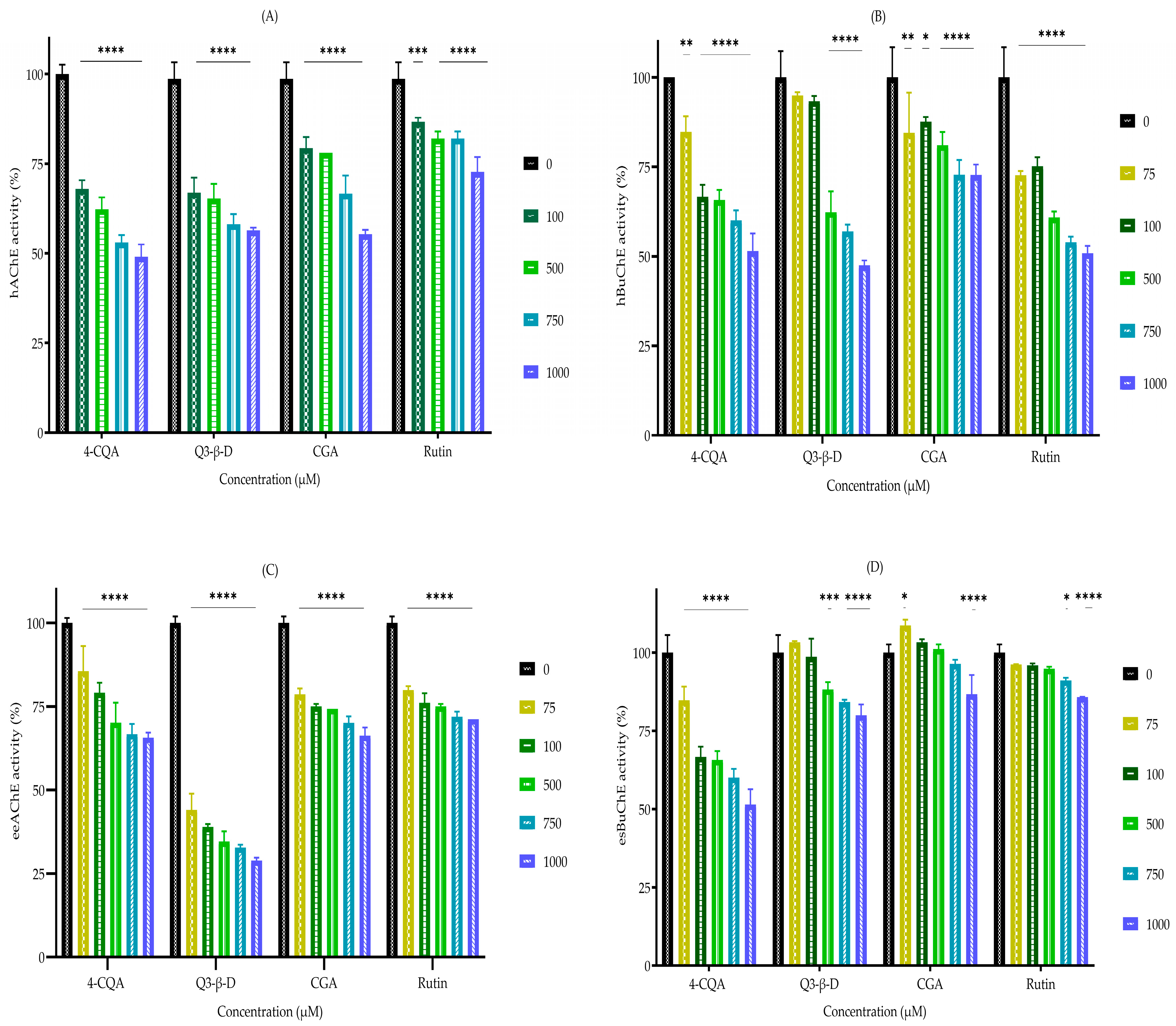
The phytochemicals were tested for their ability to inhibit AChE and BuChE using a modified version of Ellman’s assay [28]. The selected phytochemicals 4-O-caffeoylquinic acid (4-CQA), quercetin 3-beta-D-glucoside (Q3-β-D), chlorogenic acid (CGA), and rutin all displayed broad inhibitory effects on human acetylcholinesterase (hAChE) and human butyrylcholinesterase (hBuChE), with more potent inhibition against hAChE. Similarly, the AChE from Electrophorus electricus (eeAChE) was more potently inhibited than equine serum butyrylcholinesterase (esBuChE) (Figure 2). 4-CQA was the most potent inhibitor of hAChE, hBuChE, and esBuChE; Q3-β-D was the most potent inhibitor of eeAChE; rutin was the weakest hAChE inhibitor but the most potent hBuChE inhibitor, as shown in Figure 2 and Table 2.
The inhibitor (phytochemical) concentration that reduced acetyl- or butyrylcholinesterase activity by 50% (actual or estimated) was calculated using non-linear regression; the values are shown in Table 2. All the phytochemicals were relatively weak ChEIs when compared with the Food and Drug Administration (FDA)-approved ChEIs (rivastigmine, donepezil, and galantamine) assayed under the same conditions (Table 2 and Supplementary Figure S1). The commercial ChEI drugs were all potent AChE inhibitors with relatively low IC50 values (Table 2) and rivastigmine was also a potent BuChE inhibitor. Eserine (physostigmine), a recognized AChE inhibitor [29], was used as a positive control and exhibited potent AChE inhibitory activity; likewise, ethopropazine, a documented inhibitor of BuChE [52], was also used as a positive control, displaying potent BuChE inhibitory activity under the experimental conditions (Table 2).

3.3. In Silico Phytochemical Docking to Cholinesterases

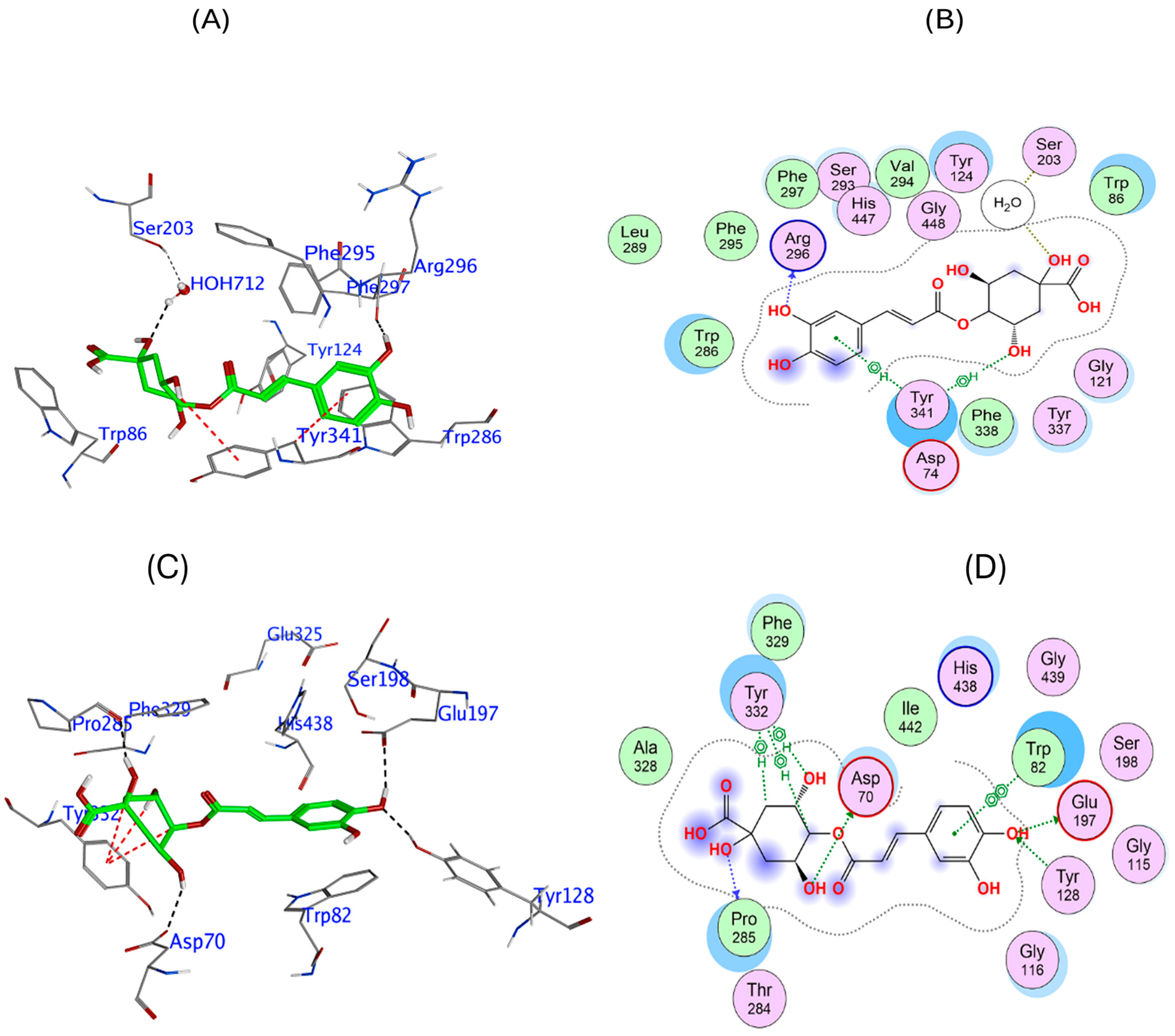
To provide further insight into the potential of the phytochemicals to act as cholinesterase inhibitors, molecular in silico docking was conducted. This docking analysis provided a guide to compound orientation at the enzymatic active site, which for hAChE, showed that 4-CQA is predicted to form strong interactions within the enzyme’s catalytic and anionic sites, forming hydrogen bonds with Arg296 and further stabilizing its position via a water molecule (HOH 712) and π interactions with Tyr341 (Figure 3A,B). These interactions spanned critical regions within the active site, potentially obstructing substrate access and thereby decreasing catalytic efficiency, with a relatively potent binding affinity of −9.12 kcal/mol (Table 3). The docking results for 4-CQA phytochemical ligand binding to hBuChE revealed a modest predicted binding strength, with interactions at the enzyme’s active site, including several hydrogen bonds with key active site residues, including Glu197 and Asp70 near the catalytic triad, as well as Pro285 and Tyr128. Additional π interactions with Tyr332 contributed to its binding energy of −7.97 kcal/mol (Figure 3C,D).
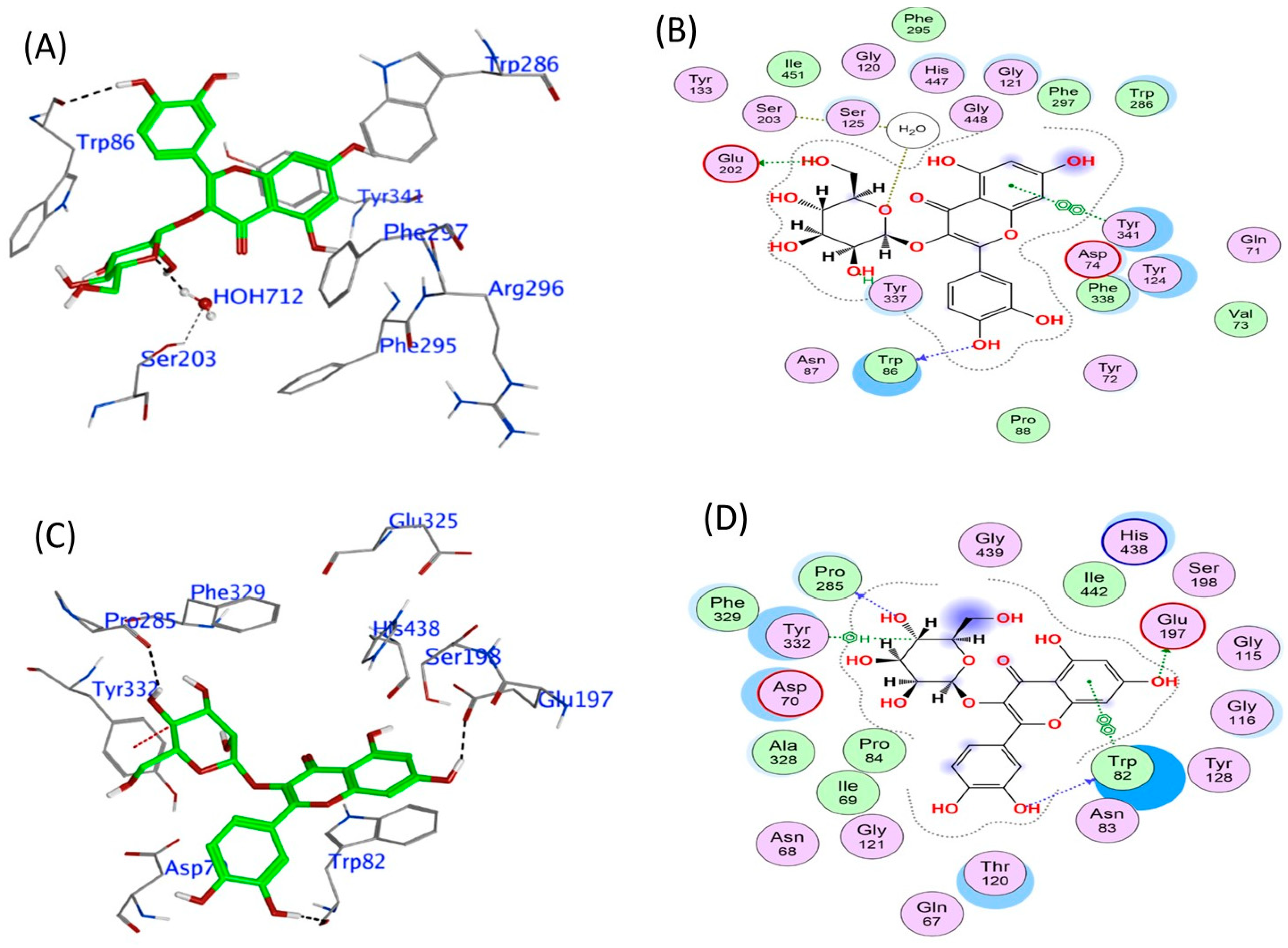
The model of Q3-β-D binding to human AChE shows that it binds to critical residues within the active site, forming hydrogen bonds with Glu202 and π-π stacking interactions with Tyr341 and Trp86. This binding effectively obstructs the access of acetylcholine to the catalytic site, thereby inhibiting its binding and hydrolysis. Such interactions could result in reduced cholinesterase enzymatic activity. Furthermore, π-π stacking interactions with Trp86 would enhance its stable binding conformation, which may hinder substrate access (Figure 4A,B), and contribute to the relatively stable predicted binding affinity of −9.96 kcal/mol (Table 3). Similarly, Q3-β-D had a strong predicted binding affinity of −10.27 kcal/mol (refer to Table 3) for human BuChE, forming hydrogen bonds with Trp82, Glu197, and Pro285. The orientation of Q3-β-D also facilitates direct engagement with catalytic residues and stabilized positioning through a π interaction with Tyr332 (Figure 4C,D).
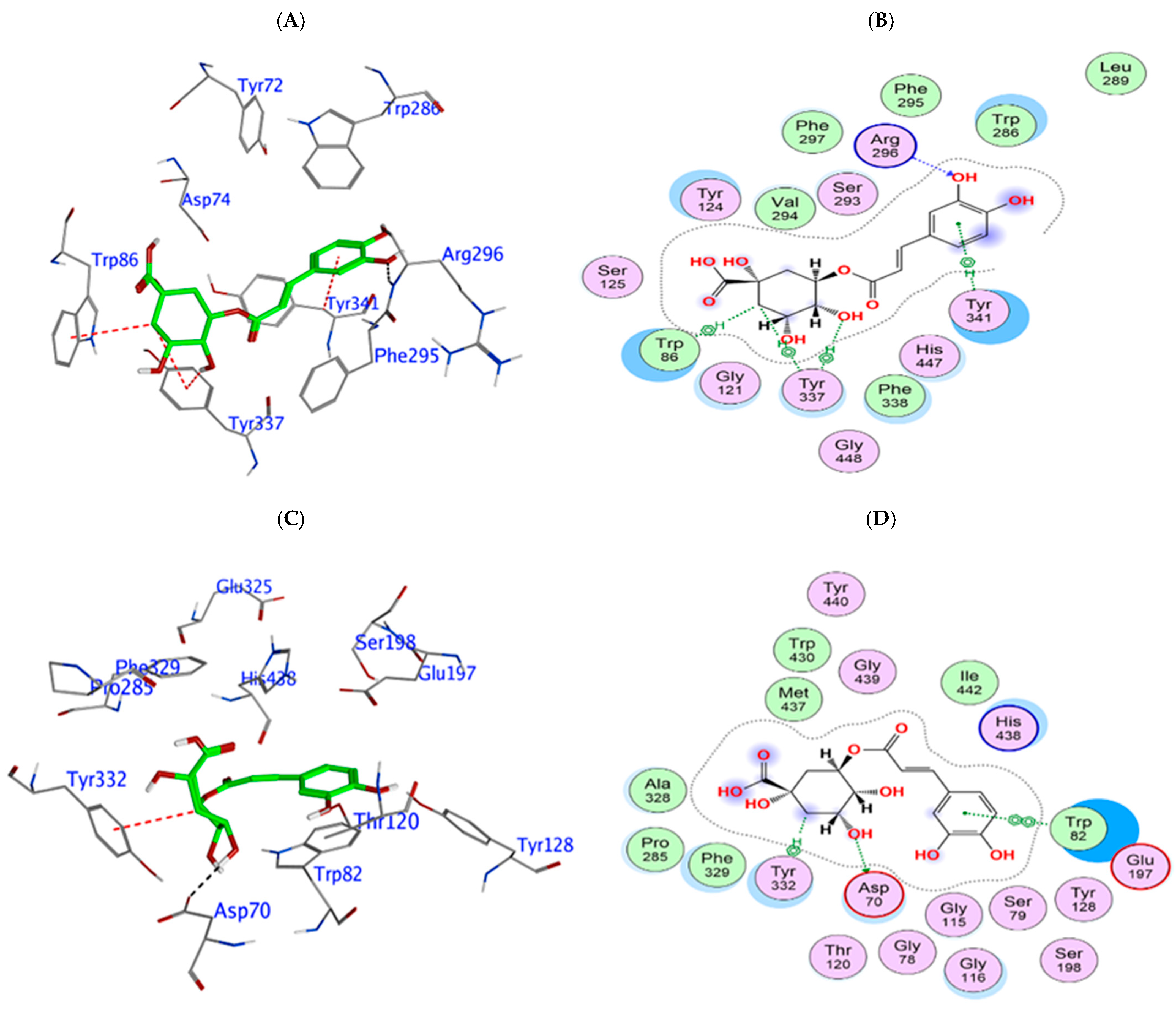
The binding energy of chlorogenic acid binding to human AChE was relatively strong and predicted to be −9.69 kcal/mol (Table 3); the binding occurs through interactions such as hydrogen bonding with Arg296 and H-π interactions with Trp86 and Tyr337 (Figure 5A,B). Its positioning within the cholinergic binding sites suggests that it could potentially disrupt the enzyme’s catalytic activity. Chlorogenic acid showed a similar and relatively strong binding profile for human BuChE (−9.69 kcal/mol, Table 3), arising from an association with Trp82 and Tyr332 (Figure 5C,D).
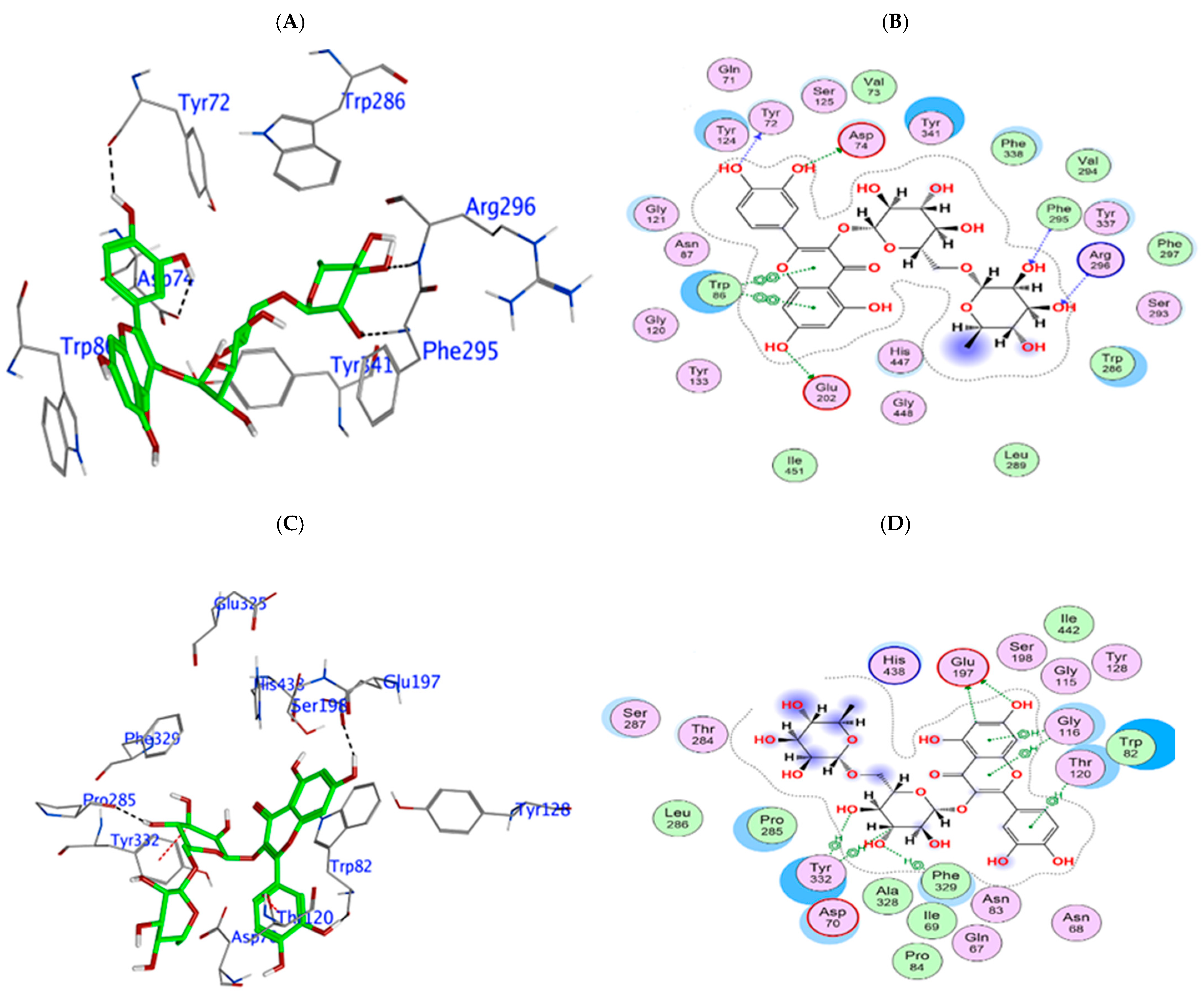
Rutin had the strongest predicted binding strength for human AChE at −14.81 kcal/mol (Table 3), with an expansive interaction profile involving multiple hydrogen bonds with catalytic residues (Asp74 and Glu202) and gating residues (Tyr72 and Arg296) (Figure 6A,B). Similarly, rutin had the highest binding affinity for hBuChE at −12.03 kcal/mol (Table 3), with an extensive interaction profile that included hydrogen bonds with Asn68, Asp70, Trp82, Glu197, and Pro285. The interaction of rutin with Tyr332 suggests additional π-π stacking, further reinforcing its fit within the active site (Figure 6C,D).
A comparative analysis of the docking interactions of 4-CQA, Q3-β-D, CGA, and rutin with hAChE and hBuChE revealed several key patterns. 4-CQA exhibited substantial interactions with both enzymes but had a stronger binding affinity for hAChE, where it engaged Arg296 and Tyr341 in the active site. With hBuChE, the interactions involved Glu197, Asp70, and other peripheral residues, which could reflect a different inhibitory mechanism. Q3-β-D demonstrated strong binding affinities for both enzymes, utilizing hydrogen bonds with essential catalytic residues such as Glu202 in hAChE and Glu197 in hBuChE. It also had additional π-π stacking and interactions with Tyr337 and Tyr332. This indicates that Q3-β-D could be an effective dual inhibitor. CGA showed similar interaction profiles with both enzymes, and its positioning in both active sites likely interfere with catalytic activity, though its inhibitory effect may be comparatively moderate. Interestingly, rutin emerged as the most potent inhibitor for both hAChE and hBuChE. Rutin displayed a broad interaction profile with both enzymes, which included hydrogen bonds with critical catalytic and peripheral residues, along with π-π stacking interactions, which result in a high likelihood for obstruction of substrate access and the formation of a stable binding conformation in the active sites of both enzymes.

3.4. Radical-Scavenging and Antioxidant Properties of the Phytochemicals

The phytochemicals 4-CQA, Q3-β-D, CGA, and rutin were evaluated for their relative radical-scavenging activities and antioxidant properties using a panel of assays, which were compared with the activities of known antioxidants: gallic acid, ascorbic acid (vitamin C), and α-tocopherol (vitamin E).

3.4.1. 2,2-Diphenyl-1-picrylhydrazyl Radical-Scavenging Assay

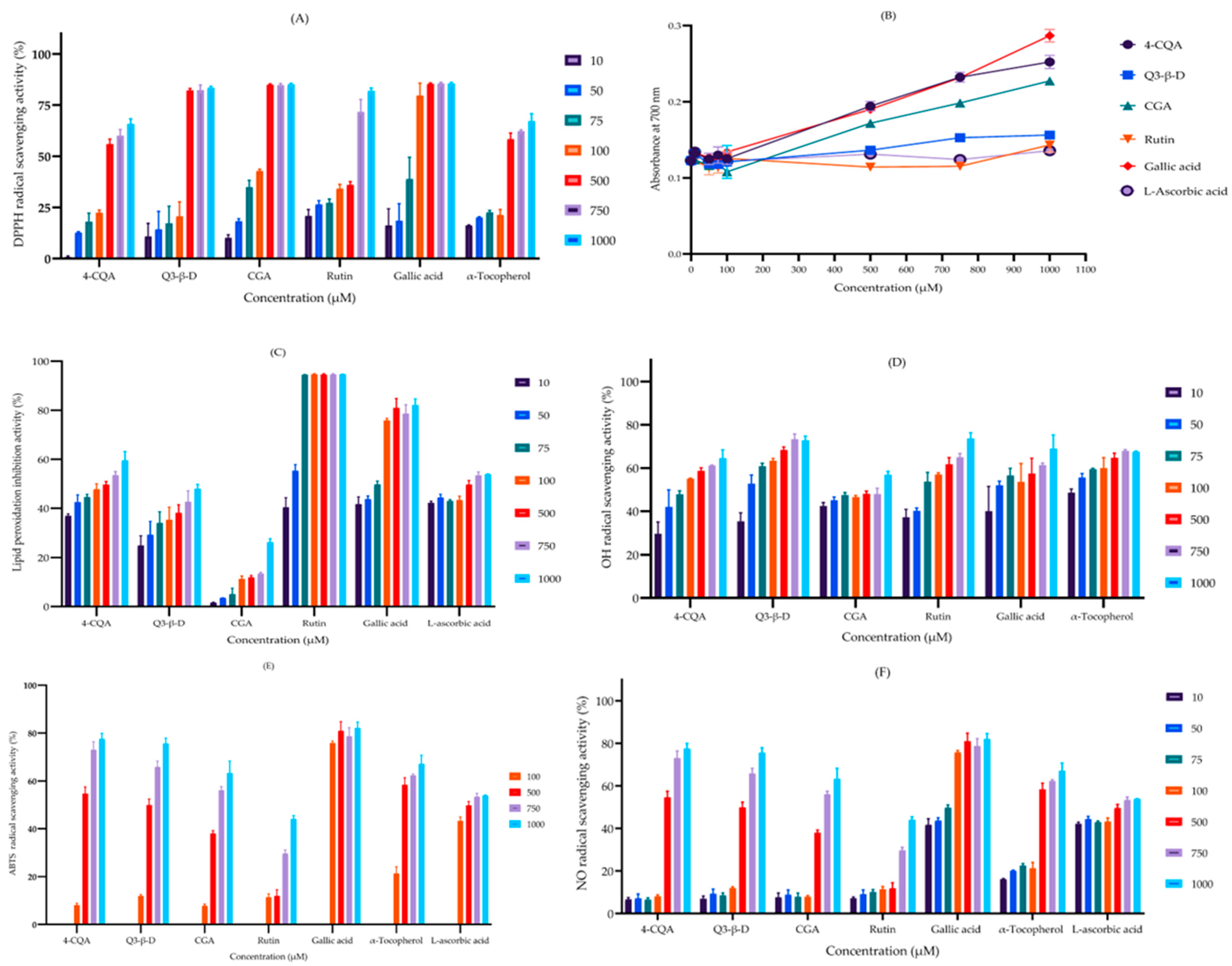
The ability of the phytochemicals (in the concentration range of 10‒1000 µM) to act as radical scavengers was assessed by measuring their ability to donate hydrogen atoms and neutralize 2,2-diphenyl-1-picrylhydrazyl (DPPH) free radicals. The relative potency of DPPH radical-scavenging activity was in the order of CGA > Q3-β-D > rutin > 4-CQA (Figure 7A). The effective drug concentration that produced a 50% effect (EC50) was calculated for each of the phytochemicals using non-linear regression and the values are shown in Table 3. When compared to the known antioxidant compound α-tocopherol, all the phytochemicals except 4-CQA displayed more potent antioxidant (radical-scavenging) capabilities, but they were all less potent than gallic acid (Figure 7A and Table 4).

3.4.2. Ferric Reducing Antioxidant Power Assay

Relative phytochemical antioxidant activity was assessed using the ferric reducing antioxidant power (FRAP) assay. The phytochemicals all displayed comparable reducing activity in the order of 4-CQA > CGA > Q3-β-D > rutin (Figure 7B). In comparison, all the phytochemicals except rutin displayed more potent antioxidant activity than α-tocopherol but were marginally less potent than gallic acid (Table 4).

3.4.3. Lipid Peroxidation Inhibition Assay

The phytochemicals were assessed using the lipid peroxidation inhibition (LPI) assay to determine their ability to limit ROS or free radical attack of lipids in cell membranes and the formation of lipid peroxides. Rutin was the most potent inhibitor of lipid peroxidation and 4-CQA also displayed high efficacy, with an EC50 value similar to L-ascorbic acid. Collectively, the order of potency for LPI was rutin > 4-CQA > Q3-β-D > CGA. Rutin and 4-CQA were more potent inhibitors than gallic acid (Figure 7C and Table 4).

3.4.4. Hydroxyl Radical-Scavenging Assay

The ability of the phytochemicals to scavenge hydroxyl radicals (•OH) was assessed. Q3-β-D and α-tocopherol had similar activities and were the most potent radical scavengers. Rutin, 4-CQA, and gallic acid also showed strong radical-scavenging abilities. OH radical-scavenging activity was in the order of Q3-β-D > rutin > 4-CQA > CGA (Figure 7D and Table 4).

3.4.5. 2,2′-Azino-bis(3-ethylbenzothiazoline-6-sulfonic acid) Radical-Scavenging Assay

An assessment of the phytochemicals’ ability to scavenge 2,2′-azino-bis(3-ethylbenzothiazoline-6-sulfonic acid) radical cations (ATBS•+) showed that the potency was in the order of 4-CQA > Q3-β-D > CGA > rutin. 4-CQA and Q3-β-D had similar efficacies as α-tocopherol but gallic acid was the most effective ABTS radical cation scavenger, with a relatively low EC50 (Figure 7E and Table 4).

3.4.6. Nitric Oxide Radical-Scavenging Assay

The ability of the phytochemicals to neutralize the nitric oxide radical (•NO) was examined and all phytochemicals displayed moderate activity in the order of 4-CQA > Q3-β-D > CGA > rutin. Notably, 4-CQA and Q3-β-D exhibited relatively strong scavenging activity, whereas the antioxidants L-ascorbic acid and gallic acid were less effective and had higher EC50 values (Figure 7F and Table 4).

4. Discussion

M. oleifera produces a diverse array of secondary metabolites with nutraceutical potential [18,19,20]. Our initial analysis of the phytochemicals in aqueous and ethanolic extracts of M. oleifera using LC-MS/MS identified a variety of bioactive compounds, including some with well-documented antioxidant properties (Table 1). Four phytochemicals, 4-O-caffeoylquinic acid, quercetin 3-β-D-glucoside, chlorogenic acid, and rutin, were further analysed and found to possess weak acetyl- and butyrylcholinesterase inhibitory activities compared with the currently prescribed ChEI drugs rivastigmine, donepezil, and galantamine. Complementary in silico analyses of the phytochemicals as ligands for AChE and BuChE substantiated their proposed dual cholinesterase inhibitor activity. Additionally, these phytochemicals also displayed useful radical-scavenging and antioxidant capabilities that were often comparable and sometimes more potent than the known antioxidants gallic acid, α-tocopherol (vitamin E), and ascorbic acid (vitamin C). Hence, the combined dual ChEI and potent antioxidant activities indicate that these phytochemicals could be used to limit some of the pathological deficits experienced by patients with AD.
Our LC-MS/MS analysis of aqueous and ethanolic extracts of M. oleifera revealed the presence of diverse chemical groups, including phenolic compounds, flavonoids, lignans, proanthocyanidins, monoterpinoids, norisoprenoids, mono- and polyunsaturated fatty acids, saturated fatty acids, and amino acids (Table 1). The identification of these compounds aligns with other analytical studies of M. oleifera, which have considered a range of plant parts [18,19,20] and documented the presence of some of the phytochemicals detailed in Table 1.
Extracts of M. oleifera display neuroprotective and cognitive enhancement capabilities [24,25,26], and this prompted us to specifically investigate whether its phytochemicals could enhance or sustain cholinergic signalling via cholinesterase inhibitor activity. The tested phytochemicals 4-O-caffeoylquinic acid (4-CQA), quercetin 3-β-D-glucoside (Q3-β-D), chlorogenic acid (CGA), and rutin all exhibited varying degrees of (in vitro) inhibitory activity against both AChE and BuChE enzymes. The inhibitory potential against hAChE was in the order of 4-CQA > Q3-β-D > CGA > rutin; this order was similar for eeAChE except Q3-β-D was more potent than 4-CQA. By comparison, for hBuChE, the phytochemicals were ranked in the order of rutin > 4-CQA > Q3-β-D > CGA and for equine serum BuChE, it was 4-CQA > Q3-β-D > rutin > CGA.
Caffeoylquinic acids (a family of compounds that are esters of caffeic acid with quinic acid), such as 4-O-caffeoylquinic acid (4-CQA), are phytochemicals that are often obtained through the consumption of fruits and vegetables as well as coffee, which have recognized anti-inflammatory and antioxidant properties [53]. Our study shows that, for the phytochemicals tested, 4-CQA displayed weak cholinesterase inhibitor activity but was the strongest inhibitor of hAChE and second-best inhibitor of hBuChE.
Flavonoids, such as quercetin 3-β-D-glucoside (Q3-β-D), constitute a broad chemical group, and are often obtained through the dietary intake of fruits and vegetables, as well as beverages, and have purported antioxidant and nutraceutical properties [54]. An assessment of the AChE inhibitory activity of members of the flavonoid class revealed that quercetin had weak but detectable AChE and BuChE inhibitor, with an IC50 of approximately 20 µM, approximately 200 times higher than that of tacrine, a compound that had been employed as a clinical ChEI [55,56,57]. Our studies using Q3-β-D (also known as isoquercetin), which has a similar structure to quercetin, showed similarly weak AChE and BuChE inhibitory activities, with IC50 values of just below 1 mM (Table 2).
Chlorogenic acid (CGA) (5-caffeoylquinic acid) is a dietary polyphenol and member of the caffeoylquinic acid family. It is structurally similar to 4-CQA and has diverse biologically active properties that include anti-inflammatory, antioxidant, as well as metabolic homeostasis modulation activities [58]. Our data suggests that CGA can act as a weak ChEI, and this concurs with other studies that have reported inhibitory activity against hAChE (IC50 of 0.41 mM) [59].
Rutin, also known as quercetin-3-rutinoside, is a flavonoid found in a number of dietary sources including teas and fruits and has a range of pharmacological and nutraceutical properties [60,61]. Rutin displayed weak dual ChEI activities (low mM IC50s) and was the most potent inhibitor of hBuChE. Similarly, studies performed using rat brain homogenates reported that rutin was capable of dual-cholinesterase inhibition in vitro, with AChE and BuChE IC50 values of 0.219 and 0.288 mM, respectively [62].
To provide further insight into the potential ChEI activity of the phytochemicals and support for our in vitro findings, molecular docking was performed to study the binding interactions of each of the phytochemicals with the active site residues of human AChE and BuChE. The docking simulations suggested that these phytochemicals interacted with critical active site residues. This provided a quantitative determination of their relative binding affinities and potential inhibitory mechanisms. Rutin displayed the highest binding affinities for both hAChE and hBuChE, with binding energies of −14.81 kcal/mol and −12.03 kcal/mol, respectively. Its ability to form extensive hydrogen bonds with catalytic and peripheral residues, combined with π-π stacking interactions, suggested a strong capacity to obstruct substrate access and stabilize its position within the active sites of these enzymes. However, in contrast to the relatively high affinity proposed by computational predictions, rutin was not the most efficient in vitro inhibitor (Table 2), although this could reflect the relatively strong predicted interaction, which could result in slow off-binding kinetics and decreased inhibitory activity, but we did not investigate this further.
The other three phytochemicals, 4-CQA, Q3-β-D, and CGA, had similar and relatively strong predicted binding affinities to hAChE: −9.12, −9.96, and −9.69 kcal/mol, respectively; these affinities are stronger (more negative) than that of galantamine at −7.7 kcal/mol [63]. However, all four tested phytochemicals displayed relatively weak in vitro cholinesterase inhibitory capacity. This highlights the difference between the dynamic nature of enzymatic interactions in vitro from the static conditions typically modelled in docking studies, as in silico simulations typically assume optimal binding conformations and do not account for factors such as enzyme flexibility and induced conformational changes.
Clearly, even at the high purity examined, these phytochemicals have inferior in vitro potencies than the current FDA-approved drugs (rivastigmine, donepezil, and galantamine) or known inhibitors (eserine and ethopropazine for AChE and BuChE, respectively) that we examined under identical kinetic conditions. Hence, these phytochemicals had IC50 values in the high nanomolar to low micromolar range, consistent with other studies that reported their in vitro efficacy values [52,64,65]. Thus, these phytochemicals would be inappropriate as therapeutic interventions if applied as a monotherapy. Nevertheless, the restoration of depleted cholinergic signalling is only one element of AD pathology, so treatments that address additional components of disease pathophysiology, such as redox stress, will likely have enhanced therapeutic utility. Therefore, we also evaluated the antioxidant properties of these phytochemicals using a panel of in vitro assays (Figure 7). The phytochemicals (4-CQA, Q3-β-D, CGA, and rutin) exhibited varying degrees of antioxidant activity, with a demonstrated ability to scavenge free radicals. 4-CQA was the most potent antioxidant in terms of performance across the panel of assays and was the most potent for three assays: FRAP, ATBS•+, and •NO. Interestingly, each of the other phytochemicals was the most powerful antioxidant in one other assay: Q3-β-D for •OH scavenging, CGA for DPPH• scavenging, and rutin for the LPI assay (Table 4). Other independent studies have also documented strong antioxidant activities for chlorogenic acid and rutin [38,44,45]. In addition, a panel of antioxidant assays performed using rutin generated IC50 values of approximately 184–938 µM [62], comparable to our results.
Despite the relatively weak ChEI activity of phytochemicals in vitro, extracts of M. olefiera have been utilized as a neuroprotective agent in vivo [22,23,24,25]. This likely reflects the effects of multiple and potentially interacting phytochemicals rather than individual agents, and these could collectively contribute to ChEI activity as well as additional mechanisms to mitigate redox stress and other AD-related pathologies, such as the accumulation of Aβ [66]. Indeed, our data indicates that the phytochemicals were each superior in one (or more) antioxidant assay. However, typically, pure chemical entities are used for randomized control trials (RCTs), such as chlorogenic acid or α-tocopherol [16,58,67,68], although several human trials with M. olefiera extracts have been undertaken to evaluate its safety and efficacy profile [69]. To date, the relative levels each of the phytochemicals within an M. olefiera extract and their contribution to its collective efficacy are unknown, but our future studies will aim to address this through more in-depth quantitative studies of M. olefiera extracts.
In summary, the findings of the current study provide evidence that phytochemicals derived from M. oleifera have weak dual ChEI activity and potent antioxidant capabilities. This supports the potential use of M. olefiera phytochemicals and/or extracts to enhance cognitive performance and as a possible means to combat some of the pathophysiological elements that arise in AD. Dietary intake or supplementation with M. olefiera (or its phytochemicals) could provide a beneficial source of nutraceuticals with sustained low dosing; it also has potential as a treatment or prophylaxis to stave off the development of AD.

5. Conclusions

Extracts of the M. oleifera plant contain a range of phytochemicals and secondary metabolites that are also commonly encountered in fruits, vegetables, and beverages such as teas and coffee. Our data show that phytochemicals from M. oleifera could have nutraceutical properties arising from their ChEI and antioxidant activities that could be beneficial as a natural treatment and/or chronic dietary supplement that can treat and limit the development or progression of neurodegenerative diseases such as AD. However, more in vivo studies and RCTs are needed to validate this proposition and confirm the phytochemicals’ safety and efficacy, as well as their potential bioavailability, pharmacokinetics, and pharmacodynamics limitations.

Supplementary Materials

The following supporting information can be downloaded at https://www.mdpi.com/article/10.3390/biomedicines13092148/s1, Supplementary Figure S1: Cholinesterase inhibition by FDA-approved drugs.

Author Contributions

Conceptualization, A.M.A. (Adel M. Aljadaan) and W.G.C.; Data curation, A.M.A. (Adel M. Aljadaan) and W.G.C.; Formal analysis, A.M.A. (Adel M. Aljadaan) and I.A.S.; Funding acquisition, A.M.A. (Adel M. Aljadaan) and W.G.C.; Investigation, A.M.A. (Adel M. Aljadaan), A.M.A. (Ayman M. AlSaadi), I.A.S., and A.W.; Methodology, A.M.A. (Adel M. Aljadaan), A.M.A. (Ayman M. AlSaadi), I.A.S., and A.W.; Project administration, A.R., D.-H.K., and W.G.C.; Resources, D.-H.K. and W.G.C.; Supervision, A.R., D.-H.K., and W.G.C.; Validation: A.M.A. (Adel M. Aljadaan), A.M.A. (Ayman M. AlSaadi), I.A.S., and A.W.; Visualization, A.M.A. (Adel M. Aljadaan) and I.A.S.; Writing—original draft, A.M.A. (Adel M. Aljadaan) and W.G.C.; Writing—review and editing, A.M.A. (Adel M. Aljadaan), A.M.A. (Ayman M. AlSaadi), I.A.S., A.W., A.R., D.-H.K., and W.G.C. All authors have read and agreed to the published version of the manuscript.

Funding

This research was funded by the Cultural Bureau and Najran University Ph.D. Scholarship, Kingdom of Saudi Arabia (A.M.A. (Adel M. Aljadaan) and A.M.A. (Ayman M. AlSaadi)).

Institutional Review Board Statement

Not applicable.

Informed Consent Statement

Not applicable.

Data Availability Statement

The data used in the production of the figures is available on request from the first author.

Conflicts of Interest

The authors declare no conflicts of interest. The funders had no role in the design of the study; in the collection, analyses, or interpretation of the data; in the writing of the manuscript; or in the decision to publish the results.

Abbreviations

The following abbreviations are used in this manuscript:
4-CQA4-O-caffeoylquinic acid
Q3-β-DQuercetin 3-β-D-glucoside
CGAChlorogenic acid

References

  1. WHO. 2021. Available online: https://www.who.int/news/item/02-09-2021-world-failing-to-address-dementia-challenge (accessed on 14 June 2025).
  2. WHO. 2023. Available online: https://www.who.int/news-room/fact-sheets/detail/dementia (accessed on 14 June 2025).
  3. Bature, F.; Guinn, B.A.; Pang, D.; Pappas, Y. Signs and symptoms preceding the diagnosis of Alzheimer’s disease: A systematic scoping review of literature from 1937 to 2016. BMJ Open 2017, 7, e015746. [Google Scholar] [CrossRef]
  4. Scheltens, P.; Blennow, K.; Breteler, M.M.; de Strooper, B.; Frisoni, G.B.; Salloway, S.; Van der Flier, W.M. Alzheimer’s disease. Lancet 2016, 388, 505–517. [Google Scholar] [CrossRef]
  5. Liang, C.S.; Li, D.J.; Yang, F.C.; Tseng, P.T.; Carvalho, A.F.; Stubbs, B.; Thompson, T.; Mueller, C.; Shin, J.I.; Radua, J.; et al. Mortality rates in Alzheimer’s disease and non-Alzheimer’s dementias: A systematic review and meta-analysis. Lancet Healthy Longev. 2021, 2, e479–e488. [Google Scholar] [CrossRef] [PubMed]
  6. Bellenguez, C.; Küçükali, F.; Jansen, I.E.; Kleineidam, L.; Moreno-Grau, S.; Amin, N.; Naj, A.C.; Campos-Martin, R.; Grenier-Boley, B.; Andrade, V.; et al. New insights into the genetic etiology of Alzheimer’s disease and related dementias. Nat. Genet. 2022, 54, 412–436. [Google Scholar] [CrossRef] [PubMed]
  7. Raulin, A.C.; Doss, S.V.; Trottier, Z.A.; Ikezu, T.C.; Bu, G.; Liu, C.C. ApoE in Alzheimer’s disease: Pathophysiology and therapeutic strategies. Mol. Neurodegener. 2022, 17, 72. [Google Scholar] [CrossRef] [PubMed]
  8. Dhapola, R.; Sharma, P.; Kumari, S.; Bhatti, J.S.; HariKrishnaReddy, D. Environmental Toxins and Alzheimer’s Disease: A Comprehensive Analysis of Pathogenic Mechanisms and Therapeutic Modulation. Mol. Neurobiol. 2024, 61, 3657–3677. [Google Scholar] [CrossRef]
  9. Igarashi, K.M. Entorhinal cortex dysfunction in Alzheimer’s disease. Trends Neurosci. 2023, 46, 124–136. [Google Scholar] [CrossRef]
  10. Lane, C.A.; Hardy, J.; Schott, J.M. Alzheimer’s disease. Eur. J. Neurol. 2018, 25, 59–70. [Google Scholar] [CrossRef]
  11. Kaufman, S.K.; Del Tredici, K.; Thomas, T.L.; Braak, H.; Diamond, M.I. Tau seeding activity begins in the transentorhinal/entorhinal regions and anticipates phospho-tau pathology in Alzheimer’s disease and PART. Acta Neuropathol. 2018, 136, 57–67. [Google Scholar] [CrossRef]
  12. Ionescu-Tucker, A.; Cotman, C.W. Emerging roles of oxidative stress in brain aging and Alzheimer’s disease. Neurobiol. Aging 2021, 107, 86–95. [Google Scholar] [CrossRef]
  13. Perluigi, M.; Di Domenico, F.; Butterfield, D.A. Oxidative damage in neurodegeneration: Roles in the pathogenesis and progression of Alzheimer disease. Physiol. Rev. 2024, 104, 103–197. [Google Scholar] [CrossRef]
  14. Houldsworth, A. Role of oxidative stress in neurodegenerative disorders: A review of reactive oxygen species and prevention by antioxidants. Brain Commun. 2024, 6, fcad356. [Google Scholar] [CrossRef] [PubMed]
  15. Singh, B.; Day, C.M.; Abdella, S.; Garg, S. Alzheimer’s disease current therapies, novel drug delivery systems and future directions for better disease management. J. Control. Release 2024, 367, 402–424. [Google Scholar] [CrossRef] [PubMed]
  16. Thangwaritorn, S.; Lee, C.; Metchikoff, E.; Razdan, V.; Ghafary, S.; Rivera, D.; Pinto, A.; Pemminati, S. A Review of Recent Advances in the Management of Alzheimer’s Disease. Cureus 2024, 16, e58416. [Google Scholar] [CrossRef] [PubMed]
  17. Oyebode, O.; Kandala, N.B.; Chilton, P.J.; Lilford, R.J. Use of traditional medicine in middle-income countries: A WHO-SAGE study. Health Policy Plan. 2016, 31, 984–991. [Google Scholar] [CrossRef]
  18. Padayachee, B.; Baijnath, H. An updated comprehensive review of the medicinal, phytochemical and pharmacological properties of Moringa oleifera. S. Afr. J. Bot. 2020, 129, 304–316. [Google Scholar] [CrossRef]
  19. Liu, R.; Liu, J.; Huang, Q.; Liu, S.; Jiang, Y. Moringa oleifera: A systematic review of its botany, traditional uses, phytochemistry, pharmacology and toxicity. J. Pharm. Pharmacol. 2022, 74, 296–320. [Google Scholar] [CrossRef]
  20. Pareek, A.; Pant, M.; Gupta, M.M.; Kashania, P.; Ratan, Y.; Jain, V.; Pareek, A.; Chuturgoon, A.A. Moringa oleifera: An Updated Comprehensive Review of Its Pharmacological Activities, Ethnomedicinal, Phytopharmaceutical Formulation, Clinical, Phytochemical, and Toxicological Aspects. Int. J. Mol. Sci. 2023, 24, 2098. [Google Scholar] [CrossRef]
  21. Nwidu, L.L.; Elmorsy, E.; Aprioku, J.S.; Siminialayi, I.; Carter, W.G. In vitro anti-cholinesterase and antioxidant activity of extracts of Moringa oleifera plants from Rivers State, Niger Delta, Nigeria. Medicines 2018, 5, 71. [Google Scholar] [CrossRef]
  22. Sutalangka, C.; Wattanathorn, J.; Muchimapura, S.; Thukham-mee, W. Moringa oleifera mitigates memory impairment and neurodegeneration in animal model of age-related dementia. Oxid. Med. Cell Longev. 2013, 2013, 695936. [Google Scholar] [CrossRef]
  23. Srivastava, G.; Ganjewala, D. An update on the emerging neuroprotective potential of Moringa oleifera and its prospects in complimentary neurotherapy. Phytomed. Plus 2024, 4, 100532. [Google Scholar] [CrossRef]
  24. Afrin, S.; Hossain, A.; Begum, S. Effects of Moringa oleifera on working memory: An experimental study with memory-impaired Wistar rats tested in radial arm maze. BMC Res. Notes 2022, 15, 314. [Google Scholar] [CrossRef] [PubMed]
  25. Onasanwo, S.A.; Adamaigbo, V.O.; Adebayo, O.G.; Eleazer, S.E. Moringa oleifera-supplemented diet protect against cortico-hippocampal neuronal degeneration in scopolamine-induced spatial memory deficit in mice: Role of oxido-inflammatory and cholinergic neurotransmission pathway. Metab. Brain Dis. 2021, 36, 2445–2460. [Google Scholar] [CrossRef] [PubMed]
  26. Ghimire, S.; Subedi, L.; Acharya, N.; Gaire, B.P. Moringa oleifera: A tree of life as a promising medicinal plant for neurodegenerative diseases. J. Agric. Food Chem. 2021, 69, 14358–14371. [Google Scholar] [CrossRef]
  27. ALNasser, M.N.; AlSaadi, A.M.; Whitby, A.; Kim, D.-H.; Mellor, I.R.; Carter, W.G. Acai berry (Euterpe sp.) extracts are neuroprotective against l-glutamate-induced toxicity by limiting mitochondrial dysfunction and cellular redox stress. Life 2023, 13, 1019. [Google Scholar] [CrossRef]
  28. Ellman, G.L.; Courtney, K.D.; Andres Jr, V.; Featherstone, R.M. A new and rapid colorimetric determination of acetylcholinesterase activity. Biochem. Pharmacol. 1961, 7, 88–95. [Google Scholar] [CrossRef]
  29. Nwidu, L.L.; Elmorsy, E.; Thornton, J.; Wijamunige, B.; Wijesekara, A.; Tarbox, R.; Warren, A.; Carter, W.G. Anti-acetylcholinesterase activity and antioxidant properties of extracts and fractions of Carpolobia lutea. Pharm. Biol. 2017, 55, 1875–1883. [Google Scholar] [CrossRef]
  30. ALNasser, M.N.; Mellor, I.R.; Carter, W.G. A Preliminary Assessment of the Nutraceutical Potential of Acai Berry (Euterpe sp.) as a Potential Natural Treatment for Alzheimer’s Disease. Molecules 2022, 27, 4891. [Google Scholar] [CrossRef]
  31. Zhang, S.; Gai, Z.; Gui, T.; Chen, J.; Chen, Q.; Li, Y. Antioxidant effects of protocatechuic acid and protocatechuic aldehyde: Old wine in a new bottle. Evid.-Based Complement. Altern. Med. 2021, 2021, 6139308. [Google Scholar] [CrossRef]
  32. Bartel, I.; Mandryk, I.; Horbańczuk, J.O.; Wierzbicka, A.; Koszarska, M. Nutraceutical Properties of Syringic Acid in Civilization Diseases-Review. Nutrients 2023, 16, 10. [Google Scholar] [CrossRef]
  33. Salau, V.F.; Erukainure, O.L.; Ibeji, C.U.; Olasehinde, T.A.; Koorbanally, N.A.; Islam, M.S. Vanillin and vanillic acid modulate antioxidant defense system via amelioration of metabolic complications linked to Fe2+-induced brain tissues damage. Metab. Brain Dis. 2020, 35, 727–738. [Google Scholar] [CrossRef]
  34. Badhani, B.; Sharma, N.; Kakkar, R. Gallic acid: A versatile antioxidant with promising therapeutic and industrial applications. Rsc Adv. 2015, 5, 27540–27557. [Google Scholar] [CrossRef]
  35. Hadidi, M.; Liñán-Atero, R.; Tarahi, M.; Christodoulou, M.C.; Aghababaei, F. The Potential Health Benefits of Gallic Acid: Therapeutic and Food Applications. Antioxidants 2024, 13, 1001. [Google Scholar] [CrossRef] [PubMed]
  36. Velika, B.; Kron, I. Antioxidant properties of benzoic acid derivatives against superoxide radical. Free Radic. Antioxid. 2012, 2, 62–67. [Google Scholar] [CrossRef]
  37. Spiegel, M.; Kapusta, K.; Kołodziejczyk, W.; Saloni, J.; Żbikowska, B.; Hill, G.A.; Sroka, Z. Antioxidant activity of selected phenolic acids–ferric reducing antioxidant power assay and QSAR analysis of the structural features. Molecules 2020, 25, 3088. [Google Scholar] [CrossRef]
  38. Wang, L.; Pan, X.; Jiang, L.; Chu, Y.; Gao, S.; Jiang, X.; Zhang, Y.; Chen, Y.; Luo, S.; Peng, C. The Biological Activity Mechanism of Chlorogenic Acid and Its Applications in Food Industry: A Review. Front. Nutr. 2022, 9, 943911. [Google Scholar] [CrossRef]
  39. Ahmadi, S.M.; Farhoosh, R.; Sharif, A.; Rezaie, M. Structure-Antioxidant Activity Relationships of Luteolin and Catechin. J. Food Sci. 2020, 85, 298–305. [Google Scholar] [CrossRef]
  40. Zhang, M.; Swarts, S.G.; Yin, L.; Liu, C.; Tian, Y.; Cao, Y.; Swarts, M.; Yang, S.; Zhang, S.B.; Zhang, K.; et al. Antioxidant properties of quercetin. Adv. Exp. Med. Biol. 2011, 701, 283–289. [Google Scholar] [CrossRef]
  41. Azeem, M.; Hanif, M.; Mahmood, K.; Ameer, N.; Chughtai, F.R.; Abid, U. An insight into anticancer, antioxidant, antimicrobial, antidiabetic and anti-inflammatory effects of quercetin: A review. Polym. Bull. 2023, 80, 241–262. [Google Scholar] [CrossRef]
  42. Septembre-Malaterre, A.; Boumendjel, A.; Seteyen, A.S.; Boina, C.; Gasque, P.; Guiraud, P.; Sélambarom, J. Focus on the high therapeutic potentials of quercetin and its derivatives. Phytomed. Plus 2022, 2, 100220. [Google Scholar] [CrossRef]
  43. Topal, F.; Nar, M.; Gocer, H.; Kalin, P.; Kocyigit, U.M.; Gülçin, İ.; Alwasel, S.H. Antioxidant activity of taxifolin: An activity–structure relationship. J. Enzym. Inhib. Med. Chem. 2016, 31, 674–683. [Google Scholar] [CrossRef]
  44. Yang, J.; Guo, J.; Yuan, J. In vitro antioxidant properties of rutin. LWT-Food Sci. Technol. 2008, 41, 1060–1066. [Google Scholar] [CrossRef]
  45. Enogieru, A.B.; Haylett, W.; Hiss, D.C.; Bardien, S.; Ekpo, O.E. Rutin as a Potent Antioxidant: Implications for Neurodegenerative Disorders. Oxid. Med. Cell Longev. 2018, 2018, 6241017. [Google Scholar] [CrossRef]
  46. Sampei, M.; Arai, M.A.; Ishibashi, M. Total syntheses of schizandriside, saracoside and (±)-isolariciresinol with antioxidant activities. J. Nat. Med. 2018, 72, 651–654. [Google Scholar] [CrossRef]
  47. Bajpai, V.K.; Alam, M.B.; Quan, K.T.; Kwon, K.R.; Ju, M.K.; Choi, H.J.; Lee, J.S.; Yoon, J.I.; Majumder, R.; Rather, I.A.; et al. Antioxidant efficacy and the upregulation of Nrf2-mediated HO-1 expression by (+)-lariciresinol, a lignan isolated from Rubia philippinensis, through the activation of p38. Sci. Rep. 2017, 7, 46035. [Google Scholar] [CrossRef]
  48. Grzesik, M.; Naparło, K.; Bartosz, G.; Sadowska-Bartosz, I. Antioxidant properties of catechins: Comparison with other antioxidants. Food Chem. 2018, 241, 480–492. [Google Scholar] [CrossRef]
  49. Yang, X.; Kang, M.-C.; Lee, K.-W.; Kang, S.-M.; Lee, W.-W.; Jeon, Y.-J. Antioxidant activity and cell protective effect of loliolide isolated from Sargassum ringgoldianum subsp. coreanum. Algae 2011, 26, 201–208. [Google Scholar] [CrossRef]
  50. Gęgotek, A.; Skrzydlewska, E. Antioxidative and Anti-Inflammatory Activity of Ascorbic Acid. Antioxidants 2022, 11, 1993. [Google Scholar] [CrossRef] [PubMed]
  51. Amat-ur-Rasool, H.; Symes, F.; Tooth, D.; Schaffert, L.-N.; Elmorsy, E.; Ahmed, M.; Hasnain, S.; Carter, W.G. Potential nutraceutical properties of leaves from several commonly cultivated plants. Biomolecules 2020, 10, 1556. [Google Scholar] [CrossRef] [PubMed]
  52. Dorling, J.L.; Clayton, D.J.; Jones, J.; Carter, W.G.; Thackray, A.E.; King, J.A.; Pucci, A.; Batterham, R.L.; Stensel, D.J. A randomized crossover trial assessing the effects of acute exercise on appetite, circulating ghrelin concentrations, and butyrylcholinesterase activity in normal-weight males with variants of the obesity-linked FTO rs9939609 polymorphism. Am. J. Clin. Nutr. 2019, 110, 1055–1066. [Google Scholar] [CrossRef] [PubMed]
  53. Alcázar Magaña, A.; Kamimura, N.; Soumyanath, A.; Stevens, J.F.; Maier, C.S. Caffeoylquinic acids: Chemistry, biosynthesis, occurrence, analytical challenges, and bioactivity. Plant J. 2021, 107, 1299–1319. [Google Scholar] [CrossRef]
  54. Waheed Janabi, A.H.; Kamboh, A.A.; Saeed, M.; Xiaoyu, L.; BiBi, J.; Majeed, F.; Naveed, M.; Mughal, M.J.; Korejo, N.A.; Kamboh, R.; et al. Flavonoid-rich foods (FRF): A promising nutraceutical approach against lifespan-shortening diseases. Iran. J. Basic Med. Sci. 2020, 23, 140–153. [Google Scholar] [CrossRef]
  55. Orhan, I.; Kartal, M.; Tosun, F.; Sener, B. Screening of various phenolic acids and flavonoid derivatives for their anticholinesterase potential. Z. Naturforsch. C J. Biosci. 2007, 62, 829–832. [Google Scholar] [CrossRef]
  56. Jung, M.; Park, M. Acetylcholinesterase Inhibition by Flavonoids from Agrimonia pilosa. Molecules 2007, 12, 2130–2139. [Google Scholar] [CrossRef] [PubMed]
  57. Khan, H.; Marya Amin, S.; Kamal, M.A.; Patel, S. Flavonoids as acetylcholinesterase inhibitors: Current therapeutic standing and future prospects. Biomed. Pharmacother. 2018, 101, 860–870. [Google Scholar] [CrossRef] [PubMed]
  58. Nguyen, V.; Taine, E.G.; Meng, D.; Cui, T.; Tan, W. Chlorogenic Acid: A Systematic Review on the Biological Functions, Mechanistic Actions, and Therapeutic Potentials. Nutrients 2024, 16, 924. [Google Scholar] [CrossRef] [PubMed]
  59. Işık, M.; Beydemir, Ş. The impact of some phenolic compounds on serum Acetylcholinesterase: Kinetic analysis of an enzyme/inhibitor interaction and molecular docking study. J. Biomol. Struct. Dyn. 2020, 39, 6515–6523. [Google Scholar] [CrossRef]
  60. Ganeshpurkar, A.; Saluja, A.K. The Pharmacological Potential of Rutin. Saudi Pharm. J. 2017, 25, 149–164. [Google Scholar] [CrossRef]
  61. Chen, R.; Hassan, H.; Rawlinson, C.; Morgan, D. Pharmacological properties of rutin and its potential uses for Alzheimer’s disease. J. Exp. Stroke Transl. Med. 2020, 13, 1–12. [Google Scholar]
  62. Ademosun, A.O.; Oboh, G.; Bello, F.; Ayeni, P.O. Antioxidative Properties and Effect of Quercetin and Its Glycosylated Form (Rutin) on Acetylcholinesterase and Butyrylcholinesterase Activities. J. Evid. Based Complem. Altern. Med. 2016, 21, NP11-7. [Google Scholar] [CrossRef]
  63. Amat-ur-Rasool, H.; Ahmed, M.; Hasnain, S.; Ahmed, A.; Carter, W.G. In Silico Design of Dual-Binding Site Anti-Cholinesterase Phytochemical Heterodimers as Treatment Options for Alzheimer’s Disease. Curr. Issues Mol. Biol. 2022, 44, 152–175. [Google Scholar] [CrossRef]
  64. Akhtar Muhammad, A.M.; Tel-Cayan, G.; Öztürk, M.; Nadeem, S.; Duru, M.E.; Itrat Anis, I.A.; Ng, S.W.; Shah, M.R. Biologically active flavonoids from Dodonaea viscosa and their structure–activity relationships. Ind. Crops Prod. 2015, 78, 66–72. [Google Scholar] [CrossRef]
  65. Amat-ur-Rasool, H.; Ahmed, M.; Hasnain, S.; Carter, W.G. Anti-Cholinesterase Combination Drug Therapy as a Potential Treatment for Alzheimer’s Disease. Brain Sci. 2021, 11, 184. [Google Scholar] [CrossRef] [PubMed]
  66. Mahaman, Y.A.R.; Feng, J.; Huang, F.; Salissou, M.T.M.; Wang, J.; Liu, R.; Zhang, B.; Li, H.; Zhu, F.; Wang, X. Moringa oleifera Alleviates Aβ Burden and Improves Synaptic Plasticity and Cognitive Impairments in APP/PS1 Mice. Nutrients 2022, 14, 4284. [Google Scholar] [CrossRef] [PubMed]
  67. Ochiai, R.; Saitou, K.; Suzukamo, C.; Osaki, N.; Asada, T. Effect of Chlorogenic Acids on Cognitive Function in Mild Cognitive Impairment: A Randomized Controlled Crossover Trial. J. Alzheimers Dis. 2019, 72, 1209–1216. [Google Scholar] [CrossRef] [PubMed]
  68. Sano, M.; Ernesto, C.; Thomas, R.G.; Klauber, M.R.; Schafer, K.; Grundman, M.; Woodbury, P.; Growdon, J.; Cotman, C.W.; Pfeiffer, E.; et al. A controlled trial of selegiline, alpha-tocopherol, or both as treatment for Alzheimer’s disease. The Alzheimer’s Disease Cooperative Study. N. Engl. J. Med. 1997, 336, 1216–1222. [Google Scholar] [CrossRef]
  69. Azlan, U.K.; Mediani, A.; Rohani, E.R.; Tong, X.; Han, R.; Misnan, N.M.; Jam, F.A.; Bunawan, H.; Sarian, M.N.; Hamezah, H.S. A Comprehensive Review with Updated Future Perspectives on the Ethnomedicinal and Pharmacological Aspects of Moringa oleifera. Molecules 2022, 27, 5765. [Google Scholar] [CrossRef]
Figure 1. Structures of the analysed phytochemicals. The structures were taken from PubChem and drawn using ChemDraw (version 23.1.1.3).
Biomedicines 13 02148 g001
Figure 2. Assessment of cholinesterase inhibition by phytochemicals. Phytochemical inhibition of cholinesterases (A) hAChE, (B) hBuChE, (C) eeAChE, and (D) esBuChE were quantified using a modified version of Ellman’s assay. Histograms represent the mean ± SEM from at least three replicate assays for each phytochemical concentration (n = 3). * p < 0.05, ** p < 0.01, *** p < 0.001, and **** p < 0.0001. Abbreviations: CGA, chlorogenic acid; 4-CQA, 4-O-caffeoylquinic acid; eeAChE, Electrophorus electricus acetylcholinesterase; esBuChE, equine serum butyrylcholinesterase; hAChE, human acetylcholinesterase; hBuChE, human butyrylcholinesterase; Q3-β-D, quercetin 3-β-D-glucoside; CGA, chlorogenic acid.
Biomedicines 13 02148 g002
Figure 3. Molecular interactions between 4-O-caffeoylquinic acid and hAChE and hBuChE. In silico model of the association of 4-O-caffeoylquinic acid with human AChE shown as a 3D representation of the interactions (A) and a 2D depiction of the molecular interactions (B). Association of 4-O-caffeoylquinic acid with human BuChE shown as a 3D representation of the interactions (C) and 2D depiction of the molecular interactions (D).
Biomedicines 13 02148 g003
Figure 4. Molecular interactions between quercetin 3-β-D-glucoside and hAChE and hBuChE. In silico modelling of the association of quercetin 3-β-D-glucoside with human AChE shown as a 3D representation of the interactions (A) and 2D depiction of the molecular interactions (B). Association of quercetin 3-β-D-glucoside with human BuChE shown as a 3D representation of the interactions (C) and 2D depiction of the molecular interactions (D).
Biomedicines 13 02148 g004
Figure 5. Molecular interactions between chlorogenic acid and hAChE and hBuChE. In silico modelling of the association of chlorogenic acid with human AChE shown as a 3D representation of the interactions (A) and 2D depiction of the molecular interactions (B). Association of chlorogenic acid with human BuChE shown as a3D representation of the interactions (C) and 2D depiction of the molecular interactions (D).
Biomedicines 13 02148 g005
Figure 6. Molecular interactions between rutin and hAChE and hBuChE. In silico modelling of the association of rutin with human AChE shown as a 3D representation of the interactions (A) and 2D depiction of the molecular interactions (B). Association of rutin with human BuChE shown as a 3D representation of the interactions (C) and 2D depiction of the molecular interactions (D).
Biomedicines 13 02148 g006
Figure 7. Assessment of phytochemical radical-scavenging and antioxidant abilities. Phytochemicals and antioxidants were assayed for their relative radical-scavenging and antioxidant activities using the (A) DPPH, 2,2-diphenyl-1-picrylhydrazyl radical scavenging assay; (B) ferric reducing antioxidant power (FRAP) activity assay; (C) lipid peroxidation inhibition activity assay (LPI); (D) hydroxyl (OH) radical-scavenging assay; (E) 2,2′-azino-bis-3-ethylbenzthiazoline-6-sulfonic acid (ABTS) radical cation-scavenging assay, and (F) nitric oxide (NO) radical-scavenging assay. Histograms represent the mean ± SEM of three replicate assays (n = 3). Abbreviations: 4-CQA, 4-O-caffeoylquinic acid; Q3-β-D, quercetin 3-β-D-glucoside; CGA, chlorogenic acid.
Biomedicines 13 02148 g007
Table 1. LC-MS results of M. oleifera extracts and compound antioxidant activities.
Name of CompoundFormulaExact MassMoringa oleifera Plant ExtractsAntioxidant ActivityReference(s)
PBSEthanol
Phenolic compounds and phenolic acids
Protocatechuic acidC7H6O4154.0268DetectedDetected[31]
Syringic acidC9H10O5198.0530DetectedDetected[32]
Vanillic acidC8H8O4168.0425DetectedDetected[33]
Gallic acidC7H6O5170.0217DetectedNot Detected[34,35,36]
4-Hydroxybenzoic acidC7H6O3138.0319DetectedDetected[36,37]
Benzoic acidC7H6O2122.0371DetectedNot Detected
2,5-Dihydroxybenzoic acidC7H6O4154.0268DetectedDetected[36,37]
Chlorogenic acidC16H18O9354.0951DetectedDetected[38]
Methyl 4-caffeoylquinic acidC17H20O9368.1108DetectedDetected-
Flavonoids
DihydrokaempferolC15H12O6288.0634DetectedDetected-
LuteolinC15H10O6286.0478Not DetectedDetected[39]
QuercetinC15H10O7302.0426Not DetectedDetected[40,41,42]
Taxifolin deoxyhexose or taxifolinC15H12O7304.0584DetectedDetected[43]
Quercetin-3-O-
rutinoside (rutin)
C27H30O16610.1519Not DetectedDetected[44,45]
Quercetin
3-O-glucoside
(isoquercitrin)
C21H20O12464.0950Not DetectedDetected-
Kaempferol
rhamnoside
C21H20O10432.1048DetectedNot Detected-
IsoorientinC21H20O11448.0996Not DetectedDetected-
Lignans
(+)-IsolariciresinolC20H24O6360.1575DetectedDetected[46]
(+)-lariciresinolC20H24O6360.1575DetectedDetected[47]
Dihydroconiferyl
alcohol
C10H14O3182.0945DetectedDetected-
Proanthocyanidins
(+)-CatechinC15H14O6290.0785Not DetectedDetected [39,48]
Monoterpenoids
(+)-Menthiafolic acidC10H16O3184.1100DetectedNot Detected-
(E,Z)−2,6-dimethyl−2,6-octadiene−1,8-diolC10H18O2170.1309Not DetectedDetected-
Norisoprenoids
(−)-LoliolideC11H16O3196.1099Not DetectedDetected[49]
Major fatty acids
Monounsaturated fatty acids
Oleic acidC18H34O2282.2558DetectedDetected-
Palmitoleic acidC16H30O2254.2247Not DetectedDetected-
Polyunsaturated fatty acids
Linoleic acidC18H32O2280.2401Not DetectedDetected-
Linolenic acidC18H30O2278.2245Not DetectedDetected-
Saturated fatty acids
Palmitic acidC16H32O2256.2403Not DetectedDetected-
Stearic acidC18H36O2284.2714Not DetectedDetected-
Amino acids
AlanineC3H7NO289.0477DetectedDetected-
LysineC6H14N2O2146.1055DetectedNot Detected-
ArginineC6H14N4O2174.1116Not DetectedDetected-
MethionineC5H11NO2S149.0511DetectedNot Detected-
PhenylalanineC9H11NO2165.0790DetectedDetected-
ProlineC5H9NO2115.0632DetectedDetected-
Glutamic acidC5H9NO4147.0532DetectedDetected-
SerineC3H7NO3105.0426DetectedDetected-
GlycineC2H5NO275.0320DetectedDetected-
ThreonineC4H9NO3119.0582DetectedDetected-
HistidineC6H9N3O2155.0694DetectedDetected-
TryptophanC11H12N2O2204.0901DetectedDetected-
TyrosineC9H11NO3181.0741DetectedDetected-
IsoleucineC6H13NO2131.0946DetectedDetected-
ValineC5H11NO2117.0788DetectedDetected-
LeucineC6H13NO2131.0948Not DetectedDetected-
Other compounds
CellotetraoseC24H42O21666.2225Not DetectedDetected-
SucroseC12H22O11342.1155DetectedDetected-
Quinic acid isomer 1C7H12O6192.0635DetectedDetected-
Vitamin CC6H8O6176.0322DetectedDetected[50]
Table 2. Comparison of inhibitory capacity of phytochemicals and known cholinesterase inhibitors.
AgentEnzyme IC50 (µM)
hAChEhBuChEeeAChEesBuChE
4-O-caffeoylquinic acid782 ± 106770 ± 116688 ± 58903 ± 139
Quercetin 3-β-D-glucoside971 ± 157931 ± 49868 ± 744029 ± 388
Chlorogenic acid1362 ± 1512049 ± 2341034 ± 13712,389 ± 4512
Rutin2677 ± 296761.4 ± 106759 ± 1046734 ± 523
Rivastigmine8.7 ± 133.6 ± 0.36.7 ± 1.63 × 10⁻⁴ ±
1 × 10⁻⁴
Donepezil1.6 ± 0.5561 ± 1323.5 ± 1.113.8 ± 0.8
Galantamine8.7 ± 2.2334 ± 236.5 ± 0.70.1 ± 0.05
Eserine2 ± 0.4-0.1 ± 0.02-
Ethopropazine-13.8 ± 0.8-1 × 10⁻² ±
3 × 10⁻⁴
Values represent the mean ± SEM of the actual or estimated IC50 values from at least three experiment trials (n = 3), as determined by non-linear regression. Abbreviations: hAChE, human acetylcholinesterase; hBuChE, human butyrylcholinesterase; eeAChE, Electrophorus electricus acetylcholinesterase; esBuChE, equine serum butyrylcholinesterase.
Table 3. The molecular docking interactions and binding affinities of the phytochemicals to human AChE and BuChE enzymes.
PhytochemicalhAChE
Binding
Energy (kcal/mol)
Key
Interactions
hBuChE
Binding
Energy
(kcal/mol)
Key
Interactions
4-CQA−9.12Arg296
(H bond),
Tyr341
(π interaction),
HOH 712
(H bond)
−7.97Glu197, Asp70 (H bond),
Pro285, Tyr128 (H bond),
Tyr332
(π interaction)
Q3-β-D−9.96Glu202
(H bond),
Trp86
(π interaction), Tyr337
(π-π stacking),
water (H bond)
−10.27Pro285, Trp82, Glu197
(H bond), Tyr332
(π interaction)
CGA−9.69Arg296
(H bond),
Trp86, Tyr337
(H-π interaction)
−9.69Glu197, Trp82
(H bond),
Tyr332
(π interaction)
Rutin−14.81Glu202, Asp74
(H bond),
Tyr72, Arg296
(H bond),
Trp86 (π-π stacking)
−12.03Glu197, Pro285, Trp82, Asn68, Asp70 (H bond),
Tyr332 (π stacking)
Table 4. EC50 values of selected phytochemicals from radical-scavenging and antioxidant assays.
AgentEC50 (µM)
DPPH•FRAPLPI•OHATBS•+•NO
4-CQA431 ± 281287 ± 45207 ± 63120 ± 30382 ± 44125 ± 30
Q3-β-D224 ± 271785 ± 107673 ± 16555 ± 12436 ± 32261 ± 77
CGA141 ± 81450 ± 543332 ± 319231 ± 80673 ± 44383 ± 99
Rutin305 ± 551941 ± 13126 ± 390 ± 211717 ± 206383 ± 87
Gallic acid86 ± 151243 ± 49372 ± 3380 ± 2353 ± 15972 ± 107
α-Tocopherol369 ± 341770 ± 100-53 ± 15414 ± 22368 ± 34
L-Ascorbic acid- 182 ± 24-526 ± 110720 ± 127
Abbreviations: ABTS•+, 2,2′-azino-bis(3-ethylbenzthiazoline-6-sulfonic acid) radical cation; 4-CQA, 4-O-caffeoylquinic acid; CGA, chlorogenic acid; DPPH•, 2,2-diphenyl-1-picrylhydrazyl radical; FRAP, ferric reducing antioxidant power; LPI, lipid peroxidation inhibition; •OH, hydroxyl radical; •NO, nitric oxide radical; Q3-β-D, quercetin 3-β-D-glucoside. -, not tested.
Disclaimer/Publisher’s Note: The statements, opinions and data contained in all publications are solely those of the individual author(s) and contributor(s) and not of MDPI and/or the editor(s). MDPI and/or the editor(s) disclaim responsibility for any injury to people or property resulting from any ideas, methods, instructions or products referred to in the content.