Review

Biological Actions of Alamandine: A Scoping Review

by
Juliane Flor
1,2,
Andresa Thomé Silveira
1,2,
Isabel Amaral Martins
1,2,
Laura Bastos Otero
1,
Flávia Moraes Silva
2,3,
Adriana Fernanda K. Vizuete
1,4,
Márcia Rosângela Wink
2,5 and
Katya Rigatto
1,2,4,*
1
Laboratório de Fisiologia Translacional, Pós-Graduação em Ciências da Saúde, Universidade Federal de Ciências da Saúde de Porto Alegre—UFCSPA, Porto Alegre 90050-170, RS, Brazil
2
Pós-Graduação em Ciências da Saúde, Universidade Federal de Ciências da Saúde de Porto Alegre—UFCSPA, Porto Alegre 90050-170, RS, Brazil
3
Departamento de Nutrição, Universidade Federal de Ciências da Saúde de Porto Alegre—UFCSPA, Porto Alegre 90050-170, RS, Brazil
4
Departamento de Ciências Básicas da Saúde, Universidade Federal de Ciências da Saúde de Porto Alegre—UFCSPA, Porto Alegre 90050-170, RS, Brazil
5
Laboratório de Biologia Celular, Universidade Federal de Ciências da Saúde de Porto Alegre—UFCSPA, Porto Alegre 90050-170, RS, Brazil
*
Author to whom correspondence should be addressed.
Biomedicines 2025, 13(8), 1957; https://doi.org/10.3390/biomedicines13081957
Submission received: 7 July 2025 / Revised: 5 August 2025 / Accepted: 6 August 2025 / Published: 11 August 2025
(This article belongs to the Section Cell Biology and Pathology)

Abstract

Objective: This scoping review aims to comprehensively map the existing literature on the mechanisms of action of Alamandine (ALA), a peptide within the renin–angiotensin system, and its effects across various physiological systems. Materials and Methods: Utilizing the Joanna Briggs Institute methodology, a thorough search of databases including PubMed, Embase, Scopus, and Web of Science was conducted up to 30 January 2025. The review focused on identifying studies that explore the biological and therapeutic roles of ALA in different contexts, incorporating in vivo, in vitro, and in silico research. Results: A total of 590 records were initially identified, with 25 meeting the eligibility criteria for inclusion in this review. China emerged as the leading contributor to the research in this area, with a significant focus on the cardiovascular system. The studies revealed that ALA exhibits a range of beneficial effects, including anti-inflammatory, vasodilatory, antifibrotic, and antiapoptotic actions. These effects are primarily mediated through the inhibition of the mitogen-activated protein kinase (MAPK) signaling pathway and modulation of the nitric oxide pathway. The review also highlighted AL’s potential in mitigating oxidative stress and its implications in treating cardiovascular diseases, fibrosis, and cancer. Conclusions: The findings suggest that ALA holds significant therapeutic potential, offering antihypertensive, anti-inflammatory, antifibrotic, and anticancer benefits without notable adverse effects, warranting further research to explore its full potential and mechanism of action.

Graphical Abstract

1. Introduction

The classical renin–angiotensin system (RAS) has long been recognized as a fundamental regulator of blood pressure, metabolism, fluid and electrolyte balance, and cardiovascular and renal homeostasis. It is activated in response to decreases in blood volume or pressure, beginning with the release of renin by the kidneys. Renin catalyzes the conversion of angiotensinogen, which is subsequently converted into angiotensin II (Ang II) by the angiotensin-converting enzyme (ACE)—the primary effector peptide of this axis. Ang II exerts potent vasoconstrictive, prohypertensive, and sodium-retentive effects through the activation of the angiotensin type 1 receptor [1]. These actions include increased sympathetic tone, aldosterone secretion, and fluid retention, all of which are essential for short-term regulation of blood pressure and volume homeostasis [2].
At the molecular level, Ang II contributes to tissue damage through several interconnected mechanisms. It induces oxidative stress via stimulation of NADPH oxidase, promotes inflammation and fibrosis through activation of intracellular signaling cascades, and modulates key pathways such as Nuclear Factor Kappa B (NF-κB), phosphoinositide 3-kinase (PI3K)/Protein Kinase B (Akt), and various mitogen-activated protein kinases (MAPKs), including extracellular signal-regulated kinases (ERKs) 1/2, c-Jun N-terminal kinase, and p38. The downstream consequences—such as endothelial dysfunction, cellular hypertrophy, and apoptosis—will be discussed in detail throughout this review, along with the corresponding literature references.
Building upon this evolving view of the RAS, ALA was identified during an investigation into the function of angiotensin-converting enzyme 2 (ACE2), a component of the RAS [3]. This discovery, alongside the identification of Angiotensin-(1-7) [Ang-(1-7)] in 1988 [4], marks a significant milestone in the interpretation of the RAS.
The classical axis coexists with a novel axis involving Ang-(1-7)-G protein-coupled receptor (Mas)/ALA-Mas-related receptor member D (MrgD), which plays a central role in the pathophysiology of various diseases. The primary mechanism by which ALA functions is through the activation of MrgD. This activation leads to various downstream effects, including vasodilation, anti-inflammatory actions [5], and antifibrotic effects [6], which closely resemble those of Ang-(1-7) [7].
Literature data demonstrate that ALA induces vasodilation both in vitro and in vivo, likely through the activation of nitric oxide synthase (NOS) to produce nitric oxide (NO) [8]. Furthermore, ALA exhibits antifibrotic effects, likely through the inhibition of transforming growth factor-β (TGF-β) signaling, thus reducing collagen deposition in the heart [9] and kidneys [10].
Building on this understanding, we propose a scoping review using an emerging methodology to synthesize existing knowledge on the topic. Although not all articles present the highest quality, considering the limited literature available, all studies discussing ALA0’s mechanisms of action were included. This method also identifies gaps, opportunities, and research priorities to guide future studies on its mechanisms of action in the body.

2. Materials and Methods

This scoping review employs the Joanna Briggs Institute methodology [11] and is registered on the Open Science Framework (OSF, DOI 10.17605/OSF.IO/CG9U4). The review follows five stages: identifying research questions, finding relevant studies, selecting eligible studies, collecting and recording data, and summarizing and reporting results. Detailed descriptions of each stage are provided below [11].

2.1. Stage 1: Identification of the Research Question

The main research question was formulated using the ‘Population’, ‘Concept’, and ‘Context’ (PCC) framework, covering both in vivo and in vitro studies. This approach focuses on identifying the primary signaling pathways that explain the ALA’s mechanism of action. In this context, we considered all body systems, including the kidneys, brain, cardiovascular system, heart, eyes, lungs, and others.
Based on the PCC framework, our main research question was the following: What mechanisms of action of ALA are described in the literature? This led to the following sub-questions:
  • In what clinical conditions and outcomes has ALA been described?
  • What are the effects of ALA?

2.2. Stage 2: Identification of Relevant Studies (Search Strategy)

All studies that evaluated the mechanism of action of ALA, regardless of the body system studied, were eligible for this scoping review. We did not impose restrictions on publication date or language. However, qualitative studies, reviews, editorials, comments, abstracts, and conference proceedings were excluded.
Initially, a search was conducted in the PubMed database to identify potential keywords for the search strategy, based on the titles, abstracts, and keywords of retrieved articles. Subsequently, a final search was performed across the PubMed, Embase, Scopus, and Web of Science databases (see Supplementary Table S1 for all search strategies). All databases were searched on 14 April 2023 and updated on 30 January 2025.

2.3. Stage 3: Selection of Studies for the Review

Endnote® (X19) was employed as reference management software to facilitate data management. Duplicate studies were removed using the software’s automated deduplication function. Subsequently, Rayyan® (https://www.rayyan.ai/) software was utilized, and two independent reviewers (A.T.S. and J.F.) assessed the eligibility of each report through a two-step process. Initially, they reviewed the titles and abstracts to select all potentially eligible articles. Later, A.T.S. and J.F. read the full texts to confirm eligibility. Any discrepancies between the reviewers were resolved through consensus, and if necessary, a third reviewer (I.A.M.) was consulted.

2.4. Stage 4: Data Extraction

The same two reviewers (A.T.S. and J.F.) independently extracted data from all eligible studies using a data extraction spreadsheet created in Microsoft Office Excel®. A pilot test of this spreadsheet was conducted using five randomly selected full texts before proceeding with data extraction, ensuring consistent data extraction by the reviewers and avoiding ambiguities and errors. Supplementary Table S2 summarizes the extracted data.

2.5. Stage 5: Summary of Data and Synthesis of Results

We used descriptive statistics to outline the characteristics of studies on ALA mechanisms of action, incorporating tables and graphs to present the collected data. This scoping review is presented in accordance with the Preferred Reporting Items for Systematic Reviews and Meta-Analyses for Scoping Reviews (PRISMAScR) checklist (Supplementary Table S3) [12].

3. Results

Selection of Studies and General Characteristics of Selected Studies

Figure 1 presents the study selection flowchart. A total of 590 records were identified across all databases. After automated deduplication and screening of titles and abstracts, 67 records remained for full-text examination and data extraction. Of these, 42 studies were excluded for the following reasons: 27 did not address the research question, 13 were posters, and 2 were abstracts. Consequently, 25 studies were deemed eligible [6,8,9,13,14,15,16,17,18,19,20,21,22,23,24,25,26,27,28,29,30,31,32,33,34]. Supplementary Table S4 provides a list of excluded studies along with justifications. Detailed characteristics of each study are presented in Supplementary Table S5. Most studies were published in 2022 (n = 6) [9,13,15,16,17,18].
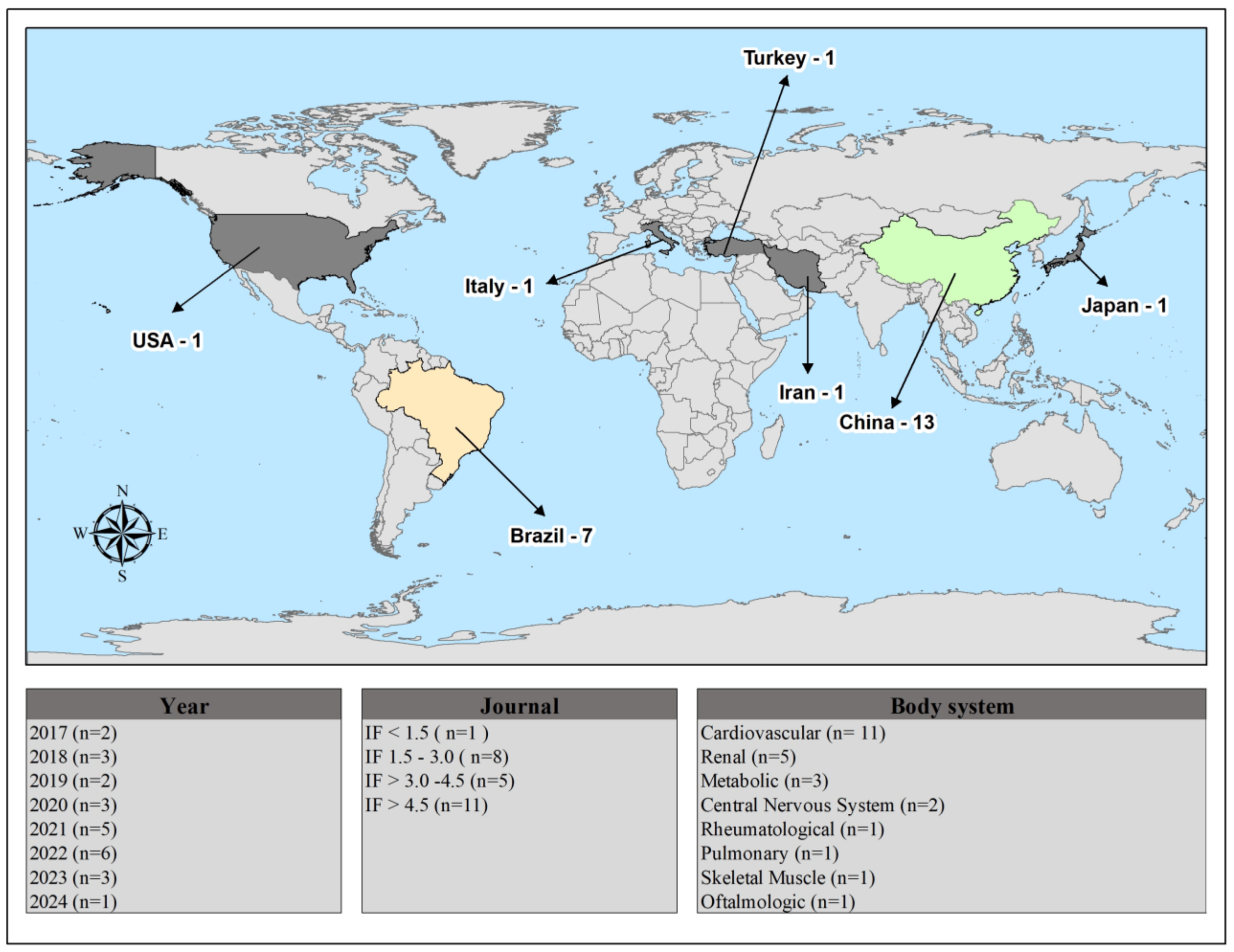
Figure 2 provides a summary of the study features, including publication year, country, impact factor, and body system. All studies were described in English and were experimental in nature. Most of the studies were published in journals with an impact factor above 4.5, with the cardiovascular system being the most frequently investigated.
Geographically, most of the research is emerging from China and Brazil, indicating a strong local interest. This may reflect strategic research priorities or funding opportunities in these regions. Other countries have contributed fewer studies, suggesting a more limited yet global interest in the topic.
Temporally, there is an observable increase in publications from the late 2010s to the mid-2020s, peaking in the early 2020s, which coincides with growing recognition of ALA’s physiological roles. The predominance of cardiovascular system studies aligns with the molecule’s established relevance in this area, but research has also expanded into renal, metabolic, and other biological systems, illustrating its multifaceted physiological impact.
These trends reflect an expanding and geographically diverse research landscape on ALA, highlighting its potential importance across multiple systems and justifying further investigation in both established and emerging regions of interest.
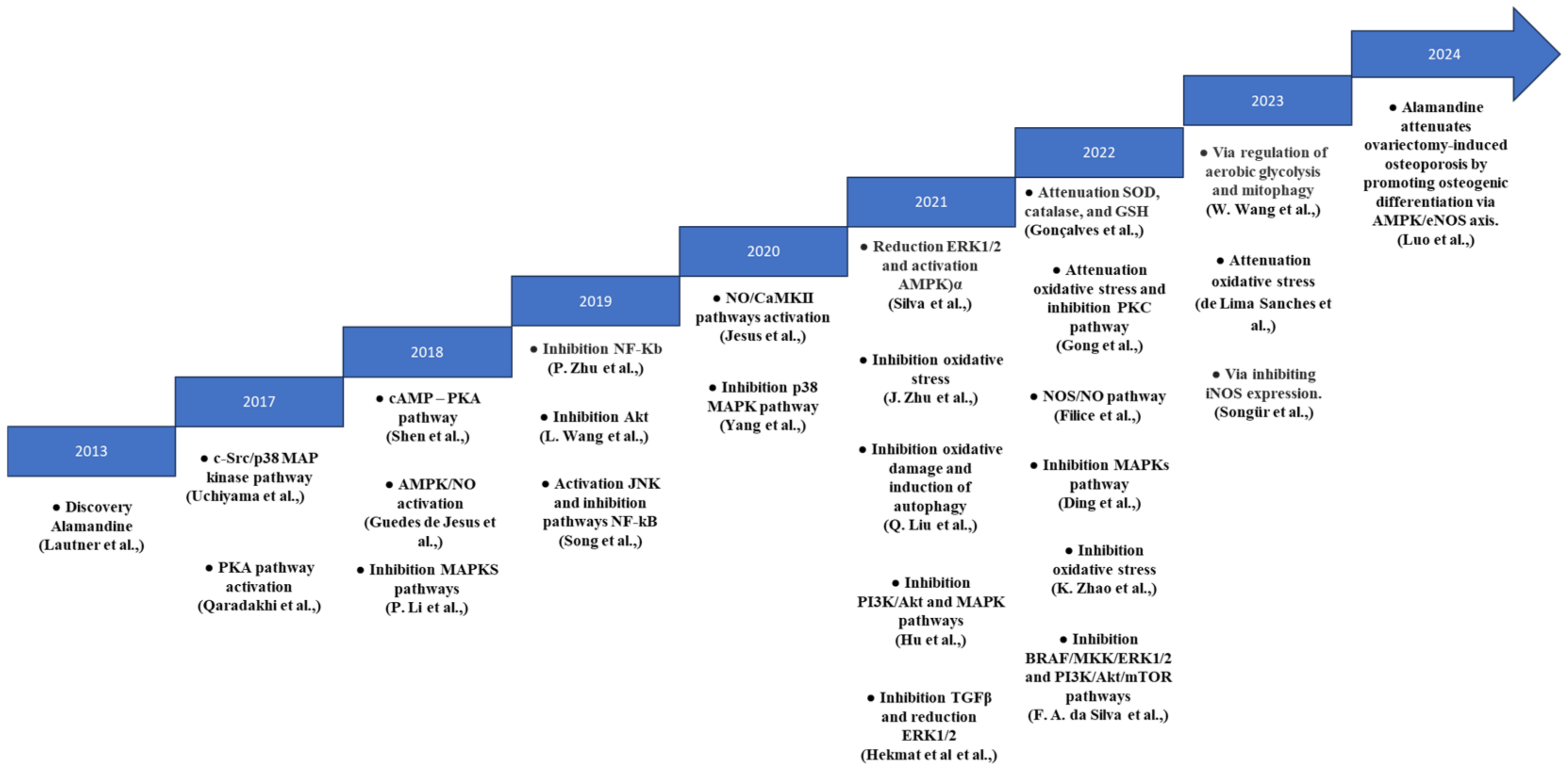
Figure 3 illustrates the timeline of significant discoveries related to ALA’s mechanisms of action since its identification in 2013. This figure highlights the evolving focus of research, particularly emphasizing its role within the cardiovascular system. Initially, research centered on ALA’s potential to enhance heart function and manage hypertension.
From 2018 onwards, studies expanded to explore its anti-inflammatory and antifibrotic properties, with particular attention to its interactions with signaling pathways like MAPK and Adenosine Monophosphate-activated protein kinase (AMPK). These discoveries underscore ALA’s therapeutic promise in addressing oxidative stress and fibrotic diseases. This timeline underscores the ongoing importance of ALA in biomedical research, highlighting the need for further studies to explore its therapeutic applications in areas such as metabolic disorders or neuroprotection.

4. Discussion

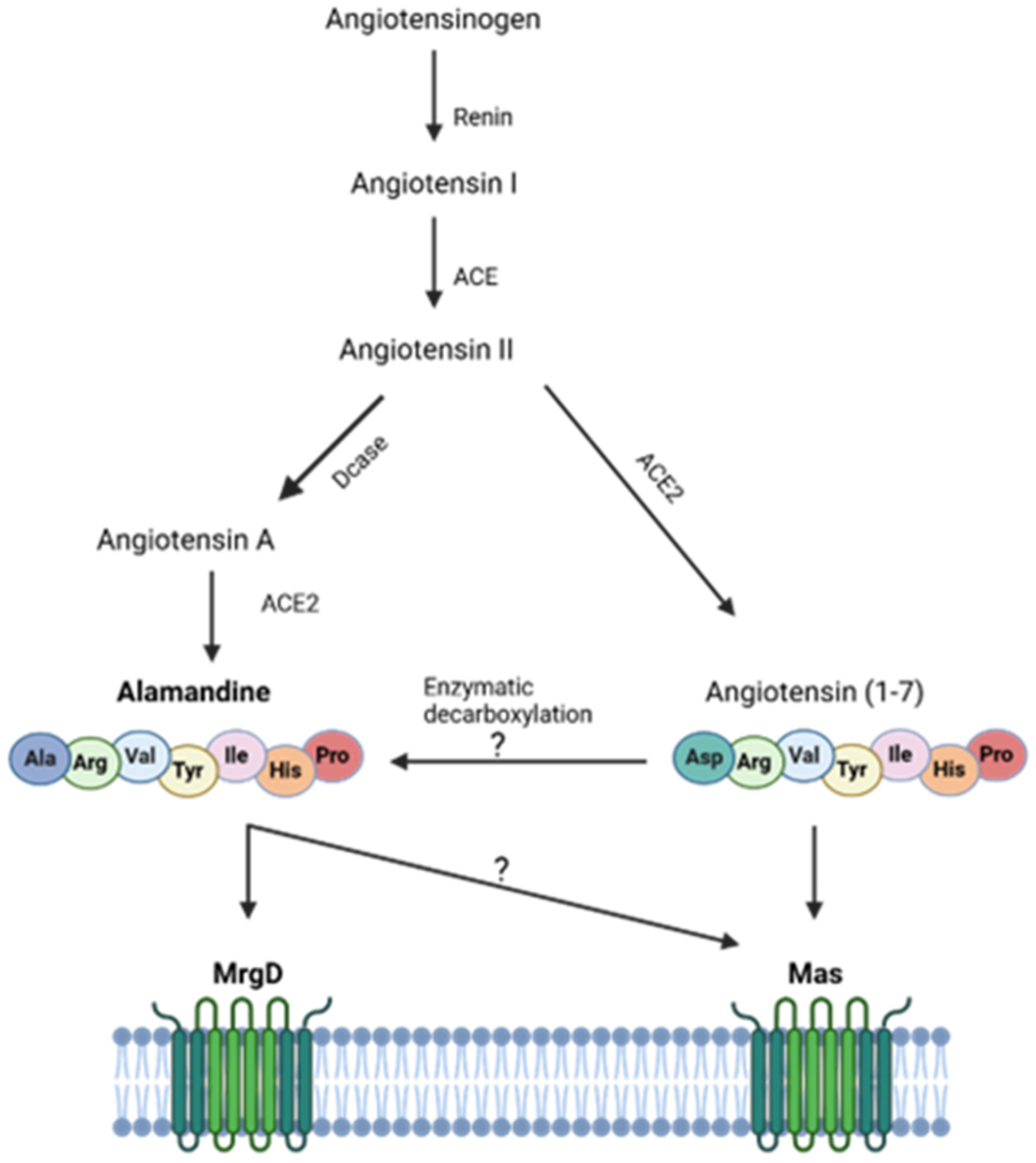
Identified in 2013 [35], ALA can be derived from angiotensin A through the action of ACE2 [36] or by the decarboxylation of Ang-(1-7) [37] (Figure 4).
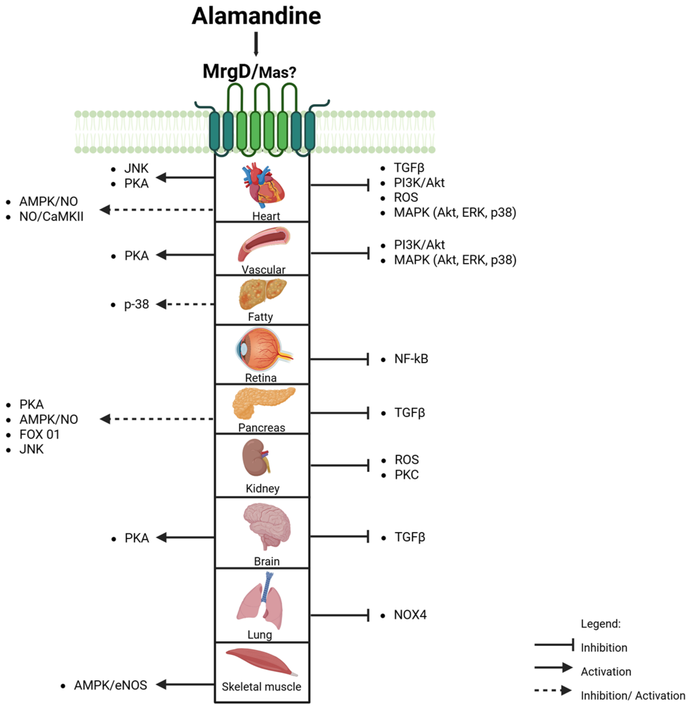
Activation of the ALA-MrgD axis is associated with vasodilation, anti-inflammatory, and antifibrotic actions. These effects are mediated by signaling pathways (Figure 5), particularly mitogen-activated protein kinases (MAPKs) and AMPK, regulating NO production and cellular energy homeostasis.

4.1. Mas and MrgD Receptors: Alamandine’s (Un)Specificity

The MrgD and Mas receptors are expressed in the dorsal root ganglia of the nervous system [38] and various organs, including the heart [8,39], brain [40,41,42], lungs [22], adipose tissue [43], vascular endothelium, arterial smooth muscle cells [44], and retina [33]. ALA’s affinity for the Mas receptor indicates a secondary receptor role [16,37].
The Mas and MrgD receptors may interact to form a functional complex. ALA binds to MrgD, promoting the dimerization of Mas and MrgD receptors, which leads to anti-inflammatory responses like reduced interleukin-6 and interleukin-1β secretion in lipopolysaccharide-activated THP-1 macrophages and M1 macrophages. Moreover, ALA promotes antiproliferative effects [45].
The complexity of this interaction is evident in cells using compounds such as PD123319, D-PRO7-ANG-(1-7), and A779, exposing the challenge of targeting specific receptors [46]. This suggests ALA’s actions may involve MrgD and AT2 receptors, not just the Mas receptor [3,32,42].

4.2. MAPK and AMPK Signaling Pathways

ALA-MrgD interaction in cardiomyocyte cultures enhances NO production [8] and phosphorylates AMPK and LKB1, key regulators of energy and cardioprotection [47]. AMPK activation is crucial for preventing cardiac injury progression to heart failure [48,49] and is linked to metabolic diseases [50,51], with bone tissue research supporting these findings [23].
Emphasizing its cardiovascular benefits, ALA exhibits a broad anti-inflammatory effect by reversing the increased phosphorylation of PI3K, Akt, ERK, c-Jun N-terminal kinase, and p38, reversing their increased activity in sepsis-associated renal injury [20]. This inhibition involves the protein kinase A (PKA) signaling pathway [52], underscoring cyclic adenosine monophosphate-dependent kinase’s role in these processes [53]. Inhibition of PKA by ALA induces cardiac remodeling [52] and promotes vasodilation, as confirmed using KT5720, a PKA inhibitor [25]. In fact, Shen et al. (2018) [25] demonstrated that regulation of the PKA pathway is essential for ALA’s effects in the brain.
Additionally, in ischemia–reperfusion, ALA treatment significantly lowers apoptosis in myocardial cells [27], possibly by attenuating the inflammatory response through inhibition of NF-κB activation [27,33]. This suppression reduces pro-inflammatory cytokine expression [20,54] and reactive oxygen species production [33] and impacts myocardial infarction models due to pressure overload [9]. It promotes protein kinase phosphorylation [20,55], affecting the balance between cellular survival and apoptosis.
The anti-inflammatory properties of ALA also extend to synovial fibroblasts, inhibiting the MAPK pathway and leading to a reduction in inflammatory mediators associated with rheumatoid arthritis [15]. Furthermore, ALA decreases matrix metalloproteinase-2, essential for TNF-alpha maturation and apoptosis control in cardiomyocytes [55]. It also reduces TGF-β, preventing connective tissue fibrosis, and plays a crucial role in mitigating hypertrophic and fibrotic pathways by inhibiting phosphorylated forms in the MAPK pathways [6].
Conversely, Ang II is known to promote phosphorylation of p38 MAPK, contributing to cardiac fibrosis [56], vascular fibrosis [32,57], cardiac stress, and hypertrophy [58,59]. The Ang-(1-7) and ALA axis improves aortic function [60], suggesting therapeutic benefits.
ALA also improves cardiac function in models of heart failure by attenuating TGF-β signaling and ERK 1 and 2 (ERK1/2) phosphorylation [26]. In cardiomyocytes, PKA inhibition may reduce MAPK activation, affecting pathways like c-Jun N-terminal kinase and p38 [6,53,61].

4.3. Nitric Oxide Production: The Therapeutic Role of ALA in Cardiovascular Health

ALA enhances endothelial nitric oxide synthase activity at serine 1177 and threonine 495, increasing NO release and highlighting its therapeutic potential for vasodilation and cardiovascular health [24,25,52]. Studies on goldfish (Carassius auratus) show that ALA enhances cardiac contractility under normoxic conditions via the NOS/NO system, revealing its sensitivity to hypoxia and broad cardiac benefits [16,62].
Furthermore, Songür et al. (2023) [28] and Hu et al. (2021) [20] showed that ALA’s anti-inflammatory, antiapoptotic, and antipyretic properties protect against acute renal injury and endotoxemia by suppressing inducible NOS (iNOS) expression, highlighting its potential in mitigating inflammatory responses. In adipose tissue, ALA activates the MrgD receptor, triggering c-Src and increasing iNOS expression [29], leading to NO production that may cause mitochondrial dysfunction and stimulate lipoprotein lipase activity, and adversely affecting adipocytes [63].
In a transgenic rat model with overexpressed renin, ALA enhances cardiomyocyte contractility via the NO/calcium/calmodulin-dependent kinase II (CaMKII) pathway, initiating NO production and activating CaMKII in vascular smooth muscle cells [21]. CaMKII phosphorylates the threonine 17 residue of phospholamban within the sarcoplasmic reticulum, increasing the activity of the sarcoplasmic reticulum Ca2+-ATPase and enhancing contractility [21,64].

4.4. Modulation of PI3K Enzyme Activity by ALA

The PI3K enzyme family is central to numerous biological processes, including cell survival, apoptosis, and cardiac function [65], playing a significant role in cardiac hypertrophy and diastolic dysfunction associated with hypertension. In primary cardiac fibroblasts, ALA’s interaction with the MrgD receptor inhibits Akt activation induced by Ang II, thereby reducing cardiac fibrosis [30,66,67].
PI3K’s role in cellular processes is broad. The inhibition of the PI3K/Akt pathway is also relevant in cancer cell lines such as Mia Paca-2 and A549. The ALA treatment shifts energy generation from anaerobic to aerobic processes, potentially slowing cancer growth. It inhibits the PI3K/Akt/mTOR pathway, essential for cancer cell growth and survival, and induces the nuclear translocation of the FoxO1 protein, affecting various cellular processes [13].
This pathway inhibition leads to the dephosphorylation and reduced activity of components like 3-phosphoinositide-dependent kinase 1, Akt1, and the mTOR receptor, as well as the BRAF/MKK/ERK1/2 signaling pathway, resulting in cell cycle arrest and apoptosis in pancreatic cancer cells. ALA reduces the phosphorylation of ERK1/2 and its effectors, NIBAN2 and Stathmin 1, highlighting its potential as a therapeutic target in cancer treatment [13,68].
Moreover, Wang et al. (2023) [31] reported that ALA inhibits glycolysis in vitro, via the MrgD axis, and downregulates hexokinase 2 (HK2), reducing the impact of TGF-β1 on lung fibroblasts [31]. ALA decreases the NADPH Oxidase 4 levels and reactive oxygen species generation in fibroblasts, which are known to contribute to the development of pulmonary fibrosis [22]. Under anaerobic conditions, the glycolysis enhancement results in lactate formation from pyruvate [69], creating a feedback loop that enhances lung fibroblast activation and upregulates glycolysis pathways such as HK2 activity and the allosteric effector of phosphofructokinase-1 by 6-phosphofructo-2-kinase/fructose-2,6-bisphosphatase 3 (PFKFB3) activity. In fact, HK2 and PFKFB3 are related to fibrosis development in several tissues: pulmonary [70,71], hepatic stellate cells [72,73], cardiac [74], kidney tubular cells [75], and renal fibroblast NRK-49F cells [76].
Conversely, ALA/MrgD counter-regulates this loop, reducing hexokinase activity [77] and decreasing glycolysis pathways [31]. Moreover, previous studies suggest that ALA-MrgD signaling downregulates the expression of HK2 and PFKFB3 genes, reinforcing the importance of changes in energetic metabolism during fibrosis and highlighting ALA’s potential role in controlling this energetic shift [31,73,75,76,78].
Mitophagy and glycolytic flux indicate a complex interaction with ALA [79]. These insights highlight ALA’s therapeutic potential by inhibiting lung fibroblast activation via TGF-β1 and autophagy proteins (Parkin/LC3) while counteracting metabolic reprogramming in bleomycin-induced fibrosis by suppressing HK2, PFKFB3, Parkin, and LC3 activation [31].
These findings show that in adipose tissue, ALA activates the NFκB and p38 MAP kinase pathways, which upregulate plasminogen activator inhibitor-1, a protein linked to adverse health outcomes [80]. Uchiyama et al. (2017) [29] demonstrated that a low dose of ALA (5.76 μg/kg) modulates leptin expression and secretion via the phospholipase C, cSrc, and p38 MAPK pathways. These findings reveal ALA’s intricate metabolic effects, indicating the need for further investigation to fully understand its mechanisms and potential applications.

4.5. Alamandine and Oxidative Stress

Evidence suggests that ALA plays a crucial role in reducing cardiac fibrosis resulting from oxygen and glucose deprivation. This effect is supported by reductions in oxidative stress and ischemic injury [9], as well as a decrease in apoptosis [9,81], which collectively lowers the risk of heart failure [82]. ALA administration is associated with enhanced activity of superoxide dismutase (SOD), catalase, and glutathione peroxidase, as demonstrated in the hippocampus of C57/Bl6 mice [17]. Furthermore, De Lima Sanches et al. (2023) [14] confirmed ALA’s antioxidant properties, showing that oral administration effectively reduced superoxide anion (O2•−) and normalized nuclear factor erythroid 2-related factor 2 levels in the right carotid artery following transverse aortic constriction.
Oxidative stress has been related to caspase-3 signaling and cardiac apoptosis activation, particularly in the doxorubicin (DOX) model [83]. Hekmat et al. (2021) [19] demonstrated increased caspase-3 activation and apoptosis in DOX-exposed groups versus controls, while ALA significantly reduces both. Zhao et al. (2022) [9] also demonstrated that ALA inhibits increases in collagen I, α-SMA, TGF-β, Bax/Bcl2, and the caspase-3/cleaved caspase-3 ratio, processes linked to cell death from oxygen–glucose deprivation in neonatal rat cardiac fibroblasts, highlighting ALA’s cardiac protective effects [9].
According to J. Zhu et al. (2021) [34], in rats subjected to ischemia–reperfusion injury, ALA treatment significantly increased SOD activity while reducing levels of malondialdehyde, NADPH oxidase activity, O2•− production, Nox expression, inflammation, and apoptosis. Additionally, Gong and colleagues [18] found that in cases of renal sodium overload, ALA mitigated the decline in SOD and glutathione peroxidase activity, as well as the increase in malondialdehyde, 8-hydroxy-2-deoxyguanosine, and O2•− levels. This effect was likely mediated by inhibiting the protein kinase C signaling pathway [18].
Despite the growing interest in ALA and its promising biological effects, important knowledge gaps remain. One major limitation is the lack of comprehensive studies examining its receptor specificity, as ALA appears to interact not only with the MrgD receptor but also potentially with Mas and AT2 receptors. Moreover, most evidence comes from experimental models using varying doses, administration routes, and disease contexts, which complicates comparisons across studies. The tissue-specific effects of ALA, as well as its dual roles, are protective in some systems but potentially detrimental in others (e.g., adipose tissue). Another limitation is the lack of clinical research, with no human studies to date assessing the safety, pharmacokinetics, or therapeutic efficacy of ALA.

5. Conclusions

Despite our limited knowledge, the discovery of Ang-(1-7) and ALA represents a significant advancement in our understanding of the RAS by introducing peptides with antagonistic actions. The recognition of these new peptides has sparked a debate and reinterpretation of the RAS. Importantly, studies indicate that ALA administration, across a wide range of dosages, does not produce detrimental effects.
It is crucial to recognize that ALA’s discovery is recent, and many mechanisms require further elucidation. Our current understanding of its role in pathophysiology and its mechanisms of action is based on the findings presented in this article. Moreover, these findings emphasize the need to consider dosage, tissue specificity, and the experimental conditions (in vivo vs. in vitro) when evaluating ALA’s biological impacts. Therefore, additional studies are necessary to explore ALA’s diverse roles in health and disease across various pathophysiological contexts.
Future research should prioritize the standardization of experimental protocols to facilitate better comparison of results and clarify the specific pathways involved in ALA signaling. These promising findings justify further preclinical studies and pave the way for translational efforts that may ultimately support clinical application.

Supplementary Materials

The following supporting information can be downloaded at: https://www.mdpi.com/article/10.3390/biomedicines13081957/s1, Table S1: Results of PubMed, Scopus, Embase and Web of Sciences search; Table S2: Summary of data extraction; Table S3: Preferred Reporting Items for Systematic reviews and Meta-Analyses extension for Scoping Reviews (PRISMA-ScR) Checklist; Table S4: References and reasons for exclusion of articles during the full-text reading phase of eligibility confirmation (n = 42); Table S5: Extraction of data from selected articles. Refs. [6,8,9,12,13,14,15,16,17,18,19,20,21,22,23,24,25,26,27,28,29,30,31,32,33,34] are cited in Supplementary Materials.

Author Contributions

Conceptualization, K.R. and J.F.; methodology, F.M.S.; validation, A.T.S., J.F., L.B.O. and I.A.M.; formal analysis, A.F.K.V.; investigation, K.R. and M.R.W.; data curation, A.T.S. and J.F.; writing—original draft preparation, J.F.; writing—review and editing, K.R., M.R.W. and J.F.; visualization, I.A.M.; supervision, K.R.; project administration, K.R. and F.M.S. All authors have read and agreed to the published version of the manuscript.

Funding

J.F., A.T.S., and I.A.M. were supported by a doctoral fellowship from Coordenação de Aperfeiçoamento de Pessoal de Nível Superior (CAPES). This research received no external funding.

Data Availability Statement

The raw data supporting the conclusions of this article will be made available by the authors upon request.

Conflicts of Interest

The authors declare no conflicts of interest.

Abbreviations

The following abbreviations are used in this manuscript:
ACEAngiotensin-converting enzyme
ACE2Angiotensin-converting enzyme 2
AktProtein Kinase B
ALAAlamandine
ANG-(1-7)Angiotensin-(1-7)
Ang IIAngiotensin II
AMPKAdenosine monophosphate-activated protein kinase
CAMKIICalmodulin-dependent kinase II
DoxDoxorubicin
eNOSEndothelial Nitric Oxide Synthase
ERK1/2Extracellular Signal-Regulated Kinases 1 and 2
HK2Hexokinase 2
iNOSInducible Nitric Oxide Synthase
JNKc-Jun N-terminal kinase
MasG protein-coupled receptor
MAPKMitogen-Activated Protein Kinase
MDAMalondialdehyde
MrgDMas-related G protein-coupled receptor member D
NF-kBNuclear Factor Kappa B
NONitric Oxide
NOSNitric Oxide Synthase
O2•−Superoxide anion
PCCPopulation Concept and Context
PI3KPhosphoinositide 3-kinase
PKAProtein kinase A
PKFB3Phosphofructo-2-kinase/fructose-2,6-bisphosphatase 3
RASRenin–angiotensin system
SHRSpontaneously hypertensive rat
SODSuperoxide Dismutase
TGFβTransforming Growth Factor-β

References

  1. Hall, J.E. Historical Perspective of the Renin-Angiotensin System. Mol. Biotechnol. 2003, 24, 27–39. [Google Scholar] [CrossRef]
  2. Suter, C.; Coote, J.H. Intrathecally administered angiotensin II increases sympathetic activity in the rat. J. Auton. Nerv. Syst. 1987, 19, 31–37. [Google Scholar] [CrossRef]
  3. Lautner, R.Q.; Villela, D.C.; Fraga-Silva, R.A.; Silva, N.; Verano-Braga, T.; Costa-Fraga, F.; Jankowski, J.; Jankowski, V.; Sousa, F.; Alzamora, A.; et al. Discovery and characterization of alamandine: A novel component of the renin-angiotensin system. Circ. Res. 2013, 112, 1104–1111. [Google Scholar] [CrossRef]
  4. Santos, R.A.; Pesquero, J.; Chernicky, C.L.; Greene, L.J.; Ferrario, C.M. Converting Enzyme Activity and Angiotensin Metabolism in the Dog Brainstem. Hypertension 1988, 11, 153–157. [Google Scholar] [CrossRef] [PubMed]
  5. Schleifenbaum, J. Alamandine and its receptor mrgd pair up to join the protective arm of the renin-angiotensin system. Front. Med. 2019, 6, 107. [Google Scholar] [CrossRef] [PubMed]
  6. Li, P.; Chen, X.-R.; Xu, F.; Liu, C.; Li, C.; Liu, H.; Wang, H.; Sun, W.; Sheng, Y.-H.; Kong, X.-Q. Alamandine attenuates sepsis-associated cardiac dysfunction via inhibiting MAPKs signaling pathways. Life Sci. 2018, 206, 106–116. [Google Scholar] [CrossRef]
  7. Fernandes, R.S.; Netto, M.R.T.; Carvalho, F.B.; Rigatto, K. Alamandine: A promising treatment for fibrosis. Peptides 2022, 157, 170848. [Google Scholar] [CrossRef]
  8. De Jesus, I.C.G.; Scalzo, S.; Alves, F.; Marques, K.; Rocha-Resende, C.; Bader, M.; Santos, R.A.S.; Guatimosim, S. Alamandine acts via MrgD to induce AMPK/NO activation against ANG II hypertrophy in cardiomyocytes. Am. J. Physiol. Cell Physiol. 2018, 314, C702–C711. [Google Scholar] [CrossRef]
  9. Zhao, K.; Xu, T.; Mao, Y.; Wu, X.; Hua, D.; Sheng, Y.; Li, P. Alamandine alleviated heart failure and fibrosis in myocardial infarction mice. Biol. Direct 2022, 17, 25. [Google Scholar] [CrossRef]
  10. Huang, Y.; Li, Y.; Lou, A.; Wang, G.Z.; Hu, Y.; Zhang, Y.; Huang, W.; Wang, J.; Li, Y.; Zhu, X.; et al. Alamandine attenuates hepatic fibrosis by regulating autophagy induced by NOX4-dependent ROS. Clin. Sci. 2020, 134, 853–869. [Google Scholar] [CrossRef]
  11. Aromataris, E.; Lockwood, C.; Porritt, K.; Pilla, B.; Jordan, Z. JBI Manual for Evidence Synthesis. 2024. Available online: https://synthesismanual.jbi.global (accessed on 30 April 2023).
  12. Tricco, A.C.; Lillie, E.; Zarin, W.; O’Brien, K.K.; Colquhoun, H.; Levac, D.; Moher, D.; Peters, M.D.J.; Horsley, T.; Weeks, L.; et al. PRISMA Extension for Scoping Reviews (PRISMA-ScR): Checklist and Explanation. Ann. Intern. Med. 2018, 169, 467–473. [Google Scholar] [CrossRef]
  13. Da Silva, F.A.; Rodrigues-Ribeiro, L.; Melo-Braga, M.N.; Passos-Silva, D.G.; Sampaio, W.O.; Gorshkov, V.; Kjeldsen, F.; Verano-Braga, T.; Santos, R.A.S. Phosphoproteomic studies of alamandine signaling in CHO-Mrgd and human pancreatic carcinoma cells: An antiproliferative effect is unveiled. Proteomics 2022, 22, e2100255. [Google Scholar] [CrossRef]
  14. De Lima Sanches, B.; Souza-Neto, F.; de Alcântara-Leonídeo, T.C.; Silva, M.M.; Guatimosim, S.; Vieira, M.A.R.; Santos, R.A.S.; da Silva, R.F. Alamandine attenuates oxidative stress in the right carotid following transverse aortic constriction in mice. Peptides 2023, 171, 171094. [Google Scholar] [CrossRef]
  15. Ding, W.; Miao, Z.; Feng, X.; Luo, A.; Tan, W.; Li, P.; Wang, F. Alamandine, a new member of the renin-angiotensin system (RAS), attenuates collagen-induced arthritis in mice via inhibiting cytokine secretion in synovial fibroblasts. Peptides 2022, 154, 170816. [Google Scholar] [CrossRef]
  16. Filice, M.; Mazza, R.; Imbrogno, S.; Mileti, O.; Baldino, N.; Barca, A.; Del Vecchio, G.; Verri, T.; Gattuso, A.; Cerra, M.C. An ACE2-Alamandine Axis Modulates the Cardiac Performance of the Goldfish Carassius auratus via the NOS/NO System. Antioxidants 2022, 11, 764. [Google Scholar] [CrossRef] [PubMed]
  17. Gonçalves, S.C.; Bassi, B.L.; Kangussu, L.M.; Alves, D.T.; Ramos, L.K.; Fernandes, L.F.; Alves, M.T.; Sinisterra, R.; Bruch, G.E.; Santos, R.A.; et al. Alamandine Induces Neuroprotection in Ischemic Stroke Models. Curr. Med. Chem. 2022, 29, 3483–3498. [Google Scholar] [CrossRef] [PubMed]
  18. Gong, J.; Luo, M.; Yong, Y.; Zhong, S.; Li, P. Alamandine alleviates hypertension and renal damage via oxidative-stress attenuation in Dahl rats. Cell Death Discov. 2022, 8, 22. [Google Scholar] [CrossRef] [PubMed]
  19. Hekmat, A.S.; Navabi, Z.; Alipanah, H.; Javanmardi, K. Alamandine significantly reduces doxorubicin-induced cardiotoxicity in rats. Hum. Exp. Toxicol. 2021, 40, 1781–1795. [Google Scholar] [CrossRef]
  20. Hu, W.; Gao, W.; Miao, J.; Xu, Z.; Sun, L. Alamandine, a derivative of angiotensin-(1-7), alleviates sepsis-associated renal inflammation and apoptosis by inhibiting the PI3K/Ak and MAPK pathways. Peptides 2021, 146, 170627. [Google Scholar] [CrossRef]
  21. Jesus, I.C.G.; Mesquita, T.R.R.; Monteiro, A.L.L.; Parreira, A.B.; Santos, A.K.; Coelho, E.L.X.; Silva, M.M.; Souza, L.A.C.; Campagnole-Santos, M.J.; Santos, R.S.; et al. Alamandine enhances cardiomyocyte contractility in hypertensive rats through a nitric oxide-dependent activation of CaMKII. Am. J. Physiol. Physiol. 2020, 318, C740–C750. [Google Scholar] [CrossRef]
  22. Liu, Q.; Zheng, B.; Zhang, Y.; Huang, W.; Hong, Q.; Meng, Y. Alamandine via MrgD receptor attenuates pulmonary fibrosis via nox4 and autophagy pathway. Can. J. Physiol. Pharmacol. 2021, 99, 885–893. [Google Scholar] [CrossRef]
  23. Luo, W.; Yao, C.; Sun, J.; Zhang, B.; Chen, H.; Miao, J.; Zhang, Y. Alamandine attenuates ovariectomy-induced osteoporosis by promoting osteogenic differentiation via AMPK/eNOS axis. BMC Musculoskelet. Disord. 2024, 25, 45. [Google Scholar] [CrossRef] [PubMed]
  24. Qaradakhi, T.; Matsoukas, M.T.; Hayes, A.; Rybalka, E.; Caprnda, M.; Rimarova, K.; Sepsi, M.; Büsselberg, D.; Kruzliak, P.; Matsoukas, J.; et al. Alamandine reverses hyperhomocysteinemia-induced vascular dysfunction via PKA-dependent mechanisms. Cardiovasc. Ther. 2017, 35, e12306. [Google Scholar] [CrossRef] [PubMed]
  25. Shen, Y.H.; Chen, X.R.; Yang, C.X.; Liu, B.X.; Li, P. Alamandine injected into the paraventricular nucleus increases blood pressure and sympathetic activation in spontaneously hypertensive rats. Peptides 2018, 103, 98–102. [Google Scholar] [CrossRef] [PubMed]
  26. Silva, M.M.; de Souza-Neto, F.P.; de Jesus, I.C.G.; Gonçalves, G.K.; Santuchi, M.d.C.; Sanches, B.d.L.; de Alcântara-Leonídio, T.C.; Melo, M.B.; Vieira, M.A.R.; Guatimosim, S.; et al. Alamandine improves cardiac remodeling induced by transverse aortic constriction in mice. Am. J. Physiol. Heart Circ. Physiol. 2021, 320, H352–H363. [Google Scholar] [CrossRef]
  27. Song, X.D.; Feng, J.P.; Yang, R.X. Alamandine protects rat from myocardial ischemia-reperfusion injury by activating JNK and inhibiting NF-κB. Eur. Rev. Med. Pharmacol. Sci. 2019, 12, 6718–6726. [Google Scholar] [CrossRef]
  28. Songür, H.S.; Kaya, S.A.; Altınışık, Y.C.; Abanoz, R.; Özçelebi, E.; Özmen, F.; Kösemehmetoğlu, K.; Soydan, G. Alamandine treatment prevents LPS-induced acute renal and systemic dysfunction with multi-organ injury in rats via inhibiting iNOS expression. Eur. J. Pharmacol. 2023, 960, 176160. [Google Scholar] [CrossRef]
  29. Uchiyama, T.; Okajima, F.; Mogi, C.; Tobo, A.; Tomono, S.; Sato, K. Alamandine reduces leptin expression through the c-Src/p38 MAP kinase pathway in adipose tissue. PLoS ONE 2017, 12, e0178769. [Google Scholar] [CrossRef]
  30. Wang, L.; Liu, C.; Chen, X.; Li, P. Alamandine attenuates long-term hypertension-induced cardiac fibrosis independent of blood pressure. Mol. Med. Rep. 2019, 19, 4553–4560. [Google Scholar] [CrossRef]
  31. Wang, W.; Zhang, Y.; Huang, W.; Yuan, Y.; Hong, Q.; Xie, Z.; Li, L.; Chen, Y.; Li, X.; Meng, Y. Alamandine/MrgD axis prevents TGF-β1-mediated fibroblast activation via regulation of aerobic glycolysis and mitophagy. J. Transl. Med. 2023, 21, 24. [Google Scholar] [CrossRef]
  32. Yang, C.; Wu, X.; Shen, Y.; Liu, C.; Li, P.; Kong, X. Alamandine attenuates angiotensin II-induced vascular fibrosis via inhibiting p38 MAPK pathway. Eur. J. Pharmacol. 2020, 883, 173384. [Google Scholar] [CrossRef]
  33. Zhu, P.; Verma, A.; Prasad, T.; Li, Q. Expression and Function of Mas-Related G Protein-Coupled Receptor D and Its Ligand Alamandine in Retina. Mol. Neurobiol. 2020, 57, 513–527. [Google Scholar] [CrossRef]
  34. Zhu, J.; Qiu, J.G.; Xu, W.T.; Ma, H.X.; Jiang, K. Alamandine protects against renal ischaemia–reperfusion injury in rats via inhibiting oxidative stress. J. Pharm. Pharmacol. 2021, 73, 1491–1502. [Google Scholar] [CrossRef] [PubMed]
  35. Villela, D.C.; Passos-Silva, D.G.; Santos, R.A.S. Alamandine: A new member of the angiotensin family. Curr. Opin. Nephrol. Hypertens. 2014, 23, 130–134. [Google Scholar] [CrossRef] [PubMed]
  36. Jankowski, V.; Vanholder, R.; van der Giet, M.; Toölle, M.; Karadogan, S.; Gobom, J.; Furkert, J.; Oksche, A.; Krause, E.; Tran, T.N.A.; et al. Mass-spectrometric identification of a novel angiotensin peptide in human plasma. Arter. Thromb. Vasc. Biol. 2007, 27, 297–302. [Google Scholar] [CrossRef] [PubMed]
  37. Tetzner, A.; Naughton, M.; Gebolys, K.; Eichhorst, J.; Sala, E.; Villacañas, Ó.; Walther, T. Decarboxylation of Ang-(1–7) to Ala1-Ang-(1–7) leads to significant changes in pharmacodynamics. Eur. J. Pharmacol. 2018, 833, 116–123. [Google Scholar] [CrossRef]
  38. Shinohara, T.; Harada, M.; Ogi, K.; Maruyama, M.; Fujii, R.; Tanaka, H.; Fukusumi, S.; Komatsu, H.; Hosoya, M.; Noguchi, Y.; et al. Identification of a G protein-coupled receptor specifically responsive to β-alanine. J. Biol. Chem. 2004, 279, 23559–23564. [Google Scholar] [CrossRef]
  39. Oliveira, A.C.; Melo, M.B.; Motta-Santos, D.; Peluso, A.A.; Souza-Neto, F.; da Silva, R.F.; Almeida, J.F.Q.; Canta, G.; Reis, A.M.; Goncalves, G.; et al. Genetic deletion of the alamandine receptor mrgd leads to dilated cardiomyopathy in mice. Am. J. Physiol. Heart Circ. Physiol. 2019, 316, H123–H133. [Google Scholar] [CrossRef]
  40. Hami, J.; von Bohlen und Halbach, V.; Tetzner, A.; Walther, T.; von Bohlen und Halbach, O. Localization and expression of the Mas-related G-protein coupled receptor member D (MrgD) in the mouse brain. Heliyon 2021, 7, e08440. [Google Scholar] [CrossRef]
  41. Jackson, L.; Eldahshan, W.; Fagan, S.C.; Ergul, A. Within the brain: The renin angiotensin system. Int. J. Mol. Sci. 2018, 19, 876. [Google Scholar] [CrossRef]
  42. Marins, F.R.; Oliveira, A.C.; Qadri, F.; Motta-Santos, D.; Alenina, N.; Bader, M.; Fontes, M.A.P.; Santos, R.A.S. Alamandine but not angiotensin-(1–7) produces cardiovascular effects at the rostral insular cortex. Am. J. Physiol. Regul. Integr. Comp. Physiol. 2021, 321, R513–R521. [Google Scholar] [CrossRef] [PubMed]
  43. Cerri, G.C.; Santos, S.H.S.; Bader, M.; Santos, R.A.S. Brown adipose tissue transcriptome unveils an important role of the Beta-alanine/alamandine receptor, MrgD, in metabolism. J. Nutr. Biochem. 2023, 114, 109268. [Google Scholar] [CrossRef] [PubMed]
  44. Habiyakare, B.; Alsaadon, H.; Mathai, M.L.; Hayes, A.; Zulli, A. Reduction of angiotensin A and alamandine vasoactivity in the rabbit model of atherogenesis: Differential effects of alamandine and Ang(1-7). Int. J. Exp. Pathol. 2014, 95, 290–295. [Google Scholar] [CrossRef] [PubMed]
  45. Rukavina Mikusic, N.L.; Silva, M.G.; Erra Díaz, F.A.; Pineda, A.M.; Ferragut, F.; Gómez, K.A.; Mazzitelli, L.; Gonzalez Maglio, D.H.; Nuñez, M.; Santos, R.A.S.; et al. Alamandine, a protective component of the renin-angiotensin system, reduces cellular proliferation and interleukin-6 secretion in human macrophages through MasR–MrgDR heteromerization. Biochem. Pharmacol. 2024, 229, 116480. [Google Scholar] [CrossRef]
  46. Assis, A.D.; do Prado Mascarenhas, F.N.; de Assis Araújo, F.; Santos, R.A.; Zanon, R.G. Angiotensin-(1-7) receptor Mas antagonist (A779) influenced gliosis and reduced synaptic density in the spinal cord after peripheral axotomy. Peptides 2020, 129, 170329. [Google Scholar] [CrossRef]
  47. Takano, A.P.C.; Diniz, G.P.; Barreto-Chaves, M.L.M. AMPK signaling pathway is rapidly activated by T3 and regulates the cardiomyocyte growth. Mol. Cell Endocrinol. 2013, 376, 43–50. [Google Scholar] [CrossRef]
  48. Beauloye, C.; Bertrand, L.; Horman, S.; Hue, L. AMPK activation, a preventive therapeutic target in the transition from cardiac injury to heart failure. Cardiovasc. Res. 2011, 90, 224–233. [Google Scholar] [CrossRef]
  49. Zhang, C.; Pan, S.; Meng, R.; Peng, C.; Xiong, Z.; Chen, B.; Chen, G.; Yao, F.; Chen, Y.; Ma, Y.; et al. Metformin attenuates ventricular hypertrophy by activating the AMP-activated protein kinase-endothelial nitric oxide synthase pathway in rats. Clin. Exp. Pharmacol. Physiol. 2011, 38, 55–62. [Google Scholar] [CrossRef]
  50. Hardie, D.G. AMPK—Sensing Energy while Talking to Other Signaling Pathways. Cell Metab. 2014, 20, 939–952. [Google Scholar] [CrossRef]
  51. Zaha, V.G.; Young, L.H. AMP-Activated Protein Kinase Regulation and Biological Actions in the Heart. Circ. Res. 2012, 111, 800–814. [Google Scholar] [CrossRef]
  52. Rababa’h, A.; Singh, S.; Suryavanshi, S.; Altarabsheh, S.; Deo, S.; McConnell, B. Compartmentalization Role of A-Kinase Anchoring Proteins (AKAPs) in Mediating Protein Kinase A (PKA) Signaling and Cardiomyocyte Hypertrophy. Int. J. Mol. Sci. 2014, 16, 218–229. [Google Scholar] [CrossRef]
  53. Peng, T.; Lu, X.; Lei, M.; Feng, Q. Endothelial nitric-oxide synthase enhances lipopolysaccharide-stimulated tumor necrosis factor-α expression via cAMP-mediated p38 MAPK pathway in cardiomyocytes. J. Biol. Chem. 2003, 278, 8099–8105. [Google Scholar] [CrossRef]
  54. Chen, X.; Li, X.; Zhang, W.; He, J.; Xu, B.; Lei, B.; Wang, Z.; Cates, C.; Rousselle, T.; Li, J. Activation of AMPK inhibits inflammatory response during hypoxia and reoxygenation through modulating JNK-mediated NF-κB pathway. Metabolism 2018, 83, 256–270. [Google Scholar] [CrossRef]
  55. Shen, J.; O’Brien, D.; Xu, Y. Matrix metalloproteinase-2 contributes to tumor necrosis factor alpha induced apoptosis in cultured rat cardiac myocytes. Biochem. Biophys. Res. Commun. 2006, 347, 1011–1020. [Google Scholar] [CrossRef] [PubMed]
  56. Yin, Q.; Lu, H.; Bai, Y.; Tian, A.; Yang, Q.; Wu, J.; Yang, C.; Fan, T.P.; Zhang, Y.; Zheng, X.; et al. A metabolite of Danshen formulae attenuates cardiac fibrosis induced by isoprenaline, via a NOX2/ROS/p38 pathway. Br. J. Pharmacol. 2015, 172, 5573–5585. [Google Scholar] [CrossRef] [PubMed]
  57. Rodrigues Díez, R.; Rodrigues-Díez, R.; Lavoz, C.; Rayego-Mateos, S.; Civantos, E.; Rodríguez-Vita, J.; Mezzano, S.; Ortiz, A.; Egido, J.; Ruiz-Ortega, M. Statins inhibit angiotensin II/smad pathway and related vascular fibrosis, by a TGF-β-independent process. PLoS ONE 2010, 5, e14145. [Google Scholar] [CrossRef] [PubMed]
  58. Nguyen Dinh Cat, A.; Montezano, A.C.; Burger, D.; Touyz, R.M. Angiotensin II, NADPH oxidase, and redox signaling in the vasculature. Antioxid. Redox Signal. 2013, 19, 1110–1120. [Google Scholar] [CrossRef]
  59. Nadal-Ginard, B.; Kajstura, J.; Anversa, P.; Leri, A. A matter of life and death: Cardiac myocyte apoptosis and regeneration. J. Clin. Investig. 2003, 111, 1457–1459. [Google Scholar] [CrossRef]
  60. Potthoff, S.; Stamer, S.; Grave, K.; Königshausen, E.; Sivritas, S.; Thieme, M.; Mori, Y.; Woznowski, M.; Rump, L.; Stegbauer, J. Chronic p38 mitogen-activated protein kinase inhibition improves vascular function and remodeling in angiotensin II-dependent hypertension. J. Renin-Angiotensin-Aldosterone Syst. 2016, 17, 147032031665328. [Google Scholar] [CrossRef]
  61. Kostenko, S.; Shiryaev, A.; Dumitriu, G.; Gerits, N.; Moens, U. Cross-talk between protein kinase A and the MAPK-activated protein kinases RSK1 and MK5. J. Recept. Signal Transduct. 2011, 31, 1–9. [Google Scholar] [CrossRef]
  62. Imbrogno, S.; Filice, M.; Cerra, M.C.; Gattuso, A. NO, CO and H2S: What about gasotransmitters in fish and amphibian heart? Acta Physiol. 2018, 223, e13035. [Google Scholar] [CrossRef]
  63. Jeon, M.J.; Leem, J.; Ko, M.S.; Jang, J.E.; Park, H.-S.; Kim, H.S.; Kim, M.; Yoo, H.J.; Lee, C.-H.; Park, I.-S.; et al. Mitochondrial dysfunction and activation of iNOS are responsible for the palmitate-induced decrease in adiponectin synthesis in 3T3L1 adipocytes. Exp. Mol. Med. 2012, 44, 562–570. [Google Scholar] [CrossRef]
  64. Beckendorf, J.; van den Hoogenhof, M.M.G.; Backs, J. Physiological and unappreciated roles of CaMKII in the heart. Basic Res. Cardiol. 2018, 113, 29. [Google Scholar] [CrossRef]
  65. Prasad, S.V.N.; Perrino, C.; Rockman, H.A. Role of phosphoinositide 3-kinase in cardiac function and heart failure. Trends Cardiovasc. Med. 2003, 13, 206–212. [Google Scholar] [CrossRef]
  66. Lin, C.-Y.; Hsu, Y.-J.; Hsu, S.-C.; Chen, Y.; Lee, H.-S.; Lin, S.-H.; Huang, S.-M.; Tsai, C.-S.; Shih, C.-C. CB1 cannabinoid receptor antagonist attenuates left ventricular hypertrophy and Akt-mediated cardiac fibrosis in experimental uremia. J. Mol. Cell Cardiol. 2015, 85, 249–261. [Google Scholar] [CrossRef] [PubMed]
  67. Zhao, Q.D.; Viswanadhapalli, S.; Williams, P.; Shi, Q.; Tan, C.; Yi, X.; Bhandari, B.; Abboud, H.E. NADPH oxidase 4 induces cardiac fibrosis and hypertrophy through activating Akt/mTOR and NFκB signaling pathways. Circulation 2015, 131, 643–655. [Google Scholar] [CrossRef] [PubMed]
  68. Roy, S.K.; Srivastava, R.K.; Shankar, S. Inhibition of PI3K/AKT and MAPK/ERK pathways causes activation of FOXO transcription factor, leading to cell cycle arrest and apoptosis in pancreatic cancer. J. Mol. Signal 2010, 5, 10. [Google Scholar] [CrossRef] [PubMed]
  69. Lu, L.; Wang, H.; Liu, X.; Tan, L.; Qiao, X.; Ni, J.; Sun, Y.; Liang, J.; Hou, Y.; Dou, H. Pyruvate kinase isoform M2 impairs cognition in systemic lupus erythematosus by promoting microglial synaptic pruning via the β-catenin signaling pathway. J. Neuroinflamm. 2021, 18, 229. [Google Scholar] [CrossRef]
  70. Zhang, S.; Sun, L.; Chen, B.; Lin, S.; Gu, J.; Tan, L.; Lin, M. Telocytes protect against lung tissue fibrosis through hexokinase 2-dependent pathway by secreting hepatocyte growth factor. Clin. Exp. Pharmacol. Physiol. 2023, 50, 964–972. [Google Scholar] [CrossRef]
  71. Yin, X.; Choudhury, M.; Kang, J.-H.; Schaefbauer, K.J.; Jung, M.-Y.; Andrianifahanana, M.; Hernandez, D.M.; Leof, E.B. Hexokinase 2 couples glycolysis with the profibrotic actions of TGF-β. Sci. Signal. 2019, 12, eaax4067. [Google Scholar] [CrossRef]
  72. Rho, H.; Terry, A.R.; Chronis, C.; Hay, N. Hexokinase 2-mediated gene expression via histone lactylation is required for hepatic stellate cell activation and liver fibrosis. Cell Metab. 2023, 35, 1406–1423.e8. [Google Scholar] [CrossRef]
  73. Zhang, Y.; Zhang, Y.; Chen, T.; Lin, Y.; Gong, J.; Xu, Q.; Wang, J.; Li, J.; Meng, Y.; Li, Y.; et al. Caveolin-1 depletion attenuates hepatic fibrosis via promoting SQSTM1-mediated PFKL degradation in HSCs. Free Radic. Biol. Med. 2023, 204, 95–107. [Google Scholar] [CrossRef]
  74. Wu, R.; Smeele, K.M.; Wyatt, E.; Ichikawa, Y.; Eerbeek, O.; Sun, L.; Chawla, K.; Hollmann, M.W.; Nagpal, V.; Heikkinen, S.; et al. Reduction in hexokinase II levels results in decreased cardiac function and altered remodeling after ischemia/reperfusion injury. Circ. Res. 2011, 108, 60–69. [Google Scholar] [CrossRef] [PubMed]
  75. Wang, Y.; Li, H.; Jiang, S.; Fu, D.; Lu, X.; Lu, M.; Li, Y.; Luo, D.; Wu, K.; Xu, Y.; et al. The glycolytic enzyme PFKFB3 drives kidney fibrosis through promoting histone lactylation-mediated NF-κB family activation. Kidney Int. 2024, 106, 226–240. [Google Scholar] [CrossRef] [PubMed]
  76. Yang, Q.; Huo, E.; Cai, Y.; Zhang, Z.; Dong, C.; Asara, J.M.; Wei, Q. PFKFB3-Mediated Glycolysis Boosts Fibroblast Activation and Subsequent Kidney Fibrosis. Cells 2023, 12, 2081. [Google Scholar] [CrossRef] [PubMed]
  77. Robey, R.B.; Hay, N.; Robey, R.B.; Hay, N. Mitochondrial Hexokinases: Guardians of the Mitochondria. Cell Cycle 2005, 4101, 654–658. [Google Scholar] [CrossRef]
  78. Zeng, H.; Pan, T.; Zhan, M.; Hailiwu, R.; Liu, B.; Yang, H.; Li, P. Suppression of PFKFB3-driven glycolysis restrains endothelial-to-mesenchymal transition and fibrotic response. Signal Transduct. Target. Ther. 2022, 7, 303. [Google Scholar] [CrossRef]
  79. Youle, R.J.; Narendra, D.P. Mechanisms of mitophagy. Nat. Rev. Mol. Cell Biol. 2011, 12, 9–14. [Google Scholar] [CrossRef]
  80. Pandey, M.; Loskutoff, D.J.; Samad, F. Molecular mechanisms of tumor necrosis factor-α-mediated plasminogen activator inhibitor-1 expression in adipocytes. FASEB J. 2005, 19, 1317–1319. [Google Scholar] [CrossRef]
  81. Kumar, D.; Jugdutt, B.I. Apoptosis and oxidants in the heart. J. Lab. Clin. Med. 2003, 142, 288–297. [Google Scholar] [CrossRef]
  82. Li, Z.; Bing, O.H.L.; Long, X.; Robinson, K.G.; Lakatta, E.G. Increased cardiomyocyte apoptosis during the transition to heart failure in the spontaneously hypertensive rat. Am. J. Physiol. Heart Circ. Physiol. 1997, 272, 41–45. [Google Scholar] [CrossRef]
  83. Dash, S.K.; Chattopadhyay, S.; Ghosh, T.; Dash, S.S.; Tripathy, S.; Das, B.; Bag, B.G.; Das, D.; Roy, S. Self-assembled betulinic acid protects doxorubicin induced apoptosis followed by reduction of ROS-TNF-α-caspase-3 activity. Biomed. Pharmacother. 2015, 72, 144–157. [Google Scholar] [CrossRef]
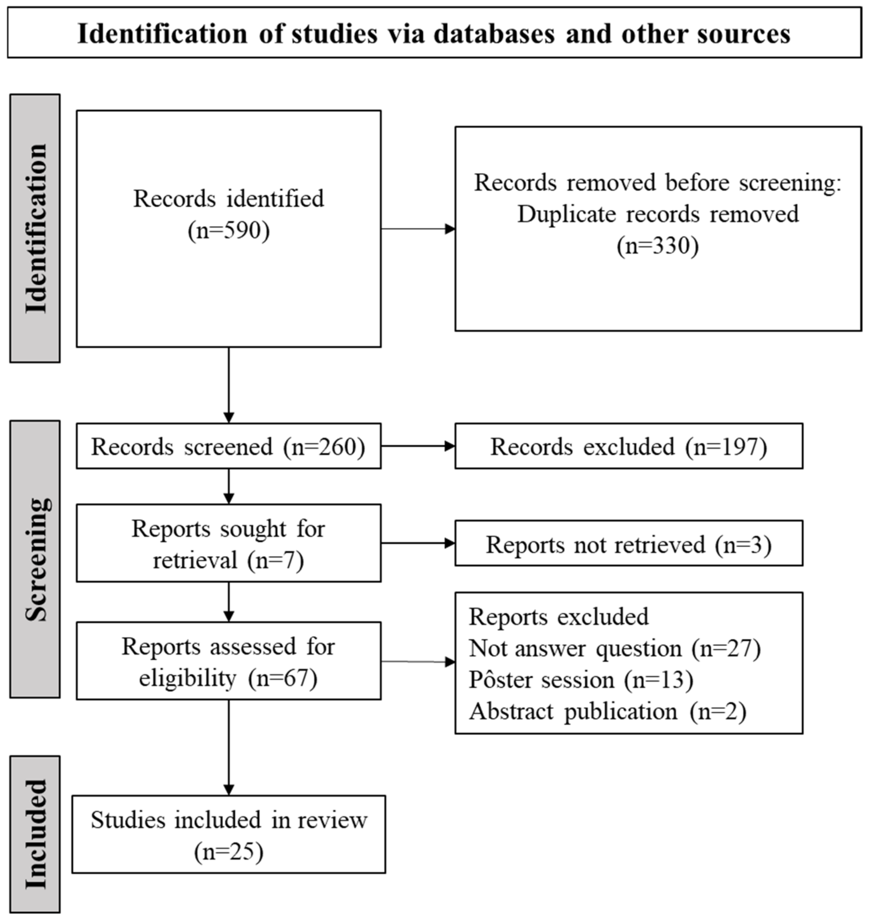
Figure 1. Visual representation of study selection for the current scoping review.
Biomedicines 13 01957 g001
Figure 2. Geographic and scientific distribution of original research articles included in this review.
Biomedicines 13 01957 g002
Figure 3. Chronological discoveries of Alamandine’s mechanisms of action [3,6,8,9,13,14,15,16,17,18,19,20,21,22,23,24,25,26,27,28,29,30,31,32,33,34].
Biomedicines 13 01957 g003
Figure 4. The classic renin–angiotensin system cascade. ACE: angiotensin-converting enzyme; ACE2: angiotensin-converting enzyme 2; Dcase: decarboxylase. Schematic representations were created with Biorender.com (YF28M19KKO).
Biomedicines 13 01957 g004
Figure 5. Organs and mechanisms activated by Alamandine. JNK—c-Jun N-terminal; PKA—protein kinase A; AMPK—adenosine monophosphate-activated protein kinase; NO—nitric oxide; TGFβ—transforming growth factor-β; PI3K—phosphoinositide 3-kinase; Akt—protein kinase B; ROS—Reactive oxygen species; MAPK—mitogen-activated protein kinase; ERK—extracellular signal-regulated kinase; NF-κB- nuclear factor kappa B; PKC—protein kinase C. Schematic representations were created with Biorender.com (CM28M18VE2).
Biomedicines 13 01957 g005
Disclaimer/Publisher’s Note: The statements, opinions and data contained in all publications are solely those of the individual author(s) and contributor(s) and not of MDPI and/or the editor(s). MDPI and/or the editor(s) disclaim responsibility for any injury to people or property resulting from any ideas, methods, instructions or products referred to in the content.