Article

Morphofunctional Profile Focusing on Strength and Ultrasound of the Upper Limbs in Female Breast Cancer Survivors: A Comparative Cross-Sectional Study Between Groups with and Without Lymphoedema and Between Ipsilateral and Contralateral Limbs

by
Ana Rafaela Cardozo Da Silva
1,
Juliana Netto Maia
1,
Vanessa Maria Da Silva Alves Gomes
1,
Naiany Tenório
1,
Juliana Fernandes de Souza Barbosa
1,
Ana Claudia Souza da Silva
1,
Vanessa Patrícia Soares de Sousa
2,
Leila Maria Alvares Barbosa
1,
Armèle de Fátima Dornelas de Andrade
1 and
Diego Dantas
1,*
1
Department of Physical Therapy, Health Sciences Center, Federal University of Pernambuco, Recife 50670-901, Brazil
2
Faculty of Health Sciences of Trairi, Federal University of Rio Grande do Norte, Santa Cruz 59200-000, Brazil
*
Author to whom correspondence should be addressed.
Biomedicines 2025, 13(8), 1884; https://doi.org/10.3390/biomedicines13081884 (registering DOI)
Submission received: 18 June 2025 / Revised: 22 July 2025 / Accepted: 31 July 2025 / Published: 2 August 2025
(This article belongs to the Section Molecular and Translational Medicine)

Abstract

Background: Breast cancer is the most common neoplasm in women. Despite effective treatments, sequelae such as decreased muscle strength, upper limb dysfunction, and tissue changes are common, highlighting the need for functional assessments during rehabilitation. This study analysed the morphofunctional profile of the upper limbs in breast cancer survivors, comparing muscle strength and ultrasound findings between groups with and without lymphoedema, as well as between ipsilateral and contralateral limbs. Methods: This cross-sectional study included female breast cancer survivors treated at an oncology physical therapy clinic. Muscle strength was measured using dynamometry (handgrip and arm flexor strength), and ultrasound assessed the thickness of the dermal–epidermal complex (DEC), subcutaneous tissue (SUB), and muscle (MT). Results: The upper limbs of 41 women were evaluated. No significant differences were observed between those with and without breast cancer-related lymphoedema (BCRL). When comparing the ipsilateral and contralateral limbs, significant reductions were observed in arm flexor strength (p < 0.001; 95% CI: −9.77 to −2.50), handgrip strength (p < 0.001; 95% CI: −4.10 to −1.22), and tissue thickness, with increased DEC thickness on the forearm (0.20 mm; p = 0.022) and arm flexors (0.25 mm; p < 0.001) of the ipsilateral limb. Conclusion: Significant differences in muscle strength and tissue structure between ipsilateral and contralateral limbs may reflect surgical and local pathophysiological effects. A trend toward reduced values for these parameters was also noted in limbs with BCRL, reinforcing the importance of future research to elucidate underlying mechanisms and guide more effective therapeutic strategies.

1. Introduction

Breast cancer is the most prevalent malignancy among women, accounting for approximately one in every four cancer diagnoses and one in every six cancer-related deaths [1]. In Brazil, an estimated 73,610 new cases are projected for the 2023–2025 period, according to the National Cancer Institute [2], underscoring the disease’s magnitude and impact.
Surgery remains a cornerstone of breast cancer treatment, significantly improving survival rates [3]. However, advances in therapeutic strategies and the resulting increase in survival rates have drawn greater attention to the long-term adverse effects of interventions such as surgery, chemotherapy, and radiotherapy [4]. The most common complications include lymphoedema, fatigue, upper limb dysfunction, and reduced muscle strength and cardiorespiratory fitness [4,5].
While essential for disease control, chemotherapy can impair muscle metabolism, leading to a loss of muscle mass and strength, particularly in the upper limbs, and a decline in overall physical fitness [6]. Radiotherapy, in turn, can indirectly alter muscle morphology, causing reductions in muscle volume and length and resulting in significant functional impact [7].
These musculoskeletal impairments can be acute or chronic, leading to dysfunctions such as reduced range of motion, decreased muscle strength, postural changes, and limitations in the activities of daily living (ADLs) [8].
These changes often lead to chronic pain and discomfort in the upper limbs, regardless of the treatment modality [9]. Beyond the physical repercussions, these alterations adversely affect quality of life, leading to significant psychosocial consequences such as symptoms of anxiety and depression and reduced social participation [10].
Functional impairment affects multiple dimensions of quality of life, including general health perception, physical performance, social roles, emotional state, body image, sexuality, and sexual satisfaction [11]. Despite the clinical relevance of complications related to muscle dysfunction, there is limited literature on the detailed characterisation of tissue and functional muscle changes in breast cancer survivors.
The early detection of morphofunctional changes is crucial. Combining objective methods, such as muscle strength assessment and tissue ultrasound, enables a more comprehensive analysis of patients’ morphofunctional profiles. This contributes to preventing and managing complications and guiding individualised rehabilitation strategies [12]. In this context, precise assessments are essential for preserving functionality and quality of life in female breast cancer survivors, allowing for early identification of patients at higher risk of morbidities and for the implementation of more effective therapeutic interventions [7,13].
This study aims to analyse the morphofunctional profile of the upper limbs in breast cancer survivors by comparing muscle strength and ultrasound findings between groups with and without lymphoedema, as well as between the ipsilateral and contralateral limbs. Integrating assessments of muscle function and tissue structure improves the clinical monitoring of these patients by providing a deeper understanding and enabling the early detection of functional changes. It can also guide interventions aimed at preserving functionality and improving quality of life.

2. Patients and Methods

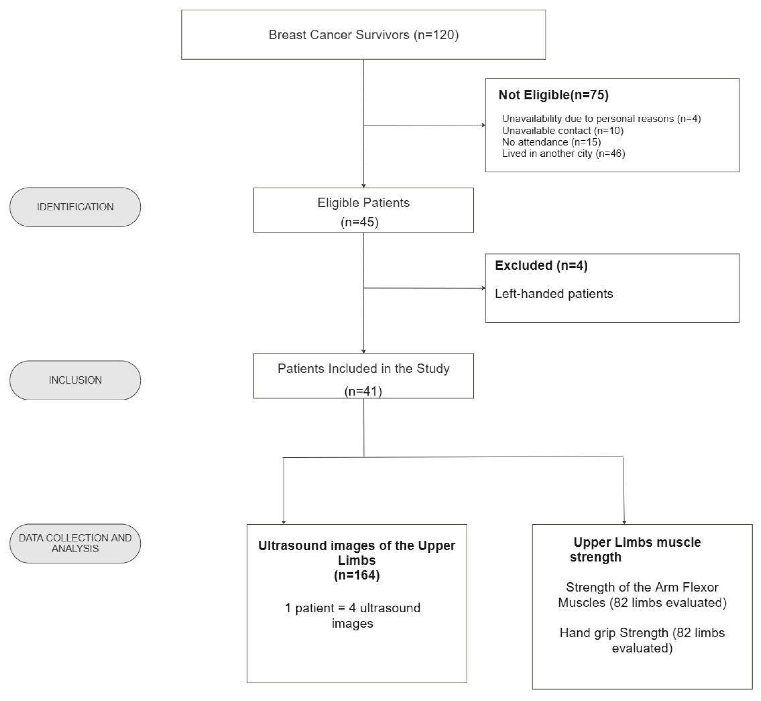
2.1. Study Population

This cross-sectional study followed the Strengthening the Reporting of Observational Studies in Epidemiology (STROBE) guidelines [14]. The research protocol was approved by the Institutional Ethics Committee (CAAE: 57624121.0.0000.5208), and all participants provided written informed consent.
This study was conducted from August 2022 to September 2023 at the Laboratory of Physical Therapy in Women’s Health and Pelvic Floor (LAFISMA), Department of Physical Therapy, Federal University of Pernambuco (UFPE).
The study population comprised female breast cancer survivors residing in Recife who were treated at LAFISMA. Inclusion criteria were women aged 40 to 70 with a history of mastectomy. Exclusion criteria included primary lymphoedema, oedema from other causes (rheumatologic, renal, or neurologic disease; orthopaedic issues; or prior vascular disease), skin conditions (erysipelas, intertrigo, or ulcers), undergoing chemotherapy or radiotherapy, and left-handedness (to ensure sample homogeneity).
Participants were recruited consecutively from the waiting list for oncology physical therapy care.
The sample size was calculated using G*Power 3.1 software (version 3.1, Düsseldorf, Germany) for a comparison between two independent groups (Student’s t-test). The effect size (d = 0.92) was estimated based on a previous study by Perez et al. that found a significant reduction in handgrip strength on the ipsilateral limb in women with breast cancer [15]. With a statistical power of 80% and an alpha of 0.05, accounting for a potential 20% sample loss or incomplete data, a minimum sample size of 39 women was required.

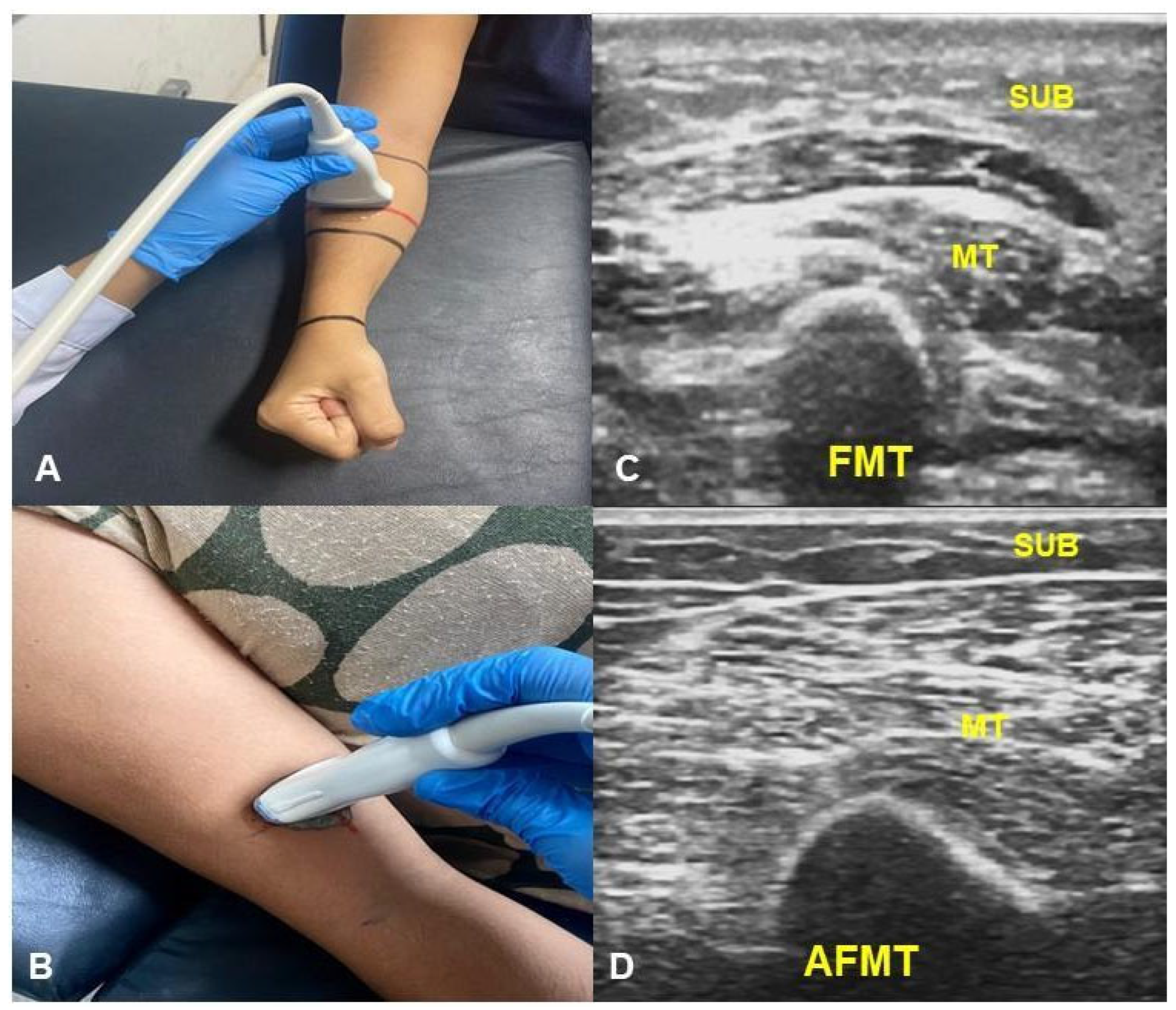
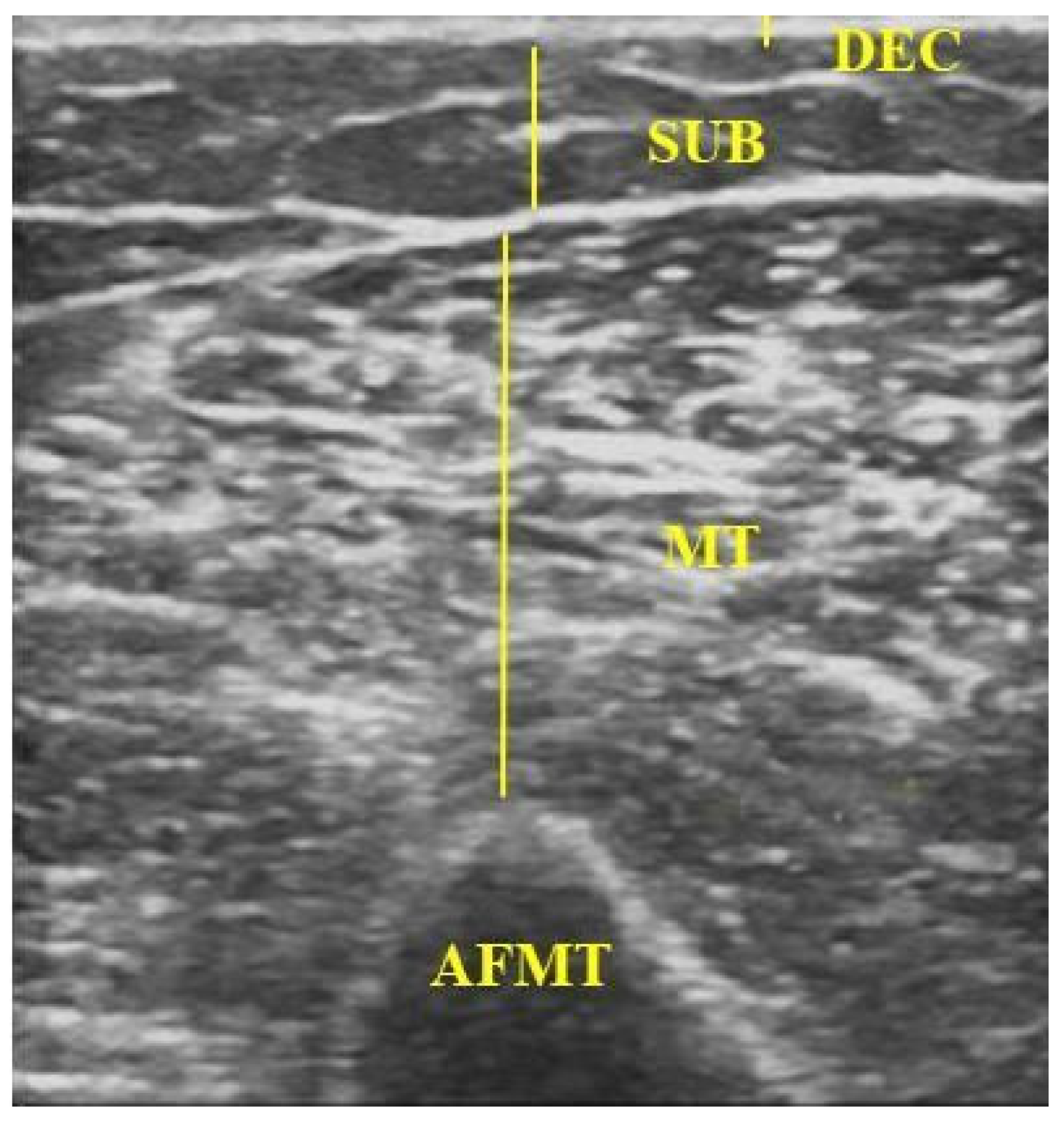
2.2. Data Collection

Data were collected at the LAFISMA facility by trained and calibrated assessors. The assessors underwent three four-hour theoretical and practical training sessions conducted by an experienced physical therapist. The training covered all study assessment procedures, including taking a medical history, performing a lymphoedema assessment, conducting an ultrasound examination, and measuring muscle strength.
Initially, participants completed a clinical form to collect personal and clinical information, including age (years), marital status, education level, time since surgery (months), type of mastectomy, and treatments received. Breast cancer-related lymphoedema (BCRL) was assessed according to the International Society of Lymphology guidelines [16]. A BCRL diagnosis was confirmed if the difference in upper limb volume was ≥200 mL or if the volume ratio was >1.04, as determined by indirect volumetry [17].
Muscle strength assessment included measurements of arm flexor muscle strength (AFMS) and absolute and relative handgrip strength (HGS). HGS was measured using a calibrated hydraulic hand dynamometer (Carci, São Paulo, Brazil). For the HGS measurement, participants were seated with their shoulder adducted and neutrally rotated, elbow flexed at 90°, forearm in a neutral position, and wrist at 0° and 30° of extension and 0° to 15° of ulnar deviation. The contralateral limb was relaxed on the thigh [18].
Assessors verbally instructed participants to exert maximal voluntary isometric force. Three sustained contractions were performed, with a one-minute rest interval between measurements [19]. Absolute HGS (AHGS) was defined as the average of the three measurements, expressed as a continuous variable in kilograms (kg). Relative HGS (RHGS) was calculated by normalising the AHGS value by the body mass index (BMI). RHGS was presented in kg/m2 [20,21].
AFMS was assessed using a portable digital dynamometer (Lafayette Hand-Held Dynamometer, Lafayette, LA, USA), which has demonstrated excellent test–retest and intra-rater reliability [22]. During the assessment, the participant was instructed to exert maximal elbow flexion force against resistance applied by the assessor at a 90° angle (±5°) to the participant’s arm. The average of three trials was recorded for mean force (N), peak force (N), and time to failure (seconds). The test was stopped when the participant could no longer maintain resistance [22].
B-mode ultrasound images were acquired using a LOGIC V5 system (GE) with a linear L6-12 transducer. Standardised parameters were used for all measurements: 8 MHz frequency, 79% gain, and 8 cm depth. In each limb, two regions of interest (ROIs) were defined, one on the forearm and one on the arm, both 10 cm from the cubital fossa [23]. Images were captured with the transducer positioned perpendicular to the skin over the muscle belly of interest (Figure 1), applying minimal pressure and using a water-based conductive gel [24]. After freezing the image, the following were measured: (i) the dermal–epidermal complex (DEC)—the linear distance from the posterior echogenic border of the epidermal entry echo to the posterior echogenic border of the dermis [25]; (ii) subcutaneous tissue (SUB)—the distance between the DEC and the muscle fascia [25,26]; (iii) muscle thickness (MT) of the forearm flexors (FMT)—the distance between the radius and the muscle interface [27]; and muscle thickness of the arm flexors (AFMT)—the distance between the muscle interface and the humeral periosteum, including the biceps brachii and brachialis muscles (Figure 2) [28,29].
For the forearm assessment, the upper limb was supported on a table and positioned with the shoulder flexed (30–45°), the elbow flexed (45°), the forearm in a neutral pronation/supination position, and the wrist in ulnar deviation (0–15°) [27]. For the arm assessment, the participant was positioned supine with the head of the bed elevated to 30° to ensure proper upper-limb alignment. The forearms were supinated with palms facing up; pillows were used to stabilise the position as needed [30].

2.3. Statistical Analyses

Statistical analyses were performed using JASP software (version 0.18.3 for Windows, Amsterdam, the Netherlands). Discrete and continuous data were presented as mean ± standard deviation, p-values, mean differences, and confidence intervals. Categorical data were presented as absolute and relative frequencies.
Data normality was assessed using the Shapiro–Wilk test. For non-parametric data, the Wilcoxon signed-rank test was used to compare muscle strength and mean ultrasound measurements between the ipsilateral and contralateral limbs, and the Mann–Whitney U test was used to compare women with lymphoedema and without lymphoedema. For parametric data, paired and independent Student’s t-tests were used, respectively. For non-parametric data, the 95% CI was calculated using the Hodges–Lehmann estimator. A p-value < 0.05 was considered statistically significant.
Spearman’s correlation test was used to analyse the association between muscle strength variables and the presence and location of lymphoedema. Correlation coefficients (rs) were interpreted according to [31] as weak (rs < 0.3), moderate (0.3 ≤ rs < 0.50), strong (0.5 ≤ rs < 0.70), or very strong (rs ≥ 0.70).

3. Results

We recruited 120 female breast cancer survivors, of whom 41 were included in this study (Figure 3). In total, we obtained 164 ultrasound images of the upper limbs, representing four per patient (two from the ipsilateral limb and two from the contralateral limb). Muscle strength was assessed using manual and digital dynamometers, resulting in the analysis of 82 upper limbs (ipsilateral and contralateral), with measurements taken separately for each device.
The mean age of the sample was 53.8 ± 7.5 years. The mean time since surgery was 76.2 ± 75.6 months. Regarding the type of surgery, 61.0% underwent a simple mastectomy, 36.5% a radical mastectomy, and 2.5% a modified mastectomy. Regarding lymphoedema, 46.3% of participants had the condition, while 53.7% did not (Table 1).
The mean strength of the arm flexor muscles was significantly lower in the ipsilateral limb (Table 2).
Similarly, peak force was lower in the limb with lymphoedema, with a mean difference of −7.55 (95% CI: −12.25 to −3.18; p < 0.001). However, there was no significant difference in time to peak force between the limbs (p = 0.936).
Ultrasound analysis revealed a significant increase in the dermal–epidermal complex thickness in the ipsilateral limb, particularly in the forearm (mean difference: 0.20 mm; p = 0.022) and arm flexor regions (mean difference: 0.25 mm; p < 0.001). Subcutaneous tissue thickness did not differ significantly (p = 0.075), whereas muscle thickness was reduced in the forearm (mean difference: −1.80 mm; p = 0.002).
As shown in Table 3, the mean strength of the elbow flexor muscles was lower in the lymphoedema group (72.63 ± 24.17) than in the non-lymphoedema group (87.3 ± 51.49). However, the mean difference of 14.72 (95% CI: −11.37 to 40.82) was not statistically significant (p = 0.367).
Likewise, absolute handgrip strength did not differ significantly (p = 0.628), with a mean of 22.03 ± 5.22 kg in the lymphoedema group and 22.88 ± 5.81 kg in the non-lymphoedema group (mean difference: 0.85 kg; 95% CI: −2.67 to 4.37).
Morphological parameters showed statistically significant differences. The dermal–epidermal complex was thicker in the lymphoedema group in both the forearm (p < 0.001) and the arm (p < 0.001). Forearm muscle thickness was significantly lower in participants with lymphoedema (9.27 ± 3.22 mm) than in those without it (12.80 ± 4.12 mm; p = 0.001).
The heatmap in Figure 4 shows the correlations between the analysed variables. The AHGS and RHGS handgrip strengths were highly correlated (rs = 0.798, p < 0.001). The absence of lymphoedema was moderately associated with a higher FMT in the ipsilateral limb (rs = 0.504, p < 0.001). Furthermore, a weak negative correlation was found between the presence of lymphoedema in the left (non-dominant) limb and AHGS (rs = −0.335, p = 0.023), while a moderate negative correlation was observed with RHGS (rs = −0.424, p = 0.006).

4. Discussion

This study aimed to analyse the morphofunctional profile of the upper limbs in breast cancer survivors by comparing muscle strength and ultrasound findings between groups with and without lymphoedema and between the ipsilateral and contralateral limbs.
The results demonstrated that the ipsilateral limb had significantly reduced muscle strength, which was associated with the structural changes identified by ultrasound. Although no statistically significant differences in strength were found between the lymphoedema and non-lymphoedema groups, relevant changes in DEC thickness in the arm and forearm, as well as decreased muscle thickness in the forearm, were identified.
The observed decrease in muscle strength in the ipsilateral upper limb indicates functional impairment. This finding aligns with the literature linking the surgical procedure to reduced strength and increased risk of shoulder joint dysfunction, which negatively affect upper limb functionality [32].
Our results showed lower HGS in the ipsilateral limb, corroborating the findings of Campos E Silva et al. [33] and earlier studies reporting reduced strength in breast cancer survivors [34,35]. Higher HGS levels are associated with better functional and psychosocial outcomes, including less disability, pain, and perceived weakness in the affected limb, as well as fewer depressive symptoms. These data reinforce the potential of HGS, both absolute and relative, as a relevant clinical marker for functional and psychosocial monitoring of patients after cancer treatment [21].
Women with breast cancer frequently experience muscle weakness and a decline in physical performance, often associated with increased rest and inactivity, which promotes disuse muscle atrophy and exacerbates functional limitations [36]. HGS assessment is, therefore, a practical and relevant tool for clinical monitoring and functional risk stratification in this patient population.
Although no statistically significant differences in muscle strength were found between the lymphoedema and non-lymphoedema groups, relevant tissue changes were identified. These findings suggest that muscle dysfunction may be a diffuse phenomenon, occurring even in the absence of lymphoedema [37], with evidence of inflammatory processes and systemic structural changes in adjacent tissues [38,39].
The study by [40] supports this view by demonstrating increased collagen deposition and CD4 expression in non-oedematous tissues, which may lead to diffuse impairment of the upper limbs.
Ultrasound enabled a detailed characterisation of structural changes, revealing increased DEC and reduced muscle thickness, primarily in the ipsilateral limb. The lack of consistent differences in subcutaneous tissue thickness may reflect the different stages of lymphoedema. Early stages are characterised by fluid accumulation and increased thickness, while advanced stages can lead to fibrosis and reduced thickness [41]. This coexistence underscores the complexity of diagnosis and clinical monitoring, highlighting the importance of US for precisely assessing the stage and severity of tissue changes.
The lymphoedema group also exhibited increased DEC thickness in the arm and forearm and reduced forearm muscle thickness compared to the non-lymphoedema group. Anatomical and functional factors, such as less efficient lymphatic drainage in the forearm and a predisposition to fluid accumulation in distal areas, contribute to these regional changes. The flatter anatomy of the forearm facilitates ultrasound visualisation of oedema, while the presence of collateral lymphatic pathways in the arm may explain the lower fluid retention in that region [25,42].
Studies suggest that US of the arm flexors, including the biceps brachii, can be a reliable marker of total muscle mass [43], reinforcing its clinical importance. Other studies have also used these muscles and the forearm for similar purposes [44,45], confirming the utility of US in detecting tissue changes associated with BCRL [46,47].
However, despite these advances, a gap remains in the literature regarding studies that integrate assessments of both muscle strength and US-measured tissue thickness in the upper limbs of female breast cancer survivors. In this study, we identified significant correlations between the structural and functional parameters of the upper limbs, including a strong association between absolute and relative handgrip strength and a moderate correlation between the absence of lymphoedema and greater forearm muscle thickness in the ipsilateral limb.
The observed relationship between muscle strength and structure highlights the importance of integrating musculoskeletal ultrasound and dynamometry for functional assessment, enabling a comprehensive and objective analysis of muscle function [48]. The clinical application of US facilitates the early detection of dysfunction, the monitoring of therapeutic responses, and a deeper understanding of the effects of oncology treatments on the underlying tissues [12].
Therefore, our findings support the need for individualised physical therapy strategies that focus on tissue structure, muscle strength, and upper limb functionality, even in the absence of clinically evident lymphoedema. However, this study has limitations, including a lack of control for lymphoedema severity and heterogeneity in post-treatment time, surgery type, and physical activity levels. These variables may influence the functional and structural outcomes, and the lack of statistical control for these factors may have contributed to the observed variations.
Longitudinal studies are needed to clarify the timeline for functional recovery and the progression patterns of musculoskeletal dysfunction. Moreover, future research should include functional assessments and account for sample heterogeneity in terms of surgery type, treatment duration, age, objective physical activity quantification, and BMI.

5. Conclusions

This study revealed significant morphofunctional changes in the upper limbs of breast cancer survivors, particularly between the ipsilateral and contralateral limbs, indicating that oncologic interventions affect both tissue structure and muscle strength. Although no significant differences were found between the lymphoedema and non-lymphoedema groups, the results suggest the need for studies with larger sample sizes and better variable control.
Integrating objective measures of strength and musculoskeletal imaging into physical therapy assessments can improve functional diagnosis, guiding more precise clinical management and personalising rehabilitation strategies. These findings reinforce the importance of continuously monitoring muscle function to prevent complications and promote functional ability and quality of life in this patient population.

Author Contributions

Conceptualisation, A.R.C.D.S., J.N.M., A.d.F.D.d.A., and D.D.; formal analysis, A.R.C.D.S., V.M.D.S.A.G., N.T., and J.F.d.S.B.; investigation, A.R.C.D.S., V.M.D.S.A.G., A.C.S.d.S., V.P.S.d.S., L.M.A.B., A.d.F.D.d.A., and D.D.; methodology, A.R.C.D.S., J.N.M., A.C.S.d.S., and D.D.; project administration, D.D.; writing—original draft, A.R.C.D.S., J.N.M., V.M.D.S.A.G., N.T., J.F.d.S.B., A.C.S.d.S., V.P.S.d.S., L.M.A.B., A.d.F.D.d.A., and D.D.; writing—review and editing, A.R.C.D.S., J.N.M., V.M.D.S.A.G., N.T., J.F.d.S.B., A.C.S.d.S., V.P.S.d.S., L.M.A.B., A.d.F.D.d.A. and D.D. All authors have read and agreed to the published version of the manuscript.

Funding

This research was funded by the Federal University of Pernambuco and the Pernambuco Science and Technology Support Foundation (FACEPE) trough postgraduate scholarship (IBPG-0081-4.08/23) and financial support for this study (APQ 0801-4.08/21 and APQ-1330-4.08/21).

Institutional Review Board Statement

This study was conducted according to the guidelines of the Declaration of Helsinki and approved by the Ethics Committee of Federal University of Pernambuco (CAAE: 57624121.0.0000.5208, approved on 25 September 2023).

Informed Consent Statement

Informed consent was obtained from all subjects involved in this study.

Data Availability Statement

The original contributions presented in this study are included in the article. Further inquiries can be directed to the corresponding author(s).

Acknowledgments

The authors would like to thank to the Federal University of Pernambuco and the Pernambuco Science and Technology Support Foundation (FACEPE) in support of this study.

Conflicts of Interest

The authors report no conflicts of interest.

Abbreviations

The following abbreviations are used in this manuscript:
AHGSabsolute handgrip strength
BCRLbreast cancer-related lymphoedema
BMIbody mass index
DECdermal–epidermal complex
RHGSrelative handgrip strength
SCLCsecondary breast cancer lymphoedema
SUBsubcutaneous tissue
FMTforearm flexors
AFMTarm flexor muscle thickness
MTmuscle thickness

References

  1. Bray, F.; Laversanne, M.; Sung, H.; Ferlay, J.; Siegel, R.L.; Soerjomataram, I.; Jemal, A. Global Cancer Statistics 2022: GLOBOCAN Estimates of Incidence and Mortality Worldwide for 36 Cancers in 185 Countries. CA Cancer J. Clin. 2024, 74, 229–263. [Google Scholar] [CrossRef]
  2. Instituto Nacional de Câncer—INCA. Estimativa 2023: Incidência de câncer no Brasil; Instituto Nacional De Câncer: Rio de Janeiro, RJ, USA, 2023; ISBN 9786588517093. [Google Scholar]
  3. Peng, X.; Lu, Z. Development and Validation of Upper Limb Lymphedema in Patients After Breast Cancer Surgery Using a Practicable Machine Learning Model: A Retrospective Cohort Study. Int. J. Gen. Med. 2024, 17, 3799–3812. [Google Scholar] [CrossRef] [PubMed]
  4. Martínez-Herrera, B.-E.; Muñoz-García, M.-G.; José-Ochoa, L.-L.; Quiroga-Morales, L.-A.; Cervántes-González, L.-M.; Mireles-Ramírez, M.-A.; Delgadillo-Cristerna, R.; Nuño-Guzmán, C.-M.; Leal-Cortés, C.-A.; Portilla-de-Buen, E.; et al. Role of Incretins in Muscle Functionality, Metabolism, and Body Composition in Breast Cancer: A Metabolic Approach to Understanding This Pathology. Biomedicines 2024, 12, 280. [Google Scholar] [CrossRef]
  5. Alstrup, M.; Johannessen, A.L.; Mohanakumar, S.; Offersen, B.V.; Hjortdal, V.E. Lymphatic Function in the Arms of Breast Cancer Patients—A Prospective Cohort Study. Plast. Reconstr. Surg. Glob. Open 2021, 9, e3779. [Google Scholar] [CrossRef]
  6. Mistry, S.; Ali, T.; Qasheesh, M.; Beg, R.A.; Shaphe, M.A.; Ahmad, F.; Kashoo, F.Z.; Shalaby, A.S. Assessment of Hand Function in Women with Lymphadenopathy after Radical Mastectomy. PeerJ 2021, 9, e11252. [Google Scholar] [CrossRef]
  7. Leonardis, J.M.; Lulic-Kuryllo, T.; Lipps, D.B. The Impact of Local Therapies for Breast Cancer on Shoulder Muscle Health and Function. Crit. Rev. Oncol. Hematol. 2022, 177, 103759. [Google Scholar] [CrossRef]
  8. Fuentes-Abolafio, I.J.; Roldán-Jiménez, C.; Campos, M.I.; Pajares-Hachero, B.I.; Alba-Conejo, E.; Cuesta-Vargas, A. Forearm Muscle Activity During the Handgrip Test in Breast Cancer Survivors: A Cross-Sectional Study. Clin. Breast Cancer 2023, 23, e175–e181. [Google Scholar] [CrossRef]
  9. Zhang, X.; Wang, C.; Fan, J.; Murakami, S.; Xie, H.; Huo, M. The Factors Influencing Shoulder Mobility Disorders in Patients after Radical Breast Cancer Surgery: A Cross-Sectional Study. Breast Care 2024, 19, 43–48. [Google Scholar] [CrossRef]
  10. Ramirez-Parada, K.; Gonzalez-Santos, A.; Riady-Aleuy, L.; Pinto, M.P.; Ibañez, C.; Merino, T.; Acevedo, F.; Walbaum, B.; Fernández-Verdejo, R.; Sanchez, C. Upper-Limb Disability and the Severity of Lymphedema Reduce the Quality of Life of Patients with Breast Cancer-Related Lymphedema. Curr. Oncol. Tor. Ont. 2023, 30, 8068–8077. [Google Scholar] [CrossRef] [PubMed]
  11. Hasanah, U.; Ahmad, M.; Prihantono, P.; Usman, A.N.; Arsyad, A.; Agustin, D.I. The Quality of Life Assessment of Breast Cancer Patients. Breast Dis. 2024, 43, 173–185. [Google Scholar] [CrossRef] [PubMed]
  12. Perez, C.S.; Mestriner, C.; Ribeiro, L.T.N.; Grillo, F.W.; Lemos, T.W.; Carneiro, A.A.; Guirro, R.R.D.J.; Guirro, E.C.O. Relationship between lymphedema after breast cancer treatment and biophysical characteristics of the affected tissue. PLoS ONE 2022, 17, e0264160. [Google Scholar] [CrossRef]
  13. Klein, I.; Friger, M.; David, M.B.; Shahar, D. Risk Factors for Long-Term Arm Morbidities Following Breast Cancer Treatments: A Systematic Review. Oncotarget 2023, 14, 921–942. [Google Scholar] [CrossRef]
  14. Cuschieri, S. The STROBE Guidelines. Saudi J. Anaesth. 2019, 13, S31–S34. [Google Scholar] [CrossRef]
  15. Perez, C.S.; das Neves, L.M.S.; Vacari, A.L.; de Cássia Registro Fonseca, M.; de Jesus Guirro, R.R.; de Oliveira Guirro, E.C. Reduction in Handgrip Strength and Electromyographic Activity in Women with Breast Cancer. J. Back. Musculoskelet. Rehabil. 2018, 31, 447–452. [Google Scholar] [CrossRef]
  16. International Society of Lymphology, 2023. Diagnostic Criteria for Breast Cancer-Related Lymphedema of the Upper Extremity: The Need for Universal Agreement|Annals of Surgical Oncology. Available online: https://link.springer.com/article/10.1245/s10434-021-10645-3 (accessed on 21 October 2024).
  17. Levenhagen, K.; Davies, C.; Perdomo, M.; Ryans, K.; Gilchrist, L. Diagnosis of Upper Quadrant Lymphedema Secondary to Cancer: Clinical Practice Guideline From the Oncology Section of the American Physical Therapy Association. Phys. Ther. 2017, 97, 729–745. [Google Scholar] [CrossRef] [PubMed]
  18. Rajendiran, S.; Pai, G.M.; Verma, V.; Rajappa, S.; Bhat, A.; Gaba, S.; Thatte, M. Normative Data of Grip Strength and Pinch Strength in the Indian Population. Indian. J. Plast. Surg. Off. Publ. Assoc. Plast. Surg. India 2024, 57, 256–262. [Google Scholar] [CrossRef] [PubMed]
  19. de Souza, C.F.; Vieira, M.C.A.; do Nascimento, R.A.; Moreira, M.A.; da Câmara, S.M.A.; Maciel, Á.C.C. Relationship between Strength and Muscle Mass in Middle-Aged and Elderly Women: A Cross-Sectional Study. Rev. Bras. Geriatr. Gerontol. Online 2017, 20, 660–669. [Google Scholar] [CrossRef]
  20. Choquette, S.; Bouchard, D.R.; Doyon, C.Y.; Sénéchal, M.; Brochu, M.; Dionne, I.J. Relative Strength as a Determinant of Mobility in Elders 67-84 Years of Age. a Nuage Study: Nutrition as a Determinant of Successful Aging. J. Nutr. Health Aging 2010, 14, 190–195. [Google Scholar] [CrossRef]
  21. Esteban-Simón, A.; Díez-Fernández, D.M.; Artés-Rodríguez, E.; Casimiro-Artés, M.Á.; Rodríguez-Pérez, M.A.; Moreno-Martos, H.; Casimiro-Andújar, A.J.; Soriano-Maldonado, A. Absolute and Relative Handgrip Strength as Indicators of Self-Reported Physical Function and Quality of Life in Breast Cancer Survivors: The EFICAN Study. Cancers 2021, 13, 5292. [Google Scholar] [CrossRef]
  22. Nepomuceno Júnior, B.R.V.; Menezes, M.P.D.S.; Santos, K.R.B.D.; Gomes Neto, M. Comparison of Methods for Evaluating Upper Limb Strength by Hand-Held Dynamometry. Rev. Bras. Med. Esporte 2021, 27, 42–48. [Google Scholar] [CrossRef]
  23. Jeon, Y.; Beom, J.; Ahn, S.; Bok, S.-K. Ultrasonographic Evaluation of Breast Cancer-Related Lymphedema. J. Vis. Exp. JoVE 2017, 119, 54996. [Google Scholar] [CrossRef]
  24. Devoogdt, N.; Pans, S.; De Groef, A.; Geraerts, I.; Christiaens, M.-R.; Neven, P.; Vergote, I.; Van Kampen, M. Postoperative Evolution of Thickness and Echogenicity of Cutis and Subcutis of Patients With and Without Breast Cancer-Related Lymphedema. Lymphat. Res. Biol. 2014, 12, 23–31. [Google Scholar] [CrossRef]
  25. Suehiro, K.; Morikage, N.; Yamashita, O.; Harada, T.; Samura, M.; Takeuchi, Y.; Mizoguchi, T.; Nakamura, K.; Hamano, K. Skin and Subcutaneous Tissue Ultrasonography Features in Breast Cancer-Related Lymphedema. Ann. Vasc. Dis. 2016, 9, 312–316. [Google Scholar] [CrossRef]
  26. Kim, S.-Y.; Lee, C.-H.; Heo, S.J.; Moon, M.-H. The Clinical Usefulness of Lymphedema Measurement Technique Using Ultrasound. Lymphat. Res. Biol. 2021, 19, 340–346. [Google Scholar] [CrossRef] [PubMed]
  27. Meza-Valderrama, D.; Sánchez- Rodríguez, D.; Perkisas, S.; Duran, X.; Bastijns, S.; Dávalos-Yerovi, V.; Da Costa, E.; Marco, E. The Feasibility and Reliability of Measuring Forearm Muscle Thickness by Ultrasound in a Geriatric Inpatient Setting: A Cross-Sectional Pilot Study. BMC Geriatr. 2022, 22, 137. [Google Scholar] [CrossRef]
  28. Hadda, V.; Kumar, R.; Hussain, T.; Khan, M.A.; Madan, K.; Mohan, A.; Khilnani, G.C.; Guleria, R. Reliability of Ultrasonographic Arm Muscle Thickness Measurement by Various Levels of Health Care Providers in ICU. Clin. Nutr. ESPEN 2018, 24, 78–81. [Google Scholar] [CrossRef]
  29. Nelson, C.M.; Dewald, J.P.A.; Murray, W.M. In Vivo Measurements of Biceps Brachii and Triceps Brachii Fascicle Lengths Using Extended Field-of-View Ultrasound. J. Biomech. 2016, 49, 1948–1952. [Google Scholar] [CrossRef]
  30. Fisher, M.I.; Capilouto, G.; Malone, T.; Bush, H.; Uhl, T.L. Comparison of Upper Extremity Function in Women With and Women Without a History of Breast Cancer. Phys. Ther. 2020, 100, 500–508. [Google Scholar] [CrossRef] [PubMed]
  31. Dancey, C.P.; Reidy, J. Estatistica Sem Matematica Para Psicologia: Usando SPSS Para Windows; Artmed: Porto Alegre, RS, Brazil, 2006. [Google Scholar]
  32. Zhang, X.-M.; Zhang, Z.-B.; Chen, W.; Wu, X. The Association between Handgrip Strength and Depression in Cancer Survivors: A Cross-Sectional Study. BMC Geriatr. 2022, 22, 111. [Google Scholar] [CrossRef] [PubMed]
  33. Campos, E.; Silva, A.C.; Bergmann, A.; Araujo, C.M.; Montenegro, A.K.S.; Tenorio, A.D.S.; Dantas, D. Association of Handgrip Strength with Quality of Life in Breast Cancer Survivors: A Systematic Review and Meta-Analysis. Asian Pac. J. Cancer Prev. APJCP 2022, 23, 3237–3245. [Google Scholar] [CrossRef]
  34. Baklaci, M.; Eyigör, S.; Tanıgör, G.; Özgür İnbat, M.; Çalışkan Kabayel, S. Assessment of Muscle Strength and Volume Changes in Patients with Breast Cancer-Related Lymphedema. Oncol. Res. Treat. 2020, 43, 584–591. [Google Scholar] [CrossRef]
  35. Fretta, T.D.B.; Boing, L.; Leite, B.; Vieira, M.D.C.S.; Moratelli, J.; Klen, J.A.; Campeiz, E.; Machado, Z.; Guimarães, A.C.D.A. Physical Functionality of the Upper Limb after Breast Cancer Surgery in Southern Brazilian Survivors: Cross-Sectional Study. Rev. Bras. Cancerol. 2021, 67, e-021168. [Google Scholar] [CrossRef]
  36. Kang, S.; Yoo, S.; Baek, H.; Lee, J.; Choi, Y.; Kim, H.; Yi, H.; Yang, E.J. Potentials of Smart Dynamometer Use for Clinical and Self-Management of Rehabilitation in Breast Cancer Survivors: A Feasibility Study. Biomed. Eng. Lett. 2019, 9, 211–219. [Google Scholar] [CrossRef]
  37. Gomes, P.R.L.; Freitas Junior, I.F.; da Silva, C.B.; Gomes, I.C.; Rocha, A.P.R.; Salgado, A.S.I.; do Carmo, E.M. Short-Term Changes in Handgrip Strength, Body Composition, and Lymphedema Induced by Breast Cancer Surgery. Rev. Bras. Ginecol. E Obstet. Rev. Fed. Bras. Soc. Ginecol. E Obstet. 2014, 36, 244–250. [Google Scholar] [CrossRef] [PubMed]
  38. Leray, H.; Malloizel-Delaunay, J.; Lusque, A.; Chantalat, E.; Bouglon, L.; Chollet, C.; Chaput, B.; Garmy-Susini, B.; Yannoutsos, A.; Vaysse, C. Body Mass Index as a Major Risk Factor for Severe Breast Cancer-Related Lymphedema. Lymphat. Res. Biol. 2020, 18, 510–516. [Google Scholar] [CrossRef] [PubMed]
  39. Yusof, K.M.; Avery-Kiejda, K.A.; Ahmad Suhaimi, S.; Ahmad Zamri, N.; Rusli, M.E.F.; Mahmud, R.; Saini, S.M.; Abdul Wahhab Ibraheem, S.; Abdullah, M.; Rosli, R. Assessment of Potential Risk Factors and Skin Ultrasound Presentation Associated with Breast Cancer-Related Lymphedema in Long-Term Breast Cancer Survivors. Diagnostics 2021, 11, 1303. [Google Scholar] [CrossRef] [PubMed]
  40. Wolf, S.; von Atzigen, J.; Kaiser, B.; Grünherz, L.; Kim, B.-S.; Giovanoli, P.; Lindenblatt, N.; Gousopoulos, E. Is Lymphedema a Systemic Disease? A Paired Molecular and Histological Analysis of the Affected and Unaffected Tissue in Lymphedema Patients. Biomolecules 2022, 12, 1667. [Google Scholar] [CrossRef]
  41. Ricci, V.; Ricci, C.; Gervasoni, F.; Giulio, C.; Farì, G.; Andreoli, A.; Özçakar, L. From Physical to Ultrasound Examination in Lymphedema: A Novel Dynamic Approach. J. Ultrasound 2022, 25, 757–763. [Google Scholar] [CrossRef]
  42. Han, N.; Cho, Y.; Hwang, J.; Kim, H.; Cho, G. Usefulness of Ultrasound Examination in Evaluation of Breast Cancer-Related Lymphedema. J. Korean Acad. Rehabil. Med. 2011, 35, 101–109. [Google Scholar]
  43. Alabadi, B.; Bastijns, S.; Cock, A.-M.D.; Civera, M.; Real, J.T.; Perkisas, S. Relation Between Ultrasonographic Measurements of the Biceps Brachii and Total Muscle Mass in Older Hospitalized Persons: A Pilot Study. J. Frailty Sarcopenia Falls 2024, 9, 25–31. [Google Scholar] [CrossRef]
  44. Baran, E.; Özçakar, L.; Özgül, S.; Aksoy, S.; Akbayrak, T. Upper Limb Sensory Evaluations and Ultrasonographic Skin Measurements in Breast Cancer–Related Lymphedema Receiving Complex Decongestive Physiotherapy. Support. Care Cancer 2021, 29, 6545–6553. [Google Scholar] [CrossRef] [PubMed]
  45. Goudarzi, S.; Whyte, J.; Boily, M.; Towers, A.; Kilgour, R.D.; Rivaz, H. Segmentation of Arm Ultrasound Images in Breast Cancer-Related Lymphedema: A Database and Deep Learning Algorithm. IEEE Trans. Biomed. Eng. 2023, 70, 2552–2563. [Google Scholar] [CrossRef] [PubMed]
  46. Azhar, S.H.; Lim, H.Y.; Tan, B.-K.; Angeli, V. The Unresolved Pathophysiology of Lymphedema. Front. Physiol. 2020, 11, 137. [Google Scholar] [CrossRef]
  47. Polat, A.V.; Ozturk, M.; Polat, A.K.; Karabacak, U.; Bekci, T.; Murat, N. Efficacy of Ultrasound and Shear Wave Elastography for the Diagnosis of Breast Cancer-Related Lymphedema. J. Ultrasound Med. Off. J. Am. Inst. Ultrasound Med. 2020, 39, 795–803. [Google Scholar] [CrossRef]
  48. Laett, C.; Gavilão, U.; do Rio, J.; Cossich, V.; de Oliveira, C.G. Relationship between Upper and Lower Limbs Muscle Explosive Strength with the Vastus Lateralis and Biceps Brachii Architecture. Rev. Bras. Ciênc. Esporte 2021, 43, e012820. [Google Scholar] [CrossRef]
Figure 1. (A) Patient and transducer positioning for ultrasound imaging of forearm flexors (FMT); (B) Ultrasound image of FMT; (C) Patient and transducer positioning for ultrasound imaging of arm flexors (AFMT); (D) ultrasound image of AFMT. SUB: subcutaneous tissue; MT: muscle thickness. Source: The authors (2005).
Biomedicines 13 01884 g001
Figure 2. Ultrasound image showing measurements of DEC, SUB, and MT of the arm flexor muscles. DEC: dermal–epidermal complex; SUB: subcutaneous tissue; MT: muscle thickness; AFMT: arm flexor muscle thickness. Source: the authors (2025).
Biomedicines 13 01884 g002
Figure 3. Flowchart of the study screening process, detailing the selection steps for female breast cancer survivors post-mastectomy. Recife/PE, 2022–2023. Source: the authors (2025).
Biomedicines 13 01884 g003
Figure 4. Heatmap of correlation coefficients among the study variables. Caption: AHGS: absolute handgrip strength; RHGS: relative handgrip strength; FMT: forearm muscle thickness; AFMT: arm flexor muscle thickness; AFMS: arm flexor muscle strength. Calculated by Spearman’s correlation test. Source: The authors (2025). Source: the authors (2025).
Biomedicines 13 01884 g004
Table 1. Sociodemographic and clinical characteristics of female breast cancer survivors post-mastectomy (n = 41).
Variablen (%)Mean (SD)
Age (years) 53.8 (7.5)
BMI (kg/m2) 27.7 (5.0)
Limb with lymphoedema
   Right (dominant)23 (56.1)
   Left18 (43.9)
Time since surgery (months) 76.2 (75.6)
Type of mastectomy
Simple25 (61.0)
Modified2 (2.5)
Radical14 (36.5)
Therapies received
Chemotherapy38 (92.7)
Radiotherapy37 (90.2)
Hormonal16 (39.0)
Lymphoedema
Yes19 (46.3)
No22 (53.7)
SD: Standard deviation; BMI: body mass index.
Table 2. Comparison of muscle strength and ultrasound parameters in the upper limbs of female breast cancer survivors post-mastectomy (n = 41).
VariablesIL
Mean (SD)
CL
Mean (SD)
p-ValueMean Difference (95% CI)
AFMS
Mean force (N)73.23 (25.61)83.97 (41.85)<0.001−5.91 (−9.77 to −2.50)
Peak force (N)92.84 (34.97)101.83 (35.21)<0.001−7.55 (−12.25 to −3.18)
Time to peak (s)2.35 (0.53)2.31 (0.53)0.9360.01 (−0.12 to 0.15)
HGS
AHGS (kg) *22.48 (5.49)25.14 (5.83)<0.001−2.66 (−4.10 to −1.22)
RHGS (kg/m2) *0.81 (0.24)0.90 (0.24)<0.001−0.09 (−0.15 to −0.04)
US
FMT (mm)
DEC1.40 (1.10)1.00 (0.20)0.0220.20 (<0.01 to 0.55)
SUB5.25 (2.01)4.51 (1.87)0.0110.65 (0.15 to 1.10)
MT11.17 (4.04)12.87(5.13)0.002−1.80 (−2.75 to −0.60)
AFMT (mm)
DEC1.25 (0.45)1.02 (0.24)<0.0010.25 (0.15 to 0.40)
SUB6.12 (3.16)5.79 (2.43)0.0750.30 (<−0.01 to 0.60)
MT15.00 (4.34)14.18 (5.00)0.9372.73 × 10−5 (−0.85 to 1.15)
Caption: Data are presented as mean (SD). SD: standard deviation; AFMS: arm flexor muscle strength; HGS: handgrip strength; US: ultrasound; AHGS: absolute handgrip strength; RHGS: relative handgrip strength; FMT: forearm muscle thickness; AFMT: arm flexor muscle thickness; CI: confidence interval; IL: ipsilateral limb; CL: contralateral limb; kg: kilograms; kg/m2: kilograms per square meter; MT muscle thickness; DEC dermal–epidermal complex; SUB subcutaneous tissue; mm: millimetres; * calculated using paired Student’s t-test.
Table 3. Comparison of muscle strength parameters in the ipsilateral limbs between groups with and without lymphoedema (n = 41).
VariablesLymphoedema (n = 19)Without Lymphoedema (n = 22)p-ValueMean Difference
(95% CI)
Mean (SD)Mean (SD)
AFMS
Mean force (N)72.63 (24.17)87.36 (51.49)0.36714.72 (−11.37 to 40.82)
Peak force (N)91.98 (33.71)101.57 (96.03)0.3029.59 (−11.25 to 30.44)
Time to peak (s)2.11 (0.54)2.43 (0.52)0.2040.31 (−0.02 to 0.65)
HGS
AHGS (kg) *22.03 (5.22)22.88 (5.81)0.628 *0.85 (−2.67 to 4.37)
RHGS (kg/m2) *0.75 (0.23)0.87 (0.25)0.1280.12 (−0.04 to 0.27)
US
FMT (mm)
DEC1.96 (1.42)0.94 (0.35)<0.0010.70 (0.40 to 1.1)
SUB5.76 (2.29)4.77 (1.69)0.3740.37 (−0.60 to 2.00)
MT9.27 (3.22)12.80 (4.12)0.001−3.71 (−5.70 to −0.90)
AFMT (mm)
DEC *1.53 (0.46)1.01 (0.27)<0.0010.51 (0.27 to 0.75)
SUB7.07 (3.94)5.30 (2.12)0.0841.2 (−0.20 to 2.70)
MT *15.76 (5.39)14.49 (3.20)0.3571.27 (−1.48 to 4.03)
Caption: Data are presented as mean (SD). SD: standard deviation; AFMS: arm flexor muscle strength; HGS: handgrip strength; US: ultrasound; AHGS: absolute handgrip strength; RHGS: relative handgrip strength; FMT: forearm muscle thickness; AFMT: arm flexor muscle thickness; CI: confidence interval; IL: ipsilateral limb; CL: contralateral limb; kg: kilograms; kg/m2: kilograms per square meter; mm: millimeters. * calculated using paired Student’s t-test.
Disclaimer/Publisher’s Note: The statements, opinions and data contained in all publications are solely those of the individual author(s) and contributor(s) and not of MDPI and/or the editor(s). MDPI and/or the editor(s) disclaim responsibility for any injury to people or property resulting from any ideas, methods, instructions or products referred to in the content.

